Algal Research 54 (2021) 102232
Available online 25 February 2021
2211-9264/© 2021 The Authors. Published by Elsevier B.V. This is an open access article under the CC BY license (http://creativecommons.org/licenses/by/4.0/).
Optimizing antioxidant activity in Agarophyton vermiculophyllum for functional packaging
Sofiia Tretiak
a,e, Jakop Schwoerbel
a,d, Ramona Bosse
b, Bela H. Buck
a,b, Ina Enders
c, Joachim Henjes
a, Dietmar Hoffmann
c, Frederike Reimold
b, Laurie C. Hofmann
a,*aAlfred Wegener Institute Helmholtz Center for Polar and Marine Research, Bussestrasse 27, 27570 Bremerhaven, Germany
bUniversity of Applied Sciences, An der Karlstadt 8, 27568 Bremerhaven, Germany
cNORDSEE GmbH, Herwigstraße 16, 27572 Bremerhaven, Germany
dUniversity of Tasmania Institute for Marine and Antarctic Studies, Australia1
eNational University of Ireland - Galway, University Road, Galway, Ireland H91 TK331
A R T I C L E I N F O Keywords:
Antioxidant activity Radical scavenging activity Macroalgae
Packaging Aquaculture
A B S T R A C T
The value of macroalgae in a healthy human diet is becoming increasingly recognized and supported throughout Europe. Macroalgae provide a rich source of vitamins, minerals, proteins, fatty acids, and antioxidants that also support the functionality of macroalgae in other industries, including cosmeceuticals, pharmaceuticals, and more recently, packaging. Sustainable aquaculture of macroalgae will be necessary to supply the increasing demand for macroalgae as a functional material, considering that natural harvests are limited and cannot keep up with demand. Different methods can be used to cultivate macroalgae, including flow-through systems or recirculating aquaculture systems (RAS) with natural or artificial seawater. The latter provides strict control over the growth conditions and water quality in order to provide a high quality and traceable product. Additionally, environ- mental conditions such as salinity, temperature, and light can be modified to optimize the concentration of functional ingredients in macroalgae. While most research efforts have focused on seasonal and geographic trends in concentrations of functional ingredients in wild macroalgae, there is less information available on optimizing these functional ingredients in aquaculture. Therefore, we performed controlled experiments to optimize the activity of antioxidants in Agarophyton vermiculophyllum (Ohmi) Gurgel, J.N.Norris et Fredericq comb. nov. (formerly Gracilaria vermiculophylla) grown in RAS with artificial seawater and commercial fertilizer.
We show that the free radical scavenging activity could be increased by 13% via high salinity, and up to 34% by increasing the light intensity, but not daily light dose, for a period of 7 days. We also monitored growth rates and the maximum quantum yield of photosystem II (Fv/Fm) and show that the conditions for optimizing antioxidant activity are not optimal for growth or photosynthesis. We therefore suggest an optimization period of 4–7 days exposure to high light on a 6:18 hour light:dark cycle prior to harvesting in order to increase antioxidant activity.
1. Introduction
Intensive use of plastic material in daily life has led to undeniable environmental pollution [1–3]. Since 1950, world plastic production has been constantly rising, and in 2017, it was estimated at 374 million tonnes [2]. The growth of the plastic industry bypasses manufacturing of many other synthetic materials [1]. However, plastic is resistant to degradation in nature and consequently accumulates in landfills and marine environments [3]. Moreover, microplastics – small fragments of
plastic – can cause harm to wildlife and accumulate up the food chain and further pose a threat to high trophic levels, including humans [4–6].
Therefore, over the last decade, there has been an increase in research involving the replacement of plastics with sustainable and biodegrad- able materials [7]. Macroalgae provide a natural, renewable resource that could potentially be used in a lot of different sectors, including the packaging industry (e.g. Evoware, OOHO). Marine algae are considered a promising raw material due to their fast growth, bioactive contents and natural, biodegradable polymers [6]. Additionally, macroalgae
* Corresponding author.
E-mail address: Laurie.c.hofmann@awi.de (L.C. Hofmann).
1 Current affiliation.
Contents lists available at ScienceDirect
Algal Research
journal homepage: www.elsevier.com/locate/algal
https://doi.org/10.1016/j.algal.2021.102232
Received 16 December 2019; Received in revised form 1 February 2021; Accepted 1 February 2021
contain a wide variety of functional ingredients with antioxidant ac- tivity that have the potential to prolong the shelf-life of materials stored in macroalgae-based products [8]. For example, an application in the food industry could reduce the main causes of food waste due to spoilage by lipid oxidation and microbial contamination. Existing strategies for preventing spoilage involve direct addition of antioxidants or packaging techniques that allow limited oxygen access [9]. Alternatively, macroalgae-based products, including functional packaging (packaging that provides benefits to the consumer) offer an antioxidant rich, natu- ral, sustainable, biodegradable material that could reduce plastic waste, increase shelf-life and provide a unique customer experience.
Macroalgae-derived polymers such as agar, carrageenan, and alginate have already been used as a base for biodegradable film development in food and pharmaceutical applications [6,7,8,10,11]. However, the complex technology and high costs involved in the polymer isolation, combined with low yields and large amounts of wasted biomass, are critical drawbacks to wider commercial use of such films in the global market [12]. Therefore, industrial solutions using the whole macroalgae biomass could be more useful [12], and additional investigations in this direction are currently underway (data not published). Either way, there is a knowledge gap regarding optimizing the functionality of seaweeds for industrial applications, particularly in the functional packaging in- dustry. Consequently, we focused on optimizing the antioxidant activity in the whole macroalgae biomass, rather than in extracts.
Macroalgae have developed mechanisms to suppress or scavenge the oxygen radicals that are inevitably produced during photosynthesis and electron transport [13]. Macroalgae encounter particularly high levels of stress in the harsh and competitive intertidal environment, and have therefore developed highly efficient defense mechanisms via the syn- thesis of antioxidant compounds. For example, macroalgae inhabiting the intertidal zones with fluctuating levels of irradiance and oxygen concentration combined with exposure to the air are able to cope with stress by synthesizing of a wide range of metabolic compounds with radical scavenging activity. These compounds include carotenoids, polyphenols, polysaccharides, squalenes and vitamins, among others [14–16]. The rapid oxidative response of macroalgae to abiotic stress [17,18] provides an opportunity to control and modify the antioxidant capacity of cultured macroalgae by exposing it to various stress condi- tions for a short period. Many studies have reported the seasonal fluc- tuations of antioxidant activity in natural macroalgae or in macroalgal extracts [8,13,19–24]. These studies provide sufficient evidence that salinity, tidal emersion, and light stress can induce oxidative stress re- sponses in macroalgae. Therefore, we manipulated salinity, desiccation, light intensity and light quality in order to increase the antioxidant ac- tivity of Agarophyton vermiculophyllum in culture in an effort to increase the functionality of this species for further industrial applications. We chose A. vermiculophyllum because it is an agarophyte that is already used in the food industry as a thickening agent and in films and coatings [25,26]. Although it is a non-native species in the Wadden Sea, it has established stable populations throughout northern Europe and can be the most abundant macroalgal species in some areas. Additionally, we focus here only on closed, recirculating land-based aquaculture systems, where propagation of additional unwanted biomass into the ecological systems is not a threat. Its broad salinity and temperature tolerance, fast growth rate, and vegetative propagation [27–29] make it an ideal candidate for aquaculture and optimization studies. The novelty of this study lies in the optimization of antioxidant activity in a cultured macroalgal species by manipulating the environmental conditions, which to our knowledge, has only been done indirectly by reducing the stocking density of the green alga Derbesia tenuissima in culture [30]. We hypothesized that short-term exposure to hypersalinity, desiccation, high light intensity, and ultraviolet radiation (UVR) would result in elevated levels of antioxidant activity in A. vermiculophyllum.
2. Materials and methods 2.1. Macroalgal culture
Agarophyton vermiculophyllum was collected from the intertidal zone during low tide on the Wadden Sea island of Sylt (List, Germany, close to the Sylter Hafen, 55◦00′49.9′′N 8◦25′53.3′′E) in March 2019 (water temperature 7 ◦C, salinity 30 ppt). Harvested seaweed was transported in a cooler to the Alfred Wegener Institute Helmholtz Center for Polar and Marine Research (AWI) in Bremerhaven for further cultivation. The epiphytes were cleaned manually from the seaweed thalli using cotton swabs and a diluted iodine solution (7.5%, Braunol, B. Braun, Melsun- gen, Germany) in filtered artificial seawater and then placed in tanks (600-L, 1.4 m wide, 1.20-m deep) for further cultivation. Each tank contained a stand pipe with a perforated bottom where compressed air was pumped from below to keep algal biomass suspended in the water column. A. vermiculophyllum was cultured in a RAS with artificial seawater at 17 ◦C, salinity 30 ppt, under natural irradiance in a green- house. The artificial seawater was enriched by adding fertilizer (Blau- korn Garden Fertilizer 2.5 L, Münster, Germany) bi-weekly at a concentration of 40 μL⋅L−1 of seawater, which resulted in a concentra- tion of approximately 300 μM N. For each experiment, 7 g (wet weight) of algae were cultivated in a climate room at 15 ◦C in 3 L clear plastic beakers (VITLAB GmbH, Grossostheim, Germany) with filtered artificial seawater enriched with Blaukorn. With warming from the lamps, the temperature inside each beaker in our experiments reached about 17 ◦C, which is close to the optimal growth temperature (20 ◦C) for A. vermiculophyllum originating from the Baltic Sea of Denmark [27,29].
Furthermore, this was the same temperature used for maintaining cul- tures in the greenhouse. It was decided not to use a higher temperature in order to prevent high epiphyte growth. The density for the algae cultivation was chosen using the stocking density recommended by Kim
&Yarish [31]. Artificial light was provided by LED lamps (Aquarius 90,
Aqua Medic Anlagenbau GmbH, Bissendorf, Germany). The intensity and day length were changed depending on the experiment (see Sections 2.2–2.4). The water in the beakers was changed once per week to maintain appropriate levels of inorganic carbon. Germanium dioxide (7 mg/L) was added in the culture tanks to prevent growth of diatoms on the algal thalli. Three separate experiments were conducted to investi- gate the effect of 1) salinity and desiccation, 2) light intensity and UVR, and 3) light dose and UVR on the free radical scavenging activity of A. vermiculophyllum.
2.2. Manipulation of salinity and desiccation
Artificial seawater was prepared by mixing salt (Seequasal-Salz, Seequasal Salz Production and Trade GmbH, Münster, Germany) with tap water. The final salinity was measured using a multiparameter meter (Orion VERSA STAR Pro, Thermo Scientific). Twenty 3 L beakers con- taining 7 g of algae were distributed to four treatments (n =5): (1) salinity 30 psu with no desiccation, (2) salinity 30 psu with desiccation, (3) salinity 40 psu with no desiccation, and (4) salinity 40 psu with desiccation. The desiccation was applied by elevating the macroalgal material above the beakers with a net for 2 h daily. All treatments were exposed to the same irradiance of 150 μmol photon m−2 s−1 ±13.5%
under a 16:8 h light:dark (L:D) cycle for 7 days. These culture conditions were similar to those used in a previous study with A. vermiculophyllum from Denmark [27], which showed that this light intensity is saturating for growth at 15 ◦C. Relative growth rates (RGR) and free radical scavenging activity were measured after 3 and 7 days of exposure (see Sections 2.5–2.6). Three grams of macroalgae was removed from each beaker for analysis of free radical scavenging activity. Chlorophyll fluorescence parameters (Fv/Fm) were measured at the beginning of the experiment at T0 (Day 0), T3 (Day 3) and T7 (Day 7; see Section 2.7 and Fig. S1 for experimental design).
2.3. Manipulation of light intensity and UVR
Two light intensities (150 and 350 μmol photons m−2 s−1 on a 16:8 h L:D cycle) were tested for their effects on radical scavenging activity.
Both of these light intensities were saturating to photosynthesis (data not shown) based on initial rapid light curves measured using pulse amplitude modulated chlorophyll fluorescence (see 2.6). The different light intensities were obtained by changing the brightness settings on the LED lamps. These conditions were maintained in 36 three-liter beakers (n =18) for 7 days. Then 8 beakers from each treatment were split into 2 groups (Fig. S2): one received additional UV treatment along with the same light irradiance used before; for the other group condi- tions remained the same as a control. Thus, by the end of the experiment, results were obtained from four different treatments: low light; low light +UVA; high light; and high light +UVA. While this design resulted in an unbalanced treatment on day 10, we used a balanced design for statis- tical analysis (n =3). The UVR was obtained by activation of channel 6 (UV; when UV light was added, all 6 channels on the LEDs were acti- vated) on the LED lamps using the 6-channel Aquarius Control accessory (Aqua Medic Anlagenbau GmbH, Bissendorf, Germany), which had a spectrum ranging from 359 to 398 nm (measured using a RAMSES UV/
VIS radiometer, TriOS Mess- und Datentechnik GmbH, Rastede, Ger- many) and therefore fell within the UVA range (see Fig. S3). The UVR intensity was 0.26 W m−2 and 0.16 W m−2 in the high and low light treatments, respectively. This contributed a total of 0.87% ±0.02 of the intensity to the low light treatment and 1.64% ±0.03 of the intensity to the high light treatment. Measurements of RGR, Fv/Fm, and free radical scavenging activity were conducted at T0, T3, T7, and T10. As biomass was removed for sampling at each time point, the corresponding beaker was excluded from further analysis during the experiment. Thus, the amount of biomass in the beaker was not artificially modified throughout the experiment and did not affect the light exposure of the macroalgae, with the exception of natural growth.
2.4. Manipulation of light dose and UVR
Thirteen beakers were exposed to 100 μmol photons m2 s−1 on a 24- hour cycle, while 18 beakers were exposed to 400 μmol photons m−2 s−1 for 6 h (6:18 L:D). Despite different intensities, the long day in the low light treatment and short day in the high light treatment resulted in a similar daily photosynthetically active radiation (PAR) dose of 8.64 mol photons m−2 day−1. In this experiment, we used slightly different light intensities (100 and 400) in the low and high light treatments so that we could compare our results to previous experiments that have looked at PAR dose effects on Gracilaria spp. [29]. After four days, UVR was added to 5 beakers from each light intensity treatment. The remaining 5 bea- kers (after removal of some beakers for sampling) from the high light treatment were switched to a 24-hour cycle, resulting in a PAR dose of 34.56 mol photons m−2 day−1. The UVR contributed a total of 0.74% + 0.02 and 1.92% +0.03 for the low and high light intensities, respec- tively (see Fig. S4 for experimental design).
2.5. Growth rates
Growth performance of A. vermiculophyllum was measured by weighing wet algae material every three to four days. Relative growth rates (RGR) were calculated using Eq. (1):
RGR=ln(Wt) − ln(W0)
t ×100% (1)
where W0 is the initial algae wet weight (g), Wt is the final wet weight (g), and t is a duration of culture (days).
2.6. Chlorophyll fluorescence
Measure and analyses of chlorophyll a fluorescence can provide knowledge on the photosynthetic organism performance. Therefore, this technique was chosen to evaluate the physiological state of A. vermiculophyllum before and during experiments. The chlorophyll a fluorescence of photosystem II (PS II) was measured by a portable pulse amplitude modulation fluorometer (Junior-PAM, Walz, Effeltrich, Ger- many). The effective quantum yield of photosystem II (Fv/Fm ratio) was measured for samples from each experimental beaker. Samples were dark-adapted prior to measurements as suggested by Hanelt et al. and Figueroa et al. [32,33] for 15 min. After dark adaptation, algae were exposed to a short (5 s) far-red light pulse [32,33] and then irradiated with increasing intensities of PAR (25; 45; 65; 90; 125; 190; 285; 420;
625 μmol photons m−2 s−1) with 30 s intervals between each PAR in- tensity. The short far-red light pulse before the rapid light curve mea- surements is required for red algae, as this pulse ensures a full oxidation of the electron transport chain, and thus, ensures steady fluorescence emission while measuring Fm [32,33].
2.7. Free radical scavenging activity
Agarophyton vermiculophyllum material used for analysis was rinsed in fresh water to remove salt and dried at 30 ◦C in an oven for 48 h. This drying protocol was chosen to mimic the handling that would most likely be used to process macroalgae in industrial applications. An extraction was prepared by weighing 0.1 ±0.001 g of dried material and grinding it in a porcelain mortar with 2 mL of ethanol (70%) [34]. From preliminary tests comparing different concentrations of ethanol used in the literature, we found that 70% ethanol produced the best extraction efficiency. The ground material was placed in 15 mL falcon tubes and then incubated in a thermal bath (SS40-2, Grant Instruments, Cam- bridge, UK) at 45 ±2 ◦C, with a constant shaking at 130 rpm for 6 h [35].
The extracts were then centrifuged (Beckman GS-15 R centrifuge) for 10 min at 4 ◦C at 2500 ×g [35,36]. Then the supernatants were carefully transferred into empty Falcon tubes. The extraction of the residue algae was repeated with an additional 2 mL of ethanol (70%) followed by the water bath for 1 h and centrifugation in order to increase extraction efficiency. Both supernatants were mixed and used for the antioxidant analyses of each sample immediately.
The ABTS radical cation (ABTS•+) decolourization assay was used [37] and adapted to the 96-well microplate [38] to determine radical scavenging activity in A. vermiculophyllum. The ABTS•+reagent was prepared by mixing 1 mL of 7 mM ABTS [2,2′-azino-bis (3-ethyl- benzothiazoline-6-sulphonic acid)] solution with 0.5 mL of 2.45 mM potassium persulfate (K2S2O8) [37]. The ABTS•+ solution was then allowed to incubate in the dark at a room temperature for at least 16 h to reach maximal absorbance and a stable radical concentration [37,38].
The ABTS•+solution was diluted with 70% ethanol until the absorbance of 0.70 at 734 nm was reached [38].
Sample extracts as well as Trolox solutions for the antioxidant standard and negative control were added (20 μL) to each well of the 96- well microplates. Three aliquots from each extract were added to separate wells. Then 280 μL of ABTS•+solution was added to each well [38]. The microplate was incubated in the dark at a room temperature for 8 min [35] and the absorbance was then recorded at 734 nm [37,38]
by the microplate reader (Infinite 200 PRO, Tecan, M¨annedorf, Switzerland). A standard curve was conducted with different dilutions of Trolox with ethanol (0 to 600 μg mL−1, R2 >0.95). Antioxidant activity was expressed as μmol Trolox equivalents (TE)⋅g−1 dry weight (DW).
2.8. Statistics
Due to the small sample size and complexity of our experiments, standard ANOVA tests were not appropriate for statistical analysis.
Therefore, the data were analyzed using the aligned rank transform
(ART) for nonparametric factorial analyses. Using this method, we could perform factorial nonparametric analyses and include repeated mea- sures where appropriate. The ARTool package was used to conduct these analyses in R version 3.6.1. Repeated Measures was used for RGR and Fv/Fm parameters, as they were measured on the same individuals over time. Time was treated as a repeated measures factor, while salinity, desiccation, light intensity, UVR, and PAR dose were treated as fixed factors. Because free radical scavenging activity required destructive sampling and was not measured on the same individuals over time, repeated measures was not applied for this response variable. When only two groups were tested (e.g. light intensity on a single day) a t-test (Welch test) was conducted. A Principle Component Analysis was applied to some data to clarify patterns in the physiological responses of the algae to environmental variables. The data were centered and scaled using the prcomp function in the package ggfortify in R version 3.6.1 and the results were visualized using the function autoplot.
3. Results
3.1. Effects of salinity and desiccation
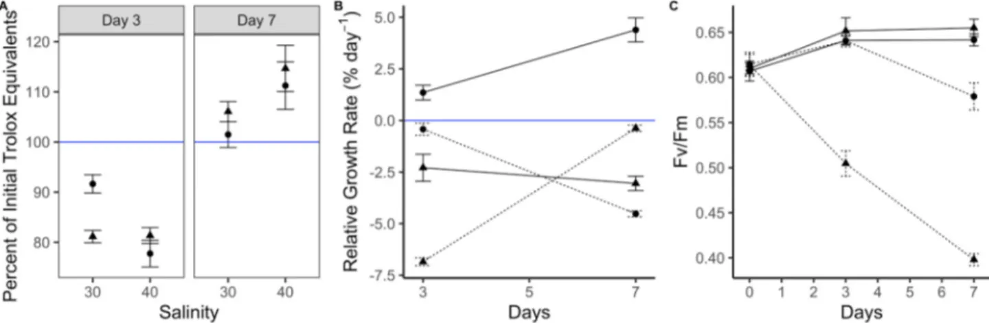
The free radical scavenging activity of A. vermiculophyllum ranged from 8.6 to 14.0 μmol TE g−1 DW. There was a significant interaction between exposure time, desiccation and salinity (ART: F = 5.8, p = 0.0054) on the percent of initial Trolox equivalents (Fig. 1A). The free radical scavenging activity first decreased in all treatments within the first 3 days, and then increased in all treatments after 7 days of exposure, but they were highest in the 40 ppt salinity treatments after 7 days, regardless of whether or not the algae were exposed to desiccation. The high salinity treatment resulted in a 13% increase compared to the initial conditions.
There was a significant interactive effect of salinity, desiccation and time on the RGR (ART: F =52.3, p =8.9e−5). The macroalgae only grew in the 30 ppt salinity treatment without desiccation, and growth was higher after 7 days than after 3 days (Fig. 1B). In all other treatments, the macroalgae exhibited no growth or a negative growth rate.
There was a significant interactive effect of salinity and desiccation (F =48.7, p =0.00012) and salinity and time (ART: F =44.2, p = 0.00016) on the Fv/Fm. Desiccation severely decreased the Fv/Fm in the high salinity treatment, and this became more severe over time (Fig. 1C). Additionally, desiccation caused an immediate decrease in the Fv/Fm in the high salinity treatment, while the negative effect of high salinity was delayed in the treatment without desiccation. In contrast,
desiccation had no effect on Fv/Fm in the 30 ppt salinity treatment.
3.2. Effects of light intensity and UVR
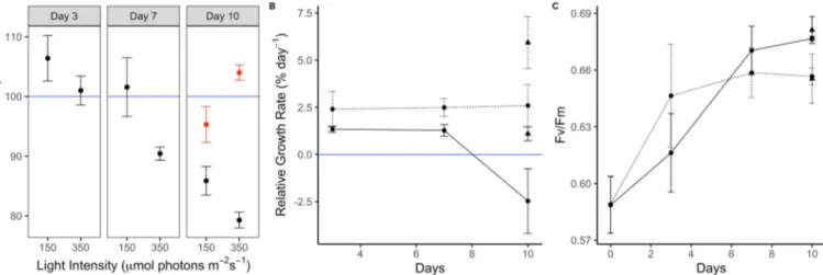
The free radical scavenging activity of A. vermiculophyllum ranged from 5.8 to 8.7 μmol TE g−1 DW. Within the first 7 days of exposure to the two light treatments, the percent of initial Trolox equivalents was significantly higher in the algae exposed to 150 μmol photons m−2 s−1 than those exposed to 350 μmol photons m−2 s−1 (ART: F =22.4, p = 0.00049, Fig. 2A) and the values generally decreased over time (ART: F
=4.8, p =0.029). Therefore, high light intensity in the absence of UVR did not result in increased free radical scavenging activity. After UVR was added, there was a significant interaction between light intensity and UVR (ART: F =17.0, p =0.0033). UVR exposure combined with high light intensity resulted in an 8% increase in free radical scavenging activity compared to the low light intensity with UVR exposure (but only 4% higher than the initial activity). In the absence of UVR exposure, the algae exposed to high light intensity had the lowest free radical scav- enging activity (Fig. 2A).
Neither light intensity nor time of exposure had a significant effect on RGR after seven days of exposure. After the addition of UVR for three days, there was a significant effect of both light intensity (ART: F =23.3, p =0.00041) and UVR (ART: F =8.7, p =0.012). The RGR was highest in the algae exposed to high light and UVR, while it was lowest in the algae exposed to low light with no UVR (Fig. 2B). In both light treat- ments, the exposure to UVR increased the RGR.
There was a significant treatment effect of light intensity on Fv/Fm
after 10 days of exposure (ART: F =5.4, p =0.039), such that Fv/Fm was higher at 150 μmol photons m−2 s−1 than at 350 μmol photons m−2 s−1 (Fig. 2C), but no significant effect of UVR (ART: F =0.15, p =0.70).
3.3. Effects of PAR dose and UVR
The free radical scavenging activity of A. vermiculophyllum ranged from 5.3 to 9.3 μmol TE g−1 DW. The macroalgae exposed to the high light intensity had significantly higher free radical scavenging activity than the algae exposed to the low light intensity (ART: F =113.3, p = 1.6e−8) on both day 4 and day 7, and this was 32% higher than the initial activity (Fig. 3A). There was a significant interactive effect between light intensity and UVR on antioxidant activity (ART: F =5.4, p =0.049, Fig. 3A). In the absence of UVR, the antioxidant activity decreased in the low light treatment, but when UVR was applied, the antioxidant activity returned to initial levels. In comparison, UVR had no effect on
Fig. 1.The response of A. vermiculophyllum to salinity and desiccation. A) The free radical scavenging activity (expressed as mean ±SE percent of initial, n =5) of A. vermiculophyllum after 3 and 7 days of exposure to 30 and 40 ppt salinity with (triangles) and without (circles) desiccation B) the mean (±SE, n =5) relative growth rate and C) mean (±SE, n =5) effective quantum yield of photosystem II (Fv/Fm) of A. vermiculophyllum after 3 and 7 days of exposure to 30 (solid lines) and 40 (dotted lines) ppt salinity with (triangles) and without (circles) desiccation. The solid blue line in A represents 100%, such that values above that line indicate an increase in antioxidant activity compared to the initial values. The solid blue line in B represents 0 growth, such that values above that line indicate positive growth, and below indicate negative growth.
antioxidant activity in the high light treatment.
There were no significant treatment effects of light intensity or UVR on RGR (Fig. 3B). There was a significant interactive effect of light in- tensity and time (ART: F =10.4, p =0.032) on RGR, such that growth rates decreased over time in the high light treatment, and increased slightly in the low light treatment. There was a significant main effect of light intensity on Fv/Fm (ART: F =21.6, p =0.0097, Fig. 3C), such that the Fv/Fm was higher in algae exposed to 100 μmol photons m−2 s−1, despite the fact that they received the same PAR dose (Fig. 3C). In contrast to the antioxidant activity, the significant interaction between UVR and light intensity (ART: F =12.2, p =0.0036) showed no effect of UVR on Fv/Fm in the low light treatment. In the high light treatment during 24-hour exposure, the Fv/Fm was significantly reduced in the absence of UVR, while the addition of UVR maintained Fv/Fm values
similar to those in the algae exposed to the lower PAR dose without UVR (Fig. 3C).
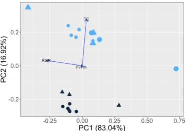
A Principle Component Analysis of antioxidant activity, growth rates, and Fv/Fm explained 91% of the variance in the first two com- ponents (Fig. 4). The first axis separated the algae exposed to low versus high light dose based on different growth rates and Fv/Fm. The second axis separated the algae exposed to different light intensities based on different antioxidant activity, and to a small extent Fv/Fm.
The effect of UVR on antioxidant activity differed depending on light intensity and dose. At low to moderate light intensities (100–350 μmol photons m−2 s−1) and dose (8–20 mol photons m−2 day−1), UVR had a significant impact on antioxidant activity, such that algae exposed to UVR had higher antioxidant activities than those not exposed to UVR (Fig. 5). On the other hand, algae exposed to high light intensity (400 Fig. 2.The response of A. vermiculophyllum to different irradiances. A) free radical scavenging activity (expressed as mean percent of initial, n =3) of A. ver- miculophyllum after 3, 7 and 10 days of exposure to low (150) and high (350) light intensity (μmol photons m−2 s−1) and after the addition of a 3-day UVR treatment (red symbols) on the 7th day B) mean (±SE, n =5) relative growth rate and C) mean (±SE, n =5) effective quantum yield of photosystem II (Fv/Fm) of A. ver- miculophyllum after 3, 7 and 10 days of exposure to low (150; solid lines) and high (350; dotted lines) light intensity (μmol photons m−2 s−1) and after the addition of a 3-day UVR treatment (triangles) on the 7th day. The solid blue line in A represents 100%, such that values above that line indicate an increase in antioxidant activity compared to the initial values. The solid blue line in B represents 0 growth, such that values above that line indicate positive growth, and values below it indicate negative growth.
Fig. 3. The response of A. vermiculophyllum to PAR dose versus light intensity. A) Mean (±SE, n =3) percent of initial Trolox equivalents after 4 days of exposure (left panel) to low (100) and high (400) light intensities, but the same PAR dose (8.6 mol photons m−2 day−1). A high PAR dose treatment (400 μmol photons m−2 s−1 for 24 h; 34.6 mol photons m−2 day−1; triangles) was added after 4 days and the effect of UVR (red symbols) for 3 days (right panel) on the percent of initial Trolox equivalents is shown. B) Mean (±SE, n =5) relative growth rates and C) mean (±SE, n =5) effective quantum yield of photosystem II (Fv/Fm) after exposure to low (100; solid lines) and high (400; dotted line) light intensities, but the same PAR dose (8.6 mol photons m−2 day−1). A high PAR dose treatment (400 μmol photons m−2 s−1 for 24 h; 34.6 mol photons m−2 day−1; triangles) and UVR (red symbols) were added after 4 days. The solid blue line in A represents 100%, such that values above that line indicate an increase in antioxidant activity compared to the initial values. The solid blue line in B represents 0 growth, such that values above that line indicate positive growth, and values below it indicate negative growth.
μmol photons m−2 s−1) at both low and high light doses had the highest antioxidant activity, and UVR did not have a significant effect.
4. Discussion
The free radical scavenging activity of A. vermiculophyllum in our study ranged from 5.3 to 14.0 μmol TE g−1 DW, which corresponds to those reported in previous studies for Gracilaria spp. and other macro- algae species [34,35]. In fact, these values are very similar to the range of antioxidant activities reported for seven macroalgal species from Spain, the highest of which (Hydropuntia cornea) was approximately 14.5 μmol TE g−1 DW [35].
Our results demonstrate that the free radical scavenging activity of A. vermiculophyllum can be increased after short-term exposure to environmental stressors such as salinity and high light intensity, but that light intensity was more important for increasing the free radical scav- enging activity than PAR dose or time of exposure. Exposure to high salinity for one week (but not 3 days) resulted in a 13% increase, high
light intensity on a long day cycle for seven days combined with a three- day UVR treatment resulted in a 4% increase, and high light intensity on a short day (6 h) cycle for 4–7 days resulted in a 32% increase in free radical scavenging activity. Therefore, short-term exposure to high light intensity on a short-day cycle was the most efficient way to increase free radical scavenging activity in A. vermiculophyllum. Exposure to high light intensity for short days induced higher antioxidant activity than low light intensity for long days, even when the PAR dose was the same, suggesting that high light intensity, rather than the total amount of photons absorbed over time, triggers a response in antioxidant activity.
This increase in antioxidant activity corresponded to a decrease in photosynthetic efficiency, but growth rates were unaffected by light intensity and remained the same when the algae received the same PAR dose. A similar trend in growth rates of A. vermiculophyllum in the Baltic Sea was reported by [29]. While few studies have investigated the different effects of PAR dose versus PAR intensity, this trend comple- ments the fact that higher rates of productivity have been found in macroalgae exposed to lower photon flux densities over a longer period [39], perhaps because the macroalgae invest more energy into photo- protection at high light intensities. Nevertheless, it is unclear why we observed higher antioxidant activity at 150 compared to 350 μmol photons m−2 s−1 in one experiment. The initial Fv/Fm was slightly lower in these algae compared to those that were exposed to 100 and 400 μmol photons m−2 s−1, suggesting that the photosynthetic efficiency and the general physiological condition of the algae differed slightly at the start of the two experiments, perhaps due to seasonal differences. It has been shown that antioxidant activity in macroalgae follows seasonal patterns [22,23], but we did not observe differences in the initial antioxidant activity based on ABTS•+of the algae before each experiment.
In addition to light intensity, light quality also affects antioxidant activity in macroalgae. Ulvan extracted from Ulva pertusa had signifi- cantly higher antioxidant activity when the algae were grown under blue light compared to white light [40]. Additionally, Ulva sp. grown under blue light had higher ABTS•+radical scavenging activity when grown under blue light compared to white light, regardless of day length [41]. Therefore, it seems both intensity, duration, and quality are important parameters that influence antioxidant activity in macroalgae, and further research should investigate whether high intensity blue light could further enhance the antioxidant activity of A. vermiculophyllum.
In a study where stocking density of Derbesia tenuissima was manip- ulated to test the effects of light availability on antioxidant activity, the authors found that stocking density, and thus, light intensity, had a significant impact on the total phenol content, DPPH radical scavenging ability, and ferric reducing antioxidant capacity, but not ABTS•+radical scavenging activity [30]. They also showed that photosynthetic effi- ciency decreased with increasing antioxidant production, which we also observed in A. vermiculophyllum. The authors suggest monitoring the Fv/ Fm to manage culture conditions for optimizing antioxidant activity in land-based macroalgal culture. The highest increase in antioxidant ac- tivity we observed (32%) corresponded to an 11.5% reduction in Fv/Fm
values (88.5% of the highest Fv/Fm values). Similarly, Magnusson et al.
[30] found that an increase in total phenol content of 20% corresponded to a 15% reduction in Fv/Fm (85% of maximum Fv/Fm). Monitoring Fv/ Fm has also been proposed as a method to detect stress early in Gracilaria cornea cultivation [42]. Therefore, monitoring Fv/Fm can be used as a tool to maintain crop health during biomass production, as well as a management tool during the optimization of antioxidant activity.
UVR had a strong effect on antioxidant activity at low light in- tensities, but not at high light intensity (>350 μmol photons m−2 s−1). In contrast to antioxidant activity, growth rates and photosynthetic effi- ciency were not significantly affected by UVR. The UVR intensity used in our study was quite low compared to studies that have investigated UVR effects on macroalgae [32,43,44], but the time of exposure was longer (up to 16 h day−1) in our study, and clearly had an effect on antioxidant activity. It would be necessary to increase the UVR in future studies to determine if the antioxidant activity could be increased even further.
Fig. 4. Principle Component Analysis of the response variables measured (Trolox equivalents, relative growth rate, and Fv/Fm). The environmental factors light intensity (100: dark blue, 400: light blue), light dose (8.64: small symbols, 34.6: large symbols) and UVR (no UVR: circles, UVR: triangles) are used to help visualize the variance in the data. The percent of the variance explained by each principle component are shown on each axis label in parenthesis.
Fig. 5. The effect of light dose on antioxidant activity. Mean antioxidant ac- tivity (±SD, n =3) of A. vermiculophyllum expressed as Trolox equivalents as a function of light dose. The effect of UVR exposure (red symbols) and light in- tensity (circles =100, triangles =150, squares =350, cross =400) are shown.
In conclusion, we showed that a four-day exposure period to high light intensity for short days (6 h) showed the highest increase in free radical scavenging activity in A. vermiculophyllum. Hypersalinity also increased free radical scavenging activity, but to a lesser extent. We therefore suggest an optimization period of 4–7 days exposure to high light on a 6:18 h light:dark cycle prior to harvesting in order to increase antioxidant activity. More research is required to investigate if higher light intensities or UVR could further increase antioxidant activity in A. vermiculophyllum, but here we provide a good starting point for increasing the functionality of A. vermiculophyllum in diverse industrial applications, including the potential extension of shelf-life for food products.
Funding
The project “Mak-Pak” is supported by funds of the Federal Ministry of Food and Agriculture (BMEL) based on a decision of the Parliament of the Federal Republic of Germany via the Federal Office for Agriculture and Food (BLE) under the innovation support programme (Project Number 28-1-A1.049-16).
CRediT authorship contribution statement
LCH & ST designed the study and drafted the manuscript, ST carried
out the study and collected the data, LCH & ST analyzed the data, JS provided technical support, BB & JH provided lab facilities and administrative support, RB, FR, IE, DH, JH and BB obtained funding for this project and revised and approved the manuscript.
Declaration of competing interest
The authors declare that they have no known competing financial interests or personal relationships that could have appeared to influence the work reported in this paper.
Acknowledgements
The authors would like to thank the team at the Center for Aqua- culture Research (ZAF), Dr. Inka Bartsch, Claudia Daniel and Andreas Wagner for laboratory supplies and technical support, as well as Dr. Inka Bartsch and Dr. Markus Molis for providing access to the climate room where the experiments were conducted. Finally we thank the two anonymous reviewers for improving the manuscript.
Appendix A. Supplementary data
Supplementary data to this article can be found online at https://doi.
org/10.1016/j.algal.2021.102232.
References
[1] R. Geyer, J.R. Jambeck, K.L. Law, Production, use, and fate of all plastics ever made, Sci, Adv. (2017) 25–29, https://doi.org/10.1126/sciadv.1700782.
[2] Plastic Europe, Plastics – the facts 2018: an analyses of European plastics production, demand and waste data. https://www.plasticseurope.org/application/
files/6315/4510/9658/Plastics_the_facts_2018_AF_web.pdf, 2018.
[3] D.K.A. Barnes, F. Galgani, R.C. Thompson, M. Barlaz, Accumulation and fragmentation of plastic debris in global environments, Philos, Trans. R. Soc. B Biol. Sci. (2009) 1985–1998, https://doi.org/10.1098/rstb.2008.0205.
[4] J.C. Prata, Airborne microplastics: consequences to human health? Environ. Pollut.
234 (2018) 115–126, https://doi.org/10.1016/j.envpol.2017.11.043.
[5] T. Efferth, N.W. Paul, Threats to human health by great ocean garbage patches, Lancet Planet. Heal. 1 (2017) e301–e303, https://doi.org/10.1016/s2542-5196 (17)30140-7.
[6] B.B. Sedayu, M.J. Cran, S.W. Bigger, A review of property enhancement techniques for carrageenan-based films and coatings, Carbohydr. Polym. 216 (2019) 287–302, https://doi.org/10.1016/j.carbpol.2019.04.021.
[7] B.B. Sedayu, M.J. Cran, S.W. Bigger, Characterization of semi-refined carrageenan- based film for primary food packaging purposes, J. Polym. Environ. 26 (2018) 3754–3761, https://doi.org/10.1007/s10924-018-1255-y.
[8] H.P.S. Abdul Khalil, C.K. Saurabh, Y.Y. Tye, T.K. Lai, A.M. Easa, E. Rosamah, M.R.
N. Fazita, M.I. Syakir, A.S. Adnan, H.M. Fizree, N.A.S. Aprilia, A. Banerjee, Seaweed based sustainable films and composites for food and pharmaceutical applications: a review, Renew. Sustain. Energy Rev. 77 (2017) 353–362, https://
doi.org/10.1016/j.rser.2017.04.025.
[9] J. G´omez-Estaca, C. L´opez-de-Dicastillo, P. Hern´andez-Munoz, R. Catal˜ ´a, R. Gavara, Advances in antioxidant active food packaging, Trends Food Sci.
Technol. 35 (2014) 42–51, https://doi.org/10.1016/j.tifs.2013.10.008.
[10] A. Farhan, N.M. Hani, Characterization of edible packaging films based on semi- refined kappa-carrageenan plasticized with glycerol and sorbitol, Food Hydrocoll.
(2017), https://doi.org/10.1016/j.foodhyd.2016.10.034.
[11] R. Jumaidin, M. Sapuan Salit, M. Jawaid, M. Ridzwan Ishak, J. Sahari, Seaweeds as renewable sources for biopolymers and its composites: a review, Curr. Anal. Chem.
(2017), https://doi.org/10.2174/1573411013666171009164355.
[12] W.M. Siah, A. Aminah, A. Ishak, Edible films from seaweed (Kappaphycus alvarezii), Int. Food Res. J. 22 (2015) 2236.
[13] K. Bischof, R. Rautenberger, Seaweed responses to environmental stress: reactive oxygen and antioxidative strategies, in: Seaweed Biol., Springer, Berlin, Heidelberg, 2012: pp. 109–132. doi:https://doi.org/10.1007/978-3-642-28451- 9_6.
[14] M.L. Cornish, D.J. Garbary, Antioxidants from macroalgae: potential applications in human health and nutrition, ALGAE. 25 (2011) 155–171, https://doi.org/
10.4490/algae.2010.25.4.155.
[15] M. Kendel, G. Wielgosz-Collin, S. Bertrand, C. Roussakis, N. Bourgougnon, G. Bedoux, Lipid composition, fatty acids and sterols in the seaweeds Ulva armoricana, and Solieria chordalis from Brittany (France): an analysis from nutritional, chemotaxonomic, and antiproliferative activity perspectives, Mar.
Drugs 13 (2015) 5606–5628, https://doi.org/10.3390/md13095606.
[16] M. Barot, N. Kumar JI, R.N. Kumar, Bioactive compounds and antifungal activity of three different seaweed species Ulva lactuca, Sargassum tenerrimum and Laurencia obtusa collected from Okha coast, Western India, J. Coast. Life Med. 4 (2016) 284–289, https://doi.org/10.12980/jclm.4.2016J5-185.
[17] P. Sampath-Wiley, C.D. Neefus, L.S. Jahnke, Seasonal effects of sun exposure and emersion on intertidal seaweed physiology: fluctuations in antioxidant contents, photosynthetic pigments and photosynthetic efficiency in the red alga Porphyra umbilicalis Kützing (Rhodophyta, Bangiales), J. Exp. Mar. Bio. Ecol. 361 (2008) 83–91, https://doi.org/10.1016/j.jembe.2008.05.001.
[18] M. Kumar, P. Kumari, C.R.K. Reddy, B. Jha, Salinity and desiccation induced oxidative stress acclimation in seaweeds, in: N. Bourgougnon (Ed.), Adv. Bot. Res., Academic Press, 2014, pp. 91–123, https://doi.org/10.1016/B978-0-12-408062- 1.00004-4.
[19] J. Coll´en, I.R. Davison, Reactive oxygen metabolism in intertidal Fucus spp.
(Phaeophyceae), J. Phycol. 35 (1999) 62–69.
[20] J. Coll´en, I.R. Davison, Reactive oxygen production and damage in intertidal Fucus spp. (Phaeophyceae), J. Phycol. (1999), https://doi.org/10.1046/j.1529- 8817.1999.3510054.x.
[21] J. Coll´en, I.R. Davison, Reactive oxygen metabolism in intertidal Fucus spp.
(Phaeophyceae), J. Phycol. (1999), https://doi.org/10.1046/j.1529- 8817.1999.3510062.x.
[22] J. Coll´en, I.R. Davison, Seasonality and thermal acclimation of reactive oxygen metabolism in Fucus vesiculosus (Phaeophyceae), J. Phycol. (2001), https://doi.
org/10.1046/j.1529-8817.2001.037004474.x.
[23] N.L. Lohrmann, B.A. Logan, A.S. Johnson, Seasonal acclimatization of antioxidants and photosynthesis in Chondrus crispus and Mastocarpus stellatus, two co-occurring red algae with differing stress tolerances, Biol. Bull. (2004), https://doi.org/
10.2307/1543211.
[24] J. Coll´en, I.R. Davison, Stress tolerance and reactive oxygen metabolism in the intertidal red seaweeds Mastocarpus stellatus and Chondrus crispus, Plant, Cell Environ. (1999), https://doi.org/10.1046/j.1365-3040.1999.00477.x.
[25] A.M.M. Sousa, A.M. Sereno, L. Hilliou, M.P. Gonçalves, Biodegradable agar extracted from Gracilaria vermiculophylla: film properties and application to edible coating, Mater. Sci. Forum 636–637 (2010) 739–744, https://doi.org/10.4028/
www.scientific.net/MSF.636-637.739.
[26] J. Reboieira, R. Ganh˜ao, S. Mendes, P. Ad˜ao, M. Andrade, F. Vilarinho, A. Sanches- Silva, D. Sousa, A. Mateus, S. Bernardino, Optimization of extraction conditions for Gracilaria gracilis extracts and their antioxidative stability as part of microfiber food coating additives, Molecules. 25 (2020) 4060.
[27] L.B. Nejrup, P.A. Staehr, M.S. Thomsen, Temperature- and light-dependent growth and metabolism of the invasive red algae Gracilaria vermiculophylla - a comparison with two native macroalgae, Eur. J. Phycol. 48 (2013) 295–308, https://doi.org/
10.1080/09670262.2013.830778.
[28] M.S. Thomsen, P.A. Staehr, C.D. Nyberg, S. Schwærter, D. Krause-Jensen, B.
R. Silliman, Gracilaria vermiculophylla (Ohmi) Papenfuss, 1967 (Rhodophyta, Gracilariaceae) in northern Europe, with emphasis on Danish conditions, and what to expect in the future, Aquat, Invasions. 2 (2007) 83–94, https://doi.org/
10.3391/ai.2007.2.2.1.
[29] F. Weinberger, B. Buchholz, R. Karez, M. Wahl, The invasive red alga Gracilaria vermiculophylla in the Baltic Sea: adaptation to brackish water may compensate for light limitation, Aquat. Biol. 3 (2008) 251–264, https://doi.org/10.3354/ab00083.
[30] M. Magnusson, L. Mata, N. Wang, J. Zhao, R. de Nys, N.A. Paul, Manipulating antioxidant content in macroalgae in intensive land-based cultivation systems for functional food applications, Algal Res. (2015), https://doi.org/10.1016/j.
algal.2015.02.007.
[31] J.K. Kim, C. Yarish, Development of a sustainable land-based Gracilaria cultivation system, Algae. 29 (2014) 217–225. doi:https://doi.org/10.4490/algae.2014 .29.3.217.
[32] D. Hanelt, C. Wiencke, W. Nultsch, Influence of UV radiation on the photosynthesis of Arctic macroalgae in the field, J. Photochem. Photobiol. B Biol. 38 (1997) 40–47, https://doi.org/10.1016/S1011-1344(96)07415-5.
[33] F.L. Figueroa, A. Israel, A. Neori, B. Martínez, E.J. Malta, A. Put, S. Inken, R. Marquardt, R. Abdala, N. Korbee, Effect of nutrient supply on photosynthesis and pigmentation to short-term stress (UV radiation) in Gracilaria conferta (Rhodophyta), Mar. Pollut. Bull. 60 (2010) 1768–1778, https://doi.org/10.1016/j.
marpolbul.2010.06.009.
[34] K. Vijayavel, J.A. Martinez, In vitro antioxidant and antimicrobial activities of two Hawaiian marine limu: Ulva fasciata (Chlorophyta) and Gracilaria salicornia (Rhodophyta), J. Med. Food 13 (2010) 1494–1499, https://doi.org/10.1089/
jmf.2009.0287.
[35] F. Alvarez-Gomez, N. Korbee, F.L. Figueroa, Analysis of antioxidant capacity and bioactive compounds in marine macroalgal and lichenic extracts using different solvents and evaluation methods, Ciencias Mar. 42 (2016) 271–288, https://doi.
org/10.7773/cm.v42i4.2677.
[36] A. Jim´enez-Escrig, E. G´omez-Ord´o˜nez, P. Rup´erez, Brown and red seaweeds as potential sources of antioxidant nutraceuticals, J. Appl. Phycol. 24 (2012) 1123–1132, https://doi.org/10.1007/s10811-011-9742-8.
[37] R. Re, N. Pellegrini, A. Proteggente, A. Pannala, M. Yang, C. Rice-Evans, Antioxidant activity applying an improved ABTS radical cation decolorization assay, Free Radic. Biol. Med. (1999), https://doi.org/10.1016/S0891-5849(98) 00315-3.
[38] P. Torres, A. Nagai, D.I.A. Teixeira, E. Marinho-Soriano, F. Chow, D.Y.A.C. dos Santos, Brazilian native species of Gracilaria (Gracilariales, Rhodophyta) as a source of valuable compounds and as nutritional supplements, J. Appl. Phycol.
(2019), https://doi.org/10.1007/s10811-019-01804-x.
[39] M.J. Desmond, D.W. Pritchard, C.D. Hepburn, Light dose versus rate of delivery:
implications for macroalgal productivty, Photosynth. Res. 132 (2017) 257–264.
[40] B. Le, J.-A. Shin, M.-G. Kang, S. Sun, S.H. Yang, G. Chung, Enhanced growth rate and ulvan yield of Ulva pertusa using light-emitting diodes (LEDs), Aquac. Int. 26 (2018) 937–946. doi:https://doi.org/10.1007/s10499-018-0260-4.
[41] J. Schwoerbel, Reproduction, Growth and Chemical Composition of Ulva sp. in Response to Different Light Treatments, University of Bremen, 2019.
[42] F.L. Figueroa, R. Santos, R. Conde-Alvarez, L. Mata, J.L. G´ ´omez Pinchetti, J. Matos, P. Huovinen, A. Schuenhoff, J. Silva, The use of chlorophyll fluorescence for monitoring photosynthetic condition of two tank-cultivated red macroalgae using fishpond effluents, Bot. Mar. 49 (2006) 275–282, https://doi.org/10.1515/
BOT.2006.035.
[43] K. Bischof, D. Hanelt, C. Wiencke, Effects of ultraviolet radiation on photosynthesis and related enzyme reactions of marine macroalgae, Planta. 211 (2000) 555–562, https://doi.org/10.1007/s004250000313.
[44] K. Bischof, I. G´omez, M. Molis, D. Hanelt, U. Karsten, U. Lüder, M.Y. Roleda, K. Zacher, C. Wiencke, Ultraviolet radiation shapes seaweed communities, Rev.
Environ. Sci. Biotechnol. 5 (2006) 141–166, https://doi.org/10.1007/s11157-006- 0002-3.