Regulation of cellular dormancy in disseminated breast cancer cells
Dissertation
zur Erlangung des Doktorgrades der Naturwissenschaften (Dr. rer. nat.) der Fakultät für Biologie und Vorklinische Medizin der
Universität Regensburg
vorgelegt von
Ana Grujovic
aus Kragujevac, Serbien 2019
Das Promotionsgesuch wurde eingereicht am 29.04.2019
Die Arbeit wurde angeleitet von Herr Prof. Dr. Christoph A. Klein Untershrift:
Table of Contents
1 Introduction ... 1
1.1 Mammary gland development and cellular organization ... 1
1.2 Stem cell hierarchy during mammary gland development... 3
1.3 Breast cancer ... 6
1.4 Metastasis ... 7
1.5 Disseminated cancer cells ... 9
1.6 Dormancy of disseminated cancer cells ... 10
1.7 Interleukin-6 signaling in physiological and pathological settings ... 11
2 Material and Methods ... 14
2.1 Methods... 14
2.1.1 Patient material ... 14
2.1.2 Animals ... 15
2.1.3 Cell culture experiments ... 19
2.1.4 Microvascular niche ... 21
2.1.5 Osteoblastic niche ... 24
2.1.6 Staining procedures... 25
2.1.7 T cell experiment ... 26
2.1.8 Whole transcriptome amplification and PCR ... 27
2.1.9 Gene expression analysis using micro array ... 30
2.1.10 Statistical analysis ... 30
2.2 Material ... 30
2.2.1 Reagents... 30
2.2.2 Consumables ... 33
2.2.3 Kits ... 33
2.2.4 Devices ... 34
3 Results ... 35
3.1 Overview of the research rationale ... 35
3.2 Balb-NeuT as an in vivo model of tumor cell dormancy ... 36
3.2.1 Short background information of previous work with the Balb-NeuT model as the starting point of the experiments ... 36
3.2.2 Search for good candidate marker for isolation of BM-DCCs ... 39
3.2.3 Improvement of detection and isolation of DCCs from the bone marrow ... 41
3.2.4 CD45 expression enables discrimination between DCCs and EpCAM-positive hematopoietic cells ... 41
3.2.5 Effect of progesterone on dissemination of cancer cells in the Balb-NeuT mice ... 43
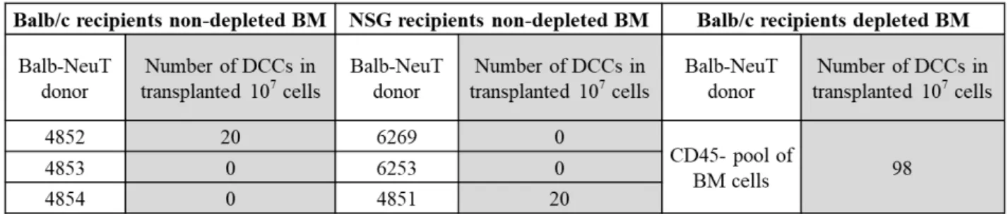
3.2.6 Bone marrow transplantation induces DCC outgrowth ... 43
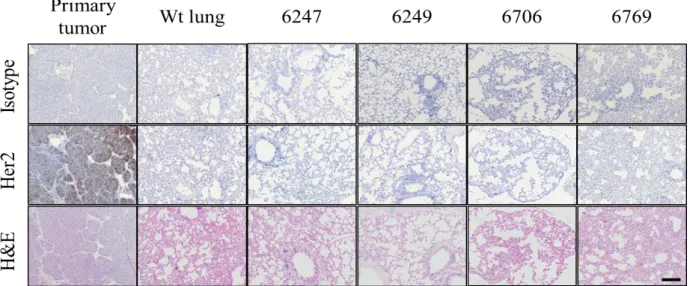
3.2.7 Analysis of DCCs in the lungs of BM recipient mice ... 45
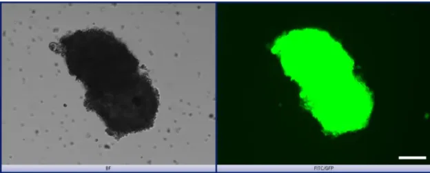
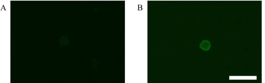
3.2.8 In vitro expansion of the mouse DCCs isolated from bone marrow ... 46
3.2.9 Effect of Collagen I on sphere forming ability of mammary cells ... 47
3.3 Establishment of the in vitro models of tumor cell dormancy ... 49
3.3.1 Implementation of the “stem cell model” for isolating dormant and proliferative epithelial cells from human mammary gland ... 49
3.3.2 Overview of samples derived from mammary reduction surgeries ... 49
3.3.3 Optimization of the cell labeling procedure ... 51
3.3.4 Density of cell seeding does not affect the frequency of sphere formation of HMECs ... 53
3.3.5 Separation of single cells from mammospheres ... 55
3.3.6 Dormant cells reside among non-divided single cells after 7 days ... 56
3.3.7 CD44 and CD24 expression in LRCs and nLRCs ... 57
3.3.8 Isolation of LRCs and nLRCs from mammosphere culture ... 58
3.3.9 Transcriptional analysis of SCs isolated from mammary gland ... 59
3.4 The effect of the vascular niche on growth of breast cancer cells ... 60
3.4.1 Microvascular niche does not affect the growth of breast cancer cells ... 60
3.4.2 Non-transformed mammary cells neither grow in the stromal nor in the microvascular niche ... 62
3.4.3 Mammary cells isolated from MVN and MSC cultures provide low quality WTA products ... 63
3.5 The effect of the osteoblastic niche on breast mammary cell growth ... 64
3.6 Analysis of the proliferation in single mammary cells ... 66
3.6.1 Establishment of proliferation marker analysis for single cells classification ... 66
3.6.2 Validation of the established proliferation markers ... 68
3.7 Determination of the proliferation status of cells isolated from dormancy models ... 70
3.7.1 Proliferation analysis of single cells isolated from stromal and vascular niches ... 70
3.7.2 Proliferation analysis of QSCs, LRCs, and nLRCs isolated from the stem cell model ... 71
3.8 Profiling the state of cellular dormancy ... 72
3.8.1 Gene expression profiling discriminates 5 groups within HMECs ... 73
3.8.2 QSCs, LRCs, and nLRCs belong to the populations of distinct differentiation states in mammary gland .. 74
3.8.3 Pathway analysis determines signaling profiles of defined groups ... 75
3.8.4 IL-6 signaling in patient derived MC-DCCs ... 76
3.9 IL-6 signaling in the mammary gland ... 78
3.9.1 MCF10A cells express IL-6 and IL-6Ra ... 78
3.9.2 IL-6 signaling regulates the frequency of stem-like MCF10A cells ... 81
3.9.3 Expression of IL-6 and IL-6Ra in stem-like and progenitor cells from MCF10A-spheres ... 82
3.9.4 Effect of IL-6 classical- and trans-signaling on proliferation and survival of cells ... 84
3.9.5 Signaling pathway analysis in MCF10A cells upon stimulation of IL-6 signaling ... 86
3.9.6 IL-6 trans-signaling increases sphere formation of HMECs ... 87
3.9.7 Expression of IL-6 and IL-6Ra in LRCs and nLRCs from HMEC-spheres ... 87
3.9.8 Expression of IL-6 and IL-6Ra in DCCs from breast cancer patients ... 88
3.9.9 Quantitative PCR analysis of gp130 in DCCs from patients with breast cancer ... 89
3.9.10 Regulation of the gp130 expression in DCCs by the bone marrow environment... 90
4 Discussion ... 94
4.1 Balb-NeuT as in vivo model of dormancy ... 95
4.2 Establishment of the in vitro models of dormancy ... 96
4.3 Studying dormancy and proliferation in BM-DCCs ... 96
4.3.1 Mammary stem cells ... 97
4.3.2 Signaling pathways in mammary stem cells ... 98
4.4 IL-6 signaling in the mammary gland ... 98
4.5 Cancer cell dormancy in the bone marrow niche ... 101
4.6 Conclusions ... 102
5 Summary ... 104
6 Literature ... 106
7 Acknowledgement ... 116
1
1 Introduction
1.1 Mammary gland development and cellular organization
Mammary gland is a very specific organ which distinguishes mammals from all other animals due to unique anatomical structure that enables production and secretion of the milk for the nourishment of the offspring. Adult human breast consists of the parenchyma, which originates from the ectoderm and the stroma, which develops from mesodermal elements (Figure 1). The parenchyma is a system of branching ducts and acini, while the stroma mainly consists of adipose tissue (Forsyth, 1991; Javed and Lteif, 2013; Medina, 1996). The mammary gland reaches its final development late after birth, during pregnancy (Inman et al., 2015; Macias and Hinck, 2012) which facilitates studying its developmental process. Most of the knowledge about embryonal development of the mammary gland arises from studies in rodents.
The stadium of the embryonal development comprises of two phases: (1) formation of the primary mammary bud and (2) development of a rudimentary mammary gland (Hughes, 1950; Inman et al., 2015). Once the mammary sprout reaches the fat pad, its further sprouting is initiated to form a rudimentary ductal tree. The ductal tree consists of epithelial and myoepithelial cells forming 15-20 secondary branches, each containing a lactiferous duct which finally leads to a nipple and enables collection and directing of the milk stream. At the time of birth, the nipple is formed from epidermal cells overlying the bud, while the rudimentary ductal tree protrudes into the surrounding fat pad. (Hens and Wysolmerski, 2005; Hogg et al., 1983; Macias and Hinck, 2012; Sakakura et al., 1976).
In the period from birth until the age of two, not many changes occur, mostly maturation of the ducts and sprouting of the vasculature. From the third year until puberty, the mammary gland remains quiescent and its development continues with the beginning of sex hormone secretion (Anbazhagan et al., 1991; Naccarato et al., 2000). Under their influence, extensive proliferation occurs which results in filling the fat pad and formation of mammary lobes. The mammary lobe comprises of the mammary
Figure 1: Anatomy of the human mammary gland. Each lactiferous duct in the mammary gland starts from the nipple and branches into ducts that end with the acini (in a non- lactating breast) or alveoli (in a lactating breast). Therefore the alveoli in a lactating breast occupy more space than in a non- lactating one, on the cost of adipose stromal tissue. Picture modified from https://theanatomybody.com/mammary- gland-anatomy/
2
ducts which end with the terminal end buds (TEB) (Hinck and Silberstein, 2005; Macias and Hinck, 2012; Paine and Lewis, 2017). Cap cells of the TEBs differentiate into myoepithelial cells, forming the outer layer, and the inner layer consist of the luminal cells (Williams and Daniel, 1983). During puberty, before pregnancy, the ends of the mammary tree are blind-ended ducts, called acini (Howard and Gusterson, 2000). These acini protrude in the fat pad, and consist of predominantly adipocytes, but also endothelial cells (which form a vascular net), fibroblasts and immune cells (Macias and Hinck, 2012;
Sheffield, 1988). At the time of puberty, estrogen secretion starts and together with the pituitary growth hormone and insulin-like growth factor-1 (IGF-1) initiates further events in the mammary gland development (Kleinberg and Ruan, 2008). Those processes involve formation of the breast bud with elevation of the nipple and enlargement of the diameter of the areola around the nipple (Marshall and Tanner, 1969). Constant increase in the size of the mammary gland during puberty is a result of changes in both, parenchymal and stromal tissue (Howard and Gusterson, 2000), with first preparation of the surrounding fibrous and fatty tissue which then enables further elongation and sprouting of the parenchymal mammary ducts (Howard and Gusterson, 2000; Russo and Russo, 2004)
Intensive growth and changing during puberty results in formation of the final structure of a double layered ducto-alveolar net in mammary parenchyma. The outer layer of cells consists of myoepithelial cells, responsible for the contraction and the guidance of milk that is produced by alveolar cells from inner layer. The alveolar cells form the inner layer together with luminal cells, lining the lumen of ducts and alveoli (Tiede and Kang, 2011; Watson and Khaled, 2008). During each menstrual cycle, upon a complex effect of hormones, the number and size of the alveoli increase, while significant increase is only seen when pregnancy occurs (Monaghan et al., 1990; Russo and Russo, 2004). All these processes are dictated mainly by the female sex hormones secreted by the ovaries, such as estrogen (inducing ductal elongation) and progesterone (responsible for side branching) (Brisken et al., 1998). Mammary ducts are branching to smaller ducts and further to very small ductuli ending with acini. All the acini arising from one terminal duct together with the surrounding stroma form the functional unit of the breast called the terminal duct lobular unit (TDLU) (Howard and Gusterson, 2000; Javed and Lteif, 2013). Again, most information about mammary growth during pregnancy and involution came from studies in mice. During pregnancy numerous changes occur in the mammary gland under the influence of progesterone and prolactin. Intensive branching of secondary and tertiary ducts is followed by alveolar development, when epithelial cells forming alveolar buds start proliferating and differentiating into milk-secreting lobules. The growth of mammary ducts and alveoli continues on a cost of stromal adipose tissue, which is reduced and disappears. Simultaneously the vascular net is branching and by late pregnancy every functional alveolus is surrounded by a capillary network (Baillo et al., 2011). A functionally mature gland is then fully developed and prepared for milk production and secretion.
The involution process starts with the weaning. At the weaning phase the produced milk is retained in the mammary epithelium, the branched mammary tree is removed and milk-producing epithelial cells
3
are disappearing. This process starts with a reversible phase of alveolar cells detachment, their accumulation in the lumen and apoptosis inducing engorgement of the alveoli. After this phase, the alveoli start to collapse, and milk supply is lost. As of that moment involution becomes an irreversible process. In this phase, tissue remodeling is most prominent while activation of matrix metalloproteinases (MMP) induces degradation of extracellular matrix (ECM) and an intensive apoptotic wave in mammary epithelial cells. As a result, the mammary gland is anatomically very similar to the original mammary gland before pregnancy, but interestingly the pattern of gene expression differs (Balogh et al., 2006; D'Cruz et al., 2002). The mammary development processes with all described phases is depicted in the Figure 2.
Figure 2: The stages of postnatal mammary gland development. At birth, the mammary epithelium is rudimentary, consisting of only a few small ducts. At the onset of puberty, expansive growth that fills the fat pad with the epithelial mammary tree happens. This growth is influenced by the growth hormones (GH), estrogen, and insulin-like growth factor-1 (IGF1). When pregnancy begins, sprouting and further branching of the existing branches continues and alveologenesis occurs under the influence of progesterone and prolactin. Prolactin stimulation continues during the stage of lactogenesis, culminating in milk production that continues until involution, the process of mammary gland remodeling back to its original adult state before pregnancy. Picture modified from (Macias and Hinck, 2012).
1.2 Stem cell hierarchy during mammary gland development
All the changes that the mammary gland undergoes during maturation and during the described cyclic differentiation process, are based on the function of undifferentiated stem cells (Stingl et al., 2006). In a first step division and differentiation of the mammary stem cells give rise to self-renewed stem cells and mammary progenitor cells. These progenitor cells continue to divide and further differentiate into matured mammary cells, while self-renewed stem cells persist and wait for another activation signal to divide again. Hence, in the parenchyma of the mammary gland, among undifferentiated stem and differentiated mature cells also progenitors reside, which are in between these two populations in regard of their differentiation status.
Early gland development is based on the expansion of the bipotent fetal mammary stem cells (fMaSCs) (Spike et al., 2012). Repeated cycles of expansion and involution of mammary glands during menstrual cycles and pregnancies clearly indicate the existence of the adult counterparts to fMaSC that are able to
4
expand and give the progeny of different lineages. These cells named adult mammary stem cells (aMaSCs) are much less abundant than fMaSC, with 1/50 and 1/14 cells in corresponding tissues respectively (Spike et al., 2012). These cells are slow cycling cells waiting for internal and external signals to activate them, renew and give the progeny instructions to develop all the changes in the adult gland (Guo, 2014; Pasic et al., 2011; Zeng and Nusse, 2010). A hierarchy of mammary cells is maintained in the adult mammary gland which, beside stem cells comprise of progenitors and differentiated mammary cells.
Thus, the mammary gland is a hierarchically structured organ (Stingl et al., 2006). The proof of pluripotency of adult mammary stem cells (MaSC) was provided in the work of Shakelton and colleagues who succeeded to reconstitute the whole mouse mammary gland upon transplantation of a single cell (Shackleton et al., 2006). Transplantation of pieces of a human mammary gland resulted in engraftment and mammary gland reconstitution in immunodeficient mice (Kuperwasser et al., 2004).
All different processes during mammary gland development are strictly controlled by growth factors and hormones and disruption of that signaling composition can lead to increased growth of the mammary gland and breast cancer (Martin, 2003). In line with the evidence that changes in the mammary gland are driven by growth and division of mammary stem cells, a corresponding subpopulation of mammary cancer stem cells (MaCSC) are believed to initiate breast cancer (Luo et al., 2015; Morrison et al., 2008).
In line with the stem cell theory that proposes that all cells in the mammary gland originate from a small proportion of stem cells, cancer stem cell theory proposes that among all cancer cell pool only a few of them act as so-called cancer stem cells (CSC), which reproduce themselves and sustain the cancer, much like normal stem cells renew and sustain organs and tissues (Yoo and Hatfield, 2008). New data, showing that the majority of malignant cells, rather than only CSCs can sustain tumors questions the stem cell theory, emphasizing it must be reevaluated. (Yoo and Hatfield, 2008). In accordance with previously stated, albeit still under controversy, nowadays both stem and progenitor cells are candidate cells-of-origin in tumorigenesis (Chen et al., 2017). Besides the theories that aberrations in less differentiated (stem and progenitors) cells cause cancer, some clues indicate that also differentiated cells, which sustain neoplastic transformation can acquire properties of less differentiated stem or progenitors and become the founders of malignancies (Bjerkvig et al., 2005). Cancer cells that leave the primary site and home to distant organs can i) undergo cell death; ii) proliferate and give rise to metastasis or iii) lodge and stay quiescent for a certain period (Aguirre-Ghiso, 2007; Goss and Chambers, 2010; Reymond et al., 2013). Such outcome we exactly see in our stem cell model where majority of cells undergo anoikis, few of them start immediate proliferation and give rise to mammospheres and rest stay quiescent but retain the potential to be reactivated. Some data show that all the cells that extravasate to distant organs first undergo the same state of dormancy (Paez et al., 2012) or proliferation (Wang et al., 2015a). The nature of the cells that acquire the ability to disseminate
5
from the primary tumor is still under investigation. It is also unknown which population in the hierarchy of mammary epithelial cells is capable to leave the primary site and lodge to distant organs.
Cellular heterogeneity and hierarchical organization present in the mammary gland are also found within mammary tumors (Bliss et al., 2018; Hassiotou et al., 2013). In line with MaSCs, MaCSCs are less differentiated cells in the cell hierarchy of mammary tumors, able to give rise to more differentiated progeny, like non-cancer stem cells (non-CSCs). Although they are not numerous, CSCs exert a very important and potent function in breast tumors. CSCs possess the features of both cancer cells and stem cells, hence they have the ability to seed tumors when transplanted into an animal host (Clarke et al., 2006). They also possess the capacity for self-renewal and the ability to differentiate into diverse specialized cell types. A pool of undifferentiated multipotent stem cells is maintained through the self- renewal process (Yu et al., 2012). Analysis of the gene expression profiles of different cell populations isolated from the mammary gland based on the expression of surface markers revealed similarities to specific subtypes of breast cancer, with CSCs being closest to claudin-low breast cancer, progenitors to basal-like breast cancer, and luminal A and luminal B showing most similarities to differentiated mammary cells (Figure 3) (da Silveira et al., 2017). The frequency of detected tumors of a certain type increases in the direction from less to more differentiated cells, but the aggressiveness of the tumors is inversely correlated (Figure 3) (da Silveira et al., 2017).
Figure 3: Connection between stem cell hierarchy and breast cancer subtypes. The mammary stem cell (MaSC) may give rise to a "committed progenitor”, which further differentiates into luminal and myoepithelial cells. MaSC profile corresponds to most aggressive claudin-low (triple-negative) breast cancer. Less aggressive basal-like breast cancers have profiles most similar to progenitor cells, while luminal breast cancers result from more differentiated cells. BC – breast cancer. Picture adapted from (da Silveira et al., 2017).
6
1.3 Breast cancer
Breast cancer is the most frequent type of cancer among women, with 2.09 million new cases and 627000 deaths in 2018 worldwide. In 2012 the number of newly diagnosed cases was 1.7 million and these numbers are constantly increasing (WHO, 2018). Breast cancer includes numerous cancer types, which are most frequently systematized using TNM (Tumor, Node, Metastasis) staging. Classification of the tumors depends on the size of the primary tumor (T), the number of the nearby lymph nodes positive for the presence of tumor cells (N), and presence of distant metastasis (M) (Roland et al., 2016).
Besides TNM staging, the histological grade of the tumors is determined. Although histological grade does not correlate to the overall survival of the patients, it can generate important information related to the clinical behavior of breast cancers (Rakha et al., 2010). The result of the histological grading of the tumor is grade 1 to 3 and is determined as a result of the three variables: 1) the frequency of mitotic cells; 2) formation of the tubular gland structures, and 3) how closely tumor cells resemble normal breast cells (nuclear grade) (Elston and Ellis, 1991). Grade 1 tumors comprise of the small tubules and cords (for ductal cancer or lobular cancer, respectively) with slowly growing, well differentiated cells.
These tumors give the best prognosis for the patient. Grade 2 tumors are moderately differentiated and based on all parameters lie in between tumors of grades 1 and 3. The worst prognosis have the patients with tumor grade 3, that is characterized with poorly differentiated cells that lack normal organization and grow rapidly, resulting in a faster spread than in tumors of lower grades (Rakha et al., 2010). In the last decades, a new approach was developed for breast cancer classification, based on the systemic investigation of expression patterns of thousands of genes using cDNA microarrays (Sorlie et al., 2001) in collectives of breast cancer samples, classified to following groups using immunohistological staining, with non-transformed mammary tissue as healthy controls. Hierarchical clustering separated the analyzed cohorts into two main branches, basal-like/estrogen receptor (ER)- and luminal/ER+
groups. ER- branch comprised of basal-like, Her2-like and normal breast-like mammary tumors, while ER+ cells comprised of several types of luminal-like tumors. Both, overall and relapse-free survival analysis showed a highly significant difference between ER- and ER+ groups, with the basal-like and Her2+ subtypes being associated with the shortest survival times (Sorlie et al., 2001; Sotiriou et al., 2003). So far, irrespective of the subtype, for the majority of the breast cancers the first line therapy is surgery followed by chemotherapy and radiation therapy, that kill the cancer cells that eventually were not seen and removed during surgery (Dhankhar et al., 2010). In some cases neoadjuvant therapy is given before the surgery to shrink tumor bulk and increase the feasibility of the conservation surgery treatments (Abbas et al., 2011; Reyal et al., 2018).
7
1.4 Metastasis
Despite the advances in diagnosis and treatment of the primary tumors (PT), no significant improvements were made during last decades in the field of metastatic disease. More than 90% of cancer related deaths are due to metastases which are resistant to conventional therapies (Fidler, 2003; Redig and McAllister, 2013; Weigelt et al., 2005). Although metastases are detected in only 6% of newly diagnosed patients, around 30% of women with breast cancer ultimately develop metastases, with bone being the most frequent metastatic organ (O'Shaughnessy, 2005; Redig and McAllister, 2013). This number did not significantly change in the last decades pointing towards the fact that this process still remains the least understood aspect of cancer biology.
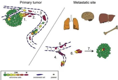
Existing knowledge shows that the metastatic cascade is a highly demanding multistep process where failure in any step will result in a failure of metastasis establishment. Therefore, only a small fraction of cancer cells acquire the ability to migrate and home to distant organs, making this a very inefficient process (Chambers et al., 2002; Valastyan and Weinberg, 2011; Weiss, 1990). The metastatic cascade starts with local invasion of cells originating from the primary tumor into the surrounding host tissue (1), when tumor cells migrate through the extracellular matrix (ECM) and stromal cells they reach lymphatic and/or blood vessels and crawl through the endothelial cells to intravasate into the circulation (2) where they face to survive in “unfriendly” conditions (shear stress, immunoevasion, flow) (3) until they reach a distant organ where they get stuck in the small blood vessels (4) and then extravasate to the distant organ (5), into the unknown hostile environment where they need to survive (6) and start proliferation in order to colonize the target organ and successfully establish a foothold for the metastasis (7) (Figure 4) (Bill and Christofori, 2015; Scully et al., 2012; Valastyan and Weinberg, 2011).
During metastatic invasion tumor cell plasticity is challenged. Therefore, cancer cells constantly undergo changes enabling them to survive and exert their function. The concept of epithelial- mesenchymal transition (EMT) preceding migration, followed by mesenchymal-epithelial transition (MET) at the distant site, a process conditional for proliferation and colonization is widely accepted in the scientific community (Bill and Christofori, 2015; Hartwell et al., 2006; Jung et al., 2008; Mani et al., 2007).
8
Figure 4: Metastatic cascade with involvement of EMT and MET. Tumor cells undergo EMT to acquire the potential to start local tissue invasion (1) and migration towards blood vessels. Upon intravasation into blood vessels (2) cells must survive (3) and arrive at target organs (4), where they are arrested in small capillary vessels mostly due to size and then they (5) extravasate.
In the distant organs tumor cells must survive in a new type of environment. Therefore they need to adapt by reversing the phenotype in MET process (6), regains proliferating phenotype to establish micro- and macrometastases (7). Picture modified from (Bill and Christofori, 2015).
As previously mentioned, breast cancer metastasizes predominantly to bones, liver, lung and brain (Jin et al., 2018; Wang et al., 2017). This organotropism is not only seen in breast cancer (Lu and Kang, 2007), but also in other cancer entities such as colorectal, prostate, lung and prostate cancers (Jin et al., 2012; Milovanovic et al., 2017; Reichert et al., 2018; Thobe et al., 2011). Based on the knowledge acquired from the autopsies of 735 women with breast cancer, Stephen Paget proposed the “seed and soil” hypothesis claiming that tumor cells (the “seed”) grew preferentially in the microenvironment of selected organs (the “soil”), and metastases can grow only when appropriate seed is planted in the corresponding soil (Paget, 1989). The basis for “seed and soil” mechanism is chemical attraction between cancer cells and factors expressed in metastatic organ. Several factors were identified to play a role, including osteopontin, osteonectin, chemokine ligand 21 (CCL21, 6Ckine) and stromal-derived factor-1 (SDF-1, CXCL12) expressed in bones (Ibrahim et al., 2000; Jacob et al., 1999; Luker and Luker, 2006; Muller et al., 2001). These chemokines are ligands for the receptors expressed on the breast cancer cells, such as C-C chemokine receptor type 4 (CXCR4, binds CXCL12) or C-C chemokine receptor type 7 (CXCR7, binds CCL21) (Luker and Luker, 2006; Mattern et al., 1996; Muller et al., 2001; Qian et al., 2018). An opposing theory was proposed by Ewing, who claimed that organ tropism is a consequence of the mechanical arrest of cancer cells in small capillaries (Chu and Allan, 2012).
Nowadays it is commonly understood that these two theories are not mutually exclusive and both factors contribute to the organ specificity of metastases (Chu and Allan, 2012; Weiss, 1992).
9
1.5 Disseminated cancer cells
During migration through the circulatory system (lymph or blood) cancer cells are referred to as circulating cancer cells (CCC) while upon extravasation and lodging to distant organs they are termed disseminated cancer cells (DCC). DCCs in mesenchymal organs such as the bone marrow or lymph nodes are detected upon using cytokeratin (CK8, CK18, and CK19, expressed only in epithelial cells) staining as a marker for DCCs. 30% of breast cancer patients are found positive for DCCs in the bone marrow (Braun et al., 2005). DCC presence in the bone marrow aspirates of patients with cancer both, breast and prostate, is a risk factor for developing metastases (Janni et al., 2011; Mathiesen et al., 2012;
Wiedswang et al., 2003). Furthermore, DCC persistence in the BM of patients diagnosed with breast cancer correlates to the decreased disease-free survival (DFS) and overall survival (OS) during the first 5 years following cancer diagnosis. (Janni et al., 2011). DCCs isolated using anti-CK staining can be used only for genomic studies, while these cells must be permeabilized prior to detection procedure and therefore the whole transcriptome is lost. Genomic analyses result in information which are insufficient in the battle against cancer as it is known that not only genetic changes, but also dysregulated gene expression contribute to the neoplastic transformation (Ferrone and Marincola, 1995; Timp and Feinberg, 2013). Therefore, epithelial cell adhesion/activating molecule (EpCAM, CD326), expressed on the surface of the cells, was introduced as a marker that enables isolation of living tumor cells.
EpCAM is overexpressed in many different human carcinomas (Baeuerle and Gires, 2007; Kimura et al., 2007; Massoner et al., 2014; Osta et al., 2004; Spizzo et al., 2004; Stoecklein et al., 2006; Went et al., 2006) and it was the first human tumor-associated antigen identified with monoclonal antibodies (Herlyn et al., 1979). Cell isolation using anti-EpCAM staining enables simultaneous isolation of both genome and transcriptome of the same cell (Klein et al., 2002) and therefore a much broader spectrum of analyses.
For a long time it was accepted that cancer cell dissemination occurs in later stages of tumor development (Koscielny et al., 1984), but the evidence from previous years strongly challenges that theory, pointing to dissemination as a process that occurs early during tumor development (Klein, 2009). Studies performed on mouse models in melanoma (Eyles et al., 2010) and breast cancer (Hosseini et al., 2016b; Husemann et al., 2008) strongly support the hypothesis on early dissemination of cancer cells and parallel progression of primary tumors and metastases. The proposed model of parallel progression (Klein, 2009) suggests that cancer cells disseminate early, as supported by evidence from melanoma, where it was shown that lymphatic dissemination occurs shortly after dermal invasion of the primary lesion at a median thickness of ~0.5 mm, a phase when the primary tumor is undetectable by standard screening techniques (Werner-Klein et al., 2018). In the early phases of tumor development, cancer cells harbor fewer genetic aberrations compared to primary tumor cells at the time of diagnosis, which in the context of early dissemination is explained by two possible scenarios: (1) DCCs harbor
10
fewer aberrations than cells from PT, indicating that DCCs progress and evolve slower or (2) DCCs harbor the same number or more aberrations than cells from the PT, indicating that DCCs progress and evolve faster than PT cells and therefore metastases arise quickly. Analyses of the DCCs from BM aspirates of breast cancer patients show that DCCs are less aberrant than PT cells and even share very few aberrations with them (Schmidt-Kittler et al., 2003), additionally supporting the first scenario and the theory of parallel evolution of tumor cells at the primary site and the metastatic organs. Although dissemination is an undeniable process in tumor biology, the relevance of early DCCs in establishment of metastases was questioned. Using the rodent models Eyles (melanoma) and Hosseini (breast cancer) with colleagues showed that metastases in both, melanoma and breast cancer evolve from early DCCs (Eyles et al., 2010; Hosseini et al., 2016b).
1.6 Dormancy of disseminated cancer cells
Initial steps of the metastatic cascade, such as invasion and dissemination are very efficient processes (Chambers et al., 2002) but compared to that, metastasis formation rate is very inefficient. This indicates that the critical step for metastasis formation is colonization of the metastatic organ. Upon extravasation to distant organs DCCs can die, resume the growth immediately or enter a state of dormancy (Aguirre-Ghiso, 2007; Chambers et al., 2002; Ranganathan et al., 2006). The relevance of the state of dormancy of DCCs was intensively studied in the last decades.
Eyles and colleagues showed that single cells spread very early during oncogenesis of melanoma, 3 weeks after the clinical onset of the primary tumor, but detectable metastases became apparent much later in life, up to 1.5 years (Eyles et al., 2010), suggesting that these early DCCs remained dormant for a long period. In line with that BM-DCCs in Balb-NeuT mice do not proliferate but can be reactivated upon transplantation into lethally irradiated siblings, showing that they retain the potential to proliferate, but reside in a dormant state (Hosseini et al., 2016b; Husemann et al., 2008). This is not only the case in rodent animal models. Existence of dormant DCCs in humans was shown in the case of a patient that was treated for melanoma and died 16 years later due to brain death without any signs of secondary disease or detectable metastases. This patient was used as a kidney donor and 2 years after transplantation both recipients died from metastases of malignant melanoma, although they were never diagnosed with melanoma. Thus, even though the donor had no signs of clinical disease, melanoma cells survived and were transferred with the graft to the recipients. Since such patients are under immunosuppression therapy to avoid graft rejection, the condition of impaired immunosurveillance was favorable for the vigorous growth of metastases (MacKie et al., 2003). Such cases support the concept of dormancy in disseminated cells. Long latency periods observed between the treatment of primary tumors and metastatic recurrence in patients are also used as a proof for the existence of clinical tumor dormancy (Klein, 2011).
11
The fate of DCCs upon extravasation depends on both intrinsic and extrinsic signals (Bloom and Zaman, 2014). Prolonged period of dormancy is induced and maintained by different mechanisms, including tumor-microenvironmental factors (such as cytokines, immunosurveillance, angiogenesis), metastasis suppressor gene activity and anti-cancer therapy (Osisami and Keller, 2013). We can discriminate two main types of tumor dormancy: (1) dormancy of single cancer cell, when cells reside in a proliferation arrested state (G0/G1 arrest) that is reversible and they can re-enter the cell cycle (reactivate proliferation) and (2) tumor mass dormancy, a phenomenon when cells do not undergo cell cycle arrest, but the rate of cell proliferation correlates to the rate of apoptosis, resulting in maintainability of the size of metastases (Kareva, 2016). This second type, tumor mass dormancy, is often a result of systemic cooperation among tumor cells and the immune system (Koebel et al., 2007; Quesnel, 2008; Vesely et al., 2011). On the contrary, different mechanisms are suspected to regulate the dormant state of a single cancer cell. For example microenvironmental stress (Adam et al., 2009a; Aguirre-Ghiso et al., 2003), cytokines and other factors secreted by stromal cells (Kobayashi et al., 2011; Lim et al., 2011; Shiozawa et al., 2010). Further, the inability of DCCs to form cytoskeletal rearrangements and interact with environmental cells (Barkan et al., 2008), but also factors like anti-tumor therapy (Chaterjee and van Golen, 2011; Schewe and Aguirre-Ghiso, 2008) can result in single DCC dormancy. The existence of different types of tumor dormancy makes the concept of metastatic dormancy even more complex, since all of the above mentioned mechanisms can be employed in maintaining the dormancy of DCCs at distant sites, but there is also the contribution of the foreign environment.
1.7 Interleukin-6 signaling in physiological and pathological settings
Interleukin 6 (IL-6) is a proinflammatory cytokine that is known to be secreted by several different types of cells. In humans it is encoded by the IL6 gene, located on chromosome 7. It belongs to a family of cytokines consisting of IL-6, IL-11, oncostatin M (OSM), leukemia inhibitory factor (LIF), ciliary neurotrophic factor (CNTF), cardiotrophin 1 (CT-1), cardiotrophin-like cytokine (CLC) and IL-27. Its main function is to mediate immune response, when secreted by T cells and macrophages, and to direct B cell differentiation. It is also known to be secreted by osteoblasts, and has a role in stimulating osteoclast maturation (Dienz et al., 2009; Eto et al., 2011; Udagawa et al., 1995; Yang et al., 2016).
Nowadays it is known that IL-6 has many different functions and plays a role in numerous inflammation related diseases (Schaper and Rose-John, 2015; Tanaka and Kishimoto, 2014; Tanaka et al., 2014).
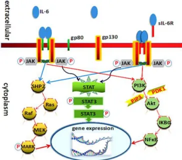
IL-6 acts by binding to the glycoprotein 130 (gp130) and can signal via two distinct pathways. The classical IL-6 signaling is mediated by direct binding of IL-6 to the heterodimeric receptor consisting of the IL-6 receptor α-chain, CD126 (gp80, IL-6Rα, IL-6Ra) and the ubiquitously expressed signal transducing receptor subunit glycoprotein 130 (gp130, IL-6Rβ) (Scheller et al., 2011) (Figure 5). Due
12
to the restricted expression of IL-6R to mainly hepatocytes and immune cells (Scheller et al., 2011), classical IL-6 signaling is limited to only few cell types. However, a soluble form of IL-6R (sIL-6R) can be produced by alternative splicing or limited proteolysis of the membrane-bound receptor. The complex of sIL-6R/IL-6 can bind to the ubiquitously expressed gp130 receptor subunit on recipient cells and thereby stimulate cells which do not express IL-6R (Rose-John and Heinrich, 1994). This type of stimulation, not mediated by a membrane receptor is referred to as trans-signaling. The binding of IL-6/IL-6R or IL-6/sIL-6R to the gp130 signaling subunit induces the homodimerization of gp130 which further triggers an intracellular signaling cascade activating several pathways including Ras/Raf, mitogen-activated protein kinase (MAPK), PI3K/Akt and Janus-activated kinase/signal transducers and activators of transcription (JAK/STAT) (Kishimoto, 2005; Murray, 2007; Streetz et al., 2003; Wegiel et al., 2008).
Figure 5: IL-6 signaling mechanism: IL-6 binds to the heterodimer receptor on the cell membrane, consisting of gp80 (carrying binding site for IL-6) and gp130 (IL-6Rβ), which then homodimerizes and activates JAK/STAT, MAPK/ERK and PI3K/Akt signaling cascades (classical signaling). In the trans-signaling pathway, sIL-6R, a soluble variant of IL-6Rα present in the intercellular space, binds IL-6. The complex IL-6/sIL-6R then binds to ubiquitously expressed gp130 and activates cells that do not express IL6R. Image taken from (Luo and Zheng, 2016).
In recent years the role of IL-6 was studied in different cancers. It was shown that IL-6 levels were increased in the sera of the patients with breast cancer, and this increase correlated with tumor stage and decreased patient survival (Bachelot et al., 2003; Kozlowski et al., 2003). Additionally, IL-6 plays a role in the biology of cancer stem cells in the mammary gland and breast tumors (Marotta et al., 2011;
Sansone et al., 2007). IL-6 and the activation of an inflammatory feedback-loop have been shown to dynamically regulate the equilibrium between cancer cells with stem-like properties and non-stem cancer cells (Marotta et al., 2011) (Sansone et al., 2007). Studies on different cancer entities pointed to a role of IL-6 signaling in development of primary tumor, but also indicated they facilitate the formation
13
of metastasis, by supporting migration and invasion of cancer cells (Browning et al., 2018; He et al., 2018; Lee et al., 2019; Tang et al., 2018).
However, the effect of IL-6 signaling on non-transformed mammary tissue, as well as on putative metastasis founder cells (DCCs) has not been fully elucidated. To further investigate the role of this cytokine on mammary cells in physiological conditions, we used the non-transformed human breast cancer cell line, MCF10A and patient derived human mammary epithelial cells (HMECs). Additionally, we examined the IL-6 signaling in DCCs isolated from the bone marrow of the patients with mammary carcinomas.
14
2 Material and Methods
2.1 Methods
2.1.1 Patient material
Human non-cancerous mammary tissue was obtained from female patients undergoing reduction mammoplasty surgeries at Caritas-Krankenhaus St. Josef, Regensburg in collaboration with Dr. Norbert Heine after informed, written consent of patients was obtained (ethics vote number 07/043, ethics committee of the University Regensburg). After verification of the non-cancerous origin of the tissue by a pathologist at the Institute of Pathology, University hospital Regensburg, mammary glands were dissociated and primary human mammary epithelial cells (HMECs) isolated. The tissues showing the signs of breast cancer were excluded from the study and not used for this work. Due to hormone changes during menopause and proposed hormonal influence in mammary stem and progenitor cells, tissue were selected based on an arbitrary age limit (45 years).
Disseminated cancer cells were obtained from bone marrow aspirates of breast cancer patients with and without distant metastases. Human mesenchymal stem cells were obtained from bone marrow aspirates of breast cancer patients or healthy donors. Written informed consent of patients was obtained and the ethics committee of the University of Regensburg (ethics vote number 07/79) approved bone marrow sampling and analysis of isolated cells.
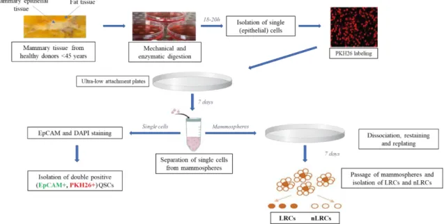
2.1.1.1 Digestion of the human mammary tissue and isolation of single epithelial cells
Mammary tissue was minced and dissociated in Ham’s F12/Dulbecco’s modified Eagle’s medium [F12:DMEM; 1:1 (v:v)] supplemented with 10 mM HEPES, 2% bovine serum albumin (BSA; Fraction V), 5 μg/ml insulin, 0,5 μg/ml hydrocortisone, 10 ng/ml cholera toxin, 300 U/ml collagenase and 100 U/ml hyaluronidase in the sterile incubator at 37°C, 5% CO2 and 7% O2 for minimum of 18 h. On the next day, the digested cell suspension was centrifuged at 210 g for 2 minutes at room temperature.
Supernatant from the first centrifugation step contained single mammary epithelial and stromal cells (fibroblasts), while in the pellet were the organoids (undigested tissue pieces of the tissue). Supernatant containing epithelial cells and fibroblasts was further centrifuged 4 minutes at 350xg to separate epithelial cell from fibroblasts. Supernatant containing fibroblasts was discarded and pellet (containing epithelial cells) was re-suspended in the basal medium, washed, and filtered through 100 m and 40
m cell strainers consecutively. Mammary epithelial cells were propagated in mammosphere medium in ultra-low attachment plates. For the propagation cells were seeded in density of 200.000 cells/ml,
15
and for the experiments where we assessed the numbers of the mammospheres cells were seeded in density of 50.000 cells/ml in 35 mm plates with 3 ml medium per plate.
For the disaggregation of the mammospheres they were collected after 7 days and centrifuged 1 minute at 100xg. Supernatant containing single cells transferred to another 50 ml falcon tube and centrifuged 5 minutes 500xg to collect quiescent single cells (QSC). Mammospheres were subjected to the mechanical/enzymatic digestion using 0,05% trypsin, 0,53 mM EDTA-4Na and 10 minutes of constant pipetting using Gilson 1 ml pipette. Trypsinization was inhibited by adding Trypsin neutralizing solution (TNS) and cells were washed and re-plated for growing second generation of mammospheres.
In the first generation of mammospheres can be found some stromal cells, but they succumb anoikis and the second mammosphere generation is “pure” and contains only mammary epithelial cells. The obtained secondary mammosphere were used for the most of the downstream assays elaborated in this work.
2.1.2 Animals
2.1.2.1 Maintenance
NOD.Cg-Prkdcscid IL2rgtmWjl/Sz (also termed NSG) or Balb/c mice were purchased from the Jackson Laboratory USA and maintained under specific-pathogen free conditions, with acidified water and food ad libitum in the research animal facilities of the University of Regensburg, Germany. Transgenic mouse model (Balb-NeuT) was obtained from Dr. Guido Forni. Maintenance in the animal facility was performed according to the European Union guidelines. All animal experiments followed EU and national institutional regulations (Government of Upper Palatinate, 55.2-2532.1-27/14). At the age of 4 weeks Balb-NeuT mice were analyzed for the presence of transgene. Female hemizygotes (neuT+/neuT-) were further cultivated and regularly inspected (twice a week) for the presence of mammary tumors, which were thereafter measured in two perpendicular diameters. Transgene negative (neuT-/neuT-) homozygotes served as wild-type Balb/c controls.
2.1.2.2 Mice dissection
Mice were sacrificed by cervical dislocation. The organs of interest (mammary glands, tumors and lungs) were divided in two parts each and one part was snap frozen while the other was embedded in paraffin. Bone marrow was prepared as described in the following section.
2.1.2.3 Paraffin embedding of tissue samples
The dissected tissue samples were fixed in a 4% paraformaldehyde (PFA) solution for minimum 12 hours. Following fixation, the samples were washed 3 times in 1x PBS, dehydrated by series of washing steps in alcohol (70%, 85% and 100% ethanol, each step 1 hour) and washed twice for 30 min in 100%
xylene. This step serves not only the removal of alcohol from the tissue, but also facilitates the
16
penetration of the paraffin during the subsequent embedding. After three incubation steps with paraffin (parablast embedding media), tissues were embedded. Paraffin embedded tissues are stored at room temperature. These samples have been used for IHC staining.
2.1.2.4 Isolation of the epithelial cells from mouse mammary gland
The mice were killed by cervical dislocation. Mammary glands were collected either from Balb/c or Balb-NeuT mice in a 50 ml tube with PBS. The tissue was minced with surgical blades to small pieces and digested in basal medium (DMEM/F12, 100 nM HEPES buffer, 10 mg/ml insulin, 0.5% BSA and 0.5x penicillin/streptomycin) with 0,1 mg/ml DNAse I 200 U/ml collagenase and 200 U/ml hyaluronidase. The tissue was digested for 1-1,5 hours at 37° C in the incubator and mixed after 20 minutes of incubation, then after 1 hour and every 10 minutes later until the organoids are disaggregated.
The cell suspension originating from the digested tissue was filtered through 40 m cell strainer and centrifuged 10 minutes at 300xg to isolate mammary epithelial cells.
2.1.2.5 Mammosphere transplantation
Before the transplantation of the GFP-labeled mammospheres (50 spheres per mouse, mammosphere generation described in paragraph 2.1.3.4) mice were anaesthetized using Midazolam 5mg/kg, Fentanyl 0,05mg/kg, Medetomidin 0,5mg/kg i.p. The skin around the mammary gland was shaved and small V- cut made around fourth left mammary gland and small pocket was made between the 4th mammary gland and skin. Mammospheres were mixed with matrigel (final concentration of Matrigel 40%) and injected into the pocket between the mammary gland and skin of the 4-6 weeks old wt (neuT-) and NSG mice. The skin was closed by a suture using polygelatin string and anesthesia was antagonized with Flumazenil 0,5mg/kg, Atipamezol 2,5mg/kg, Naloxon 1,2mg/kg s.c. The wound was disinfected with Braunol and Hansaplast spray. Mammosphere recipients were sacrificed 4 or 8 weeks after transplantation. Curative surgery or sacrifice of mice was done when the diameter of tumors was between 5-10 mm.
2.1.2.6 Bone marrow processing for staining and single cell isolation
Tibias and femurs were collected from NSG, Balb/c or Balb-NeuT mice and held in PBS. The soft tissue surrounding the bones was removed. Epiphyses were removed using scissors and the bone marrow was flushed out with 1x PBS into a 50 ml tube using a 26G needle. Afterwards, the cells were pooled and centrifuged at 200x g for 10 min. The supernatant was discarded and the pellet was resuspended in 9 ml of 1x PBS. Cell suspension was slowly and carefully overlaid onto 6 ml of 65% percoll in a 15 ml tube to form a layer and centrifuged for 20 min at 1000x g to remove erythrocytes and granulocytes.
Interphase was carefully collected using a 5 ml pipette and transferred into a new 50 ml tube. The tube was filled up with 1x PBS and centrifuged for 10 min at 500x g for washing the cells. The cells were resuspended in 5 ml of 1x PBS and counted. For the preparation of slides for staining, 500.000 cells
17
were placed on each adhesion slide. For the isolation of EpCAM+ cells, bone marrow was first subjected to CD45 depletion (section 2.1.2.8).
2.1.2.7 Bone marrow transplantation
Balb/c (9 weeks old) and NSG (6 weeks old) mice were irradiated previous to BM transplantation.
Balb/c mice were irradiated twice, two days before transplantation and on a transplantation day 5-6 hours before BM transplantation, with 5 Gy. NSG mice were irradiated on transplantation day 5-6 hours before BM transplantation with 2 Gy. Bone marrow was isolated from 8-10 weeks old Balb-NeuT donors as described and naïve bone marrow cell suspension (without removing erythrocytes and granulocytes on 65% Percoll) was injected i.v. to previously irradiated mice (7-10x106 BM cells/mouse).
For the transplantation of the CD45-depleted BM, after isolation BM suspension was subjected to two rounds of CD45 depletion and the efficiency of the elimination of CD45+ cells was examined by FACS.
To each of previously irradiated Balb/c mice (as described above) were injected 6,5x10⁶ CD45-depleted BM cells. On the following day one Balb/c mouse was sacrificed and 2-5 x10⁶ BM cells were injected i.v.
2.1.2.8 CD45 depletion
Bone marrow of Balb/c and Balb-NeuT mice was isolated and CD45+ cells were depleted after the instructions of the vendor. Briefly, Cells were resuspended in MACS buffer (90 µl per 107 cells) and anti-mouse CD45 beads were added (10 µl per each 107 cells). After 15 minutes incubation at 4ºC cells were washed with 2 ml of 1x PBS and centrifuged 10 min at 300xg. Supernatant was removed and cells resuspended in MACS buffer (500 µl per 108 cells). Column was rinsed once with 2 ml buffer and afterwards cells were added to the column. CD45+ cells are kept on the column and CD45- fraction collected in the tube. Column was washed twice, with 2 ml and 1 ml of MACS buffer respectively, CD45- cells (flow-through) were counted, centrifuged and resuspended in 1x PBS so that we get the needed density.
2.1.2.9 Progesterone treatment
For the progesterone treatment pellets of 5 mg with 21 days releasing time were implanted as proscribed by vendor. Briefly, small neck was made on the lateral side of the neck of the recipient mice (Balb/c and Balb-NeuT) and the pocket was formed 2 cm behind the cut using forceps. Pellets were placed into the pockets using forceps and the cut was sutured. No alcohol disinfectant was used as it can activate releasing of the pellet content before it is placed in the body of the recipient. After 4 or 8 weeks recipient mice were sacrificed.
18 2.1.2.10 Bone marrow staining for DTC detection
At least 106 cells per mouse were stained to detect positive cells and 0,5x10⁶ cells per mouse to control the Isotype positivity (Isotype control). Blocking solution (5% rabbit serum in 1x TBS) was added to the slides to rehydrate the cells and to block unspecific binding of antibodies to the cells. After 20 min the blocking solution was discarded and primary antibody against CK 8 and 18 was added and incubated for 60 min. The primary antibody was discarded and slides were washed 3 times for 3 min in 1x TBS.
Then slides were incubated with the secondary antibody for 25 min, and washed 3 times for 3 min in 1x TBS followed by incubation with ABC complex for 25 min. Finally, the development system of the BCIP/NBT for alkaline phosphatase enzymatic substrate was added for 10 min. The slides were washed 3 times for 3 min and screened for CK8/18 positive cells. The positive cells were typically violet-to- black in color. TUBO, a tumor cell line derived from a primary mammary tumor of Balb-NeuT mice and expresses CK8/18, was used as a positive control.
2.1.2.11 Haematoxylin and eosin staining
Paraffin embedded tissue was cut into 5 µm sections onto poly-L-lysine-coated slide. Sections were dewaxed in Xylol for 10 min twice and rehydrated first in 100% ethanol for 3 min and then in 80%
ethanol for 3 min. The sections were washed in PBS for 1 min thrice and incubated with hematoxylin for 45 sec. The slides were rinsed in tap water for a short duration and washed in tap water for 30 min in a glass cuvette. Eosin (0.1%) was added to the tissue sections and after 2 min they were washed with ddH2O for 1 min. The stained sections were dehydrated in 70% ethanol for 2 min, 100% ethanol for 2 min and finally in xylol for 15 min. Mounting gel was added to the tissue sections and a cover slip was placed carefully on the gel avoiding bubbles and were left to dry overnight.
2.1.2.12 Immunohistochemistry
For Her2 immunohistochemistry of tissues, 5 µm sections of paraffin blocks were collected onto poly- L-lysine-coated slides. Samples were dewaxed by two 5-min washes in xylene and rehydrated with graded alcohol by 5-min washes and a final wash in water. A standard Tris-EDTA buffer and pressure cooking was the antigen retrieval procedure and then sections were blocked in 0.3% H2O2 in TBS and 10% normal goat serum. Sections were incubated for 1 hour with primary antibody and after washing secondary antibody was added based on manufacturers suggested dilution. After washing with PBS, sections were stained using the ABC detection system (Vector laboratory) according to the manufacturer’s instruction. Visualization was performed with chromogen reagent (Dako) according to manufacture instructions.
19
2.1.3 Cell culture experiments
2.1.3.1 Storage and propagation of the cell lines
The identity of all used cell lines was controlled by ATCC recommended DNA fingerprinting and they were negative for mycoplasma contamination as confirmed by regular routine tests. Cell lines were preserved in liquid nitrogen in medium containing 50% FCS, 40% complete growth medium for propagation and 10% DMSO. Cell lines were propagated until 70-80% confluence and re-plated afterwards. Cell lines were de-attached using Trypsin/EDTA and re-plated in an appropriate cell density.
The non-tumorigenic mammary epithelial cell line MCF 10A (ATCC, 50) was cultured in Ham’s Dulbecco’s modified Eagle’s/F12 (DMEM/F12) medium supplemented with 5% horse serum, 1%
penicillin/streptomycin, 20 ng/ml EGF, 0.5μg/ml hydrocortisone, 10 μg/ml insulin and 0.1 µg/ml cholera toxin. MCF 10A-GFP cells were generated by transducing MCF 10A cells with pRRL.sin.cPPT.hCMV-GFP.WPRE. hTERT-HME1-derived cell line HME-EGFR (generously provided by Alberto Bardelli, Italy) was maintained in DMEM/F-12 medium supplemented with 10%
fetal calf serum, 20 ng/ml EGF, 0.5 µg/ml hydrocortisone, 10 µg/ml insulin and 1%
penicillin/streptomycin. Murine embryonic fibroblasts C3H10T1/2 (a generous gift from M. Wicha, University of Michigan, USA) cells were grown in DMEM medium supplemented with 5% fetal calf serum, 2mM glutamine, 1% penicillin/streptomycin. T4-2 and HUVEC cells were a kind gift from Dr.
Cyrus Ghajar (Laboratory for the Study of Metastatic Microenvironments, Fred Hutchinson Cancer Research Center, Seattle, WA, USA). T4-2 cells were propagated in collagen-coated cell culture dishes in DMEM/F12 supplemented with 250 ng/ml Insulin, 10 µg/ml apo-Transferrin, 2,6 ng/ml sodium- selenite, 10-10 M β-estradiol, 1,4x10⁻⁶ M Hydrocortisone and 5 µg/ml Prolactin. HUVECs were propagated in complete EGM-2 medium (EGM-2 Kit, Lonza) and used maximum until passage 11. All cell lines were kept at 37°C and 5% CO2 in a fully humidified incubator.
2.1.3.2 Preparation of collagen coated plates
PureCol collagen is approximately 97% Type I collagen with the remainder being comprised of Type III collagen and it is used for the coating of the dishes for culture of the cell lines that need additional support from the matrix similar to their physiological environment. PureCol (stock concentration 3 mg/ml) was diluted in ice cold 1x PBS to working concentration of 67 g/ml. This solution was added to the cell culture flask at least a day before cell seeding. Per one 75 cm2 flask was added 10 ml of the collagen solution and it was incubated overnight at 4ºC. These dishes can be stored at 4ºC for 14 days.
Before seeding cells the liquid was removed (carefully not to disturb the bottom layer of collagen) and gently washed with prewarmed growth medium.