Design, Synthesis and Characterization of ABCG2 Inhibitors with a Focus on Water Solubility and Stability in Plasma
D ISSERTATION
Z UR E RLANGUNG DES D OKTORGRADES DER N ATURWISSENSCHAFTEN (D R . RER . NAT .)
AN DER F AKULTÄT C HEMIE UND P HARMAZIE DER U NIVERSITÄT R EGENSBURG
VORGELEGT VON
F RAUKE A NTONI AUS E RLANGEN
2020
Die vorliegende Arbeit entstand unter Anleitung von Herrn Prof. Dr. Armin Buschauer, der leider viel zu früh verstorben ist, und Herrn Prof. Dr. Günther Bernhardt an der Fakultät für Chemie und Pharmazie der Universität Regensburg.
Promotionsgesuch eingereicht am: 30. September 2020 Tag der mündlichen Prüfung: 09. Dezember 2020
Prüfungsausschuss: Prof. Dr. Dominik Horinek (Vorsitzender)
Prof. Dr. Günther Bernhardt (Erstgutachter)
Prof. Dr. Pierre Koch (Zweitgutachter)
Prof. Dr. Joachim Wegener (Drittprüfer)
Publications
Articles and Patents
F. Antoni, D. Wifling, G. Bernhardt, Water-soluble inhibitors of ABCG2 (BCRP) – a fragment-based and computational approach, Eur. J. Med. Chem., (2020) 112958.
F. Antoni, M. Bause, M. Scholler, S. Bauer, S.A. Stark, S.M. Jackson, I. Manolaridis, K.P. Locher, B. König, A. Buschauer, G. Bernhardt, Tariquidar-related triazoles as potent, selective and stable inhibitors of ABCG2 (BCRP), Eur. J. Med. Chem., 191 (2020) 112133.
F. Antoni, G. Bernhardt, Derivatives of nitrogen mustard anticancer agents with improved cytotoxicity, Arch. Pharm., n/a (2020) e2000366.
S. Huber, F. Antoni, C. Schickaneder, H. Schickaneder, G. Bernhardt, A. Buschauer, Stabilities of neutral and basic esters of bendamustine in plasma compared to the parent compound: kinetic investigations by HPLC, J. Pharm. Biomed. Anal., 104 (2015) 137-143.
H. Schickaneder, C. Schickaneder, A. Buschauer, G. Bernhardt, S. Huber, F. Antoni, Preparation of bendamustine derivatives and related compounds for use in treating cancer, WO2015091827A1 (2015).
Oral Presentations
F. Antoni, G. Bernhardt, Approaches to ABCG2 inhibitors with drug-like properties, Barrier and Transporter Meeting (2018, Bad Herrenalb).
F. Antoni, G. Bernhardt, Fragment-based discovery of novel ABCG2 inhibitors, Doctoral Colloquium of the Graduate School ChemPharm (2017, Regensburg).
Poster Presentations
F. Antoni, G. Bernhardt, A fragment-based approach to ABCG2 modulators, Summer School Medicinal Chemistry (2018, Regensburg).
F. Antoni, G. Bernhardt, A fragment-based approach to ABCG2 inhibitors with drug-like properties, Frontiers in Medicinal Chemistry (2018, Jena).
F. Antoni, S.A. Stark, M. Scholler, G. Bernhardt, B. König, A. Buschauer, Triazole-type
ABCG2 modulators, DPhG Annual Meeting (2016, München).
S.A. Stark, F. Antoni, M. Scholler, G. Bernhardt, A. Buschauer, B. König, Synthesis and
characterization of triazole-type ABCG2 modulators, Summer School Medicinal Chemistry
(2016, Regensburg).
Contents
1 General Introduction ... 1
1.1 Membrane Transport and ABC Transporters ... 2
1.2 Structure and Mechanism of ABCG2 ... 5
1.3 Functions and Substrates of ABCG2 ... 8
1.3.1 Endogenous Functions ... 9
1.3.2 Barrier Functions ... 9
1.3.3 Function at the Blood-Brain Barrier ... 11
1.3.4 Function in Multidrug Resistance in Cancer ... 12
1.4 Applications of ABCG2 Inhibitors ... 13
1.4.1 Applications as Molecular Tools ... 13
1.4.1.1 Tools for Fundamental Research ... 13
1.4.1.2 Tools for Drug Discovery and Development ... 15
1.4.1.3 Clinical Diagnostic Tools ... 16
1.4.2 Applications as Prospective Drugs ... 16
1.4.2.1 Drugs for Improving Oral Bioavailability ... 16
1.4.2.2 Drugs for Improving Brain Penetration ... 17
1.4.2.3 Drugs for Overcoming Multidrug Resistance in Cancer ... 18
1.5 Reported ABCG2 Inhibitors and Problem Statement ... 20
1.6 Scope and Objectives of the Thesis ... 22
1.7 References ... 24
2 Tariquidar-Related Triazoles as Potent, Selective and Stable Inhibitors of ABCG2 (BCRP) ... 33
2.1 Introduction ... 34
2.2 Results and Discussion ... 36
2.2.1 Synthesis ... 36
2.2.2 Inhibition of the ABCG2, ABCB1 and ABCC1 Transport Activity ... 39
2.2.3 Effect on the ABCG2 ATPase Activity ... 42
2.2.4 Thermostabilization of ABCG2 ... 46
2.2.5 Cytotoxicity and Reversal of Drug Resistance ... 47
2.2.6 Stability in Blood Plasma ... 48
2.3 Conclusion ... 49
2.4 Experimental Section ... 51
2.4.1 Chemistry ... 51
2.4.1.1 General Experimental Conditions ... 51
2.4.1.2 Synthesis Protocols and Analytical Data ... 52
2.4.2 Biology ... 75
2.4.2.1 General Experimental Conditions ... 75
2.4.2.2 Cell Culture ... 76
2.4.2.3 Inhibition of ABCG2: Hoechst 33342 Transport Assay... 77
2.4.2.4 Inhibition of ABCB1: Calcein-AM Transport Assay ... 77
2.4.2.5 Inhibition of ABCC1: Calcein-AM Transport Assay ... 78
2.4.2.6 ABCG2 ATPase Assay ... 78
2.4.2.7 Size-Exclusion Chromatography-Based Thermostability Assay ... 80
2.4.2.8 Chemosensitivity Assay ... 81
2.4.2.9 Chemical Stability Assay (in Blood Plasma)... 81
2.5 Supplementary Material... 83
2.5.1 NMR-Spectra of Key Compounds ... 83
2.5.2 Chromatograms (Purity Control) of Key Compounds ... 85
2.6 References ... 88
3 Water-Soluble Inhibitors of ABCG2 (BCRP) – A Fragment-Based and Computational Approach ... 93
3.1 Introduction ... 94
3.2 Results and Discussion ... 97
3.2.1 Design and Synthesis ... 97
3.2.2 Molecular Docking ... 102
3.2.3 Biological Characterization ... 103
3.2.3.1 Inhibition of the ABCG2, ABCB1 and ABCC1 Transport Activity ... 103
3.2.3.2 Cytotoxicity and Reversal of Drug Resistance ... 109
3.2.3.3 Effect on the ATPase Activity ... 110
3.2.3.4 Stability in Blood Plasma ... 112
3.2.4 Aqueous Solubility ... 113
3.2.4.1 Kinetic Solubility ... 114
3.2.4.2 Thermodynamic Solubility ... 118
3.3 Conclusion ... 123
3.4 Experimental Section ... 125
3.4.1 Chemistry ... 125
3.4.1.1 General Experimental Conditions ... 125
3.4.1.2 Synthesis Protocols and Analytical Data ... 126
3.4.2 Molecular Docking ... 167
3.4.3 Biology ... 168
3.4.3.1 General Experimental Conditions ... 168
3.4.3.2 Cell Culture ... 169
3.4.3.3 Inhibition of ABCG2: Hoechst 33342 Transport Assay... 170
3.4.3.4 Inhibition of ABCB1: Calcein-AM Transport Assay ... 170
3.4.3.5 Inhibition of ABCC1: Calcein-AM Transport Assay ... 170
3.4.3.6 Chemosensitivity Assay ... 170
3.4.3.7 ABCG2 ATPase Assay ... 170
3.4.3.8 Chemical Stability Assay (in Blood Plasma)... 172
3.4.4 Solubility ... 172
3.4.4.1 General Experimental Conditions ... 172
3.4.4.2 Kinetic Solubility Assay ... 173
3.4.4.3 Thermodynamic Solubility Assay ... 173
3.5 Supplementary Material... 175
3.5.1 Figures 3.13, 3.14 and 3.15 ... 175
3.5.2 NMR Spectra ... 176
3.5.3 Chromatograms (Purity Control) ... 213
3.6 References ... 226
4 Summary ... 233
5 Annex: Derivatives of Nitrogen Mustard Anticancer Agents with Improved Cytotoxicity ... 237
5.1 Introduction ... 238
5.2 Results and Discussion ... 241
5.2.1 Synthesis ... 241
5.2.2 Cytotoxicity ... 242
5.2.2.1 Bendamustine and Derivatives ... 243
5.2.2.2 Isobendamustine and Derivatives ... 244
5.2.2.3 Chlorambucil and Derivatives ... 245
5.2.2.4 Melphalan and Derivatives ... 246
5.3 Conclusion ... 247
5.4 Experimental Section ... 249
5.4.1 Chemistry ... 249
5.4.1.1 General Experimental Conditions ... 249
5.4.1.2 Synthesis Protocols and Analytical Data ... 250
5.4.2 Biology ... 257
5.4.2.1 General Experimental Conditions ... 257
5.4.2.2 Cell Culture ... 258
5.4.2.3 Chemosensitivity Assay ... 258
5.5 Supplementary Material... 260
5.5.1 NMR Spectra ... 260
5.5.2 Chromatograms (Purity Control) ... 268
5.6 References ... 271
Abbreviations ... 273
Acknowledgements ... 277
1 General Introduction
1.1 Membrane Transport and ABC Transporters
Biological membranes are selectively permeable structures, which form the interface between a cell or an organelle and its environment. Membrane transport constitutes one of the most fundamental processes in all living cells – it allows the maintenance of cellular homeostasis [1].
Due to the enormous chemical diversity of the molecules required by or expelled from the cell, it comes to no surprise that the transport mechanisms across biomembranes are multitudinous.
In Escherichia coli, for example, about 10% of the entire genome encodes membrane-bound and soluble proteins involved in transport processes [2]. The simplest of all transport forms is passive diffusion. While some gases, water and other small polar molecules as well as unpolar compounds can freely permeate a lipid bilayer, ions and larger polar molecules like glucose cannot do so and require transport proteins, such as channel proteins or carriers/transporters, to mediate the passage [3-6]. This process is called passive transport or facilitated diffusion. In case a solute is to be moved against an electrochemical gradient, active transport under the consumption of energy is necessary [4,6]. Active transporters are either driven by an electrochemical potential difference at the biomembrane (secondary active transport) or by the hydrolysis of an energy-carrying molecule, mainly adenosine triphosphate (ATP) (primary active transport) [4,6]. Membrane ATPases are classified into four categories: F-ATPases are mitochondrial or chloroplast ATP synthases; P-ATPases, for instance sodium-potassium pumps or calcium pumps, build an electrochemical gradient; V-ATPases form a proton gradient and are found within many membranes of cell organelles, such as vacuoles in plant cells or endosomes [7]; and ATP-binding cassette (ABC) transporters translocate myriad substrates – inorganic and organic, charged and neutral – across cellular membranes [7-10]. The latter embody by far the largest and most diverse of the transporter families [7,8] and one of the largest protein families in general [11].
The substrates of ABC transporters are beyond number – and so are their functions. They are widespread in all three domains of life. In bacteria and archaea, ABC importers enable the uptake of various nutrients and ABC exporters sequester virulence factors or antibiotics [8,12].
The majority of eukaryotic family members function in the direction of export. On one hand,
ABC transporters excrete endogenous metabolites from secretory epithelial cells, such as bile
salts in the liver [9,10,13]. On the other hand, at physiological barriers, e.g. the intestinal barrier
or the blood-brain barrier (BBB), they protect cells from the entry of diverse xenobiotics,
including toxic and carcinogenic substances, but also a wide range of drugs such as cytostatics,
antipsychotics or antiepileptic agents [9,10,14,15]. The diversity of the biological roles played
by ABC transporters is reflected by their extensive involvement in diseases. A malfunction of
ABC transporters is the cause of various severe genetic disorders, such as bile transport
disorders or cystic fibrosis [9,10,13]. Moreover, ABC transporters confer drug resistance (MDR) in cancer [16] or brain diseases [17,18] and they modulate the bioavailability of drugs [9,10,14,15].
Although ABC transporters deserve much attention because of their functions in the human body, they were first discovered and characterized in detail in bacteria, as early as in the 1970s [12,19-22]. Only later it was demonstrated that these proteins are not restricted to prokaryotes.
In 1976, Juliano and Ling discovered a drug resistance-related glycoprotein in a mammalian cell line and called it P-glycoprotein (P-gp), because it appeared to confer drug resistance by making the cellular membrane less permeable [23]. It was not until 1986 that studies revealed a strong homology between the DNA sequence of P-gp and the bacterial transport proteins discovered before and it was suggested that P-gp functions as an energy-dependent export pump [24,25]. In the same year, it was recognized by Higgins et al. that highly conserved ATP- binding subunits characterize a large superfamily of transport proteins [26]. The designation
‘ABC transporters’ was first introduced in 1990 by Higgins’ group who named these proteins after their most characteristic feature – the aforementioned ATP-binding cassette [8,27].
Subsequent genome sequencing and phylogenetic analyses confirmed that the ABCs, or
nucleotide binding domains (NBDs), are monophyletic, i.e. they have a common evolutionary
origin [8,11,26,27]. The NBDs are characterized by highly conserved motifs including the
Walker A and B motifs, common to many nucleotide binding proteins [28], and others like the
signature motif, which is distinctive of ABC proteins [11,29]. Noteworthily, the ABC protein
superfamily also comprises a second, smaller subfamily of cytosolic, non-transporter ABC
proteins, which are dedicated to ‘housekeeping’ processes such as protein synthesis [11]. By
contrast with the NBDs, the transmembrane domains (TMDs) of ABC transporters are variable
and polyphyletic. Bioinformatic analyses showed that the TMDs of exporters have at least three
different ancestors [30]. The archetypal ABC transporter comprises two TMDs, each composed
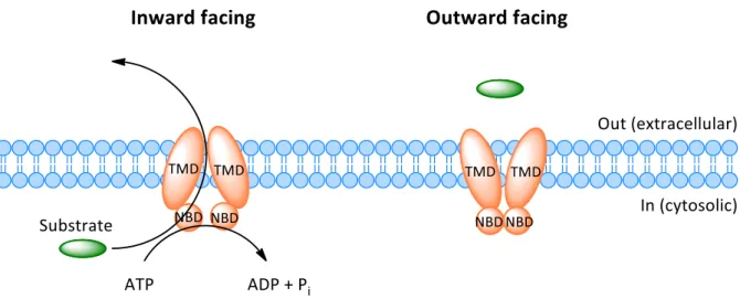
of four to eleven (mostly six) transmembrane helices, and two cytosolic NBDs (Figure 1.1)
[8,11,27,31-34]. In archaea and bacteria, these four domains often exist as distinct subunits,
whereas in eukaryotes usually one NBD and one TMD are fused into half-transporters or all
four domains reside in a single polypeptide chain and form a full-transporter [8,11,31-34]. ABC
importers additionally comprise an extracytoplasmic solute-binding protein involved in the
capture of substrates [12,35].
Figure 1.1. Cartoon of the structural organization of an archetypal ABC exporter composed of two TMDs and two NBDs. Two
conformational states of the transporter – inward facing and outward facing – are depicted to demonstrate the alternating access mechanism of transport. Hydrolysis of ATP to ADP and inorganic phosphate (P
i) powers the transport.
In the last two decades structural biology techniques – X-ray crystallography and cryogenic electron microscopy (cryo-EM) – have provided numerous structures of bacterial and human ABC transporters and enabled the visualization of conformational states at high resolutions.
The structural organization and mechanism of ABC transporters is depicted in Figure 1.1.
Generally, the two TMDs form a translocation pore across the membrane and feature the substrate binding site, whereas the NBDs bind and hydrolyze ATP [10,36-38]. In most ABC transporters the concomitant conformational changes in the NBDs induce a switching of the TMDs between an inward facing and outward facing conformation, thereby translocating the substrate [10,36-38]. It should be noted that there are mechanistic differences between individual ABC transporters, pre-eminently concerning the events at the TMDs, which reflects the variability in the TMDs and differing substrate specificities; even outward-only mechanisms have been proposed for some bacterial subtypes [39].
To date 51 human ABC proteins are known [40]. Due to the progress of the Human Genome
Project and to other mass sequencing efforts in the 1990s, 30 human ABC proteins had been
identified until 1999 [41]. They were divided into seven subfamilies based on similarity in gene
structure (half- vs. full-transporter), on succession of domains, and on sequence homology in
the NBDs and TMDs [34,42-45]. The Genome Nomenclature Committee of the Human
Genome Organization (HUGO) developed a nomenclature system for the human ABC
transporter family, which was implemented in 1999 [37,41]. The seven subfamilies were
assigned the designations ABCA to ABCG. The A and C subfamilies are composed of full-
transporters, the D and G subfamily of half-transporters and the B subfamily contains both full-
and half- transporters; the E and F subfamilies are non-transporter ABC proteins and have no
TMDs [34,42-45]. The aforementioned transporter P-gp – the first eukaryotic ABC transporter
to be discovered – was termed ABCB1. The human ABC transporter subtypes are of differing
medical relevance. The major drug transporters are ABCB1, ABCG2 and ABCC1 – this triad of efflux pumps seems to mostly account for MDR in cancer [16,46,47] and brain diseases [17], and they play a leading role in tissue defense and the bioavailability of drugs [14,15]. At the BBB, which is one of the most restrictive tissue barrier found within the human body, the predominant transporter was shown to be ABCG2 [48-50].
1.2 Structure and Mechanism of ABCG2
In 1990, Chen et al. discovered a novel drug resistance-related membrane protein in a human, doxorubicin-resistant breast cancer cell line [51]. The gene encoding this protein was cloned by Doyle et al. in 1998, who identified the latter as an ABC transporter and named it breast cancer resistance protein (BCRP) [52]. Almost simultaneously, two further articles were published reporting on nearly identical genes expressed in the placenta [53] and in a mitoxantrone- resistant colon carcinoma cell line [54], respectively. Accordingly, the genes were termed ABCP (for the expression in the placenta) and MXR (for mitoxantrone resistance). When the three sequences were eventually compared, they were recognized as essentially identical.
Subsequently, the HUGO Genome Nomenclature Committee termed this transporter ABCG2 [55].
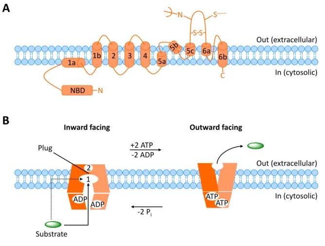
The human ABCG subfamily comprises five half transporters [40] (as in all mammals, only rodents have one more) with an NBD at the N-terminus and a TMD at the C-terminus – this is the reverse configuration compared to other ABC subfamilies, where the NBD is at the C-terminus and the TMD at the N-terminus [34,42,44]. The ABCG2 gene spans over 66 kb and encodes a 75 kDa glycoprotein, composed of 655 amino acids [55,56]. Membrane topology analysis of the ABCG2 protein showed that the TMD consists of six transmembrane helices with the N- and C- termini located intracellularly (Figure 1.2A) [57]. As a half-transporter, ABCG2 must dimerize to form a functional transporter, with disulfide bridges contributing to homodimer stabilization [56].
The three-dimensional structure and the transport mechanism of ABCG2 remained a
conundrum for almost three decades after its discovery. Generally, integral membrane proteins
of mammalian origin are challenging targets for crystallographic studies, because they are often
refractory to crystallization and/or difficult to express and purify in sufficient quantities for
crystallization [58]. As a case in point, analyses of ABCG2 by electron crystallography
provided only low resolution data, precluding insights into the structural details of the transport
process [59-62]. In silico homology models did not provide a remedy, since they were not
accurate, as demonstrated later by high-resolution techniques [63]. After years of speculations
and hazy structural images, a new era was recently inaugurated with the ‘resolution revolution’
in single-particle cryogenic electron microscopy (cryo-EM), mainly driven by direct electron detectors, better microscopes, and improved image processing software [38,58,62]. These novel technologies have made it possible to determine the atomic structure of ABCG2 as well as a broad range of biological macromolecules without crystallization [38,58,62]. In 2017, Locher’s group published the first, long-desired high-resolution structure of ABCG2, determined by cryo-EM [63]. One year later, the same group publicized the cryo-EM structures of ABCG2 in complex with two different inhibitors, one of them presented in the thesis in hand, providing a structural basis of small-molecule inhibition of ABCG2 [64]. Later that year, Locher’s laboratory provided cryo-EM structures of an ABCG2 mutant trapped in ATP-bound and substrate-bound states, further elucidating the transport mechanism of ABCG2 [65].
The aforementioned studies confirmed conjectures that ABCG2 exhibits an inward open conformation and functions according to the above-mentioned ‘switch model’ for ABC transport (Figure 1.1). The established membrane fold of the ABCG2 monomer with six transmembrane helices (TMs 1-6) was refined as depicted in Figure 1.2A. The NBD of each monomer is linked to TM1a via a highly charged amino acid sequence. The longest extracellular loop (EL3), between TM5c and TM6a, contains a single N-glycosylation site and three cysteine residues, two of which form an intramolecular disulfide bond and the third an intermolecular disulfide bridge to the second monomer. The two monomers form a homodimer with twofold symmetry (Figure 1.2B). The TMD interface is formed by TM2 and TM5 of opposing ABCG2 monomers. At the interface, ABCG2 displays two cavities – the first of these (cavity 1) is accessible from the cytoplasmic side of the membrane and is separated from a second, extracellular site (cavity 2) by a plug formed by two leucine residues of opposing monomers.
Cavity 1 was assigned the role of a multidrug binding pocket, accommodating both substrates and inhibitors of ABCG2, the slit-like shape of the cavity favoring flat, polycyclic and hydrophobic molecules. The NBDs are in functionally ‘open’ conformation in the absence of a substrate, with a gap between the catalytically relevant motifs, but they remain in contact.
[63,65]
Upon the binding of a substrate and ATP, conformational changes convert the transporter to an outward facing state (closed NBDs, rotated TMDs with collapsed cavity 1, open ‘leucine plug’
and cavity 2), the substrate being moved to cavity 2, from where it is expelled into the
extracellular space (Figure 1.2B). The binding of two ATP molecules provides the power stroke
for these conformational changes, which are induced via strict conformational coupling of the
NBDs and TMDs. ATP hydrolysis to ADP and inorganic phosphate (P
i) resets the transporter
to the inward facing conformation. [63,65]
Figure 1.2. Cartoon of the structure and mechanism of ABCG2. (A) Membrane topology of the ABCG2 monomer. Cystein
residues forming intra- and intermolecular disulfide bonds are indicated, as is the
N-glycosylation site. (B) Transportmechanism of the ABCG2 homodimer. The substrate enters cavity 1 from the cytoplasm or the inner leaflet of the membrane, the ‘leucine plug’ blocking the access to cavity 2. ATP binding induces the closing of the NMD interface, conversion of the homodimer from the inward to the outward facing state and the release of the substrate to the outside. Hydrolysis of ATP to ADP and P
iis accompanied by the return of the transporter to the inward facing state.
Small-molecule inhibitors were shown to bind to the central multidrug binding site (cavity 1), just as substrates do. Either one or two molecules – depending on inhibitor size and shape – occupy almost the entire volume of cavity 1, thereby preventing the binding of other molecules, such as transporter substrates; in this sense, the inhibitors act competitively. Simultaneously, they lock the inward open conformation, and due to the conformational coupling of the TMBs to the NBDs, the closing of the NBDs and the ATPase activity, i.e. the ATP hydrolysis, are inhibited. The studies by Locher’s group also shed light on the question as to why certain compounds act as substrates, while others are inhibitors of ABCG2. The data corroborated the theory that the key determinant is the binding affinity. Whereas substrates appeared to bind deep in cavity 1, inhibitors were found to fill the cavity completely (one or two molecules) and immobilize the transporter like ‘wedges’, the numerous contacts to the TM helices explaining the increased binding affinity. [64]
N
C NBD
1a 1b 2 3 4
5a
5c 6a
6b
Out (extracellular)
In (cytosolic)
A
B
S S N S
Out (extracellular)
In (cytosolic) 1
2
ADP ADP
Substrate
Plug +2 ATP
-2 ADP Inward facing
ATP ATP Outward facing
-2 P
i1.3 Functions and Substrates of ABCG2
Soon after the discovery of ABCG2 in multidrug-resistant cancer cells, the expression of the protein in normal human tissue was researched in several studies, both on the RNA and on the protein level, the latter by using antibodies against ABCG2, such as 5D3 [66,67]. The tissue distribution pattern emerging from these reports – very similar to ABCB1 – implied that ABCG2 plays a role in the protection against xenobiotics and in the secretion/excretion of endobiotics, apart from conferring MDR to cancer cells [66,67]. Indeed, subsequent studies on abcg2-knockout mice and on loss-of-function polymorphisms of ABCG2 in humans confirmed these physiological functions [68,69].
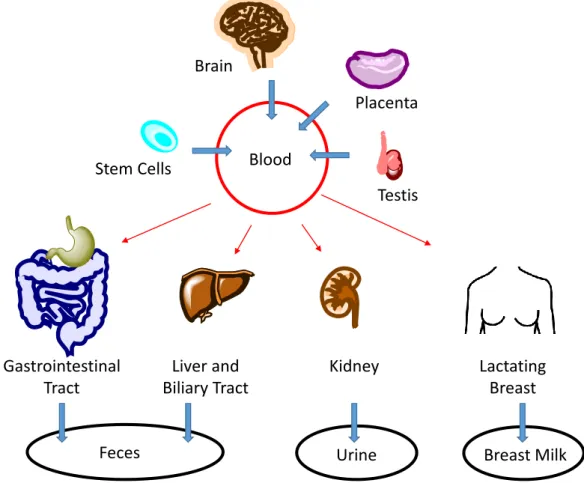
Figure 1.3 shows a schematic overview of the ABCG2 localization in the human organism. The protein is expressed in the apical plasma membrane of epithelial cells throughout the body. It is located in the gastrointestinal tract, the liver and biliary tract, and the kidney, limiting the absorption and promoting the elimination of substrates. The highest expression was observed in the placenta, but also the testes and the brain are protected by ABCG2. The role of ABCG2 in the (lactating) breast is still somewhat enigmatic. Besides epithelial cells, (hematopoietic) stem cells express ABCG2 in the plasma membrane. [66,67,70].
Figure 1.3. Schematic overview of ABCG2 expression in normal tissue. Blue arrows indicate the direction of substrate
transport.
Brain
Liver and Biliary Tract Gastrointestinal
Tract
Kidney Stem Cells
Testis
Lactating Breast Placenta
Blood
Feces Urine Breast Milk
1.3.1 Endogenous Functions
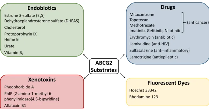
Various studies suggest that transporters involved in drug resistance also have physiological functions: the secretion/excretion of metabolites, nutrients, antioxidants, microbiome products, bile salts, neurotransmitters, other neuro-active molecules, hormones, and signaling molecules [13]. In the case of ABCG2, several endogenous substrates (Figure 1.4) have been discovered.
Studies identified sulfated conjugates of steroid hormones as ABCG2 substrates, suggesting an important role in the biliary and renal excretion of these hormones [71] (steroids are converted to water-soluble conjugates for excretion). Also the precursor of steroid hormones, cholesterol, and other sterols are extruded by ABCG2 [72]. Furthermore, it was discovered that ABCG2 is involved in the cellular export of porphyrins and hemes and their biliary excretion, possibly protecting the liver and other cells from an accumulation of these compounds, which is toxic and tends to occur under hypoxic conditions [73]. The abundant expression of ABCG2 in (hematopoietic) stem cells and the ability to survive under hypoxic conditions are thought to be related [73]. Moreover, ABCG2 is an important urate transporter, allowing excretion in the kidney and gastrointestinal tract, and its malfunction is associated with the gout disease [74].
On a final note, ABCG2 also secretes vitamin B
2(riboflavin) into the breast milk and bile [68].
This finding elucidates the presence of ABCG2 in the mammary gland a little, however it is still controversially discussed why a xenobiotic transporter is used to secrete vitamins into breast milk [68].
1.3.2 Barrier Functions
Various lipophilic compounds rapidly pass through biological membranes. By recognizing and removing (often toxic) xenobiotics from the cell, ABC transporters – mainly ABCG2, ABCB1 and ABCC1 – form our cellular defense system [14,75]. ABCG2 is a gatekeeper at all major physiological barriers [14,75]. The first hurdle after ingestion are the pre-systemic barriers, namely the intestinal barrier (gut-blood barrier) and the hepatic barrier (blood-bile barrier).
Among the exogenous substrates of ABCG2 (Figure 1.4) are the following dietary toxins:
pheophorbide A, a phototoxic chlorophyll metabolite structurally related to porphyrin, which
occurs in plant-derived foods [76], PhIP (2-amino-1-methyl-6-phenylimidazo(4,5-b)pyridine),
a carcinogen formed during the frying and cooking of meat [77], and aflatoxin B1, a carcinogen
produced by certain molds [78]. Studies indicate that ABCG2 prevents or limits the absorption
of the latter compounds in the gut, and mediates the efflux into the bile. Xeno-substrates that
have managed to enter into the systemic circulation are excreted by ABCG2 at the renal barrier
(blood-urine barrier). Also distribution to organs is limited by ABCG2 – it is highly expressed
at the BBB and the blood-testis barrier, protecting these especially sensitive organs. Moreover,
at the blood-placental barrier (maternal-fetal barrier), ABCG2 defends the fetus against toxic materials ingested by the mother. [14,70,75]
The flipside of the coin is that ABCG2, as well as ABCB1 and ABCC1, also accepts numerous drugs as substrates – with severe consequences. Firstly, the oral bioavailability of such drugs is often poor. For instance, the widely used anticancer drug topotecan exhibits low oral bioavailability of around 30%, which was attributed mainly to its property as an ABCG2 substrate [79,80]. Therefore, intravenous application is standard for many ABCG2-transported drugs, which reduces a patient’s quality of life and increases hospital costs, in view of the fact that the patient cannot administer the drug at home [79]. Secondly, drugs that are ABCG2 substrates have only very limited access to the brain – a major obstacle in the treatment of many brain disorders such as HIV infection of the brain or brain cancer, patients with a primary or metastatic brain tumor having a dismal prognosis [17,80]. And thirdly, eponymous for BCRP, expression of the protein in cancer cells confers MDR [70].
Drugs which are substrates of ABCG2 (Figure 1.4) comprise – but are not limited to – anticancer agents such as mitoxantrone, topotecan, methotrexate, and the tyrosine kinase inhibitors imatinib, gefitinib and nilotinib [70,81]; antibiotics like erythromycin [81]; antivirals including anti-HIV drugs such as lamivudine [81]; anti-inflammatory agents like sulfasalazine [81]; and the antiepileptic drug lamotrigine [82]. Noteworthily, also some fluorescent dyes are transported by ABCG2, for instance Hoechst 33342 and rhodamine 123 [81].
Taken together, over 200 transport substrates of ABCG2 have been identified, which considerably overlap with ABCB1 [83].
Figure 1.4. Selected substrates of ABCG2.
ABCG2 Substrates Endobiotics
Estrone 3-sulfate (E
1S)
Dehydroepiandrosterone sulfate (DHEAS) Cholesterol
Protoporphyrin IX Heme B
Urate Vitamin B
2Xenotoxins
Pheophorbide A
PhIP (2-amino-1-methyl-6- phenylimidazo(4,5-b)pyridine) Aflatoxin B1
Drugs
Mitaxantrone Topotecan Methotrexate
Imatinib, Gefitinib, Nilotinib Erythromycin (antibiotic) Lamivudine (anti-HIV)
Sulfasalazine (anti-inflammatory) Lamotrigine (antiepileptic)
Fluorescent Dyes
Hoechst 33342 Rhodamine 123
(anticancer)
1.3.3 Function at the Blood-Brain Barrier
The BBB is one of the most restrictive barriers found within the body [84], which comes to no surprise, considering that the brain is our most critical and most sensitive organ [85]. The BBB is formed primarily by a single monolayer of endothelial cells lining the brain capillaries (Figure 1.5), and represents a dual barrier: physical and biological [84,85]. In contrast to endothelia in peripheral capillaries, these endothelial cells are connected by tight junctions, making the BBB the densest physical barrier in the body (together with the urinary bladder) [17,84,85]. Therefore, paracellular transport of substances into the brain is negligible and the brain endothelium has to be penetrated via the transcellular route, which is restricted biologically by the expression of efflux pumps on the surface of the endothelial cells [17,18,84].
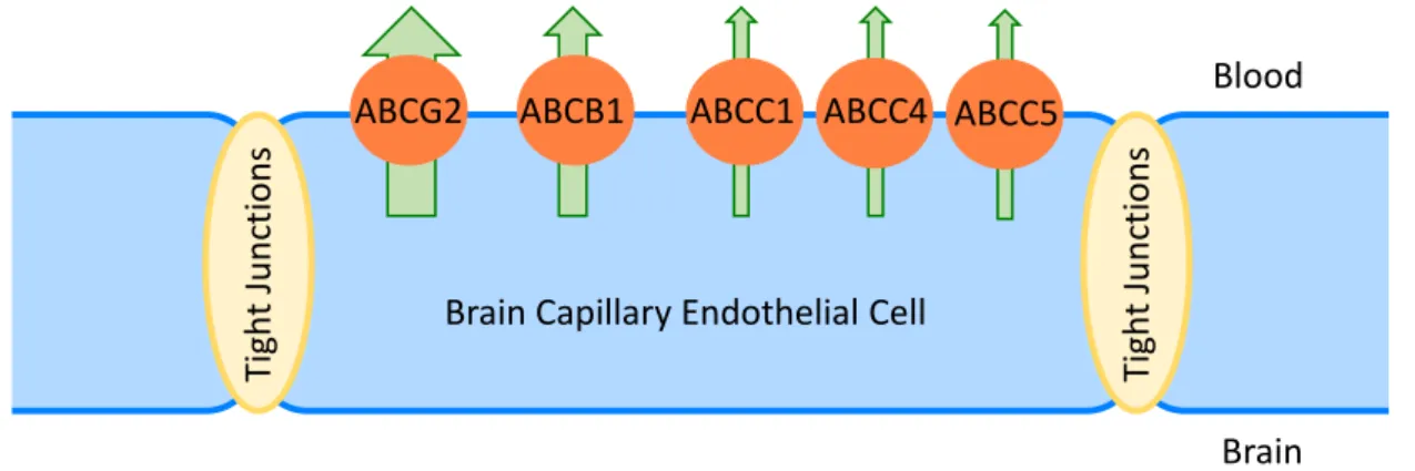
Quantitative (transcriptomic and proteomic) analyses of transporter expression in human brain microvessels revealed that ABCG2 is the most expressed efflux transporter at the BBB, followed by ABCB1 and, to a much lesser extent, by some members of the ABCC subfamily (Figure 1.5) [48-50]. It is worth mentioning that in mice, by contrast, ABCB1 was found to be predominant at the BBB, which led to the initial assumption that this is also the case at the human BBB. This interspecies difference questions the reliability of extrapolating animal data for predicting brain distribution of ABCG2 and ABCB1 substrates in humans [48-50].
Figure 1.5. Expression of ABC transporters (biological barrier) in brain capillary endothelial cells, which are connected by
tight junctions (physical barrier) and constitute the principal cellular element of the BBB.
The BBB is almost impermeable to drugs – approximately 98% of small molecule and all large molecule medications are normally excluded from the brain [84]. This is a key mechanism of drug resistance in brain diseases – a complex phenomenon, to which ABCG2 and ABCB1 contribute in two ways: firstly, through their constitutive expression at the BBB and secondly by overexpression (at the BBB or in the target tissue) in some brain diseases, which can be triggered by the activation of nuclear receptors [17,18]. A detailed description of the ABC transporter regulation can be found in an excellent review [86].
Blood
Brain
Ti ght Jun cti ons
ABCG2 ABCB1 ABCC1 ABCC4 ABCC5
Brain Capillary Endothelial Cell
Ti ght Jun cti ons
In short, the expression of ABCG2 and ABCB1 has been shown to be the main reason for the difficulties in treating brain diseases [17]. Therefore, effective strategies to overcome the restrictions by the BBB, especially ABCG2 and ABCB1, have to be developed.
1.3.4 Function in Multidrug Resistance in Cancer
MDR in cancer is the ability of cancer cells to become simultaneously resistant to multiple, structurally and functionally unrelated drugs – a significant impediment to successful chemotherapy [16,87]. There are several mechanisms that cause drug resistance, including reduced drug uptake, e.g. by endocytosis, activation of detoxifying systems, such as DNA repair and cytochrome P450 oxidases, and defective apoptotic pathways [16]. The most commonly encountered mechanism is an increased efflux of cytostatics mediated by ABC transporters, mainly ABCG2 and its functional homologs ABCB1 and ABCC1 [46]. The notorious activity of ABCG2 in extruding anti-tumor drugs in a variety of cancer cell lines in vitro has been firmly established. By contrast, the clinical relevance of ABCG2 is still sketchy and there have been conflicting reports [70,83]. Studies on clinical samples usually examine the expression of ABCG2, either on the RNA or protein level, and correlate the latter with the outcome of a drug treatment regimen. Most of such studies were carried out for hematological malignancies. There is considerable evidence that ABCG2 (as well as ABCB1) is associated with a poor result in acute myeloid leukemia (AML) and diffuse large B-cell lymphoma, whereas for acute lymphoid leukemia the evidence is less convincing [70,83,88]. Yet, even for AML there exist contradictory studies [70]. Also (leukemic) cancer stem cells express high levels of ABCG2 (and ABCB1), which has been found to correlate with a meager response to chemotherapy [70,83,88]. In solid tumors, the picture is mixed; some studies of solid tumors showed correlation between ABCG2 expression and prognosis, such as for lung or gastric cancer, while others did not. For many solid tumor types, there are not enough studies to draw compelling conclusions [70,83,88].
After almost half a century of research on drug transporters, launched with the discovery of
ABCB1 in the 1970’s, the role of ABC transporters in clinical MDR has still not yet been
clarified, especially that of ABCG2, the latest major drug transporter discovered. Several
reasons have been invoked for this deficiency. A great problem is the detection of ABCG2
expression. The detection is variable across laboratories and often not validated at all or
validated with a cell line expressing extremely high levels of ABCG2, which one would not
expect in clinical samples [70,89]. Besides, ABCG2 is often detected on the RNA level, which
does not always directly correlate with the protein level [90]. In addition, ABCG2 has several
polymorphic variants, which was not taken into consideration in all studies [90]. A second
major challenge is the complexity of MDR mechanisms. Often, the expression of only one
transporter was examined, though the expression of multiple ABC transporters in a single tumor type is common [47,70]. In addition, studies indicate that several other survival factors, such as anti-apoptotic proteins, are up-regulated along with ABCG2, sometimes via the same regulatory machinery [90]. Thus it was suggested that ABCG2 expression by itself is not the sole mechanism behind MDR but rather a marker [90], which is supported by the intriguing observation that ABCG2 expression correlates positively with a resistant phenotype even if the drugs employed are not ABCG2 substrates [83,90].
Apart from chemotherapy, ABCG2 expression is also linked to the failure of photodynamic therapy, since some tumor-selective photosensitizers are substrates of ABCG2 [91].
All in all, studies have shown many correlative links between ABCG2 expression and clinical outcome, yet these have not proved causal. Despite this caveat, it is interesting that the tumor types most refractory to chemotherapy are among those with the highest levels of ABCB1 or ABCG2 expression [47], which suggests that overcoming these transporters may contribute to overcoming MDR in cancer.
1.4 Applications of ABCG2 Inhibitors
In parallel to mechanistic and functional studies, research has also focused on finding inhibitors of ABCG2. Given the wide distribution of the protein in the human body, its various functions including the involvement in diseases and drug response, and all the still open questions in these matters, several applications for ABCG2 inhibitors are obvious. On one hand, they are needed as molecular tools to gain further insight into the mechanism of ABCG2, its role in the human body, and its interaction with drugs. On the other hand, they may be of interest for improving the oral bioavailability of drugs and for overcoming drug resistance in brain diseases and in cancer.
1.4.1 Applications as Molecular Tools
1.4.1.1 Tools for Fundamental Research
Comprehension of the molecular mechanism of ABC transporters is essential for understanding
and modulating their function. High-resolution structural data offer an opportunity to model
compounds into the binding pocket, which may facilitate a structure-based inhibitor and drug
design or the identification of substrates [63]. ABC transporters are highly dynamic systems
and a single structure is merely a snapshot of one conformational state of the protein as it moves
its substrate through the membrane [38]. Thus, more than one structural image is necessary in
order to understand the transport mechanism in detail, especially also with the protein trapped
in a substrate-bound, ATP-bound or inhibitor-bound state. Therefore, inhibitors of ABC
transporters suitable for ‘complexing’ with the respective transporter in structural biology experiments are required. In the case of ABCG2, structures of the protein in complex with two different inhibitors have been published (see Section 1.2) [64], one of which will be presented in Chapter 2 of this thesis.
Fundamental research into the functions of ABCG2 depends heavily on experiments with abcg2-knockout mice. The use of selective ABCG2 inhibitors, i.e. a chemical instead of a genetic disruption of the ABCG2 function, complements this approach. Moreover, the application of dual ABCB1/ABCG2 inhibitors is an alternative to double-knockout mice [90,92].
Although the results of animal studies are valuable, the extrapolation to humans is difficult due to interspecies differences. Experiments in humans are restricted to non-invasive methods, positron emission tomography (PET) being used most frequently [93]. For this purpose, a PET imaging probe that interacts with the ABC transporter is necessary: a substrate or inhibitor labeled e.g. with
11C or
18F [93]. Usually, transporter imaging probes used in PET studies are not molecules designed de novo, but are marketed drugs or natural food constituents (or metabolites thereof), which are repurposed for this application [93]. The first in vivo evidence for ABC transport activity at the human BBB was produced via PET imaging by Sasongko et al. in 2005. The distribution of the ABCB1-selective substrate [
11C]verapamil (an antiarrhythmic drug) to the brain was increased during the inhibition of ABCB1 by cyclosporin A (an immunosuppressant drug) – a proof of ABCB1 activity at the human BBB [94]. This is an excellent example of how inhibition of an ABC transporter enabled pioneering fundamental research.
The visualization of ABCG2 at the human BBB is hampered by the lack of an ABCG2-selective
substrate probe. Langer’s group, who has conducted extensive research on imaging ABC
transporter functions, developed a protocol in which they used a dual ABCB1/ABCG2 substrate
probe and eliminated the influence of ABCB1 efflux by inhibiting this transporter with
tariquidar (Figure 1.6) [95,96]. For lack of a clinical ABCG2 inhibitor, another obstacle, they
performed experiments on subjects who were either non-carriers or heterozygous carriers of an
ABCG2 polymorphism associated with a reduced function of the protein [96]. Indeed, the
distribution of the substrate to the brain was increased in the carriers of the polymorphism – the
first verification of ABCG2 activity at the human BBB in vivo, reported only four years ago
[96]. To date, clinical ABCG2 inhibitors are still quite scarce and selective as well as dual
ABCG2/ABCB2 inhibitors (depending on the experimental design) are urgently needed for the
PET experiments with labeled substrate probes, as strikingly demonstrated by the elaborate
workaround applied by Langer and co-workers. Furthermore, selective ABCG2 inhibitors
amenable to PET labeling are of interest as PET imaging agents [97].
1.4.1.2 Tools for Drug Discovery and Development
During the discovery and development of new drugs, interactions of the latter with ABC transporters need to be assessed for several reasons. Firstly, it is likely that these drug efflux pumps limit the absorption and distribution of potential medications and promote their elimination; in case of anticancer agents, the uptake by cancer cells may be restrained. Thus, pharmaceutical companies include ABCB1 and ABCG2 transporter assays in early screens in drug discovery to eliminate drug candidates that are substrates of these proteins [98], which can be considered ‘antitargets’ in this respect [99]. Secondly, by contrast, compounds that do have substrate properties are selected in case drugs are not intended to enter the brain. Third generation antihistaminic agents, for example, were designed as substrates of ABC transporters and hence are devoid of central side effects like sleepiness [99,100]. And thirdly, relevant in (pre-) clinical drug development, substrate and inhibitor properties of drugs can cause drug- drug interactions (DDIs). When two drugs which interact with the same ABC transporter are co-administered, one drug may induce transporter inhibition or saturation and thereby change the disposition of the other drug [101]. This phenomenon often causes severe adverse drug reactions in patients [102]. Therefore, the importance of assessing drug-transporter interactions was emphasized in a statement of the International Transporter Consortium in 2010 [102], on the basis of which regulatory authorities – the US Food and Drug Administration (FDA) and the European Medicines Agency (EMA) – recommend in their guidelines that all investigational drugs should be evaluated as substrates or inhibitors of ABCB1 and ABCG2 [103,104].
In the assessment of drug-transporter interactions, prototypical ABC inhibitors are important
molecular tools. For instance, in vitro trans-epithelial transport assays, complemented with the
use of selective inhibitors, allow probing for ABC transporter substrates [102]. On the
examination of DDIs, in vivo studies may be recommended if a drug candidate turns out to be
an ABC transporter substrate or inhibitor in vitro [102]. Regulators do not endorse animal
studies due to pronounced interspecies differences [105] – DDI studies in humans are the ‘gold
standard’ [106]. Healthy volunteers are treated with the drug of interest and an ABC inhibitor,
in case the drug is a substrate. Vice versa, if the drug in question is an inhibitor, it is
administered together with a substrate. Then, the drug concentration in the blood is assessed
and compared to the results achieved when the drug is dosed alone [106]. Some DDIs may lead
to changes in the organ distribution of drugs (e.g. brain, liver, kidneys) without affecting plasma
concentrations. To elucidate potential DDIs on a tissue level, imaging techniques such as PET
can be used. If the drug of interest is an inhibitor, it could be given in an unlabeled form to
assess its influence on the distribution of a PET imaging substrate probe [107]. If the drug is a
substrate, it may be radiolabeled and used in combination with a transporter inhibitor [107].
As is the case in fundamental research, the shortage of clinically applicable, specific model inhibitors and substrates of ABC transporters as well as probes suitable for imaging studies is well recognized, and represents an area of research that warrants moving forward with drug- transporter interaction and DDI studies [102].
1.4.1.3 Clinical Diagnostic Tools
Though the role of ABCG2 expression in MDR has not yet been fully elucidated, ABCG2 has been identified as a tumor marker, which could be exploited in the diagnosis of cancer [108].
By analogy with ABCB1 [93], the detection of ABCG2 expression in tumors could be achieved by imaging techniques such as single-photon emission computed tomography (SPECT) or PET, using a labeled substrate probe and an ABCG2 inhibitor, which should increase the accumulation of the imaging probe in ABCG2 expressing tumors. Measuring ABCG2 expression in tumors may also be useful for the prediction and assessment of MDR and response to chemotherapy [47]. This would be a valuable application of clinical ABCG2 inhibitors.
1.4.2 Applications as Prospective Drugs
To date, no inhibitor targeting an ABC transporter has received marketing authorization by regulatory agencies [47,99,109], though some marketed drugs addressing other targets show off-target, inhibitory activity at ABC transporters [109]. Nevertheless, several potential therapeutic applications of ABCG2 inhibitors have been and are still being explored.
1.4.2.1 Drugs for Improving Oral Bioavailability
In the light of ABCG2 expression at the intestinal and hepatic barrier, which was shown to limit
the oral bioavailability of (anticancer) drugs, the idea of using ABCG2 inhibitors to improve
oral bioavailability has been investigated [79,80,110,111]. Preceding preclinical and clinical
studies on ABCB1 had already been successful, the most prominent example being the
improvement of the oral bioavailability of the anticancer agent paclitaxel when combined with
the ABCB1 inhibitor cyclosporin A [79,80,110,111]. Similarly, the oral bioavailability of
topotecan was improved when co-administered with the dual ABCB1/ABCG2 inhibitor
elacridar (Figure 1.6) in clinical research [112,113]. Since topotecan is a selective substrate for
ABCG2, the results suggest that the interaction of the two compounds is due to inhibition of
ABCG2 [112,113]. Given the co-expression of ABCB1 and ABCG2 at the intestinal and
hepatic barrier and their overlapping substrate ‘specificities’, dual ABCB1/ABCG2 inhibition
is of interest for dual substrates, as demonstrated in a clinical trial using the dual
ABCB1/ABCG2 substrate irinotecan in combination with the tyrosine kinase inhibitor
gefitinib, which also inhibits ABCB1 and ABCG2 [114]. Together, these findings encourage
the application of ABCG2 inhibitors to improve oral bioavailability. The reservation must be
made, however, that ABCG2 inhibition can potentially increase brain uptake, which would be undesirable because of possible (neuro)toxic side effects, yet advantageous in case the tumor is located in the brain [111].
1.4.2.2 Drugs for Improving Brain Penetration
Due to the restrictions by the BBB, the development of drugs targeting the brain shows the
highest failure rates in phase I and II clinical trials: 93% to 98% [84]. Clinical strategies to
overcome the BBB comprise physical methods such as the administration of drugs directly into
the brain region of interest, requiring invasive surgery, and osmotic disruption, e.g. with a
mannitol solution, implying increased and unwanted leaking of plasma material into the brain,
which is severely toxic for the brain [84]. Neither method is ideal. An alternative in order to
specifically increase the brain penetration by drugs which are substrates of ABC transporters is
the inhibition of these efflux pumps by the use of inhibitors. In 2002 a first proof-of-concept
study in mice demonstrated that the paclitaxel concentration in the brain, which is very low
after an intravenous injection, was increased by co-administration with the ABCB1 inhibitor
valspodar [115]. Whereas paclitaxel alone did not affect the volume of an implanted brain
tumor, co-administration with valspodar reduced the tumor volume by 90% [115]. Several
follow-up preclinical studies in mouse or rat models with different substrate/ inhibitor
combinations confirmed the principle and showed that it is also applicable to ABCG2
[111,116]. In most cases, however, ABCB1 and ABCG2 work in concert to transport a substrate
drug from the brain back into the blood, which can be explained by their prominence and
substrate overlap [111,116]. Studies with knockout mice showed that these two export pumps
have a cooperative effect at the BBB: the removal of either transporter alone does not result in
a significant increase in brain penetration of dual substrates, because it is compensated by the
respective other transporter, whereas the complete removal of both efflux pumps has a
disproportional effect on brain uptake [111,116]. Consequently, the largest increase in brain
accumulation of dual substrate drugs should be achieved by the inhibition of both ABCB1 and
ABCG2 [111,116]. This was verified in extensive preclinical research in rodents, mostly using
the dual inhibitor elacridar in combination with various dual substrate drugs, not only
cytostatics but also drugs for neurological disorders [111,116,117]. Because of interspecies
differences, these results do not necessarily translate to humans. The confirmation that this
principle – inhibition of ABC transporters to increase drug penetration – is applicable to humans
was provided by PET imaging. The aforementioned PET studies, proving the activity of
ABCB1 and ABCG2 at the human BBB, were recently followed by a couple of clinical trials
focused on improving drug exposure to the brain. [
11C]Erlotinib, a tyrosine kinase inhibitor,
was used as a PET substrate probe. Its brain uptake is limited as it is a substrate of both ABCB1
and ABCG2. The first such study, two years ago, was not successful, which was attributed to
the use of elacridar as a dual inhibitor, as practiced before in numerous preclinical trials [118].
Elacridar is a much more effective inhibitor of ABCB1 than it is of ABCG2, meeting the needs of the rodent BBB, where ABCB1 predominates over ABCG2. Since the situation is vice versa in humans, it was speculated that a better result could be achieved using a dual inhibitor with high potency at ABCG2. Indeed a proof-of-concept study carried out by Langer’s group only one year ago accomplished a much better outcome [119]. They found out that erlotinib is not only a dual substrate, but at a supratherapeutic dose it functions as an inhibitor of ABCB1 and ABCG2. Using erlotinib, they were able to achieve an increase in brain distribution of [
11C]erlotinib [119]. However, the clinical application of supratherapeutic doses of erlotinib may be hampered by safety concerns [119], which is why further research on clinically applicable, potent dual ABCB1/ABCG2 inhibitors is desirable.
Together, the preclinical and clinical data make a compelling case for the application of ABCG2 and dual ABCB1/ABCG2 inhibitors as drugs to improve brain penetration.
1.4.2.3 Drugs for Overcoming Multidrug Resistance in Cancer
As the human ABC transporters were first discovered in cancer cells, where they extrude anticancer drugs, the use of ABC inhibitors to improve the response to chemotherapy stood to reason. Already in the 1980’s, less than 10 years after the discovery of ABCB1, the first clinical trials on combination chemotherapy – an anticancer drug together with an ABCB1 inhibitor – were carried out and dozens more followed [46]. Nevertheless, this is by far the most uncertain and most disputed potential application of ABC inhibitors – no wonder, considering that the role of ABC transporter expression in MDR is still unclear.
Initially, drugs which had been approved for other indications and noted to inhibit ABCB1 (first
generation inhibitors) were used in clinical trials, for instance verapamil or cyclosporin A
[46,47,120,121]. Owing to their low inhibitory potency, higher doses than those required for
their individual therapeutic activity were applied, which consequently led to unacceptable toxic
side effects [46,47,120,121]. Second generation inhibitors were analogs of the initial agents
developed solely for the purpose of altering drug resistance, the cyclosporin derivative
valspodar being tested most frequently in clinical trials [46,47,120,121]. These inhibitors were
more potent and less toxic, but induced pharmacokinetic interactions that limited the clearance
and metabolism of anticancer drugs and thereby elevated their plasma concentrations beyond
acceptable toxicity, for instance by inhibiting cytochrome P450 enzymes [46,47,120,121]. This
usually prompted dose reductions of the anticancer agents down to ineffective levels
[46,47,120,121]. Third generation inhibitors, such as elacridar and tariquidar, were designed
with increased specificity to overcome these disadvantages [46,47,120,121]. Indeed, they
displayed either no or only minor pharmacokinetic interactions with different anticancer agents,
yet several clinical studies failed to show a conclusive benefit of the addition of an ABCB1 inhibitor to standard therapy and some were closed early owing to toxicity [46,47,120,121].
Therefore, the conduction of further trials was generally discouraged. However, the point must be made that the clinical trial designs were often flawed. Typically, ABCB1 was not assayed in tumors and there was no selection of patients whose tumors displayed overexpression of ABCB1 [46,120,122]. Furthermore, many studies enrolled patients following multiple lines of therapy, so that drug resistance was likely to be multifactorial, yet other drug efflux mechanisms or a lack of uptake mechanisms were not controlled [46,120,122]. Thus the idea of overcoming MDR by inhibiting ABCB1 was not adequately tested [46,120,122].
Given the lack of success with the preceding trials on ABCB1 inhibitors and the restricted knowledge about which cancers express ABCG2, only very few clinical trials aimed at overcoming ABCG2-mediated drug resistance have been carried through, and the picture is very similar to ABCB1. In the last decade, a couple of trials investigated the combination of topotecan with the tyrosine kinase inhibitor lapatinib, which also inhibits ABCG2 [88,123,124].
Unfortunately, the results did not demonstrate an advantage over the application of topotecan alone, although ABCG2 expression was detected [88,123-126]. This failure was attributed mainly to other mechanisms being involved in the resistance and also toxicity was a problem in some cases [88,123,124].
It is worth noting that an explanation for toxicity problems despite the lack of pharmacokinetic interactions could be the ABCB1/ABCG2 inhibition in cells, where these transporters are known to play a protective role, e.g. in bone marrow stem cells, yet in many cases there was no clear evidence for such an effect [120,123].
Taken together, there is still no definite answer to the question if inhibitors of ABC transporters can effectively reverse drug resistance in humans. It could be speculated, however, that in case proper patient populations were selected, whose tumors show ABC transporter-dominated resistance, successful reversal of drug resistance by ABC inhibitors might be feasible [46,47,120,124]. Given the expression of multiple ABC transporters in tumors, compounds that inhibit ABCB1 and ABCG2 (and ABCC1) may be advantageous [120].
Moreover, apart from chemotherapy, also photodynamic therapy (PDT) could profit from ABCG2 inhibition. As mentioned above, a couple of PDT agents were shown to be ABCG2 substrates and high ABCG2 expression was correlated with decreased potency of the latter [91].
Thus, ABCG2 inhibitors may improve the response to PDT.
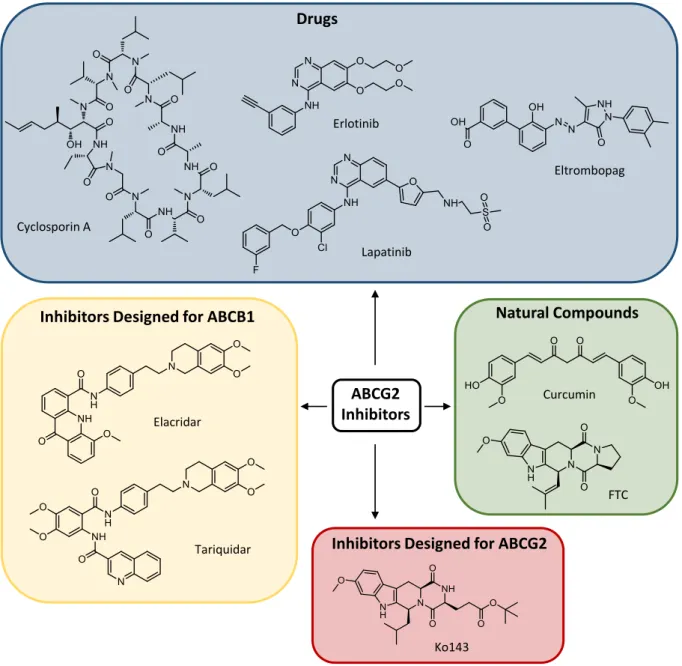
1.5 Reported ABCG2 Inhibitors and Problem Statement
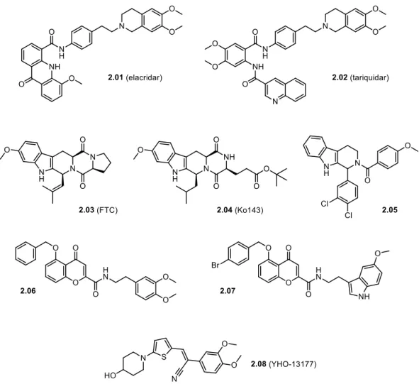
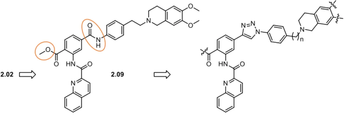
The research into inhibitors of ABC transporters began in 1981 with the serendipitous finding that the calcium channel blocker verapamil inhibited ABCB1-mediated drug efflux in resistant tumor cells [116,127]. Subsequently, numerous chemicals were screened for their potential to inhibit ABCB1 [116] and, as outlined in the previous section, a couple of ABCB1 inhibitors with higher potency were developed, in particular the two benzanilides elacridar (by Glaxo) [128] and tariquidar (by Xenova) [129] (Figure 1.6). Both are non-marketed, experimental compounds, which have been used in clinical trials.
After the discovery of ABCG2, the ability of the hitherto developed ABCB1 inhibitors to impede also ABCG2-mediated drug efflux was investigated. Indeed, both elacridar [130] and tariquidar [131] happen to inhibit ABCG2, albeit less potently than ABCB1. The first selective inhibitor of ABCG2 reported was the fungal toxin fumitremorgin C (FTC) [132,133]
(Figure 1.6), which, however, is precluded from the use in vivo due to its severe neurotoxic side effects. A number of other natural and dietary compounds were identified as ABCG2 inhibitors, primarily flavonoids [134,135]. Of these constituents, only curcumin (Figure 1.6) was evaluated clinically as an ABCG2 inhibitor [103,136]. This substance was also shown to inhibit ABCB1 and ABCC1, yet with lower potency [136]. Disadvantageously, it shows poor absorption and extensive metabolic instability [137]. Furthermore, various drugs on the market were investigated for interaction with ABCG2. The immunosuppressant cyclosporin A (Figure 1.6) is a very unspecific ABCG2 inhibitor, since it also inhibits ABCB1 and other transporters, as well as cytochrome P enzymes [92,138]. With tyrosine kinase inhibitors emerging as anticancer drugs in the beginning of this century, they were evaluated in this regard and some turned out to be ABCG2 inhibitors, notably erlotinib [139] and lapatinib [103,140] (Figure 1.6), which were used in some of the clinical trials mentioned in Section 1.4. At higher concentrations, they inhibit ABCB1 as well [103,139,140]. Recently, the platelet-increasing agent eltrombopag (Figure 1.6) was demonstrated to be an ABCG2 inhibitor [92,141]. However, the use of drugs for in vivo experiments may cause safety problems due to the effects they were originally designated for.
Of course, medicinal chemists did not remain idle and synthesized compounds designed solely
for the purpose of inhibiting ABCG2. Soon after the discovery that the mycotoxin FTC is a
selective ABCG2 inhibitor, analogs thereof, featuring lower toxicity and higher potency, were
reported [142]. In particular Ko143 [142] (Figure 1.6), which is still among the most potent
ABCG2 inhibitors published so far, has to be pointed out. Later it was shown that it is not
concentrations [143]. A drawback of Ko143 is its poor metabolic stability caused by the labile ester group [143,144]. Since the report on Ko143, many more ABCG2 inhibitors have been created, mostly derivatives of the aforementioned compounds. They were extensively reviewed elsewhere [135,145,146] and some prominent examples will be highlighted in Chapter 2.
Nevertheless, to the best of the author’s knowledge, none of these have been developed for the use in humans. Hence, the aforementioned drugs and dietary compounds have to be repurposed, with all the detriments indicated above. For clinical DDI studies, for instance, the FDA recommends the use of cyclosporin A, eltrombopag and curcumin as ABCG2 inhibitors [147].
In some articles on the subject, the utilization of lapatinib is suggested [92,103].
Figure 1.6. Structures of selected inhibitors of ABCG2.
ABCG2 Inhibitors Drugs
Inhibitors Designed for ABCB1 Natural Compounds
Inhibitors Designed for ABCG2
Cyclosporin A
Lapatinib Erlotinib
Eltrombopag
Curcumin
FTC
Ko143 Elacridar
Tariquidar