Pharmacological and Chemogenetic
Characterization and Modulation of the Brain Neuropeptide S and Oxytocin
Dissertation zur Erlangung des Doktorgrades der Naturwissenschaften (Dr. rer. nat.)
an der Fakultät für Biologie und Vorklinische Medizin der Universität Regensburg
vorgelegt von Thomas Grund
aus Arnstadt
im Jahr 2017
Summary I
Summary
Anxiety is a natural response to a real or perceived threat that has been conserved throughout evolution. From this perspective, sensation of anxiety is viewed as an adaptive behavioral state, which occurs in response to signals of danger. However, excessive or inappropriate anxiety can become pathological. Anxiety disorders represent one major burden of our modern society, and the available treatment options are limited by adverse side effects.
During the last decades, research centered on anxiety disorders has focused on neuropeptides.
Neuropeptide S (NPS) and oxytocin (OXT) represent potential neuropeptide candidates for the treatment of various psychopathologies, including anxiety disorders due to their potent anxiolytic profile. Both signal through G protein-coupled receptors and thereby regulate complex neuronal signaling pathways. How these pathways contribute to behavioral and physiological effects of NPS and OXT remains to be elucidated. Therefore, in the present thesis I aimed to reveal, (i) whether NPS acts on the brain OXT system to exert its anxiolytic properties, (ii) whether the anxiolytic profile of NPS is mediated in a Gq pathway-dependent manner, and (iii) to characterize the spatiotemporal characteristics of chemogenetically induced OXT neurons activation.
NPS receptors (NPSR) are found within the hypothalamic paraventricular nucleus (PVN), where OXT is synthesized besides the supraoptic and accessory nuclei. Besides anxiolysis, NPS and OXT share other effects, such as the reversal of social fear, the attenuation of aggressive-like behavior as well as the inhibition of food-intake and anti-nociceptive properties as demonstrated in rodents. These behavioral similarities, together with the neuroanatomical overlapping of the NPSR and OXT expression within the PVN, led me to hypothesize that NPS effects are mediated by acting on the OXT system within the PVN. Herein, I present a chain of evidence that the effects of NPS within the PVN are mediated via actions on local OXT neurons
Summary II
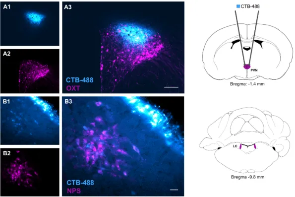
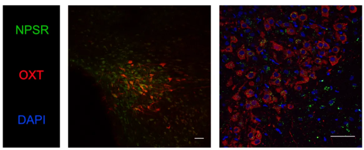
in male Wistar rats. Retrograde tracing revealed that NPS-immunoreactive neurons originating within the Locus coeruleus innervate the PVN. Moreover, fluorescence-activated cell sorting identified NPSR expression in PVN-OXT neurons. NPS reliably induced transient Ca2+-influx in a subpopulation of OXT neurons - an effect mediated via the NPSR. Moreover, intracerebroventricular (icv) NPS evoked a significant release of OXT within the PVN as assessed by microdialysis in combination with a highly sensitive radioimmunoassay. Both, chemogenetic silencing of the endogenous OXT system using designer receptors exclusively activated by designer drugs (DREADD) as well as pharmacological blockade of brain OXT receptors (OXTR) abolished the anxiolytic effect of NPS infused icv. These findings provide the first evidence for an intra-hypothalamic mechanism involving NPSR-expressing OXT neurons in the potent anxiolytic profile of NPS, and fill an important gap in our neurophysiological understanding of brain neuropeptide interactions in the context of regulation of emotional behaviour within the hypothalamus.
Next, I performed studies in order to extend our knowledge on the intraneuronal mechanisms underlying the anxiolytic profile of NPS by studying signaling pathways downstream of NPSR activation. Here, bilateral microinfusion of NPS into the medial amygdala (MeA) of male adult Wistar rats reduced anxiety-related behavior on the elevated plus maze and the open field.
Moreover, icv infusion of NPS evoked expression as well as increased phosphorylation of Ca2+/calmodulin-dependent kinase II in the amygdala, and subsequent phosphorylation of the mitogen-activated protein kinase ERK1/2. Importantly, NPS-induced anxiolysis in the MeA was prevented by local inhibition of phospholipase C using the specific inhibitor U73122. In contrast, local pharmacological blockade of adenylyl cyclase signaling using the specific inhibitor 2’,5’-dideoxyadenosine failed to inhibit the anxiolytic effect of NPS infused into the MeA. Hence, NPS promotes acute anxiolysis within the MeA dependent on NPSR-mediated phospholipase C signaling.
Finally, in order to validate an innovative and highly specific tool for activating OXT neurons
Summary III
within the PVN in a behavioral context, I performed a series of experiment using the DREADD technique. Acute chemogenetic activation of PVN-OXT neurons resulted in enhanced release of OXT within the PVN within one hour as assessed using intracerebral microdialysis. Further, DREADD activation reduced anxiety-related behavior in the light/dark box, and increased self- grooming behavior in the home cage. In addition, chemogenetic activation of PVN-OXT neurons marginally improved ethanol-induced locomotor deficits.
In summary, my experimental data advance our understanding of the mechanisms by which NPS promotes anxiolysis and the temporal release patterns of OXT following chemogenetic activation and its behavioral relevance.
Zusammenfassung IV
Zusammenfassung
Aus evolutionsbiologischer Sicht ist Angst eine konservierte, natürliche Reaktion auf eine reale oder gefühlte Bedrohung. Allerdings können exzessive bzw. unangepasste Angstzustände ein pathologisches Ausmaß annehmen. Angsterkrankungen repräsentieren daher eine der Hauptbelastungen unserer modernen Gesellschaft. Da die derzeitigen Behandlungsmethoden limitiert und von Nebenwirkungen geprägt sind, rückten Neuropeptide in den letzten Jahrzehnten mehr und mehr in den Fokus der Wissenschaft. Aufgrund ihrer potenten, angstlösenden Wirkung repräsentieren Neuropeptid S (NPS) und Oxytocin (OXT) zwei mögliche Kandidaten zur Behandlung von Angsterkrankungen. Beide Neuropeptide binden an G-Protein- gekoppelte Rezeptoren und regulieren dadurch komplexe neuronale Signalwege. Daher war das Ziel meiner Doktorarbeit zu erforschen (i) welchen Einfluss OXT-Neurone im Hypothalamus auf die angstlösende Wirkung von NPS haben, (ii) welche intrazellulären Signalkaskaden der anxiolytischen Wirkung von NPS zugrunde liegen, und (iii) den zeitlichen Ablauf einer chemogenetischen Aktivierung der OXT-Neurone zu charakterisieren.
NPS-Rezeptoren (NPSR) werden im hypothalamischen Nucleus paraventricularis (PVN) exprimiert und zeigen eine morphologische Überlappung mit OXT-Neuronen, die sowohl im PVN also auch dem Nucleus supraopticus und den Nucleus accessorius lokalisiert wurden.
Neben ihrer angstlösenden Wirkung reduzieren sowohl NPS als auch OXT soziale Angst und aggressives Verhalten in Ratten und Mäusen. Darüber hinaus mindern beide Neuropeptide die Nahrungsaufnahme und hemmen die Schmerzwahrnehmung in Ratten. Aufgrund der ähnlichen Verhaltenseffekte beider Neuropeptide sowie der neuroanatomischen Überlappung der neuronalen NPSR- und OXT-Expression untersuchte ich die Fragestellung, ob NPS- induzierte Effekte über das Oxytocin-System im PVN vermittelt werden. Mittels einer retrograden Markierung konnte ich zeigen, dass NPS-immunoreaktive Neurone aus dem Locus
Zusammenfassung V
coeruleus zum PVN projizieren. Darüber hinaus bewies Fluoreszenz-aktivierte Zellsortierung, dass der NPSR in OXT-Neuronen des PVN exprimiert wird. In einer Subpopulation von OXT- Neuronen induzierte NPS durch die Bindung an seinen Rezeptor einen Anstieg des intrazellulären Calcium-Spiegels. Durch intrazerebrale Mikrodialyse in Verbindung mit einem hochsensitiven Radioimmunoassay für OXT konnte ich zeigen, dass NPS die somato- dendritische Freisetzung von OXT im PVN erhöht. Um einen kausalen Zusammenhang zwischen der NPS-induzierten Stimulation der OXT-Neurone und deren Effekt auf die angstlösende Wirkung von NPS zu untersuchen, habe ich eine chemogenetische Inhibierung der OXT- Neurone sowie eine pharmakologische Blockade der OXT-Rezeptoren (OXTR) durchgeführt.
Meine Ergebnisse demonstrieren, dass das OXT-System im PVN eine essentielle Rolle für die angstlösende Wirkung von NPS spielt.
Um das Wissen über die intraneuronalen Wirkmechanismen von NPS zu erweitern, habe ich die NPSR-gekoppelten Signalwege untersucht. Meine Experimente zeigten, dass eine bilaterale Mikroinfusion von NPS in den Nucleus amygdalae medialis (MeA) das Angstverhalten männlicher Wistar-Ratten sowohl auf der elevated plus maze als auch in der open field box reduziert. Zusätzlich demonstrierten die Untersuchungen, dass icv infundiertes NPS zur Expression und Phosphorylierung der Calcium/Calmodulin-abhängigen Kinase II und zur Phosphorylierung der Mitogen-aktivierten Proteinkinase ERK1/2 in der Amygdala führt. Durch die Inhibierung der Phospholipase C mittels des spezifischen Blockers U73122 wurde die NPS- induzierte Anxiolyse gehemmt. Im Gegensatz dazu verhinderte die pharmakologische Blockade der Adenylatcyclase nicht die angstlösende Wirkung von NPS. Daher kann geschlussfolgert werden, dass die akute NPS-induzierte Anxiolyse von einem Phospholipase C-gekoppelten Signalweg in der MeA abhängig ist.
Im Hinblick auf die Validierung einer innovativen und hoch spezifischen Methode zur Aktivierung der OXT-Neurone im PVN und den damit verbundenen Verhaltenskontext, habe ich die zeitliche Abfolge einer chemogenetischen Stimulation der OXT-Neurone untersucht.
Zusammenfassung VI
Dadurch konnte ich zeigen, dass die chemogenetische Aktivierung innerhalb einer Stunde zum Höhepunkt der OXT-Freisetzung führt, das Angstverhalten in der Light/Dark Box mindert und zu einer Zunahme des Putzverhaltens führt. Darüber hinaus untersuchte ich den Einfluss der chemogenetisch aktivierten OXT-Neurone auf die Bewegungsaktivität während eines Alkohol- induzierten Rauschzustandes. Hierbei hatte die endogene OXT-Freisetzung allerdings nur einen marginal förderlichen Effekt auf den Bewegungsablauf.
Zusammengefasst erweitern meine experimentellen Ergebnisse unser Verständnis im Hinblick auf den zugrundliegenden Mechanismus, der zur NPS-induzierten Anxiolyse führt, und den zeitlichen Verlauf der chemogenetisch induzierten Aktivierung von OXT-Neuronen im PVN sowie deren Einfluss auf definierte Verhaltensweisen.
Table of contents VII
Table of contents
Summary ... I Zusammenfassung ... IV Table of contents ... VII List of abbreviations ... X
1 Introduction ... 13
1.1 Definition of an emotion termed anxiety ... 13
1.2 Categories and comorbidity of anxiety disorders ... 13
1.3 Drug treatment of anxiety disorders ... 14
1.4 Neuropeptides ... 17
1.5 The brain neuropeptide S system ... 18
1.5.1 Discovery of Neuropeptide S receptor and its endogenous ligand by GPCR deorphanization ... 18
1.5.2 Expression patterns of Neuropeptide S ... 19
1.5.3 NPS receptor distribution and intracellular signaling ... 20
1.5.4 Single nucleotide polymorphism in NPSR gene ... 21
1.5.5 Neurobiological actions of NPS ... 22
1.6 The brain oxytocin system ... 24
1.6.1 Synthesis and release of oxytocin ... 24
1.6.2 Brain OXTR distribution and intracellular signaling ... 27
1.6.3 Central effects of OXT ... 28
1.7 Characterization of distinct cellular populations by fluorescent signals ... 30
1.7.1 Fluorescence-activated cell sorting ... 31
1.7.2 Measuring Ca2+ dynamics in living cells by genetically encodable Ca2+ indicators ... 31
1.8 Recent technologies to selectively regulate cellular activity ... 34
1.9 Aims and outline of the present study ... 37
2 Materials and methods ... 40
2.1 Animals and husbandry ... 40
2.2 Surgical procedures ... 40
2.2.1 Intracerebral infusion of recombinant adeno-associated virus ... 41
2.2.2 Implantation of guide cannulas ... 42
2.2.3 Implantation of a microdialysis probe ... 42
2.2.4 Implantation of jugular vein catheter ... 43
2.3 Drug infusion in conscious rats ... 43
2.4 Experimental design - Chapter I ... 45
Retrograde tracing of NPS-immunoreactive neurons ... 45
Fluorescence-activated cell sorting ... 45
Table of contents VIII
Immunofluorescent staining of NPSR ... 46
Characterization of NPSR knockout vs. wild type mice ... 47
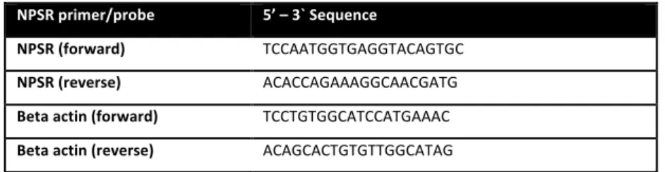
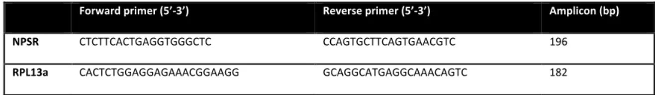
NPSR mRNA expression ... 47
Genotyping of NPSR1 knockout mice ... 49
Specificity of NPSR antibodies ... 49
Ex-vivo Ca2+ imaging in PVN-OXT neurons using GCaMP6s ... 50
Pharmacological inhibition of the OXTR system ... 53
Chemogenetic silencing of PVN-OXT neurons ... 53
2.5 Experimental design - Chapter II ... 55
Bilateral infusion of NPS into the MeA on anxiety-related behavior ... 55
Effects of icv NPS on Ca2+/calmodulin-dependent kinases and ERK1/2 in the amygdala ... 55
Effect of simultaneous blockade of both phospholipase C and adenylyl cyclase on NPS-induced anxiolysis ... 56
Specific effects of blockade of either PLC or the AC signaling on NPSR-mediated anxiolysis in the MeA ... 57
2.6 Experimental design - Chapter III ... 58
Monitoring of chemogenetically induced intracerebral OXT-release ... 58
Monitoring of chemogenetically induced peripheral OXT-release ... 59
Behavioral consequences induced by chemogenetic activation of the endogenous OXT system ... 59
Wire-hanging test ... 60
Righting-reflex test ... 60
2.7 Anxiety-related behavior ... 61
2.7.1 Light-dark box ... 61
2.7.2 Open field ... 61
2.7.3 Elevated plus maze ... 61
2.8 Verification of cannula and microdialysis probe placement ... 62
2.9 Immunofluorescent staining ... 62
2.10 Statistical analysis ... 63
3 Results ... 65
Chapter I – NPS evokes anxiolysis by acting on paraventricular OXT neurons ... 66
1. LC-NPS afferents towards the hypothalamic PVN ... 66
2. PVN-OXT neurons express NPSR ... 67
3. NPS evokes transient Ca2+ influx in PVN-OXT neurons ... 70
4. NPS evokes release of OXT within the PVN ... 73
5. Selective inhibition of OXTR and chemogenetic silencing of OXT neurons within the PVN prevent NPS-induced anxiolysis ... 74
Chapter II – NPSR activation is anxiolytic in a Gq pathway-dependent manner ... 79
1. Effect of bilateral infusion of NPS into the MeA on anxiety-related behavior79 2. Effects of icv NPS on Ca2+/calmodulin-dependent kinase IIɑ and ERK1/2 in the amygdala ... 80
3. Effects of simultaneous blockade of both PLC and AC on NPS-induced anxiolysis ... 81
Table of contents IX 4. Effects of specific blockade of either PLC or AC on NPSR-mediated anxiolysis
in the MeA ... 83
Chapter III – Characterization of DREADD-induced activation of OXT neurons in the PVN85 1. Monitoring of central and peripheral OXT release following chemogenetic OXT neuron activation within the hypothalamus ... 85
1.1 Chemogenetic OXT neuron activation evokes OXT release within the PVN 87 1.2 Chemogenetic activation of hypothalamic OXT neurons failed to increase measurable OXT release within lateral septal nuclei ... 89
1.3 Chemogenetically evoked activation of hypothalamic OXT neurons increases peripheral OXT release ... 90
2. Chemogenetically activation of OXT neurons induces anxiolysis and self- grooming behavior ... 92
3. Effect of chemogenetic PVN-OXT neuron activation on EtOH-induced locomotor impairments ... 93
4 General discussion ... 95
Chapter I: NPS-induced anxiolysis is mediated via the OXT system within the hypothalamic PVN ... 96
Chapter II: NPS induces acute anxiolysis by PLC-dependent signaling within the MeA .. 103
Chapter III: Efficiency of DREADD system in PVN-OXT neurons ... 107
Perspectives and future directions ... 113
Acknowledgement ... 118
Curriculum vitae ... 120
List of publications ... 121
References ... 122
List of abbreviations X
List of abbreviations
AAV adeno-associated virus rAAV recombinant AAV AC adenylyl cyclase AN accessory nuclei ANOVA analysis of variance AP anteroposterior axis
BAPTA 1,2-bis(o-aminophenoxy)ethane-N,N,Nʹ,Nʹ-tetraacetic acid CaMK Ca2+/calmodulin-dependent kinase
cAMP cyclic adenosine monophosphate cDNA complementary DNA
CHO Chinese hamster ovary (cell line) CNO clozapine N-oxide
CRF corticotropin-releasing factor CRF-R CRF receptor
CTB-488 Cholera toxin subunit B coupled to Alexa Fluor 488 DAG 1,2-diacylglygerol
DDA 2',2'-dideoxyadenosine
DEPC-H2O diethylpyrocarbonate-treated water DMSO dimethylsulfoxide
DREADD designer receptor exclusively activated by designer drugs DV dorsoventral axis
EPM elevated plus maze ER endoplasmic reticulum
ERK1/2 extracellular signal-regulated kinase 1/2
EtOH ethanol
FACS fluorescence-activated cell sorting GABA gamma-aminobutyric acid
List of abbreviations XI
GAD generalized anxiety disorder GCaMP genetically encoded Ca2+ indicator GFP green fluorescent protein
GIRK G protein-coupled inwardly rectifying potassium channel GPCR G protein-coupled receptor
HEK293T Human embryonic kidney 293 T (cell line) hM3Dq Gq protein-coupled DREADD
hM4Di Gi protein-coupled DREADD icv intracerebroventricular
ID inner diameter
Ile isoleucine
ip intraperitoneal
IP3 inositol-1,4,5-triphosphate KOR kappa opioid receptor KORD KOR designer receptor
LC locus coeruleus
LDB light/dark box
LDCV large-dense core vesicle MAOI monoamine oxidase inhibitor MAPK mitogen-activated protein kinase MeA medial nucleus of the amygdala
MEK1/2 mitogen-activated protein kinase kinase ML mediolateral axis
mRNA messenger RNA
NPS neuropeptide S
NPSR neuropeptide S receptor
OD outer diameter
OF open field
OXT oxytocin
OXTR oxytocin receptor OXTR-A OXTR antagonist
XII
PBS phosphate-buffered saline PCR polymerase-chain reaction
PFA paraformaldehyde
PI3K phosphoinositide 3-kinase PLC phospholipase C
PVN paraventricular nucleus rAAV recombinant AAV RNA ribonucleic acid RPL13a ribosomal protein L13a rpm rounds per minute
RT room temperature
RT-PCR reverse transcription PCR SALB Salvinorin B
SEM standard error of the mean SNP single nucleotide polymorphism
SNRI selective serotonin/noradrenalin reuptake inhibitor SOCE store operated Ca2+ entry
SON supraoptic nucleus
SSRI selective serotonin reuptake inhibitor
TBS tris(hydroxymethyl)aminoethane-buffered saline TBS-T TBS supplemented with 0.1 % Tween-20
TCA tricyclic antidepressants
TRPV2 transient receptor potential vanilloid type 2
Veh vehicle
YFP yellow fluorescent protein
1 Introduction 13
1 Introduction
1.1 Definition of an emotion termed anxiety
Anxiety is a psychological and physiological state characterized by emotional, somatic, and behavioral imbalances (Seligman et al., 2000). It is a state characterized by dreaded feelings about something that appears intimidating an individual. In the actual or imaginary presence of psychological stress, anxiety can create feelings of fear, worry, and uneasiness (Bouras &
Holt, 2007). The main function of anxiety is to act as a signal of danger, threat, or motivational conflict, and to trigger appropriate adaptive responses (Steimer, 2002). Activation of the sympathetic nervous system resulting in increased heart rate, elevated blood pressure, enhanced vigilance, and increased autonomic and neuroendocrine activation may enable individuals to cope with a precarious situation (Steimer, 2002). These physiological adaptations are important and lead to fight-or-flight responses. Therefore, anxiety is considered to be a natural reaction to a stressor (Sokolowska & Hovatta, 2013), which enhances an individual’s lifespan especially in the case of the presence of predators and risky situations (Price, 2003).
However, when anxiety becomes pathological and interfere with the ability to cope successfully with various challenges and stressful events, it may fall under the classification of an anxiety disorder (Lewis et al., 2010).
1.2 Categories and comorbidity of anxiety disorders
The term anxiety disorder refers to panic disorders, post-traumatic stress disorders, agoraphobia, social anxiety disorders, obsessive-compulsive disorders, specific phobias and generalized anxiety disorders (GAD) with a lifetime prevalence of about 28 % (Kessler et al., 2005). These anxiety disorders have specific symptoms, but all of them are dealing with
1 Introduction 14
excessive, irrational fear and dread (Nutt & Ballenger, 2003). Unlike the relatively mild, brief anxiety caused by a stressful event (such as speaking in public or a first date), anxiety disorders last at least six months (Kessler et al., 2005; Kessler & Wang, 2008). Anxiety disorders commonly occur along with other mental or physical illnesses making the diagnosis of anxiety disorders difficult (Nutt & Ballenger, 2003). This is for instance exemplified by comorbid alcohol or substance abuse, which may mask anxiety symptoms or make them worse (Merikangas et al., 1998; Grant et al., 2004; Luthi & Luscher, 2014). Furthermore depressive- like behavior, psychosomatic discomfort, eating disorders, as well as suicidal thoughts accompany anxiety disorders (Steimer, 2002). A reinforcing factor is that impaired social interactions lead to isolating oneself from one ́s social environment, which results in social isolation and enhance the side effects mentioned above (Teo et al., 2013). Moreover, anxiety disorders require increased rates of chronic medical conditions (Scott et al., 2007).
Steinhausen and colleagues report alarming figures: 14 % of the European population suffers from anxiety disorders, which constitute the second leading cause of mental illness after depression (Wittchen et al., 2011). In 2010, mental illnesses have caused costs around 74.4 billion Euro in the health system of the European Union (Gustavsson et al., 2011). To reduce these costs, and to improve the quality of life of those suffering from anxiety disorders, several research groups are working to develop appropriate therapeutic approaches.
1.3 Drug treatment of anxiety disorders
In December 1951, Paul Charpentier synthetized the tricyclic antidepressant (TCA) chlorpromazine from synthetic histamines (Ban, 2007). The psychiatric effects were first noticed in a hospital in Paris in 1952 and, then, widely used as an antipsychotic drug all over the world. The TCAs with their 3-ring molecular structure are primarily used in the clinical treatment of mood disorders such as major depression and anxiety disorders by inhibiting
1 Introduction 15
both serotonin and norepinephrine reuptake from the synaptic cleft primarily by binding to and modulating serotonin and norepinephrine transporters (Feighner, 1999). With respect to GAD, imipramine was found to exert anxiolytic-like effects (Rickels et al., 1993) and to decrease the risk of relapse (Mavissakalian & Perel, 1999). The major limiting factor to the more widespread use of TCAs are their side effects, which includes anti-cholinergic and anti- adrenergic effects such as sedation, sexual dysfunction, dry mouth, constipation and their well-documented toxicity in overdose (Ravindran & Stein, 2009).
Based on the knowledge that an imbalanced serotonin and norepinephrine system play a crucial role in the development of psychiatric disorders, monoamine oxidase inhibitors (MAOI), selective serotonin reuptake inhibitors (SSRI), and serotonin/norepinephrine reuptake inhibitors (SNRI) were developed (Ravindran & Stein, 2009). MAOIs are another class of older antidepressants that has been investigated for anxiety disorders (Ravindran & Stein, 2009).
The enzyme monoamine oxidase is irreversibly inhibited and thereby blocks the degradation of monoamines such as serotonin and norepinephrine in the synaptic cleft. This results in an overall increased availability of these brain neurotransmitters. However, the efficacy and potency of MAOI is controversially discussed in the literature, since the effectiveness is limited to a subpopulation of patients suffering from anxiety disorders (Frank et al., 1988; Shestatzky et al., 1988; Kosten et al., 1991).
More specifically, SSRIs and SNRIs inhibit the reuptake of the neurotransmitters via the presynaptic serotonin/norepinephrine transporter pump, resulting in increased levels of brain serotonin and norepinephrine. Currently, six SSRIs are available for clinical use including fluoxetine and, most recently, escitalopram. Due to their tolerability, efficacy and safety they became the gold standard for the treatment of GAD. Moreover, escitalopram is effective for both acute and long-term treatment for GAD (Davidson et al., 2004; Goodman et al., 2005;
Allgulander et al., 2006). The most prominent SNRI available for clinical use is venlafaxine, which is considered as alternative first-line agent for the treatment of anxiety disorders. Tzanis
1 Introduction 16
and colleagues demonstrated that venlafaxine was effective to prevent relapse during a six- month follow-up (Ferguson et al., 2007). However, it points out that drugs targeting the serotonin and/or norepinephrine system demonstrate a slow onset of actions and thereby require a constant drug intake over months.
In 1955, Leo Sternbach discovered an antipsychotic drug by chance which core structure is based on a benzene ring and a diazepine ring. A few years later, benzodiazepines were made available as diazepam (also called Valium; Hoffmann-LaRoche)(Wick, 2013). Benzodiazepines enhance signaling of the neurotransmitter gamma-aminobutyric acid (GABA) by binding to the GABAA receptor resulting in increased total conductance of chloride ions. From this follows a decreased excitability of neurons that brings about sedative, muscle relaxant, anxiolytic and hypnotic properties. Based on their fast onset of action and tolerability, benzodiazepines are commonly used. On the other side, benzodiazepine treatment is dangerous in overdose (due to oversedation, cognitive impairment, loss of locomotor coordination), comprises a risk of tolerance and dependence in long-term use. Moreover, individuals discontinuing benzodiazepine use may experience uncomfortable withdrawal symptoms. Thus, benzodiazepines and SSRIs are prescribed simultaneously to treat anxiety disorders. Once the major symptoms are attenuated by fast-acting benzodiazepines, SSRIs stabilize patient’s mood for a longer period (Gross & Hen, 2004).
So far, the development of therapeutics for anxiety disorders has generally focused on the improvement of acute symptoms and relapse prevention with little discussion of primary prevention (Mathew et al., 2008). Therefore, it is important to investigate neuropeptide systems and their involvement in anxiety disorders as this might help in the quest to i) understand the underlying mechanisms, which are responsible for the emergence of an anxiety disorder and ii) to develop new pharmacological tools to treat anxiety disorders.
1 Introduction 17
1.4 Neuropeptides
Neurons use many chemical signals to convey information, including more than 100 different neuropeptides (Hokfelt et al., 2003). The human genome contains 90 genes that encode precursors of neuropeptides. Neuropeptides are small protein-like molecules composed of 3- 100 amino acids, which are synthesized at ribosomes in the perikaryon (Hokfelt et al., 2003).
The peptides are stored in vesicles and released either autocrine, paracrine or neuroendocrine by various stimuli at axon collaterals and dendrites and then bind to G protein-coupled receptors (GPCR) presented at plasma membrane of the target cell.
Neuropeptides also function as neuromodulators: These neuronal signaling molecules exert influence on the activity of the brain in specific ways and are thus involved in particular brain functions, like analgesia, reward, food intake, and learning and memory (Strand, 1999).
Due to the multifactorial etiology of anxiety disorders, neuropeptides have been recognized to play a crucial role in the development of an anxiety-like phenotype, the onset of an anxiety disorder and their potential as a pharmacological tool. Thus, neuropeptides are a “hot spot” in research for which reason a growing number of publications over the last decades focus on orexin, galanin, vasopressin, corticotropin releasing hormone, neuropeptide Y, substance P, prolactin, oxytocin (OXT) and neuropeptide S (NPS) (Mathew et al., 2008). Understanding the underlying mechanisms resulting in a more or less anxious phenotype may lead to discover the causes of anxiety disorders and open up the possibility to develop new therapies and drugs.
1 Introduction 18
1.5 The brain neuropeptide S system
1.5.1 Discovery of Neuropeptide S receptor and its endogenous ligand by GPCR deorphanization
GPCRs are a main component in the regulation of cellular homeostasis. Activated by natural ligands, they induce the activation of various signaling cascades. At least 800 seven- transmembrane receptors participate in diverse physiological and pathological functions (Tang et al., 2012). Historically, ligands were discovered first, but the advent of molecular biology reversed this trend (Civelli, 2012). Bioinformatics of DNA sequences gave rise to about 140 GPCRs whose natural ligands remain unknown. Thus, these GPCRs are called “orphan receptors”. The natural ligand is discovered by so-called “deorphanization”, a process that is based on reverse pharmacology (Pausch, 1997; Chung & Civelli, 2006; Civelli et al., 2006; Suga
& Haga, 2007).
GPR154 (also known as GPRA, PGR14, ASRT2 and VRR1) and its ligand were first reported in the patent literature (Sato et al., 2002). Searching public DNA databases, Reinscheid and colleagues identified a genomic sequence encoding for the GPR154 ligand, which was highly conserved among various species including humans, rats and mice (Xu et al., 2004). The precursor protein contains a hydrophobic signal peptide that is immediately followed by the initiator methionine. Moreover, a pair lysine/arginine motif might serve for proteolytic cleavage to process the peptide (Reinscheid & Xu, 2005b). The mature ligand consists of 20 amino acids (N-SFRNGVGSGVKKTSFRRAKQ-C), whereas the amino-terminal residue is a serine.
According to the nomenclature that has been used earlier (Shimomura et al., 2002), the ligand was named NPS.
1 Introduction 19
1.5.2 Expression patterns of Neuropeptide S
NPS is expressed in all vertebrates with the exception of fish (Reinscheid, 2007). In the rat brain, NPS mRNA expression was examined using in situ hybridization. NPS mRNA is expressed discretely in a few brain areas with strongest expression in the locus coeruleus (LC), principle sensory 5 nucleus and the lateral parabrachial nucleus (Xu et al., 2004). Moderate NPS expression is present in the dorsomedial hypothalamic nucleus and the amygdala (Xu et al., 2004). Moreover, double in situ hybridization revealed that NPS mRNA colocalizes predominantly with vesicular glutamate transporter (VGLUT) mRNA, indicating that these cells are glutamatergic (Xu et al., 2007). A small number of NPS neurons coexpresses choline acetyltransferase mRNA, suggesting presence of acetylcholine (Xu et al., 2007). However, NPS mRNA expression does not colocalize with tyrosine hydroxylase or glutamate decarboxylase 67 (GAD67, marker for GABAergic neurons) mRNA. Moreover, a colocalization of NPS mRNA and corticotropin-releasing factor (CRF) mRNA is present in the lateral parabrachial nucleus.
Densitometry analysis of rat NPS-immunoreactive fibers revealed that NPS-positive nerve endings innervate the thalamus, hypothalamus, septal nuclei and septal border zone as well as amygdala (Adori et al., 2015).
In mice, Pape and colleagues showed that NPS neurons coexpress the CRF receptor 1 (CRF-R1) and are in close proximity to CRF-containing fibers (Jungling et al., 2012). They demonstrated that CRF depolarizes NPS neurons through activation of CRF-R1. In vivo, acute immobilization stress leads to an activation of NPS neurons indicated by increased expression of the immediate early gene cFos (Jungling et al., 2012). Their data provide evidence for a direct interaction between the CRF and NPS system. In accordance, forced swim stress increases the expression of cFos in NPS-immunoreactive neurons suggesting a role for NPS and its involvement in stress system (Liu et al., 2011). In 2011, Singewald and colleagues measured in vivo release of NPS following stress exposure. Microdialysis performed within the basolateral amygdala in male Sprague-Dawley rats revealed increased NPS-release during and after forced
1 Introduction 20
swim stress suggesting that NPS might play a regulatory role in stress responsiveness (Ebner et al., 2011).
1.5.3 NPS receptor distribution and intracellular signaling
The NPS receptor (NPSR) is a typical GPCR composed of a 7-transmembrane domain. The highest degree of similarity with respect to homology is found with vasopressin and OXT receptors (OXTR) (Reinscheid & Xu, 2005b). Using in situ hybridization, Reinscheid and colleagues demonstrated NPSR mRNA expression widely throughout the central nervous system with moderate to high levels of NPSR expression found in thalamus and several nuclei of the hypothalamus, including the paraventricular hypothalamic nucleus as well as cortex and amygdala (Xu et al., 2004; Reinscheid & Xu, 2005b). Contrary, within the brainstem the NPSR mRNA was detected at low levels. Similar expression patterns have been demonstrated at mRNA levels in mouse brain (Clark et al., 2011). Thus, the expression of NPSR mRNA in anxiety- associated neurocircuits of the limbic system indicates that the NPS system may interact with other neurotransmitter and neuropeptide systems.
The pharmacological characteristics of NPSR activation were examined in both, Chinese hamster ovary (CHO) cells and human embryonic kidney 293 T (HEK293T) cells (Reinscheid et al., 2005; Camarda et al., 2009; Liao et al., 2016). In vitro, cells stably transfected with NPSR construct demonstrate reliable increases in intracellular Ca2+ levels and cyclic adenosine monophosphate (cAMP) levels at low nanomolar concentrations of NPS (Reinscheid et al., 2005). This suggests that the NPSR is coupled to both Gq and Gs proteins (Xu et al., 2004;
Reinscheid et al., 2005). In hippocampal NPSR-transfected neurons, researchers detected a biphasic time course of NPSR-mediated Ca2+ influx with a fast and slow component suggesting two major Ca2+ routes via intracellular stores and extracellular space (Erdmann et al., 2015). In Ca2+-free solution the slow component was drastically reduced suggesting Ca2+ influx from the extracellular space. However, it is not known which Ca2+ channel(s) manage(s) the entry of the
1 Introduction 21
divalent ion upon NPS administration. Moreover, NPS induces phosphorylation and thereby an activation of the mitogen-activated protein kinase (MAPK) pathway (Reinscheid et al., 2005).
1.5.4 Single nucleotide polymorphism in NPSR gene
Two studies identified a possible panic disorder susceptibility locus on human chromosome 7p15 (Knowles et al., 1998; Logue et al., 2003). In 2004, Kere and collogues described a number of single nucleotide polymorphisms (SNP) in the NPSR gene that is located on chromosome 7p14-15 (Laitinen et al., 2004). Analysis of genomic DNA from blood samples of a Japanese cohort revealed that the functional NPSR A/T polymorphism (SNP database accession number rs324981), which leads to an amino acid exchange of asparagine into isoleucine (Ile) at position 107, is associated with panic disorders in male patients (Okamura et al., 2007).
Paradoxically, this SNP leads to a gain-of-function in NPSR functioning by increasing its sensitivity to NPS about tenfold (Reinscheid & Xu, 2005a). Moreover, Therien and colleagues demonstrated an increased expression of Ile107 allele that caused an increased cell surface expression in vitro (Bernier et al., 2006). This effect was recently confirmed by Neumann and colleagues using a rat model selectively bred for high anxiety-related behavior; here a synonymous SNP was found (Slattery et al., 2015). It is speculated that the Ile107 allele characterized by increased agonist sensitivity might lead to an overstimulation of neuronal circuits that are modulated by NPS (Okamura et al., 2007; Raczka et al., 2010). A second population-based study of NPSR SNPs in a Swedish cohort confirmed a correlation between NPSR polymorphisms and anxiety disorders (Donner et al., 2010). Moreover, a multilevel approach was applied to further elucidate the role of NPS/NPSR system in the etiology of human anxiety (Domschke et al., 2011). Herein, the A/T polymorphism was found to be associated with panic disorders, too. Moreover, the T risk allele was related to increased anxiety sensitivity. As the amygdala has a pivotal role in transforming stressful events into anxiety (Roozendaal et al., 2009), Domschke and colleagues found the above-mentioned
1 Introduction 22
functional SNP in the human NPSR gene to be associated with altered amygdala responsiveness to aversive stimuli in healthy European participants free from any life-time history of psychiatric disorders (Dannlowski et al., 2011). Hence, this might contribute to the development of an anxiety disorders (Dannlowski et al., 2011).
1.5.5 Neurobiological actions of NPS
Neurons from lateral parabrachial nucleus (which amongst others harbors a cluster of NPS- immunoreactive cells) project to ventromedial and paraventricular hypothalamic nuclei, amygdala and periaqueductal gray (Gauriau & Bernard, 2002). Since the NPSR is highly abundant in limbic brain regions such as amygdala, hypothalamus, bed nucleus of the stria terminalis, raphe nucleus and ventral tegmental area, NPS might influence emotional behaviors and body homeostasis (Reinscheid & Xu, 2005a).
Reinscheid and colleagues demonstrated strong anxiolytic-like activity of intracerebroventricularly (icv) infused NPS in mice subjected to elevated plus maze (EPM), light/dark box (LDB) and open field (OF) (Xu et al., 2004). Moreover, the anxiolytic activity was confirmed by a marble-burying test, a model that is not biased by locomotor activity (Xu et al., 2004; Paneda et al., 2009). Since 2006, the anxiolytic action of NPS was confirmed using various tests in mice and rats following icv (Leonard et al., 2008; Rizzi et al., 2008; Vitale et al., 2008; Wegener et al., 2011; Slattery et al., 2015) and local infusions (Smith et al., 2006) as well as intranasal applications (Ionescu et al., 2012; Lukas & Neumann, 2012; Dine et al., 2015).
Based on Pavlovian conditioning, an aversive stimulus such as an electric foot shock is associated with a neutral stimulus (e.g. tone), a process termed acquisition phase. This cued fear conditioning paradigm is based on associative fear memory that is tested by presentation of conditioned stimulus alone. The freezing behavior during extinction phase indicates fear response of an animal. However, repeated presentation of the conditioned stimulus without
1 Introduction 23
aversive stimulus leads to gradual extinction of fear response. NPS was found to reliably reverse cued fear in both mice and rats (Jungling et al., 2008; Slattery et al., 2015; Zoicas et al., 2016). This effect is mediated by NPS-evoked increase in glutamatergic transmission to intercalated GABAergic neurons in the amygdala (Jungling et al., 2008; Pape et al., 2010). More specifically, NPS blocked the expression of fear-potentiated startle (Fendt et al., 2010). In addition, NPS facilitates extinction of social fear and reduces avoidance behavior in a social defeat model in mice (Zoicas et al., 2016). Although NPS enhances object recognition (Okamura et al., 2011), it failed to prolong social memory in rats suggesting that NPS has memory-enhancing effects in a non-social context (Lukas & Neumann, 2012). Reinscheid and colleagues demonstrated an attenuation of NPS-induced memory enhancement by blocking the adrenergic signaling, suggesting an interaction between NPS system and the brain noradrenergic system to promote arousal (Okamura et al., 2011).
NPS effects were also investigated in the context of aggressive-like behavior. Neumann and colleagues demonstrated anti-aggressive effects of icv infused NPS in rats selectively bred for low anxiety-like behavior (Beiderbeck et al., 2014). This effect was confirmed in mice subjected to the resident/intruder test (Ruzza et al., 2015). Herein, NPS facilitated anti-aggressive effects that were abolished in the presence of the non-peptidergic NPSR antagonist SHA-68.
In 2005, Sticker-Krongrad and colleagues described an anorexigenic effect of NPS. In Long- Evans rats, icv infused NPS strongly inhibited chow intake in overnight fasted rats (Beck et al., 2005). Moreover, they demonstrate that this effect is independent of neuropeptide Y, ghrelin and leptin pathways. A second study confirms the findings as both icv and intra-PVN infusion of NPS reduced food intake, too (Smith et al., 2006). In 2009, Ciccocioppo and colleagues identified the hypothalamic PVN as an important site of NPS’ anorexic effect. Moreover, the NPS effects on reduction of food intake were completely blocked in the presence of a NPSR antagonist (Fedeli et al., 2009). In addition, it was demonstrated that the NPS-induced decrease of food intake were not modified by CRF-R antagonists indicating that NPS’ effects
1 Introduction 24
are independent of the hypothalamic CRF system (Fedeli et al., 2009; Peng et al., 2010).
Patients suffering from chronic pain have an increased prevalence to develop an anxiety disorder (Hasnie et al., 2007; Kroenke et al., 2009; Newcomer et al., 2010; Reme et al., 2011).
The periaqueductal gray is an important component of pain system (Fields et al., 1991).
Moreover, this brain region has been demonstrated to express high levels of NPSR mRNA (Xu et al., 2007). In 2009, Wang and colleagues demonstrated an anti-nociceptive effect of NPS in a dose-dependent manner using hot-plate test and a formalin test, an effect that was prevented in the presence of a NPSR antagonist (Li et al., 2009; Peng et al., 2010). In 2014, Mao and colleagues demonstrated that sciatic nerve injury increases anxiety-like behavior, a condition that was accompanied by a decreased NPS expression (Zhang et al., 2014). However, exogenously applied NPS reversed pain-induced anxiety. Herein, they argue that NPS enhances GABAergic transmission in the amygdala and thus reduces anxiety-like behavior.
In summary, the brain NPS/NPSR system promotes anxiolysis, reverses cued and social fear, enhances memory, facilitates anti-aggressive effects, reduces food intake and has anti- nociceptive properties. Thus, the NPS/NPSR system is a strong regulator of both emotional behaviors and body homeostasis.
1.6 The brain oxytocin system
1.6.1 Synthesis and release of oxytocin
In 1906, Sir Henry Dale discovered that extracts from the posterior pituitary stimulated uterus contractions of a cat. It was also Henry Dale who coined out the name oxytocin (OXT) from the Greek words “ὀξύς τόκος”, meaning swift birth. Accordant to the Greek name, the most prominent peripheral effect of OXT is the induction of uterine contractions facilitating birth (Fuchs & Poblete, 1970) and, moreover, maintaining well-known neuroendocrine effects such
1 Introduction 25
as milk ejection and orgasm (Rhodes et al., 1981; Gimpl & Fahrenholz, 2001).
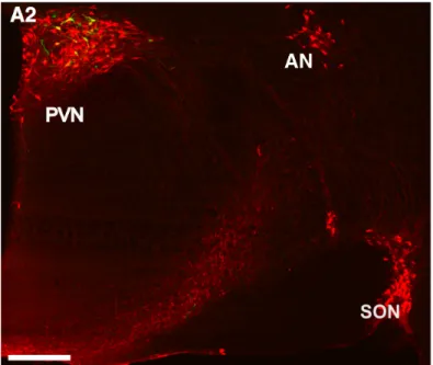
The nonapeptide OXT is synthetized by neurons located in the hypothalamic PVN, the supraoptic nucleus (SON), and accessory nuclei (AN) of the hypothalamus (Fig. 1) (Rhodes et al., 1981; Swanson & Sawchenko, 1983). Stored in large-dense core vesicles (LDCV) along with their respective carrier proteins called neurophysins, OXT is released from magnocellular neurons with a diameter of 20-30 µm into the periphery via axons forming neurohemal contacts within the neurohypophysis (Gimpl & Fahrenholz, 2001; Meyer-Lindenberg et al., 2011).
Figure 1: OXT neurons located within hypothalamic PVN, SON and AN. Scale bar represents 500 µm. Adapted from Eliava et al., 2016.
Moreover, magnocellular PVN-OXT neurons innervate forebrain structures such as the nucleus accumbens (Dolen et al., 2013) and the central nucleus of the amygdala (CeA). Within the CeA, Grinevich and colleagues demonstrated that OXT release from local axonal endings specifically controls region-associated behaviors such as decreased freezing responses in fear-conditioned
1 Introduction 26
rats (Knobloch et al., 2012).
Electron microscopic profiles demonstrated that dendrites of magnocellular OXT neurons have a strong peptide immunoreactivity (Armstrong, 1995) revealing abundant LDCV in somatodendritic structures (Pow & Morris, 1989). The first evidence that dendrites could be a substantial source of peptide release came from studies of the magnocellular neurons of the supraoptic nucleus demonstrating that central and peripheral release of vasopressin and OXT differ in temporal dynamics and do not necessarily act in a linear fashion (Neumann et al., 1993a; Ludwig, 1998; Ludwig & Leng, 2006).
In contrast to magnocellular neurons, parvocellular OXT neurons terminate mainly in spinal chord and brainstem (Swanson & Sawchenko, 1983). It has been proposed that OXT from parvocellular OXT axonal terminals contributes to the modulation of cardiovascular functions, breathing, and feeding behavior (Mack et al., 2002; Petersson, 2002; Atasoy et al., 2012).
Recently, a subpopulation of parvocellular OXT neurons located within the PVN has been demonstrated to control the activity of magnocellular OXT neurons within the SON and to suppress nociception in order to facilitate analgesia (Eliava et al., 2016).
The release of LDCVs is evoked by a rise in intracellular Ca2+ levels (Hokfelt, 1991).
Magnocellular neurons synthesizing OXT are densely filled with LDCV, representing 85% of the total volume of the neuron and thus contain very large amounts of OXT (Stoop, 2012; Stoop et al., 2015). Under physiological conditions, OXT-filled LDCVs are bound to F-actin (Ludwig et al., 2016). Once OXT neurons are activated (e.g. by transient Ca2+ influx), F-actin is rapidly and reversibly depolymerized to G-actin, the OXT-filled vesicle is transported to the membrane, and following binding of a SNARE complex, the vesicle fuses with the plasma membrane resulting in the release of OXT into the extracellular space (Ludwig et al., 2016). The Ca2+ that triggers release of OXT can originate from extracellular and intracellular sources such as N-type voltage-gated Ca2+ channels which appeared to be particularly important for dendritic OXT-
1 Introduction 27
release (Fisher & Bourque, 1996), N-methyl-D-aspartate receptors that influence burst firing of OXT neurons (Hu & Bourque, 1992), and intracellular Ca2+ stores like the endoplasmic reticulum (Lambert et al., 1994). Similarly, dendritic release of OXT also depends on the increase in intracellular Ca2+ (Neumann et al., 1993b; Lambert et al., 1994; Ludwig et al., 2002;
Landgraf & Neumann, 2004; Ludwig & Leng, 2006). Moreover, dendritic release is accompanied by “priming”, an effect facilitated by internal Ca2+ mobilization which makes secretory vesicles available for release in response to electrical stimuli (Ludwig & Leng, 2006).
Once released within the brain, OXT has a half-life time of 20 min in the cerebrospinal fluid (Ludwig & Leng, 2006); the half-life in blood was assessed to be about 1.5 min (Higuchi et al., 1986). Its degradation is mainly operated by aminopeptidases (Stoop, 2012).
Altogether, these studies demonstrate that OXT neurons show widespread central projections of hypothalamic OXT neurons, underlining the evolution of a fine-tuned network composed of various projections originating from hypothalamic areas (Viviani et al., 2011; Stoop, 2012).
1.6.2 Brain OXTR distribution and intracellular signaling
The OXTR is a GPCR composed of a 7-transmembrane domain. Most notably, the OXTR expression is located in cortical areas, the olfactory system, the basal ganglia, the limbic system (e.g. lateral septum, amygdala, subiculum), the thalamus and hypothalamus, the brain stem and the spinal cord (Gimpl & Fahrenholz, 2001). The OXTR is functionally coupled to a Gq
protein that stimulates the activity of phospholipase C (PLC). Consequently, PLC cleaves inositol-4,5-bis-phosphate (PIP2) into 1,2-diacylglycerol (DAG) and inositol-1,4,5-triphosphate (IP3), whereas DAG stimulates protein kinase C, and IP3 increases intracellular Ca2+ levels (Gimpl & Fahrenholz, 2001; van den Burg & Neumann, 2011). The latter is of great importance in the regulation of firing patterns and excitability and, consequently, neurosecretion, gene transcription, and protein synthesis. In most cellular systems, the OXT-induced rise in intracellular Ca2+ levels is more prominent in the presence of extracellular Ca2+ (Clapham,
1 Introduction 28
2007). This suggests that OXT might increase intracellular Ca2+ levels via receptor- or voltage- gated channels (Zhong et al., 2008). Recently, it has been shown that OXT stimulation promotes Ca2+ influx via transient receptor potential vanilloid type 2 (TRPV2) channels in a phosphoinositide 3-kinase (PI3K)-dependent manner (van den Burg et al., 2015). Moreover, Ca2+ influx from extracellular space is necessary for MAPK kinase (MEK1/2) phosphorylation and the anxiolytic effect of OXT within the PVN. In addition, Neumann and colleagues demonstrated that an OXT-induced phosphorylation of MEK1/2 is necessary to promote anxiolysis in male (Blume et al., 2008) as well as in female virgin and lactating rats (Jurek et al., 2012).
1.6.3 Central effects of OXT
Local infusion of synthetic OXT into the hypothalamic PVN and the central amygdala promotes acute anxiolytic activity in both males and females (Neumann et al., 2000; Bale et al., 2001;
Blume et al., 2008; Neumann, 2008; van den Burg & Neumann, 2011; Jurek et al., 2012; van den Burg et al., 2015). Besides induction of lordosis behavior in females and erectile effect of OXT in males in response to the expectation of copulation (McCarthy et al., 1994; Argiolas &
Melis, 2004), mating-induced release of endogenous OXT similarly reduces the state of anxiety-related behavior (Waldherr & Neumann, 2007).
Furthermore, OXT has been demonstrated to reduce pain perception (Juif et al., 2013; Juif &
Poisbeau, 2013), which is maintained by a population of parvocellular OXT neurons that prevent inflammatory pain processing by inhibition of sensory spinal cord neurons (Eliava et al., 2016).
In 1991, Verbalis and colleagues discovered an anorexic effect following central OXT infusion.
This effect was confirmed using a specific OXTR agonist, whereas an OXTR antagonist (OXTR-A) blocked the anorexic effect in food-deprived rats (Olson et al., 1991a; Olson et al., 1991b).
1 Introduction 29
Moreover, OXT was found to attenuate ethanol-induced motor impairments in rats by preventing ethanol actions at delta-subunit of the GABAA receptor (Bowen et al., 2015). In addition, central OXT infusion inhibits ethanol-induced dopamine release in the nucleus accumbens and reduces ethanol consumption after long-term voluntary ethanol intake (Peters et al., 2016).
Besides OXT’s non-social effects, Kelsch and colleagues demonstrated that OXT enhances social recognition by modulating cortical control of early olfactory processing (Oettl et al., 2016). Moreover, social fear represents a main symptom of social anxiety disorder that can be mimicked in the social fear-conditioning paradigm (Toth et al., 2012; Toth & Neumann, 2013).
Fittingly, synthetic OXT promotes extinction of social fear (Zoicas et al., 2014). In extension, OXT has been shown to facilitate pro-social behavior and it prevents social avoidance in both rats and mice (Lukas et al., 2013). In addition, OXT increases social investigation and mediates rodent social memory in regions of the limbic system, namely lateral septum and medial amygdala (Lukas & Neumann, 2013). Furthermore, OXT has been shown to be a crucial regulator of pair bonding (Insel & Shapiro, 1992; Carter et al., 1995; Insel & Hulihan, 1995; Cho et al., 1999) and maternal behavior (Pedersen & Prange, 1979; Neumann et al., 2000; Bosch et al., 2004; Bosch et al., 2005; Pedersen et al., 2006). Lastly, OXT has been demonstrated to play a major role in the regulation of stress responsiveness and the regulation of hypothalamic- pituitary-adrenal axis in response to a variety of emotional, but also physical and pharmacological stressors (Neumann, 2008; Neumann & Slattery, 2016).
Taking into account that OXT release is maintained by magnocellular and parvocellular neurons, by axonal and/or somatodendritic release, the regulatory role of Ca2+ influx and its influence on OXT release and the numerous cell types expressing OXTRs might explain the distinct impact of OXT in brain-region specific behaviors (Lee et al., 2009).
1 Introduction 30
1.7 Characterization of distinct cellular populations by fluorescent signals
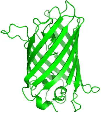
In 2008, the Nobel Prize in Chemistry was awarded to Osamu Shimomura, Martin Chalfie and Roger Y. Tsien “for the discovery and development of the green fluorescent protein (GFP)”
(Figure 2). Originally derived from jellyfish Aequorea victoria, GFP is frequently used as a reporter gene and redefined fluorescent microscopy (Arun et al., 2005; Yuste, 2005).
Figure 2: Green fluorescent protein (GFP). Typical beta-barrel structure of the green-light emiting protein originally derived from Aequorea victoria.
Based on the properties of GFP, genetic mutants have been examined for their fluorescent activity such as the yellow fluorescent protein (YFP). To date, improved versions of YFP are Citrine, Ypet and Venus. The latter contains a novel amino acid substitution transforming the protein to the most powerful fluorescent protein to date (Miyawaki et al., 1997). Tagging proteins of interest with Venus allows identification of a specific cell population with highest precision.
1 Introduction 31
1.7.1 Fluorescence-activated cell sorting
Recently, Grinevich and colleagues constructed an adeno-associated virus (AAV) expressing Venus under the control of an OXT promoter fragment and infused it into the PVN, SON and AN of female Wistar rats. More than 97 % colocalization of OXT and Venus has been observed (Knobloch et al., 2012) underlying the high specificity of this construct. Thus, Venus-labeling of OXT-immunoreactive neurons provides the basis for precise cell sorting.
Fluorescence-activated cell sorting (FACS) is a specialized form of flow cytometry. Hereby, a heterogeneous cell population is sorted cell-by-cell. First, each cell enters a single droplet as soon it leaves the nozzle tip. Since each fluorophore has a characteristic peak excitation and emission wavelength following excitation by laser, a detector characterizes each single cell.
Depending on the fluorescent signal of the cell, the drop is electronically charged. Next, drops containing a single cell pass two deflection plates that either attract or repel the cells accordant to their charge into collection tubes. Following FACS, cells can be cultured or analyzed further for specific expression patterns, thereby allowing characterization of a distinct cellular population.
1.7.2 Measuring Ca2+ dynamics in living cells by genetically encodable Ca2+
indicators
Ca2+ is one of the most abundant mineral compounds of the vertebrate body. In form of hydroxyapatite, Ca2+ is stored in bones and teeth and thus gives stability and strength. Outside of bones Ca2+ acts as a versatile second messenger. It is involved in muscle contraction, exocytosis, gene expression, proliferation, differentiation, blood clotting and apoptosis (Krebs, 1998; Clapham, 2007).
The Ca2+ concentration within a non-stimulated cell is low. A reason for this phenomenon is that this divalent ion precipitates with phosphate. Thus, cells have to compartmentalize,
1 Introduction 32
chelate, or extrude it (Clapham, 2007). Concentrations of free intracellular Ca2+ are regulated by a large variety of channels, exchangers and pumps on both the plasma membrane and intracellular storage organelles such as endoplasmic as well as sarcoplasmic reticula or mitochondria (Berridge et al., 2000). However, about 0.1 % of the Ca2+ are located in the extracellular space and available as free, biologically active ions. Ca2+ enters the cell via diverse set of Ca2+-permeable channels and thus regulates intracellular signaling cascades and cellular activity. Differences in the amplitude, frequency and location of Ca2+ can encode a variety of messages that are decoded by a number of Ca2+ binding proteins.
In order to measure cellular activity, enormous effort has been devoted to develop tools for Ca2+ imaging in living cells. Such indicators allow dissecting spatial and temporal Ca2+ signaling processes. Small molecule Ca2+ indicators based on the chelating character of 1,2-bis(o- aminophenoxy)ethane-N,N,N’,N’-tetraacetic acid (BAPTA) are frequently used to measure changes in intracellular Ca2+ levels. However, loading cells in vivo with these Ca2+ indicators is invasive and does not allow chronic timescales. Thus, genetically encoded Ca2+ indicators (GCaMP) have been developed based on the most prominent representative of intracellular Ca2+-binding proteins named calmodulin (Cheung, 1980). Calmodulin is a small, ubiquitous adaptor protein, which is evolutionary highly conserved and expressed in all eukaryotes (Clapham, 2007). The Ca2+-binding EF-hand has a typical helix-loop-helix structure. Its affinity for Ca2+ is increased by interaction with target proteins. When Ca2+ binds, the shape of the calmodulin domains changes, thus triggering the ability to remodel active sites (Hoeflich &
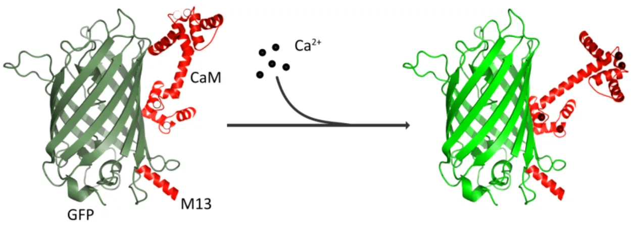
Ikura, 2002). Based on this knowledge, Ca2+ indicators are composed of calmodulin, an alpha- helix of M13 protein and a circularly permutated GFP (fusion of N- and C-terminus). Once Ca2+
is chelated by calmodulin, the latter undergoes a conformational change leading to a deprotonation of GFP. Hence, the chromophore changes its spectral properties and thus emits light (Figure 3) (Wang et al., 2008; Akerboom et al., 2009).
1 Introduction 33
Figure 3: Principle of GCaMP. Following Ca2+ binding, calmodulin (CaM) undergoes a conformational change and thereby deprotonates green fluorescent protein (GFP). Hence, its spectral properties are changed and GFP emits light.
GCaMP expression is introduced into living cells using AAVs that allow genetically specified expression under the control of a promoter fragment. In 2004, the first transgenic mouse expressing GCaMP was reported (Ji et al., 2004). However, the sensitivity, thermostability and response kinetics were slow in comparison to BAPTA-based indicators (McCombs & Palmer, 2008). Since neural activity evokes fast changes in intracellular Ca2+ levels (Tank et al., 1988;
Kerr et al., 2000; Sabatini et al., 2002), efforts have been made to optimize GCaMP system.
Using structure-based mutagenesis and neuron-based screening, Kim and colleagues developed a family of ultrasensitive fluorescent Ca2+ sensors (GCaMP6) that outperformed other sensors in cultured neurons and in zebrafish, flies and mice in vivo (Chen et al., 2013).
GCaMP6 reliably detects single action potentials in neuronal somata and, hence, provides a new window into the organization and dynamics of neural circuits over multiple spatial and temporal scales.
In conclusion, rapid intracellular Ca2+ dynamics can be monitored using genetically engineered ultrasensitive fluorescent proteins and thus allow imaging of neuronal activity in and ex vivo over a large timescale.
1 Introduction 34
1.8 Recent technologies to selectively regulate cellular activity
In 1979, Francis Crick predicted that in order to elucidate neuronal codes that specify behavior and perception ‘‘a method (is needed) by which all neurons of just one type could be inactivated, leaving the others more or less unaltered’’ (Crick, 1979). In the past decade, emerging synthetic biology technologies have been developed and thereby revolutionized selective targeting of specific neuronal populations similar like a remote control (Rogan &
Roth, 2011). The most prominent techniques are optogenetics, which is based on the activation of light-sensitive channels using an optrode, and chemogenetics, which uses engineered GPCRs that are activated by biologically inert drugs (Boyden et al., 2005;
Armbruster et al., 2007).
The term chemogenetics refers to a designer receptor exclusively activated by designer drugs (DREADD). In 2007, Roth and colleagues created a mutated human muscarinic acetylcholine receptor, which lost its affinity for its natural ligand (Armbruster et al., 2007), but has an increased affinity towards clozapine N-oxide (CNO), an otherwise chemically inert drug.
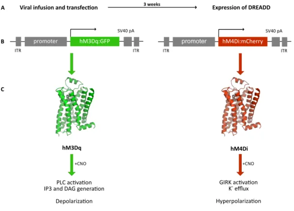
Usually, DREADDs are introduced into cells using viral vectors such as AAV. The selective targeting is achieved by cell type-specific promoter fragment to drive DREADD expression (Fig.
4). In order to verify successful transfection and expression of DREADD in vivo, the construct encodes for a reporter gene (such as mCherry, GFP, tdTomato, mCitrine or Venus). Moreover, the expression can further be controlled using recombinase-based system (CRE recombinase- dependent manner), which allows DREADD expression in a subpopulation of a certain cell type (Boender et al., 2014). Once CNO is applied intraperitoneally via osmotic minipumps, chow pellets, or drinking water, it crosses the blood-brain barrier and binds with low nanomolar affinity to its receptor (Armbruster et al., 2007; Dong et al., 2010; Urban & Roth, 2015; Urban et al., 2016).
1 Introduction 35
DREADDs, much like endogenous GPCRs, are either coupled to inhibitory (Gi) or excitatory (Gq, Gs) signaling cascades (Whissell et al., 2016). CNO-evoked activation of the modified human M4 class muscarinic DREADD via Gi-signaling (hM4Di receptor) induces neuronal silencing by transactivation of G protein-coupled inwardly rectifying potassium channels (GIRK). Thus, the efflux of K+ induces hyperpolarization resulting in decreased neuronal firing. Conversely, CNO- induced activation of the modified human M3 class muscarinic DREADD via Gq-signaling (hM3Dq receptor) induces neuronal activation characterized by increased PLC activity, elevated IP3 and DAG levels, and a rise in intracellular Ca2+ concentration (Whissell et al., 2016).
Figure 4: Theory of designer receptors exclusively activated by designer drugs (DREADD). (A) Following cellular transfection, DREADDs are expressed within three weeks. (B) Cell type-specific promoter fragments achieve selective targeting and expression of hM3Dq or hM4Di in combination with a reporter gene (GFP, mCherry). (C) Both receptor types are activated by clozapine N-oxide (CNO) resulting in altered intracellular signaling and cellular activity inducing depolarization in a Gq- and hyperpolarization in a Gi-dependent manner.