1
Mathematics of the Ruperto-Carola University of Heidelberg, Germany for the degree of
Doctor of Natural Sciences
Presented by Agnieszka Sadowska
Diploma: Master of Biotechnology, University of Gdansk Born in Gdansk, Poland
Oral examination:
profilin1 and profilin2 in mice and the role of the profilin ligand Mena in neuronal cell function and mouse behavior
Referees: Prof. Dr Jochen Wittbrodt Prof. Dr Klaus Unsicker
3
I want to thank my supervisor Walter Witke for giving me the possibility to carry on this project in his group.
I would like to express my gratitude Dr Anne Cecile Trillat for teaching me the methodology used for neuronal cultures and showing an incredible passion for neuroscience. Thank you Cecile for your support to the whole idea as well as for your critical eye looking at my results and their interpretation, but above all for your presence.
I would like to thank the members of The Phenotyping Core Facility, EMBL Monterotondo Karin Gale and Janice Carter help in Phenotyping Analysis of the Mena KO mice.
I would like to thank Pietro Pilo Boyl for involvement in teaching me what is the perfection and for critical comments and discussions on this thesis. Grazie Pietro.
I would like to than Jakky Kelly-Barrett for the injection of my ES cells and generation two of my knock-in mice.
I would love to thank all the members of the Witke group, those with whom I have started in EMBL: James Sutherlad, wonderful friend and teacher, Laura Spinardi for being always next to me, Ralph Gareus for help and critics, Alessia Di Nardo for her Profilin2 KO mice, and all the present members: Christine, Denise, Ekaterina and Emerald who were making my time at the bench enjoyable.
In addition I would like to thank Craig and Mark for making me always laugh and for being present in emergency situations.
And most importantly I would love to thank my boyfriend Claudio for his patience and love, my parents and my sister Edyta that were always ready to listen to me and my friends Magda, Xenka and Aga with whom I could share this experience.
Abbreviations i
List of figures ii
Summary iv
1. Introduction 1
2. Results 14
2.1 Functional redundancy between profilin1 and profilin2 in mice 14
2.1.1 Conditional replacement of the mouse profilin1 gene by the human profilin1/ mouse profilin2 (IRES-hP1/mP2) cassette 14
2.1.2 Generation of the IRES-hP1/mP2 knock-in mouse 16
2.1.3 Profilin2 expression from the IRES-hP1/mP2 knock-in allele 17
2.1.4 Alternative replacement strategy by direct knock-in of the profilin2 cDNA into the profilin1 locus 18
2.1.5 Generation of profilin2 cDNA knock-in mice 19
2.1.6 Profilin2 expression from the profilin2 cDNA knock-in allele 20
2.1.7 Studies on functional redundancy between profilin1 and 2 in mice, conclusions 20
2.2 Role of the profilin ligand Mena in neuronal cell development, brain physiology and behavior 21
2.2.1 Morphology and cytoskeletal organization of Mena KO hippocampal neurons 21
2.2.2 Mena does not play a role in glutamate and NMDA mediated neurodegeneration 24 2.2.3 Mena KO neurons have an increased sensitivity to Menadione-induced oxidative stress 26
2.2.4 Consequences of the Mena mutation on behavior 27
2.2.5 Animals 27
2.2.6 Breeding strategy and Mena KO survival 28
2.2.7 Time line of behavioral tests 28
2.2.8 Body weight 30
2.2.9 Home cage activity and circadian rhythm are not altered in Mena KO mice 32
2.2.10 Mena KO mice display decreased locomotor activity in an open field test 33
2.2.11 Motor coordination, balance and motor learning in Mena KO mice 35
2.2.12 Grip test – muscle strength 37
2.2.13 Tail suspension test 39
2.2.14 Time spent and distance traveled in the center of the open field 40
2.2.15 Operant task- learning and memory 41
2.2.16 Phenotype of Mena KO mice – summary 43
2.2.17 Comparison of Mena and profilin2 phenotypes in mice. Is there a common pathway? 44
ii
4. Materials and Methods 56
4.1 Molecular Biology 56
4.1.1 Cloning strategy used for the targeting of the human profilin1/mouse profilin2 cassette into profilin1 locus 56
4.1.2 Cloning strategy used for targeting profilin2 cDNA into profilin1 locus 59
4.1.3 Validation of the recombinase recognition sites by transformation of Cre and Flp expressing bacteria with NA9-IRES-hP1/P2-NEO and NA9-P2cDNA-NEO targeting constructs 60
4.1.4 Genomic DNA isolation from mouse-tail biopsy and PCR analysis of mutant mice 61
4.1.5 Genomic DNA isolation from ES cells and Southern blot analysis 64
4.2 Cell biology 64
4.2.1 ES cell culture and transfection 64
4.2.1a ES cell transfection 65
4.2.1b Selection of ES cell clones 65
4.2.2 Neuronal primary cultures and in vitro neurodegeneration assays 65
4.2.2a Primary culture of cortical and hippocampal neurons 65
4.2.2b Determination of neurite growth by digital image analysis 66
4.2.2c Immunofluorescent staining of hippocampal neurons 66
4.2.2d In vitro neurodegeneration assays 67
4.2.2e Cytosine arabinoside treatment 67
4.2.2f Oxidative stress - Menadione insults 68
4.2.2g NMDA/Glutamate-insults 68
4.2.2h Determination of cell viability via MTS 68
4.3 Biochemistry 69
4.3.1 Preparation of protein lysates from cell lines and mouse tissues for western blot analysis 69
4.3.1a Primary hippocampal neuron and ES cell lysates 69
4.3.1b Tissue lysates 69
4.3.1c Western blot analysis 69
4.3.2 Primary and secondary antibodies used for western blot and immunostaining assays 70
4.3.3 List of dyes used for immunostaining assays 71
4.4 Mice 71
4.4.1 Mena KO mice 71
4.4.2 Profilin2 KO mice 71
4.4.3 Housing conditions of the animals used for primary behavioral phenotyping 72
4.5.1 Infra Mot – exploratory activity in the home cage 72 4.5.2 Open Field – measurement of the locomotor activity 73 4.5.3 Rota Rod – motor coordination, balance and motor learning 74 4.5.4 Grip test - measurement of muscle strength 75 4.5.5 Tail suspension test – depression and stress response 75 4.5.6 Operant Task - learning and memory 75
5. Bibliography 77
i
[(Ca2+)i] intracellular calcium ion 3’UTR 3’ untranslated region 5’UTR 5’ untranslated region
aa amino acid
ADF actin depolymerizing factor ANOVA analysis of variance
ATP adenosine 5’-triphosphate BSA bovine serum albumine Ca2+ calcium ion
cDNA complementary deoxyribonucleic acid DNA desoxyribonucleic acid
dNTPs deoxyribonucleotides DTT dithiothreitol
EDTA ethylenediaminotetraacetate
EGTA ethyleneglycole-bis[β-aminoethyl ether]-N,N,N’,N’-tetraacetate EVH1 Ena/VASP homology domain1
EVH2 Ena/VASP homology domain2 F-actin filamentous actin
FCS fetal calf serum G-actin globular actin
GTP guanosine 5’-triphosphate
HEPES N-2-hydroxyethylpiperazine-N’-2-ethane sulfonic acid IPTG isopropylthio-β-D-galactoside
kb kilobase
kDa kilodalton
LB Lauria-Bertani medium
mRNA messenger RNA
MTS 3-(4,5-dimethylthiazol-2-yl)-5-(3-carboxymethoxyphenyl)-2-(4- sulphophenyil)-2H tetrazolium, inner salt
NMDA N-methyl-D-aspartate
PAGE polyacrilamide - gel electrophoresis PBS phosphate buffered saline
PCR polymerase chain reaction PH pleckstrin homology domain
PIP2 phosphatidylinositol-4,5-bis phosphate PLP poly-L-proline
RNA ribonucleic acid
RT room temperature
SDS sodium dodecylsulphate
X-GAL 5-bromo-4-chloro-3-indolyl-β-D-galactoside
Figure 1 Histological comparison of WT and Mena KO adult brains
Figure 2 Representation of the structural organization of highly related vertebrate proteins Mena, VASP and EVL and their invertebrate orthologs: Ena, VASP and UNC-34.
Figure 3 Strategy used for the replacement of profilin1 by the IRES-hP1/mP2 allele.
Figure 4 Schematic drawing of the PCR strategy used for screening of the IRES-hP1/mP2 knock-in mice
Figure 5 Western blot analysis of protein extracts from thymus, spleen and brain of IRES- hP1/mP2 knock-in mice
Figure 6 Targeting strategy of the profilin2 cDNA into profilin1 locus.
Figure 7 Western blot analysis of the protein extracts isolated from thymus, spleen and brain of profilin2 cDNA knock-in mice
Figure 8 Morphological analysis of E16.5 cultured hippocampal neurons Figure 9 Glutamate and NMDA effect on cortical neurons
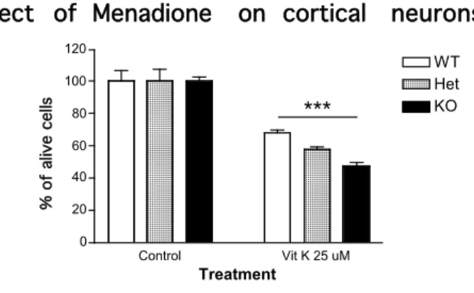
Figure 10 The oxidative stress effect on cortical neurons upon Menadione treatment
Figure 11 Schematic drawing of the behavioral task panel calendar used to assess Mena mice primary behavioral phenotype
Figure 12 Schematic drawing of the behavioral task panel used to investigate the effect of the Mena mutation in mice
Figure 13 Body weight measurement
Figure 14 Front legs muscle weight measurement Figure 15 Fat pads weight measurement
Figure 16 Locomotor and circadian activity of wild type (WT) and Mena KO (KO) mice over a 24h time period for 5 consecutive days.
Figure 17 Locomotor activity (mm) in a 60 min open field Figure 18 Rota Rod – motor coordination and motor learning Figure 19 Grip test - muscle strength
Figure 20 Grip strength of the front legs divided by front legs muscle weight Figure 21 Tail suspension test - depression and stress response
Figure 22 Center of the open field – stress response Figure 23 Operant task- learning and memory
Figure 24 Confirmation of the presence and position of expected restriction sites in NA9-IRES- hP1/P2-NEO targeting construct
iii
transformation of the targeting vectors NA9-IRES-hP1/P2-NEO and NA9-P2cDNA- NEO into Cre and Flp bacteria
Figure 27 TSE Infra Mot apparatus – measurement of the exploratory activity of small laboratory animals in their home cage
Figure 28 Open field apparatus – measurements of the locomotor activity
Figure 29 TSE Rota Rod apparatus for the analysis of motor coordination and balance and motor learning
Figure 30 Apparatus for the measurement of grip strength (TSE) Figure 31 MED-Associate, TSE operant behavior system
Table 1 List of proteins detected in profilin1 and profilin2 protein complexes by MALDI mass spectroscopy and western blot in the mouse brain
Table 2 Results of the breeding between mice heterozygous for Mena mutation
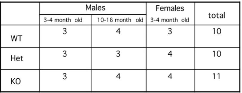
Table 3 Composition of the population of Mena mice subjected to the primary behavioral phenotyping panel
Table 4 Primary behavioral phenotype of Mena KO mice – summary
Table 5 Summary of the Mena and Profilin2 KO primary behavioral phenotypes in comparison to wild type mice
Table 6 Collection of primary and secondary antibodies that were used for immunostaining and western blot assays
Profilin1 and profilin2 are two actin-binding proteins. Biochemically, proflin1 and profilin2 are similar in respect to their interactions with actin, phosphatidylinositol-4,5-bisphosphate (PIP2), and proteins containing poly-L- proline rich motives among which the Ena/VASP protein family members such as Mena. While profilin1 is highly expressed throughout development and adulthood in most tissues including brain, profilin2 is the neuronal specific isoform. Lack of profilin1 results in early embryonic lethality, while profilin2 KO mice are viable and show behavioral abnormalities.
In this thesis I aimed to address three major questions:
1. Is there functional redundancy between profilin1 and profilin2 in vivo?
2. What is the role of the profilin ligand Mena in neuronal cell function and mouse behavior?
3. Is there a common functional pathway for profilin2 and Mena?
In order to address functional redundancy between profilin1 and 2 I generated two different knock-in mouse lines in which profilin1 was substituted by mouse profilin2. However, for unknown reasons expression of profilin2 from the transgene was not detectable in tissues isolated from mice targeted with both knock-in strategies.
My studies on Mena KO mice suggested an involvement of this protein in re- organization of the actin cytoskeleton and axon path-finding. In hippocampal neurons isolated from Mena KO mice, I showed that Mena is normally inhibiting the outgrowth of dendritic processes and cell spreading. In the mutant animals these alterations lead to axonal path-finding defects and behavioral abnormalities. My behavioral analysis showed that lack of Mena leads to impairment of locomotor activity, motor coordination and balance as well as to alteration in stress response.
The severity of the phenotype was found to be age-dependent.
Since Mena and profilin2 are known to interact in vitro I tried to investigate if both proteins act in common physiological pathways. Therefore, I compared the neuronal cell phenotype and the behavior of Mena KO and profilin2 KO mice.
Interestingly, the morphological alterations are very similar in hippocampal neurons from Mena KO and profilin2 KO mice (longer primary processes and faster spreading). In terms of behavior, both KO lines showed alterations in locomotor activity, impairments in motor coordination and balance as well as altered stress response. The overlap and the differences of phenotypes suggest that profilin2 and Mena are linked in common functional pathways, but also that Mena and profilin2 have unique functions in mouse brain physiology.
Profilins
Cells undergo continuous changes in shape and motility. These processes are initiated in response to either external or internal stimuli and accomplished by the cytoskeleton. In particular the microfilament cytoskeleton is considered to be a highly sensitive recipient of messages conveyed by various signaling pathways.
In response to such information the three-dimensional organization of the actin cytoskeleton or the equilibrium between actin filaments and the monomeric actin pool undergoes continuous changes. These events are spatially and temporally controlled.
Two decades ago Carlsson and co-workers discovered profilin, a small 12-16 kDa protein that directly interacts with actin, acidic phospholipids and several other proteins involved in various signal transduction pathways. Since then profilin was identified in all Eukaryotes investigated, such as Protozoa, echinodermata, insects, plants and mammals. Even Vaccinia virus contains a profilin like gene (Blasco et al. 1991). In yeast and Drosophila one profilin gene is expressed while, Arabidopsis, Acanthamoeba, Dictyostelium, Physarum, and plants encode more than one profilin isoform on distinct genes. It was long believed that mammalian species contain only one profilin isoform, however during the execution of a random human cDNA cloning project, a second profilin gene - profilin2 was discovered. To date three profilin isoforms have been identified in mammals.
Mouse profilins are encoded on three different genes and called profilin1, profilin2 and profilin3.
The amino acid sequence identity between two isoforms within any given organism is between 54% and 83%; for example, the human profilin1 and profilin2
2
are 65% identical at the amino acid level but only 35% identical to profilins from lower eukaryotes and about 20% identical to plant profilins. In spite of the considerable degree of sequence variation between profilins from different species, their biochemical properties have been extremely well conserved throughout evolution (Lambrechts et al. 1997). It has been shown that plant and bovine profilin is able to rescue the defects in cell shape, cytokinesis and development that were observed in Dictyostelium profilin null mutant (Karakesisoglou et al. 1996; Schluter et al. 1998).
Biochemical features of profilins
Biochemically, profilin was identified by its ability to bind to poly-L-proline stretches and was shown to form a 1:1 complex with monomeric G-actin (Carlsson et al. 1977). The formation of this complex might represent a mechanism to sequester G-actin from the free cellular pool, although upon binding to actin monomers, profilin in vitro increases the rate of nucleotide exchange on the actin monomer, thereby charging the monomer with ATP, and possibly enhancing actin filament dynamics (Goldschmidt-Clermont et al. 1992). In vitro data show that profilin does not solely act as a sequestering protein for G-actin. Kinetics studies of actin polymerization have shown that profilin can accelerate actin filament growth if free barbed filament ends are available (Pantaloni and Carlier 1993). This finding suggests that in vivo profilin might actually promote actin polymerization.
The only known physiological compounds able to release actin from the profilin-actin complex are the phosphoinositides, especially phosphatidylinositol- 4,5-bisphosphate (PIP2), since the domains responsible for profilin interaction with actin and PIP2 overlap (Lassing and Lindberg 1985). Profilin binds PIP2 with high affinity and is able to inhibit the non-tyrosine-phosphorylated form of phospholipase C-
γ ,
suggesting that profilin may also play a role in signaltransduction through tyrosine kinases and phospholipids (Goldschmidt-Clermont et al. 1991). In addition, the profilin-actin complex in vitro binds with high affinity to poly-L-proline stretches, suggesting that in vivo profilin might bind to proline rich proteins. This property could serve other functions depending on the ligand. The Arp2/3 complex (Machesky et al. 1994), Ena/VASP family members (Reinhard et al. 1995; Gertler et al. 1996), gephyrin (Mammoto et al. 1998), Diaphanous (Watanabe, 1997) and dynamin (Damke et al. 1994) have been shown to interact with profilins. The profilin ligands appear to play a role in the control of cell motility and actin dynamics such as fibroblast migration as well as axon guidance or platelet activation (Aszodi et al. 1999; Lanier et al. 1999; Bear et al. 2002;
Loureiro et al. 2002). They were found concentrated at the leading edge of cells suggesting a role in regulating the dynamics of the major protrusive actin structures: lammelipodia thin membrane sheets and membrane spikes known as filopodia (Rottger et al. 1999).
The role of profilin in actin-based motility
Many studies on profilin role in actin - based motility were taking advantage of intracellular pathogens. Certain intracellular pathogenic bacteria such as Shigella flexneri, Listeria monocytogenes, Rikettsia conorii as well as an enveloped virus, Vaccinia virus (Cudmore et al. 1995), use actin-based motility for their intra- and intercellular movement. Intracellular movement is strictly coupled to a polarized actin polymerization process at one end of the bacterium, resulting in the formation of an actin comet tail or rocket tail. Specific bacterial surface proteins have been identified to be involved in this movement for example VirG or IcsA for S. flexneri, and ActA for L. monocytogenes (Bernardini et al. 1989). These proteins are anchored in the bacterial membrane, transversely to the peptidoglycan layer, and have an intracellular domain, which recruits cellular
4
proteins like F-Actin, Arp2/3 complex, N-WASP, actin depolimerizing factor (ADF or cofilin) and capping proteins. Profilin is recruited to sites of active cytoskeletal assembly through the association with N-WASP, a signaling protein that links multiple signaling components to direct actin-dependent events (Suetsugu et al.
1998). In addition, profilin associates with the Arp2/3 complex (Mullins et al.
1998), which is present in lammelipodia and other dynamic, actin-based structures (Machesky et al. 1997) and appears to initiate actin polymerization during the intracellular motility of the pathogenic bacterium Listeria monocytogenes (Reinhard et al. 1995; Welch et al. 1997). Profilin also interacts with members of the Ena/VASP protein family, including VASP - Vasodilator-Stimulated Phosphoprotein - (Reinhard et al. 1995) and Mena - mammalian Enabled - (Gertler et al. 1996). Like the Arp2/3 complex, VASP and Mena are recruited by the Listeria protein ActA to sites of actin polymerization, where they colocalize with profilin (Theriot and Mitchison 1993; Gertler et al. 1996; Niebuhr et al. 1997).
Inhibiting profilin binding to VASP (Kang et al. 1997) or VASP binding to ActA (Smith et al. 1996) impairs Listeria motility, suggesting a role of profilin in accelerating the actin-dependent motility of the pathogen.
Apart from pathogen motility, profilin has been shown to be important for motility of eukaryotic cells. For example, D. discoideum mutants deficient for profilin show defects in F-actin content, cytokinesis and development (Haugwitz et al. 1994). Yeast lacking profilin show defects in cell shape and actin localization (Haarer et al. 1990). Actin cables are no longer visible, the polarity of the cells is lost, and the normal axial budding of haploid cells becomes random. In Drosophila, genomic deletions of the chickadee locus (Drosophila profilin) resulted in a late embryonic lethal phenotype indicating that profilin is essential for fly development.
Lack of profilin during Drosophila oogenesis leads to a failure in proper assembly of nurse cell actin filament bundles and abnormal mitosis (Cooley et al. 1992;
Manseau et al. 1996). Using an RNA interference approach (RNAi) in C. elegans it was shown that profilin PFN-1 is required for assembly and stability of the cortical
actomyosin cytoskeleton as well as for oogenesis and fertilization (Karakesisoglou et al. 2000).
Profilins in mouse
To date, three profilins profilin1, 2 and 3 were identified in mouse, each encoded on a distinct gene (Kwiatkowski and Bruns 1988; Honore et al. 1993;
Braun et al. 2002). Profilin1 is highly expressed in most tissues except skeletal muscle, while profilin2 is present predominantly in neuronal tissues and at lower levels in uterus and kidney (Witke et al. 1998). Two spliced variants of profilin2 profilin2A and profilin2B have been identified (Di Nardo et al. 2000). Since profilin2A is the main brain specific profilin2 isosform in mice, I will simply refer to profilin2A as profilin2 in following work. Recently a novel profilin isoform, profilin3, was detected in mouse, rat and human tissues (Hu et al. 2001; Braun et al. 2002).
Mouse profilin3 expression is a testis specific isoform (Braun et al. 2002).
During embryonic development profilin1 is expressed highly at all stages including embryonic stem (ES) cells. Profilin2 is expressed at very low level in ES cells and in early embryos, with peak expression around day E13. The increase in the expression level correlates with rapid brain development around E13.
Mouse profilin1 and profilin2 share 62% identity at the amino acid level. The biochemical properties of these two profilin isoforms are very similar with respect to actin binding, PIP2 binding and affinity for poly-L-proline (Gieselmann et al.
1995; Lambrechts et al. 1995).
6
Role of profilin1 and profilin2 in mice
The functional differences between profilin isoforms are not yet known and their role may be dependent on the tissue specific expression, on the total intracellular concentration of the protein or on the specific complexes formed with their ligands. In order to study the role of mouse profilins in vivo conventional profilin1 and profilin2 knockout mice were generated in our laboratory.
Mutation of the profilin1 gene leads to an embryonic lethal phenotype: KO embryos die before implantation probably from defects in cytokinesis (Witke et al.
2001). Heterozygous mice for profilin1, which express 50% of profilin1, are viable and have a normal longevity.
Profilin2 deficient mice are viable and show normal brain anatomy. Profilin2 KO hippocampal neurons spread fasted after plating and produce longer dendritic processes. Profilin2 mutant mice show behavioral abnormalities, such as impaired maternal behavior, altered stress response and hyperactivity (Di Nardo, PhD thesis, Pilo Boyl unpublished data).
Profilin1 and profilin2 are both expressed in mouse brain
In the mouse brain the expression level of profilin2 is three times higher compared to the expression of profilin1. Because in brain both profilins are expressed, this tissue serves as an ideal model to study the general functions of profilins as well as specific activities of profilin2 (Witke et al. 2001). Using MALDI mass spectrometry protein complexes formed by profilin1 and profilin2 were identified (Witke et al. 1998). Interestingly, these complexes were very different from each other and different from the ones described in lower eukaryotes (Machesky et al. 1994). The components of profilin complexes are involved in signal transduction, endocytosis and synaptic recycling. The major components of the profilin1 complex in mouse brain are: clathrin, valosine containing protein
(VCP), hsp70 and actin. The most prominent component of profilin2 complex is dynamin1, a protein known to be involved in clathrin-mediated vesicle formation (Damke et al. 1994). In primary mouse hippocampal neurons profilin2 and dynamin1 co-localize at vesicular structures, supporting the idea that the profilin2 complexes exist in vivo and might play a role in vesicle recycling (Witke et al.
1998). Synapsin1A/B, synapsin2A/B, ROCK2 a downstream effector of Rho signaling pathway, and Mena were detected as other abundant components of profilin2 complex in the mouse brain. Mena has been shown to have an important function in regulating cytoskeletal dynamics and recent work has suggested that Mena might link profilin to this process. Table1 summarizes the proteins detected in profilin1 and profilin2 brain complexes.
Profilin1 complex Profilin2 complex
Actin Actin
Tubulin ROCK2
clathrin POP
Hsp 70 HEM
Mena, VASP Dynamin1
VCP Synapsin1A/B
Synapsin2A/B Hsp70 Mena,VASP
Table1 List of proteins detected in profilin1 and profilin2 protein complexes by MALDI mass spectroscopy and western blot in the mouse brain.
8
Mena, a member of the Ena/VASP protein family, and its interaction with profilins
An important question was raised by the biochemical data on profilin complexes and the in vivo findings in the KO mice, whether the specific complexes account for the different phenotype of the mutant mice. In this respect, the observations made in the double mutant for profilin1 and the ligand Mena is particularly informative. The brains of Mena KO mice exhibit striking abnormalities in the corpus callosum and hippocampal commissure. Fibers in the corpus callosum fail to project medially and to cross the midline, therefore there is no proper connection formed between the two hemispheres (Lanier et al. 1999). The failure of Mena-deficient growth cones to choose the correct path suggests that Mena has a role in growth cone's ability to read or respond adequately to the guidance cues.
A
B
E
D
A C F
Figure 1 Histological comparison of WT and Mena KO adult brains Matched section of WT and Mena KO brains were analyzed by silver staining.
(A,C,E) Coronal section through wild type (WT) brain, where structures of pre- and post commissural fornix (fopr and fop, respectively) and the hippocampal commissure (hc) are shown.
(B,D,F) In Mena KO brains many of the above structures are abnormal or missing.
(D and F) A few fibers from the corpus callosum appear to project ventrally and cross just above the dorsal fornix (arrow head).
(D and F) Cells are visible in the midline (arrow), but fibers of the hippocampal commissure do not appear to cross as they do in the wild type control (compared to C and E)
Lanier et al., 1999
Mice heterozygous for profilin1 gene and deficient for Mena expression die prenatally from cephalic tube closure defects. This suggests the existence of a common pathway where the two proteins are acting (Lanier et al. 1999).
The Ena/VASP protein family is a structurally conserved family found in vertebrates, invertebrates and Dictyostelium. This family is composed of Drosophila Enabled (Ena), C. elegans UNC-34/Enabled, vertebrate EVL (Ena/VASP- like protein), VASP and Mena. Ena/VASP proteins are implicated in cell motility (Bear et al. 2000; Goh et al. 2002). Mena and all the other Ena/VASP proteins share a highly conserved tripartite structure: a proline rich core flanked by two defined regions called Ena/VASP homology domains 1 and 2, EVH1 and EVH2 (Figure 2).
EVH1
EVH2 EVH1
EVH1 EVH2
EVH2
PRO PRO
PRO P P P
P P P
Q-Rich Drosophila Ena
Dictyostelium VASP C.elegans unc-34
Invertebrate homologs
EVH1 PRO P EVH2
P P
FAB
TML COCO
LERER P
+EXON
EVH1 PRO EVH2
P
P FAB
TML COCO
P
EVH1 PRO TML FAB EVH2 COCO
P P
iEXON
Conserved PKA site
P
Phosphotyrosine Phothreonine/
phosphoserine Mena
EVL
VASP
Verterbrate family
EVH1, EVH2 - Ena/Vasp homology domain COCO - coiled – coil region
TML – thymosin-like-motif FAB – F-actin binding region PRO – proline rich region LERER - a.a sequence Q-Rich - glutamate rich region
+EXON –neuronal specific alternative ragion
EVH1
EVH2 PRO
P P
P FAB TML
COCO LERER
P
+EXON EVH1
EVH2 PRO
P
P
FAB TML COCO
P
EVH1 EVH2FAB TML PRO
COCO P P
iEXON
Conserved PKA site
P
Phosphotyrosine Phothreonine/
phosphoserine
Mena EVL
VASP
Verterbrate family
Figure 2 Representation of the structural organization of the highly related vertebrate proteins Mena, VASP and EVL and their invertebrate orthologs: Ena, VASP and UNC-34.
All members share a conserved domain structure: PRO, a proline rich core, flanked by two distinct regions called Ena/VASP like domain (EVH1 and EVH2). All verterbrate family members are substrates for Ser/Thr protein kinase A and G. Ena is a substrate for Abl tyrosine kinase and contains six phosphorylation sites.
10
The N-terminal EVH1 domain binds directly to a consensus motif and plays an essential role in targeting Ena/VASP proteins to focal adhesion sites, while the central proline-rich region serves as a docking site for several SH3 (Src-homology 3) and WW domain-containing proteins like Abl, IRSp53 and FE65 as well as for profilin (Lambrechts et al. 2000). The EVH2 domain is required for oligomerization (Bachmann et al. 1999) and binding of actin (Bachmann et al. 1999; Walders- Harbeck et al. 2002). In fibroblasts, Ena/VASP proteins regulate the protrusive step of motility by controlling the geometry of actin networks within lamellipodia (Bear et al. 2002). However, these findings seem to contradict the enhanced intracellular actin based motility of L. monocytogenes caused by presence of Ena/VASP and profilin (Loisel et al. 1999; Grenklo et al. 2003). Since distinct regions within the Ena/VASP molecule are required for lammelipodial protrusion of fibroblasts and Listeria motility, the observed paradox might be explained by the different functions performed by the Ena/VASP proteins in these two actin- dependent processes. Ena/VASP proteins are also implicated in many other actin- dependent processes, including axon guidance (Gertler et al. 1990), neural tube closure (Lanier et al., 1999), attenuation of platelet aggregation (Aszodi et al.
1999), T-cell activation, phagocytosis (Krause et al. 2000), cell–cell adhesion and the intracellular movement of the bacterial pathogen L. monocytogenes (Vasioukihin et al., 2001).
EVH1 domain. The structure of EVH1 domain of Ena/VASP has been solved and has revealed a direct binding to the consensus motif (D/E)-FPPPP-X(D/E)(D/E) (Niebuhr et al. 1997). This recognition sequence is identified in a number of cellular proteins, which preferentially localize at focal adhesion sites. The interaction of Ena/VASP through EVH1 domain is confirmed for the cytoskeletal proteins zyxin and vinculin (Beckerle 1997) as well as for the transmembrane axon guidance molecules Robo (Godenschwege et al. 2002) and Semaphorin6A-1 (Klostermann et al. 2000). The EVH1 domain of Ena/VASP proteins is involved in the association of Mena and VASP with sites of an actin tail assembly on L.
monocytogenes surface by direct binding to proline-rich motifs of the bacterial ActA protein. This binding would stimulate intracellular movement of Listeria (Smith et al. 1996).
The proline rich region (PRO region) of Ena/VASP proteins contains binding sites for profilin and SH3 and WW domain-containing proteins. Deletion of this region has no effect on the ability of Ena/Vasp proteins to localize and to support normal rates of fibroblast movement. This indicates that this region is not essential for the function of Ena/VASP within lammelipodia during this type of motility. However, the PRO region, by interaction with profilin, recruits profilin-actin complexes to rapidly assemble the actin tail during Listeria movement and lack of the PRO region in Ena/VASP proteins reduces speed of L . monocytogenes.
Considering the genetic interaction between Ena and Abl in Drosophila (Gertler et al. 1990) it is possible that the physical interaction between the PRO region and the SH3 domain of Abl kinase may play a role in connecting Abl signaling to Ena/VASP protein function. Also Dictyostelium mutagenesis studies revealed an important role of the PRO region in filopodia formation, suggesting its requirement for initiation or stability of these structures.
EVH2 domain. The EVH2 domain is composed of three conserved regions: a G- actin binding region known as thymosin-like-motif (TML), an F-actin binding region (FAB) and a coiled-coil region (COCO). In addition, phosphorylation sites for PKA/PKG kinases were found within EVH2 domains of Mena, VASP and EVL.
Deletion of the TML region leads to cellular miss-localization of the Ena/VASP proteins and their diffused distribution throughout the cytoplasm and weak detection in the focal adhesions as well as along the leading edge of lammelipodia.
The FAB region binds to F-actin and plays a crucial role in targeting Ena/VASP proteins to the leading edge. The FAB is also involved in proper regulation of cell motility possibly by maintaining the anti-capping activity of Ena/VASP proteins (Bear et al. 2002). The C terminus region of EVH2 domain contains the coiled-coil motif that mediates oligomerization of Ena/VASP proteins (Bachmann et al.
12
1999), which seems to be important for in vitro actin nucleation and the binding to various Ena/VASP ligands.
Ena/VASP proteins regulate axonal outgrowth
Actin - based motility is critical for neuronal development and neural crest cell migration. Genetic studies performed on several models revealed that Ena/VASP proteins are required for normal axon pathfinding. In wild-type Drosophila, the intersegmental nerve group b (ISNb) subset of motor axons defasciculate from the intersegmental nerve just adjacent to the ventral nerve cord and innervate their target muscles (Vactor et al. 1993). In Ena mutant flies, the ISNb neurons ignore the signals to turn and normally innervate their muscle targets and instead continue processing dorsally (Wills et al. 1999). In C. elegans, mutation in UNC-34/Ena gene leads to misguidance of the motor axons and premature termination of axons in the ventral nerve cord (Yu et al., 2002).
Goals of my PhD thesis
Biochemical similarities of profilins with respect to actin, PIP2 and poly-L- proline binding as well as rescue experiments performed on Dictyostelium mutants lacking profilin using plant and bovine profilins (Karakesisoglou et al. 1996;
Schluter et al. 1998) have led to hypothesize a functional redundancy between profilin1 and profilin2 genes in mice. Due to the importance of this question the first part of my PhD thesis was aimed at addressing a possible in vivo functional redundancy between profilin1 and profilin2. For this purpose I performed a study where profilin1 was substituted by profilin2 in mice. Considering the dramatically different phenotypes shown by the two KO mice for profilin1 and profilin2, which
might be due to differences in developmental expression pattern, tissue distribution as well as subcellular localization, this approach would have addressed the biochemical functional redundancy between the two proteins as well as their functional specificity.
In the second part of my thesis I focused on the consequences of Mena gene inactivation on neuronal cell development and behavior. Therefore, I analyzed the morphology of Mena KO hippocampal neurons and tested the behavior of Mena KO mice.
In the last part, I was interested in possible common pathways for profilin2 and Mena in vivo. In order to address this issue I compared the neuronal cell phenotype and the behavior of profilin2 and Mena KO mice.
2.1 Functional redundancy between profilin1 and profilin2 in mice
Studies on tissue distribution and developmental expression of profilin1 and profilin2 as well as similarities with respect to their interactions with actin, phosphoinositides (PIP2) and poly-L-proline rich motives suggest the possibility of functional redundancy between the two genes. In order to address this question, I decided to replace profilin1 by profilin2 in vivo. The mouse model generated was meant to express profilin2 in all the tissues that normally express profilin1.
2.1.1 Conditional replacement of the mouse profilin1 gene by the human profilin1/mouse profilin2 (IRES-hP1/mP2) cassette
In order to replace profilin1 by profilin2 in vivo, the targeting construct that carries the human profilin1/mouse profilin2 cassette was generated as described in (4.1.1). Human and mouse profilin1 share 98% identity in amino acid sequence and similar affinity for actin, PIP2, poly-L-proline stretches. The availability of a specific antibody against human profilin1 (2H11) allowed me to easily identify the functionality of the knock-in allele. Translation of the hP1/mP2 from the knock-in allele is controlled by an IRES (Internal Ribosome Entry Site) sequence. Due to the design of the targeting construct, transcription results in a mRNA composed of human profilin1 and mouse profilin2 followed by the BGH 3’UTR and polyadenylation signal. The expression of mouse profilin2 is achieved only after conditional removal of hP1. The NEO resistance cassette can be removed upon FLP
15
induced recombination (4.1.3). The final targeting construct contained a 0.7 kb short homology arm and a 3.8 kb long homology arm for the recombination in the genomic locus of mouse profilin1, as shown in Figure 3A. ES cells were electoporated as described with the linearized targeting vector (4.1.1; 4.2.2a).
After selection, 143 clones were picked, expanded, and genomic DNA prepared.
The clones were tested by Southern blot using HindIII digest and the external NcoI/BamHI probe, as shown in Figure 3A. A band of 10 kb was expected from the wild type allele and a band of 8.7 kb in case of an homologous recombination event due to the presence of a new HindIII site introduced together with the human profilin1 coding sequence. Four ES cell clones showed the right pattern for homologous recombination (Figure 3B). Single copy integration was subsequently confirmed using a neomycin probe (data not shown).
Ex1 Ex2 Ex3
HindIII BamHI BamHI HindIII
BGHpA
BamHI BamHI
Ex1 Ex2 Ex3
IVSIRES hP1 mP2 NEO
ATGTGA HindIII
10kb NcoI
NcoI/BamHI probe
HindIII BamHI BGHpA BamHI HindIII
Ex1 Ex2 Ex3
IVSIRES hP1 mP2 NEO
ATGTGA HindIII
8kb
Profilin1 WT locus
IRES-hP1/mP2 targeting construct
IRES-hP1/mP2 targeted allele
A
IRES - Internal Ribosome Entry Site hP1 - human profilin1
mP2 - mouse profilin2 Ex- exon
BGHpA - bovine growth hormone 3’UTR and polyadenylation signal NEO - neomycin resistance cassette
LoxP site Frt site
Figure 3 Strategy used for the replacement of profilin1 by the IRES-hP1/mP2 allele.
A) Schematic drawing representing the organization of the wild type locus of mouse profilin1, the targeting construct and the targeted allele. The genomic regions for homologous recombination are 0.7kb (left arm) and 3.8kb (right arm).
B) Southern blot analysis of genomic DNA from a targeted and a wild type ES cell clone digested with HindIII. With the external NcoI/BamHI probe the expected 8.7kb band for homologous recombinats was identified.
2.1.2 Generation of the IRES-hP1/mP2 knock-in mouse
One targeted ES cell clone (#50) was injected into C57 blastocysts, and four chimeras were generated. After intercrosses performed between chimeras and wild type C57 famales, agouti coat color mice were obtained indicating that germline transmission had occured. Agouti offspring were genotyped by PCR amplification of human profilin1 and the NEO genes using specific pairs of primers as shown in Figure 4.
BGHpA Ex1 Ex2 Ex3
IVSIRES hPI PII NEO
ATG
BGHpA Ex1 Ex2 Ex3 IVSIRES PII
ATG
FLP recombination CRE recombination
400bp 350bp
700bp
IRES-hP1/P2NEO allele
IRES-ΔhP1/P2ΔNEO allele LoxP site
Frt site
BGHpA Ex1 Ex2 Ex3
IVSIRES hPI PII NEO
ATG
BGHpA Ex1 Ex2 Ex3 IVSIRES PII
ATG
FLP recombination CRE recombination
400bp 350bp
700bp
IRES-hP1/mP2NEO allele
IRES-ΔhP1/mP2ΔNEO allele
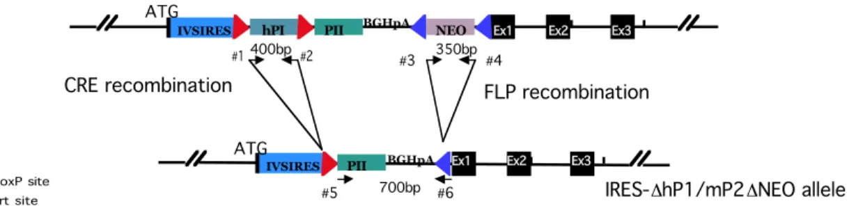
Figure 4 Schematic drawing of the PCR strategy used for screening of the IRES-hP1/mP2 knock- in mice.
The localization of specific primer pairs is illustrated. Primers #1 and #2 amplify the hP1 cDNA, primers #3 and #4 amplify a fragment of the NEO resistance cassette and primers #5 and #6 serve for mP2-BGHpA amplification after the FLP recombination (4.1.3). While the amplification of hP1 and the NEO fragment confirms transmission of the knock-in allele, the presence of the mP2-BGHpA band is specific for the deletion of the NEO cassette in the knock-in allele.
#5 #6
#1 #2 #3 #4
NcoI/BamHI probe
B
17
2.1.3 Profilin2 expression from the IRES-hP1/mP2 knock-in allele
In order to test the functionality of the IREShP1/mP2 knock-in allele western blot analysis was performed on mouse tissues before and after the deletion of the NEO resistance cassette. Deletion of the NEO cassette was achieved by crossing the IRES-hP1/mP2 mice with a FLP deleter strain (IRES-hP1/P2 ∆NEO). Subsequently IRES-hP1/P2 ∆NEO mice were crossed with Cre deleter mice in order to excise the human profilin1 (hP1) gene and induce mouse profilin2 (mP2) expression (IRES-P2
∆NEO∆hP1). Since thymus and spleen have high expression levels of profilin1 (P1) and since mP2 should be transcribed under the control of profilin1 promoter, those tissues were tested for the expression of P2 in mice heterozygous for the knock-in allele (Figure 5).
3003 ab P1-T ab coomassie
thymus spleen brain
WT IRES hP1/P2 NEO
IRES hP1/P2 ΔNEO
IRES P2 Δ NEO
ΔhP1
WT IRES hP1/P2 NEO
IRES hP1/P2 ΔNEO
IRES P2 Δ NEO
ΔhP1
WT IRES hP1/P2 NEO
IRES hP1/P2 ΔNEO
IRES P2 Δ NEO
ΔhP1
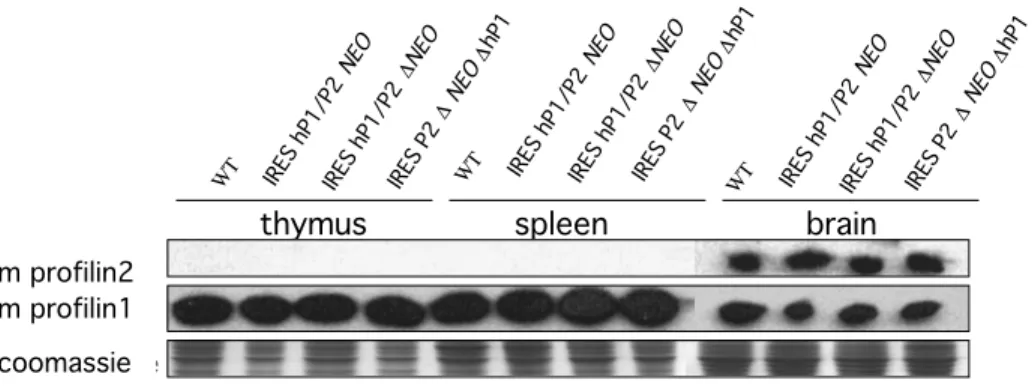
Figure 5 Western blot analysis of protein extracts from thymus, spleen and brain of IRES- hP1/mP2 knock-in mice.
No profilin2 was detected in thymus and spleen extracts after replacement of the endogenous profilin1 by the IRES-hP1/mP2 cassette using the 3003 antibody. Endogenous expression of profilin2 in the brain of these mice was not altered. Profilin1 expression detected with P1-T antibody shows the expected pattern, before and after each deletion. Equal loading was confirmed by coomassie staining.
m profilin2 m profilin1 coomassie
No profilin2 expression was detected in spleen and thymus of heterozygous IRES- hP1/mP2 knock-in mice. Endogenous profilin2 expression was not altered in the brain of knock-in mice. Profilin1 expression level from the WT allele was not altered in these mice.
2.1.4 Alternative replacement strategy by direct knock-in of the profilin2 cDNA into the profilin1 locus
Since IRES sequences do not always work reliably (Paulous et al. 2003) this might be an explanation for the absence of profilin2 expression. Therefore, I decided to use a second approach, where the coding sequence for profilin2 is fused directly to the ATG of profilin1 (4.1.2). In this construct the profilin1 promoter structure is maintained and the profilin1 translation start site is used.
The NEO resistance cassette can be conditionally removed by FLP recombination (Figure 6A). The targeting vector was electroporated into ES cells and 196 clones were picked and expanded (4.2.1a).
A
BGHpA
BamHI BamHI
Ex1 Ex2 Ex3
P2 NEO
ATG
Ex1 Ex2 Ex3
HindIII BamHI BamHI HindIII
10kb NcoI
NcoI/BamHI probe
HindIII BamHI BGHpA BamHI HindIII
Ex1 Ex2 Ex3
P2 NEO
ATG
12kb
P2 - mouse profilin2 Ex- exon
BGHpA - bovine growth hormone 3’UTR and polyadenylation signal
NEO - neomycin resistance cassette Frt site
Profilin1 WT locus
Profilin2 cDNA targeting construct
Profilin2 cDNA targeted allele
A
19
Figure 6 Targeting strategy of the profilin2 cDNA into profilin1 locus.
A) Schematic drawing representing the profilin1 wild type region, the targeting construct and the targeted allele where profilin1 is substituted by the profilin2 cDNA fused to the start codon of profilin1.
B) Southern blot analysis of genomic DNA from homologous recombinant ES cell and wild type clones digested with HindIII.
After probing with NcoI/BamHI external probe the right pattern confirming homologous recombination event was identified.
After HindIII digestion of ES cell genomic DNA and Southern blot analysis with the external NcoI/BamHI probe, two bands were identified, one of 10 kb, belonging to the WT allele and one of 12 kb belonging to the targeted allele (Figure 6B). The shift in size results from the insertion of the profilin2 cDNA and neomycin resistance cassette (Figure 6A). Five ES cell clones were correctly targeted. Single copy integration into the profilin1 locus was confirmed by probing with a NEO probe (data not shown).
2.1.5 Generation of profilin2 cDNA knock-in mice
Two correctly targeted ES cell clones (#3 and #7) were injected into C57 blastocysts. Eighteen male chimeras were generated and mated with wild type C57 females for germline transmission. Agouti offspring were genotyped by PCR using a specific pair of primers amplifying a fragment of the neomycin resistance cassette. The design of the targeting construct outlined in Figure 6A enables the conditional removal of the NEO cassette upon FLP recombination (P2cDNAΔNEO).
The same PCR strategy that was used for the screening of IRES-hP1/P2ΔNEO mice was applied to genotype the P2cDNAΔNEO mice (primers #5 and #6 in Figure 4).
NcoI/BamHI probe
A)
B
2.1.6 Profilin2 expression from the profilin2 cDNA knock-in allele
Western blot analysis was performed on the tissues isolated from the profilin2 cDNA knock-in mice before and after deletion of the NEO resistance cassette.
Profilin2 expression in thymus and spleen of the profilin2 cDNA knock-in mice was udetectectable, suggesting that also the P2 cDNA knock-in approach was not successful (Figure 7).
3003 ab P1-T ab coomassie
thymus spleen brain
WT P2cDNA NEO
FLP P2cDNA ΔNEO
WT P2cDNA
NEO
FLP P2cDNA ΔNEO
WT P2cDNA NEO
FLP P2cDNA ΔNEO
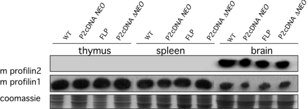
Figure 7 Western blot analysis of the protein extracts isolated from thymus, spleen and brain of profilin2 cDNA knock-in heterozygous mice.
3003 polyclonal antibody recognizing the C terminus of mouse profilin2 did not detect profilin2 expression in thymus and spleen isolated from profilin2 cDNA knock-in mice, regardless of the deletion of the NEO cassette. Probing with P1-T antibody shows that profilin1 expression is not changed in these mice. Presence of the Flp transgene did not interfere with profilin expression.
Coomassie staining shows equal sample loading.
2.1.7 Studies on functional redundancy between profilin1 and 2 in mice, conclusions
The approaches used to substitute profilin1 by profilin2 in vivo the IRES- hP1/mP2-NEO and the P2cDNA knock-in strategies did not result in detectable expression of profilin2 from the mutated alleles.
m profilin2 m profilin1 coomassie
21
2.2 Role of the profilins ligand Mena in neuronal cell development, brain physiology and behavior
Mice where profilin1 expression is reduced to 50% in the Mena null background die in the course of embryogenesis due to the defects in cell migration related to neuronal tube closure. Mena expression is localized in brain, fat, ovaries and testes. As profilin2 is brain specific, I was interested in studying the interaction between Mena and Profilin2 in vivo. I decided to compare the profilin2 and Mena KO mouse models with respect to neuronal cell and behavioral phenotype in order to bring new elements to demonstrate the hypothesis that the two proteins act on a common pathway. The behavioral analysis performed on profilin2 KO mice by Alessia di Nardo and Pietro Pilo Boyl in our laboratory showed hyperactivity, altered response to stress and age-dependent impairment in balance and coordination. In vitro cultured hippocampal neurons, isolated from Profilin2 KO mice, showed that the lack of profilin2 stimulates neurite outgrowth. In the following chapters I will focus on the effects of the Mena mutation in neuronal cells development and physiology and its further implications for mouse behavior.
2.2.1 Morphology and cytoskeletal organization of Mena KO hippocampal neurons
A number of morphological changes, which occur during the establishment of neuronal polarity, are dependent on actin cytoskeleton dynamics. Growth cone movement to its synaptic target is powered by dynamic rearrangements of the actin cytoskeleton. Filopodial spikes punctuate to the growth cone periphery. The intervening space between filopodia contains a lammelipodium-like branched actin filament network. Actin polymerization in the growth cone drives membrane protrusion. The Ena/VASP protein family is involved in the overall cell motility by
regulating of the actin filament length at the growth cone periphery (Bear et al.
2002). Mena is concentrated at filopodial tips in the growth cone (Lanier et al.
1999), where it may regulate filopodial extension and stability in a manner analogous to that proposed for melanoma cell filopodia (Svitkina et al. 2003).
According to these observations, neurons deficient for the Mena gene might exhibit alterations in the response to the guidance cues, related to changes in the integrity of growth cone formation and function.
In order to elucidate the role of Mena in neuronal cells a comparative analysis between wild type (WT) and Mena KO (KO) hippocampal neurons was performed. Intercrosses between heterozygous mice for the Mena mutation were set up and at day E16.5 embryos were dissected and genotyped, and hippocampal neurons were prepared as described (4.2.2a). At this stage hippocampal neurons are undifferentiated. Shortly after plating, cells attach to the extracellular matrix and pass through specific morphological changes in a defined sequence that eventually leads to neuronal polarization. Initially neurons are unpolarized (stage1).
After 6-12 hours in culture short neurites appear, followed by the outgrowth of minor processes that do not show differences in morphology (stage2). Within 48 hours in culture one of the processes elongates more extensively than others: the establishment of the axon defines cell polarization (stage3). After 12 - 16 days in culture hippocampal neurons are able to form functional synapses as they have gained all the features of mature neurons (Craig and Banker 1994). In my studies, I determined neurite length and cell body area to compare neuronal differentiation in the wild type and Mena deficient hippocampal neurons. Neurons were followed by light microscopy at four different time points after plating: 6, 24, 48 and 72 hours (4.2.2b) Phase contrast pictures were captured and data regarding neurite length and cell body area were collected in seven separate neuronal preparations and analyzed with Image J 1.29X (NIH, Baltimore, USA) and Stat View 5.0 (Abacus Concept, Berkley, USA) for statistical analysis. Figure 8A contains representative images of neurons. The majority of the hippocampal neurons deficient for Mena
23
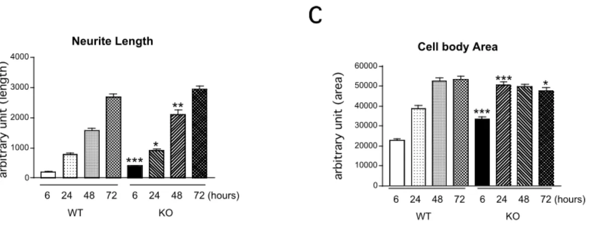
produced primary processes already within 6 hours after plating while neurons isolated from WT mice merely settled. 6h, 24h and 48h after plating the length of the primary processes in Mena KO neurons were significantly longer than those in WT cells (6h, +150%, P<0.001; 24h, +32%, P<0.05, 48h, +37%, P<0.01, one- way ANOVA, Figure 8B). However 72h after plating the length of the dendritic processes between Mena KO and WT neurons were similar. Measurement of the cell body area revealed that Mena KO neurons 6h and 24h after plating are spread significantly more compared to WT cells (6h, +60%, P<0.001; 24h, +30%, P<0.001, one-way ANOVA, Figure 8C). 48h after plating measurements of the cell body area of WT and Mena KO neurons appeared to be equal. Cell body area of Mena KO neurons seems to reach a plateau in size around 24h in vitro while WT cells keep spreading for another 24h.
6h 24h 48h 72h
A
Results
Neurite Length
0 1000 2000 3000 4000
6 24 48 72 6 24 48 72 (hours) WT KO
*** *
**
L e n g t h ( a r b i t r a r y u n i t )
Cell body Area
0 10000 20000 30000 40000 50000 60000
6 24 48 72 6 24 48 72 (hours) WT KO
***
***
A r e a ( a r b i tr a r y un it )
*
*P < 0.05, **P < 0.001, ***P < 0.05 compared to WT at the same time point
arbitrary unit (length)
*P<0.05; **P<0.01; ***P<0.001 compare to the WT
Neurite Length
0 1000 2000 3000 4000
6 24 48 72 6 24 48 72 (hours) WT KO
*** *
**
L e n g t h ( a r b i t r a r y u n
Cell body Area
0 10000 20000 30000 40000 50000 60000
6 24 48 72 6 24 48 72 (hours) WT KO
***
***
A re a ( a r b i t r a r y un i t )
*
*P < 0.05, **P < 0.001, ***P < 0.05 compared to WT at the same time point
arbitrary unit (area)
*P<0.05; **P<0.01; ***P<0.001 compare to the WT
Figure 8 Morphological analysis of E16.5 cultured hippocampal neurons.
A) Representative images (light microscopy) of the hippocampal neurons isolated from WT and Mena KO mice 6h, 24h, 48h and 72h after plating.
B) Neurite length of Mena KO neurons 6h, 24, 48h after plating is significantly longer compared to the WT cells (P<0.001;P<0.05; P<0.01, one-way ANOVA). At the latest time point examined (72h) neurite length is comparable between WT and Mena KO neurons.
C) Cell body area of the Mena KO neurons 6h and 24h after plating is significantly larger compared to the WT cells (P<0.001, one-way ANOVA). At 48h and 72h time points cell body area is comparable between WT and Mena KO neurons.
2.2.2 Mena does not play a role in glutamate and NMDA mediated neurodegeneration
A growing body of evidence suggests that in addition to the regulation of the cell structure and motility, actin filaments may be involved in the modulation of ion channels function (Johnson and Byerly 1993; Rosenmund and Westbrook 1993; Berdiev et al. 1996). Several actin-binding proteins like gelsolin, spinophilin and PSD-Zip45 (Homer 1c) have been implicated in coupling actin cytoskeleton dynamics with neurotransmission (Furukawa et al. 1997; Smith et al. 1999; Usui et al. 2003). Gelsolin for example is involved in the opening and closure of NMDA receptors and voltage dependent calcium channels (Furukawa et al. 1997).
Therefore I was interested in whether Mena might play a role in ion channels regulation through modulation of the actin cytoskeleton.
B C
*P<0.05, **P<0.01,***P<0.001 compared to WT