• Keine Ergebnisse gefunden

Crosstalk between membrane trafficking and cell adhesion: The role of the SNARE protein TI-VAMP in neuronal morphogenesis

N/A
N/A
Protected

Academic year: 2021

Aktie "Crosstalk between membrane trafficking and cell adhesion: The role of the SNARE protein TI-VAMP in neuronal morphogenesis"

Copied!
131
0
0

Wird geladen.... (Jetzt Volltext ansehen)

Volltext

(1)

Crosstalk between membrane trafficking and cell adhesion:

The role of the SNARE protein TI-VAMP in neuronal morphogenesis

I n a u g u r a l - D i s s e r t a t i o n zur

Erlangung des Doktorgrades

der Mathematisch-Naturwissenschaftlichen Fakultät der Universität zu Köln

vorgelegt von

Philipp Alberts

aus Coesfeld, Westfalen

2004

(2)

Berichterstatter : Professor Jonathan C. Howard, Institut für Genetik, Universität zu Köln

Professor Thomas Langer, Institut für Genetik, Universität zu Köln

Dr Thierry Galli, Institut Fer-à-Moulin, Paris

Tag der mündlichen Prüfung in Form der Disputation: 15.01.2004

(3)

Abstract

The membrane trafficking pathway mediated by the SNARE protein Tetanus neurotoxin-Insensitive Vesicle Associated Membrane Protein (TI-VAMP) in neurons is still unknown. In this work, I show that TI-VAMP expression is necessary for neurite outgrowth in PC12 cells in culture. TI-VAMP interacts with plasma membrane and endosomal target SNAREs suggesting that TI-VAMP mediates a recycling pathway. This view is supported by the direct demonstration that TI-VAMP recycles from and to the plasma membrane. L1, a cell-cell adhesion molecule involved in axonal outgrowth, colocalizes with TI-VAMP in the developing brain, neurons in culture, and PC12 cells. Plasma membrane L1 is internalized into the TI-VAMP-containing compartment. Silencing of TI-VAMP results in reduced expression of L1 at the plasma membrane and impaired L1- but not N-Cadherin-mediated adhesion. Futhermore, the TI-VAMP-compartment is specifically recruited to L1 bead-cell junctions in an actin-dependent manner suggesting that axon guidance cues like L1-ligation act by controlling cytoskeletal and membrane dynamics in a coordinated manner. In conclusion, TI-VAMP mediates the intracellular transport of L1 and L1-mediated adhesion controls this membrane trafficking, thereby suggesting that cross-talk between membrane trafficking and cell-cell adhesion plays a central role in coordinating axonal outgrowth and pathfinding.

Zusammenfassung

Die Rolle des vesikulären Transports in Neuronen, welcher von dem SNARE Protein

Tetanus neurotoxin-Insensitive Vesicle Associated Protein (TI-VAMP) vermittelt wird, ist

unklar. In dieser Arbeit ist gezeigt, das die Expression von TI-VAMP für effizientes

Auswachsen von Neuriten notwendig ist. TI-VAMP interagiert mit target SNARE Proteinen

der Plasma Membran und des endosomalen Systems. Es kann gezeigt werden, das TI-

VAMP-positive Vesikel mit der Plasmamembrane fusionieren. Daher kann davon

ausgegangen werden kann, dass TI-VAMP den Austausch von Proteinen und Lipiden

zwischen der Zelloberfläche und einem intrazellulären, endosomalen System vermittelt. Das

Zell-Zell Adhesionsmolekül L1, welches eine wichtige Rolle in der Gehirnentwicklung spielt,

kolokalisiert mit TI-VAMP im embryonalen Gehirn, in in vitro-kultivierten Neuronen sowie in

der Zellinie PC12. L1 wird von der Plasmamembran in ein intrazelluläres, TI-VAMP-positives

Membrankompartiment aufgenommen. Die Inhibierung der TI-VAMP-Expression provoziert

eine verminderte Expression von L1 an der Zelloberfläche und selektive Instabilität von L1-

vermittelten adhesiven Kontakten. Weiterhin rekrutieren L1-abhängige Kontakte TI-VAMP-

positive Vesikel, ein Phänomen, welches von dem Aktincytoskelett abhängig ist. Diese

Beobachtungen legen die Hypothese nahe, dass Signale wie zum Beispiel die Aktivierung

des Adhesionsmolküls L1, welche das Navigieren von Axonen während der

Gehirnentwicklung ermöglichen, die Dynamik des Cytoskeletts und von Membranverkehr in

koordinierter Weise kontrollieren. Es lässt sich der Schluss ziehen, dass TI-VAMP den

intrazellulären Transport von L1 vermittelt und das gleichzeitig L1-vermittelte Adhesion die

Dynamik dieses Membrantransportweges kontrolliert. Der Austausch zwischen

Membranverkehr und Zelladhäsion könnte von entscheidender Wichtigkeit für Phänomene

wie das koordinierte Auswachsen von Axonen entlang vorbestimmter Pfade während der

embryonalen Hirnentwicklung sein.

(4)

Abbreviations

ARF ADP ribosylation factor ATP Adenosine 5’-triphosphate BFA Brefeldin A

BSA Bovine serum albumin CAM Cell Adhesion Molecule

DMEM Dulbecco’s modified Eagle’s medium EDTA Ethylenediamine -tetraacetic acid ER Endoplasmic reticulum

GST Glutathione-S-transferase GTP Guanosine 5’-triphosphate

IgCAM Cell Adhesion Molecule of the Immunoglobulin Superfamily IgG Immunoglobulin class G

kDa kilo Dalton

MVB Multivesicular body MVE Multivesicular endosome

NSF N-ethylmaleimide-sensitive factor oN over Night

PAGE Polyacrylamide gel electrophoresis PBS Phosphate buffered saline

PCR Polymerase chain reaction RT Room Temperature

SDS Sodium dodecylsulphate siRNA small inhibitory RNA

SNAP Soluble NSF attachment protein SNARE SNAP receptor

Stx Syntaxin

Syb 2 Synaptobrevin 2

SNAP-25 Synaptosome associated protein of 25 kDa TBS Tris buffered saline

TGN Trans Golgi network

TI-VAMP Tetanus neurotoxin Insenstive VAMP

(5)

TMD Transmembrane domain TeNT Tetanus Neurotoxin

Tris Tris[Hydroxymethyl]aminomethane

VAMP Vesicular Membrane Associated Protein

(6)

Table of contents

I. Introduction 1

I.1 Membrane trafficking in eukaryotic cells: a historical overview 3 I.2 SNARE proteins in membrane trafficking: the current view 8

I.2.1The formation of cargo loaded vesicles by coat proteins 8

I.2.2The targeted delive ry of vesicles by tethering factors and Rab proteins 9 I.2.3 SNARE proteins and membrane fusion 11

I.2.5 Spatial and temporal control of SNARE dependent membrane fusion 15 I.2.6 The role of membrane trafficking in neuronal morphogenesis 17

I.3 Aims of the thesis 23

II. Materials and methods 26

II.1. Materials 26

II.1.1. Antibodies, clones, and reagents 26 II.1.2. Plasmids 27

II.2. General molecular biology methods 27

(7)

II.2.1. PCR 27

II.2.2. Restriction digestion 27

II.2.3. Agarose gel electrophoresis 28 II.2.4. DNA Ligation 28

II.2.5. Annealing Oligonucleotides 27 II.2.6. Bacterial culture media 28

II.2.7. Transformation of Competent Bacteria 28 II.2.8. Plasmid Purification 28

II.2.9. Production of GST-fusion proteins 29 II.3. Tissue culture 29

II.3.1. Culture of cell lines 29

II.3.2. Preparation and culturing of primary hippocampal and cortical/striatal neurons neurons 30

II.3.3. Culture of hippocampal neurons on L1-substrate 30 II.4. Biochemical methods 31

II.4.1. Immunoprecipitation 31 II.4.2. SDS-PAGE 31

II.4.3. Western Blotting 32

II.4.4. Subcellular fractionation 33 II.5. Cell biological methods 34

II.5.1. SiRNA treatment of PC12 cells 34 II.5.2. Antibody uptake assays 34

II.5.3. Qualitative and quantitative immunocytochemistry 35 II.5.4. Bead-cell adhesion assay 37

II.5.5. Video microscopy 38

(8)

III. Results 39

Part I: TI-VAMP mediates L1 trafficking 39

III.1. Characterization of the TI-VAMP monoclonal antibody Cl158.2 39 III.2. The expression of TI-VAMP is developmentally regulated 41

III.3. TI-VAMP forms complexes with SNAP-25, syntaxin 1, syntaxin 7, and vti1b in vivo and recycles to the plasma membrane in neuronal cells 43 III.4. TI-VAMP is essential for neurite outgrowth 47

III.5. The IgCAM L1 is a cargo molecule of the TI-VAMP compartment 49 III.6. TI-VAMP is required for L1-mediated adhesion 58

Part II: L1 controls TI-VAMP-mediated transport 60

III.7. TI-VAMP accumulation in axonal growth cones is actin dependent 60 III.8. L1-dependent adhesion controls TI-VAMP-mediated trafficking 65 III.9. L1-, but not N-Cadherin-mediated adhesive contacts induce actin-

dependent recruitment of the TI-VAMP compartment 74

III.10. Specific recruitment of the TI-VAMP compartment to the plasma

membrane at L1-dependent contacts 78

(9)

IV. Discussion 87

IV.1. TI-VAMP expression is developmentally regulated and required for neurite outgrowth 85

IV.2. TI-VAMP mediates a recycling pathway in neuronal cells 89 IV.3. TI-VAMP is required for L1 function 91

IV.4. TI-VAMP-localization in growth cones depends on actin dynamics 94 IV.5. Regulation of the TI-VAMP-compartment in growth cones by L1

signalling 95

IV.6. Specific recruitment of TI-VAMP by L1-, but not N-Cadherin junctions 98

IV.7. Neurite outgrowth and neuronal polarity 99

V. Litterature 102

VI. Deutsche Zusammenfassung 119

(10)

I. Introduction

The structure of the nervous system is of great complexity. A high number of distinct neuronal cell types interact with each other via a sophisticated network of specific interactions, sometimes over long distances. At the same time, communication points between neurons are not static, but can be modified, which ultimately allows for the fascinating properties of the nervous system, including learning and memory.

For insight into the complex function of the brain, the molecular mechanisms that warrant brain development have to be elucidated. What are the molecular events that mediate the correct wiring of the highly diverse neuronal connections? It is known that the pattern of connections between neurons is organized by guidance cues which are correspondingly of highest diversity. For example Cadherins, which play a fundamental role in brain development, constitute a large gene family (Yagi and Takeichi, 2000). It is most interesting that the genomic organization of a subgroup of Cadherins, the so called protocadherins, shows similarities to the genomic organization of B- and T-cell receptors of the lymphoid system (Wu and Maniatis, 1999). The well known genomic reorganization of B- and T-cell receptors is the fundamental principle by which the almost unlimited diversity within the immune system is generated, and a similar mechanism might be involved in the generation of diversity during brain development.

Yet, inherent to the generation of complexity, a fundamental problem arises; how to establish specificity in an environment which is of highest diversity?

Neurons have to grow axons and dendrites to their specific target along specific pathways. Contact formation has to occur at the right place and the right time, and during its journey the growing neurite has to ignore a number of cues, that are recognized by others as their target. Thus, neurons have to be equipped with a highly sensitive apparatus that reads and interprets cues and that allows advance while preventing the premature establishment of contacts.

For all these reasons, studying the molecular mechanisms of neurite

outgrowth is of fundamental importance in order to understand the complex wiring of

the nervous system. A huge number of studies on the role of attractive and repulsive

(11)

cues and their effects on the neuronal cytoskeleton has led to an impressive knowledge as to how these molecules might work during brain development. In contrast, the role of membrane trafficking during neuronal morphogenesis has received little attention, although it is the delivery of membrane which ultimately allows the neuron to grow. Clearly, for site-directed growth to occur, the delivery of membrane has to be tightly connected to the signaling molecules that guide axons and dendrites during brain development

One reason for the poor knowledge about membrane trafficking in neuronal

differentiation might be that molecular markers for specific neuronal membrane

compartments with a function in membrane trafficking were sparse. This has

changed with the discovery of SNARE proteins, which are essential players in

membrane trafficking of eukaryotes. Specific SNARE proteins are expressed in

specific membrane compartments and thus the study of a given SNARE protein will

lead to a deeper understanding of membrane trafficking pathways and their function

(see Fig I1). Therefore, the goal of this thesis was to characterize the role of TI-

VAMP/VAMP7, a recently discovered SNARE protein, in the differentiation of

neuronal cells.

(12)

Figure I1) Subcellular localization of mammalian SNARE proteins

The mammalian SNAREs that have been studied so far localize to distinct subcellular compartments in the secretory pathway. (Red, syntaxin family; blue, VAMP family; green, SNAP-25 family; black, others. CCP, clathrin-coated pits; CCV, clathrin-coated vesicles; DCV, dense core vesicles; IC, intermediate compartment; RER, rough endoplasmic reticulum; SER, smooth endoplasmic reticulum;

SNAP-25, 25 kDa synaptosome-associated protein; TGN, trans-Golgi network; V, vesicles; VAMP, vesicle-associated membrane protein.) (from Chen and Scheller, 2001).

In this introduction, I will first give an overview of the general molecular mechanisms of membrane trafficking, as they can be found from yeast to human.

Secondly, I will present the current knowledge of the role of membrane trafficking in

neuronal morphogenesis.

(13)

I.1 Membrane trafficking in eukaryotic cells: a historical overview

Based on morphological and pulse chase studies on the secretory process in pancreatic cells, George Palade suggested in 1975 (Palade, 1975) that granules release their protein content into the extracellular lumen via a process termed ”exocytosis” or ”membrane fusion”. The membrane of the granule fuses with the plasma membrane and the content is released into the extracellular medium while the diffusion barrier between the cytosol and the extracellular medium is maintained. Moreover, exocytosis was observed to occur in a site-directed and stimulus-dependent way. Thus, granules fuse with the apical, but not the basolateral plasma membrane and hormones can trigger the release of secretory granules in exocrine cells. The astonishing fidelilty by which granules fuse only with the plasma membrane despite the existence of numerous intracellular organelles at similar distance led to the suggestion that ”complementary recognition sites in each membrane may be involved in binding preliminary to fusion” (Palade, 1975). The work on the secretory process in pancreatic cells provided the conceptual framework for later studies: a vectorial flow of proteins and lipids between different compartments of the cell exists, and this flow is mediated by discrete transport carriers or vesicles. The secretion of granules is tightly regulated in space and can be regulated in time by stimulus -secretion coupling indicating a high degree of plasticity in membrane trafficking.

The identification of the key molecular components, that correspond to the ”complementary recognition sites” predicted by Pallade, has been achieved by the convergence of two different approaches, yeast genetics (Novick et al., 1980) and in vitro reconstitution of intracisternal transport in the Golgi stack of a mamalian cell line (Balch et al., 1984). Randy Schekman and co-workers took advantage of the fact that yeast cells defective in secretion accumulate newly synthesized proteins and lipids while plasma membrane growth ceases, inducing an increase in cell density.

Thus, temperature sensitive mutant cells were selected by simple density gradient

centrifugation separating dense and therefore secretion defective cells from non-

defective cells. This approach led to the identification of 23 distinct gene products

(14)

necessary for secretion and thus cell growth in yeast, called sec 1-23 (Novick et al., 1980).

Ultrastructural analysis of the mutant strains grown at the restrictive temperature revealed that the different sec mutants could be classified into three different categories: accumulation of ER structures (1), accumulation of so called Berkeley bodies, similar to Golgi (2) or accumulation of vesicles of 80-100 nm in diameter (3) (Novick et al., 1980). To assess the order in which the individual gene products were required for secretion, double mutant strains of the different sec mutants were generated and a comparative analysis of the single and double mutant strains was performed. It was observed that defects in genes producing an ER phenotype were always dominant over genes producing a Berkeley body or 80- 100nm vesicle phenotype, indicating that the ER mutants are blocked before the Golgi and 100 nm vesicle stage. In turn Golgi mutants were dominant over the 100 nm vesicle stage. This analysis allowed for the establishment of an order of events in the secretory pathway in yeast (Novick et al., 1981). Interestingly, secretion in yeast was essentially identical to the order of events in the secretory pathway in mamalian pancreatic cells (Palade, 1975). Moreover, this work opened the way for the search for key molecules controlling different steps in the secretory pathway in yeast.

Jim Rothman and colleagues reconstituted intra-Golgi transport in vitro to analyze the molecular confinements of this transport mechanism. A thorough analysis of this transport assay led to the discovery of numerous key molecules in membrane trafficking.

Transport between Golgi membranes is mediated by vesicular

intermediates (Balch et al., 1984). Fusion of these vesicles with the acceptor

membranes could be inhibited when the non-hydrolyzable GTP-analogue GTP-γS

was added or by pretreatment of the cytosol with the cysteine alkylating agent NEM

(N-ethylmaleimide) (Orci et al., 1989). Both treatments led to the accumulation of

vesicles, yet of different morphology. Whereas GTP-γS treatment leads to the

accumulation of vesicles carrying a coat, NEM treatment leads to the accumulation of

uncoated vesicles. Treatment with both agents leads to the accumulation of coated

vesicles only, demonstrating that GTP-γS action precedes the function of the NEM-

sensitive component in vesicular trafficking (Orci et al., 1989). The accumulation of

coated vesicles upon GTP-γS treatment allowed for the biochemical purification of

(15)

these vesicles and analysis of the coat components. The target of GTP-γS is a cytosolic GTP-binding protein called ARF (Serafini et al., 1991), whereas the coat seen by electron microscopy is made of seven proteins termed COP (Waters et al., 1991). As will be seen later, ARF-dependent recruitment of coat proteins to the donor membrane is an essential step in the formation of most transport vesicles in the eukaryotic cell (Chavrier and Goud, 1999).

The NEM-sensiti ve cytosolic component, essential for intra Golgi transport after coat recruitment and vesicle formation, was found to be a 76kDa protein that forms homotetramers and has ATP binding and hydrolyzing activity. The purified protein restored transport in a NEM-blocked transport assay, and was by itself NEM sensitive. (Wilson et al., 1989) Importantly, the NSF protein (for NEM Sensitive Factor) was the mammalian homologue to the sec18 gene identified by Novick and Schekman (Block and Rothman, 1992). Thus, the authors were able to test the hypothesis, that a similar fusion machinery might operate in yeast and mammals by replacing mammalian cytosol with cytosol from wildtype or sec18 yeast mutants. Indeed, cytosol from WT yeast could restore intra-Golgi traffick in vitro and a NEM sensitive factor was required, whereas cytosol generated from sec18 mutants failed to restore transport (Wilson et al., 1989).

This was the first direct evidence that a general fusion machinery exists, which is conserved from yeast to mammals. Even plant cytosol could operate in the Golgi transport assay, which extended the generality of the finding to all eukaryotic cells (Paquet et al., 1986). It was surprising that NSF in yeast was essential for protein export from the ER and NSF in mammalian cells for intra-Golgi transport, yet they were functionally interchangeable. This led to the conclusion, that NSF is a general fusion protein which must in some way interact with factors specific for different transport steps (Block and Rothman, 1992).

Whereas NSF could be found associated with membrane, purified NSF

would not bind to Golgi membranes, unless cytosol was added. This triggered the

search and purification of the Soluble NSF Attachment Proteins or SNAPs. The

homologous proteins α -, β-, and γ-SNAP could be purified from bovine brain based

on their ability to attach NSF to Golgi membranes and thus restore transport in the in

vitro assay (Clary et al., 1990). As with NSF, a yeast mutant ( sec17) identified in the

screen of Novick and Schekman failed to restore SNAP activity, but could be rescued

(16)

by addition of purified mammalian SNAP further strengthening the view, that a conserved fusion machinery was operating (Clary et al., 1990).

Salt stripped Golgi membranes would bind NSF only when purified α-, or β-SNAP was present indicating the existence of a membrane integral SNAP REceptor or SNARE protein. Purified NSF and SNAP bound to the SNARE could be extracted from Golgi membranes with detergent as a stable 20S particle. Thus, four proteins were isolated from bovine brain as SNAP receptors which had been previously purified and cloned as abundant transmembrane proteins of the presynaptic terminal: Synaptobrevin, Syntaxin 1 and SNAP-25 (Söllner et al., 1993b).

Further characterization of the 20S particle revealed that all three SNARE proteins identified from bovine brain were present in a single 20S particle together with NSF and SNAP, and that ATPase activity of NSF dissociates the complex (Söllner et al., 1993a). This finding strongly suggested an important role for SNARE proteins in a late step in membrane fusion, since NSF was known function after vesicle formation.

Intriguingly, Synaptobrevin localizes to synaptic vesicles, whereas Syntaxin and SNAP-25 are expressed on the synaptic plasma membrane, thus complex formation between these proteins should occur when the synaptic vesicle has approached the presynaptic membrane (Baumert et al., 1989; Bennett et al., 1992; Oyler et al., 1989;

Rothman, 1994). An essential role for SNARE proteins in neurotransmitter release had been proposed shortly before (Schiavo et al., 1992). Clostridial neurotoxins which completely block neurotransmitter release are in fact proteases which cleave the synaptic SNARE proteins Synaptobrevin, SNAP-25 or Syntaxin 1 (Blasi et al., 1993a; Blasi et al., 1993b; Schiavo et al., 1992). Taking all these observations into consideration, Rothman and colleagues suggested a molecular mechanism, which would explain vesicle docking and fusion in molecular terms, the so called SNARE hypothesis (Rothman and Warren, 1994; Söllner et al., 1993a; Söllner et al., 1993b).

Each transport step within the eukaryotic cell is mediated by an original pair of SNAREs which reside on the vesicle (the v-SNARE) and the target membrane (the t- SNARE). Complex formation between cognate v- and t-SNAREs would provide the specificity of vesicular docking to the target membrane. Fusion of the vesicle with its target membrane would then be mediated by the concerted action of the general fusion proteins SNAP and NSF.

The predictions of the SNARE hypothesis as they were formulated in

1993 did not hold true in all details, as will be described below. Nevertheless, the

(17)

SNARE hypothesis provided the conceptual framework that stimulated and guided studies that aimed at understanding the molecular mechanisms of membrane trafficking.

I.2 SNARE proteins in membrane trafficking: the current view

A common theme of vesicular trafficking is that cargo-loaded transport vesicles are formed in a donor membrane and transported to their specific target organelle. Fusion of the vesicle with its target results in the vectorial transport of cargo. As mentioned above, the step of NSF/SNAP and SNARE dependent vesicle fusion with the target membrane could be distinguished biochemically in the in vitro Golgi transport assay from earlier events like vesicle formation. Therefore the intial events in membrane trafficking will be discussed briefly before introducing the function of SNARE proteins in membrane fusion.

I.2.1 The formation of cargo loaded vesicles by coat proteins

The recruitment of cargo proteins destined to leave a donor compartment in transport vesicles is mediated by direct and indirect interactions with cytosolic factors which form a coat. Coat formation ultimately leads to vesicle formation by direct coat-dependent deformation of the membrane or coat dependent recruitment of specialized fission proteins (Farsad and De Camilli, 2003; Kirchhausen, 2000). The ARF-dependent recruitment of coat proteins, as it was described by Rothman and colleagues (see above), is a paradigm for coat formation (Chavrier and Goud, 1999).

Eight different coats, which are mainly multi-subunit protein complexes, have been

identified in mammalian cells and most of which are conserved from yeast to

mammals (Bonifacino and Lippincott-Schwartz, 2003). Different coat complexes

serve different trafficking pathways which coincides with their differential localization

throughout the cell. Coats forming at the different membrane compartments can be

subdivided into two major groups, those that functionally depend upon the structural

protein clathrin or those that are clathrin independent. The first group includes the

adaptor protein complexes (AP) 1 and 2, which are multisubunit complexes made of

(18)

four different polypeptides. GGAs (Golgi-localized, ?-ear-containing, ARF-binding proteins) constitute a protein family of three members (GGA 1, 2 and 3) which form monomeric coats and function via clathrin recruitment like AP1 and 2. AP-3 and AP-4 are homologue to AP-1 and 2, but function in a clathrin independent manner like the complexes COP I and COP II.

Demonstrating the essential role of coat formation in the early secretory pathway several sec mutants were later on identified as genes coding for subunits of COPI and II (Kirchhausen, 2000; Novick et al., 1980). Mutants of clathrin in mammalian cells are lethal, (Wettey et al., 2002) whereas yeast mutants show delayed growth, but are still viable (Payne and Schekman, 1985). This is similar with adaptor proteins. Mouse mutants of subunits of AP-1 and 2 are lethal and mutants of AP subunits in Drosophila and Caenorhabditis show a strong phenotype (Zizioli et al., 1999; Gonzalez-Gaitan et al., 1997; Meyer et al., 2000), whereas yeast cells mutant in all AP subunits are still viable (Huang et al., 1999). Thus, in contrast to yeast, in multicellular organisms the efficiency of AP-mediated trafficking pathways seems to be indispensable for proper development.

Coat independent pathways are known to exist in the endocytic pathway, particularly macropinocytosis and phagocytosis (Bonifacino and Lippincott-Schwartz, 2003), but they do not seem to be able to compensate for loss of the major endocytic pathway, which depends on clathrin (Wettey et al., 2002).

I.2.2 The targeted delivery of vesicles by tethering factors and Rab proteins

After coat-dependent cargo selection and vesicle formation, the transport

carrier finds its way towards its specific target membrane. Long-range movements of

vesicles and organelles are mediated by specific motor proteins of the dynein or

kinesin family, which bind to microtubules. Specific members of kinesin interact with

their specific cargo organelle to mediate transport to its final destination (Peretti et al.,

2000; Setou et al., 2000). For example KIF4, a neuronal motor protein of the kinesin

family, mediates long-range anterograde axonal transport of specific transport

carriers from the cell body towards the tip of the axon, whereas other vesicles

transported in the same direction are apparently independent of KIF4 (Peretti et al.,

(19)

2000). Following the microtubule -dependent long-range transport, many vesicles will undergo local movements on F-actin, which are powered by members of the myosin protein family. Again, specific myosin isoforms exist, which mediate movement of specific target organelles on the actin cytoskeleton (Hammer and Wu, 2002).

How do transport vesicles interact specifically with their target membrane? It was originally proposed that specific pairing between SNARE proteins would result in the docking of the vesicle to its target membrane followed by the final fusion reaction (Söllner et al., 1993a). This seems not to be the case. Inactivation of SNARE proteins of the synaptic SNARE complex with botulinum neurotoxins blocks neurotransmitter release in the giant-squid nerve terminal, but the number of vesicles docked to the active zone is in fact doubled, and not diminished (Hunt et al., 1994). Genetic inactivation of synaptic v- and t-SNAREs in Drosophila showed that vesicles were still docked to the presynaptic plasma membrane, but were unable to release neurotransmitter in the perisynaptic cleft (Broadie et al., 1995).

The molecular nature of docking factors which act independently and upstream of SNARE proteins was first described in yeast. In vitro reconstitution of ER-to-Golgi transport with purified proteins showed that vesicles can dock to Golgi membranes even in the absence of functional SNARE proteins mediating this transport step. Instead, docking requires the concerted action of two cytosolic proteins: a GTPase of the Rab family, Ypt1p, and another protein called Uso1p (Cao et al., 1998). The active GTP bound form of Ypt1p mediates the recruitment of Uso1p to the target membrane (the Golgi) (Cao and Barlowe, 2000). The following Uso1p- dependent binding of vesicles to the Golgi membrane was termed ”tethering” (Cao et al., 1998). The mammalian homologue of Uso1p, p115, was shown to act in ER to Golgi transport, and its action to be controlled by Rab 1, the mammalian homologue of Ypt1p (Allan et al., 2000).

The importance of tethering factors in vivo became clear when the gene products of sec 3, 5, 6, 8, 10, 15, together with Exo70 and Exo84, where shown to form a multisubunit tethering complex called the exocyst (Novick et al., 1980;

TerBush et al., 1996). Similar to Uso1p and YPT1p, formation of the exocyst at the

plasma membrane in yeast is controlled by a Rab protein called Sec 4 (TerBush et

al., 1996). A mammalian homologue of the exocyst has been isolated, which plays a

role similar to the yeast exocyst in recruiting exocytotic vesicles (Grindstaff et al.,

1998). Now, tethering factors, which are recruited by the action of Rab proteins, have

(20)

been detected in a number of trafficking steps (Zerial and McBride, 2001).

Interestingly, Rab proteins are also involved in the organization of organelle transport along the cytoskeleton, highlighting the importance of Rab proteins in a number of membrane trafficking steps (Hammer and Wu, 2002).

I.2.3 SNARE proteins and membrane fusion

Once docked, a donor vesicle fuses with its target membrane resulting in content mixing or release into the extracellular medium, in the case of exocytosis.

SNARE proteins play a critical role in this last step of membrane trafficking. The

proteins Synaptobrevin, Syntaxin 1 and SNAP-25 are the founding members of a

superfamily of SNARE proteins (Weimbs et al., 1998) which is expressed in all

eukaryotic organisms analysed so far. SNAREs are membrane anchored either by a

single transmembrane domain like Synaptobrevin and Syntaxin 1 or via cystein linked

palmitoylation like SNAP-25. The hallmark of all SNARE proteins is a stretch of about

sixty amino acids containing conserved heptad repeat sequences that form coiled-

coil structures, wheras N-terminal sequences and the transmembrane domains show

very little homology (Fasshauer et al., 1998). The crystal structure of the synaptic

core complex revealed that the coiled-coil domain of synaptobrevin, syntaxin and two

coiled coil domains provided by SNAP-25 form a bundle of four α-helices aligned in

parallel (See Figure I2) (Sutton et al., 1998). The bundle of α -helices forms sixteen

layers of interacting amino acid side chains, which are hydrophobic except the central

or zero layer. The central layer is made of hydrophilic amino acids, which are three

glutamines (Q) contributed by SNAP-25 and syntaxin 1 and one arginine (R)

contributed by synaptobrevin (Sutton et al., 1998). Based on the crystal structure and

sequence comparisons, SNARE proteins were reclassified from t- and v-SNAREs

into Q-SNAREs which contribute the glutamine to the central ionic layer and R-

SNAREs which contribute the arginine (Fasshauer et al., 1998). Four distinct

subgroups of SNARE proteins can be distinguished based on homologies of their

coiled-coil domains: a syntaxin family of Qa-SNAREs, a SNAP-25 family of Qb-

SNAREs homologous to the N-terminal coiled coil of SNAP-25, a SNAP-25 family of

Qc-SNAREs homologous to the C-terminal coiled-coil, and a synaptobrevin family of

R-SNAREs.

(21)

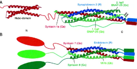
The detailed analysis of a SNARE complex operating in endosomal fusion showed that the N- and C-terminal coiled-coil domain provided by SNAP-25 for the synaptic core complex can be provided by two different polypeptides (Antonin et al., 2000). Furthermore, the crystal structure of this endosomal complex consisting of the R-SNARE VAMP-8 and the Q-SNAREs Syntaxin 7, Syntaxin 8 and Vti1b showed striking similarity to the structure of the synaptic core complex (Antonin et al., 2002).

Based on the analysis of a number of SNARE complexes from different fusion steps within the cell, the current model predicts that the structure and function of SNARE complexes is conserved and consists of one R-SNARE and three Q-SNAREs (Chen and Scheller, 2001)(see Figure I2).

Figure I2) Conserved Structure of SNARE complexes

(A) The core of the synaptic SNARE complex consists of an extended four-helix bundle structure that contains one helix of Synaptobrevin 2 (R-SNARE), one helix of Syntaxin 1a (Qa-SNARE), and two helices of SNAP-25 (Qb- and Qc-SNAREs). The two SNAP -25 helices, and the N-terminal three-helix bundle Habc-domain and the SNARE-motif of Syntaxin 1a are connected by flexible linker regions.

Syntaxin 1a and Synaptobrevin each contain a C-terminal TM domain, whereas SNAP-25 is attached to the membrane by palmitoyl modifications. (B) The core of the endosomal SNARE complex consists of a very similar four-helix bundle structure. It is composed of one helix each of Endobrevin (R- SNARE), of Syntaxin 7 (Qa-SNARE), of Vti1b (Qb-SNARE), and of Syntaxin 8 (Qc-SNARE) . Syntaxin 7, Vti1b, and Syntaxin 8 carry large N-terminal domains, probably all constituting three-helix bundle structures. All four endosomal SNAREs hold a TM domain adjacent to their respective SNARE motif . The cylinders represent the TM regions that are linked by a short sequence of unknown structure to the respective SNARE helices. The curved lines represent extended flexible linker regions (from Fasshauer, 2003).

Genome analysis of yeast, worm, fly and human revealed several

interesting points: a set of twenty to thirty related SNARE proteins, that can be

detected in yeast, worm, fly and humans seems to be sufficient to mediate the main

intracellular trafficking events. Moreover, the closest related members in the different

(22)

species reside in similar subcellular compartments. In human, additional SNARE proteins are expressed, which raises the total number of SNAREs detected in the human genome to 35 (Bock et al., 2001). This might indicate the use of tissue- specific SNARE proteins for tissue specific membrane trafficking pathways and a diversification of transport steps in mammals.

The SNARE hypothesis predicts a cognate pair of SNARE proteins for each trafficking step within the cell. Consistent with the prediction, different SNARE proteins can be found on different subcellular compartments, indicating that specific SNARE proteins might indeed be involved in specific transport steps. This view is supported by genetic data. Yeast mutants of the SNARE proteins sec22p and sec9p block the secretory pathway at an early timepoint and late timepoint, respectively (Brennwald et al., 1994; Hay et al., 1997; Novick et al., 1980). Mutations of neuron specific SNAREs involved in the highly specialized pathway of neurotransmitter release in fly, worm and mice lead to a block of evoked synaptic transmission and are lethal (Deitcher et al., 1998; Nonet et al., 1998; Schoch et al., 2001; Washbourne et al., 2002). In contrast, mouse mutants of two ubiquitous SNARE proteins involved in endosomal trafficking, cellubrevin and vti1b, are viable and show normal development without any obvious or only very mild phenotype, respectively (Atlashkin et al., 2003; Yang et al., 2001). Thus certain SNARE proteins are dispensable in vivo and lack of these proteins can be compensated. Earlier studies showed that inactivation of cellubrevin leads to reduced but not complete inhibition of Transferrin receptor recycling, already indicating that alternative recycling pathways exist, which are presumably mediated by other v-SNAREs (Galli et al., 1994).

What is the exact function of SNARE proteins? SNARE proteins form

trans core complexes during membrane fusion (see figure I3). The core complex

formation is accompanied by major conformational changes in the coiled coil

domains of the SNARE proteins. Whereas SNARE domains are unstructured in

solution, combining appropriate SNARE motifs leads to spontaneous formation of

four helical bundles which is accompanied by a major release of free energy

(Fasshauer et al., 2002; Hayashi et al., 1994; Yang et al., 1999). Formation of

SNARE complexes was proposed to proceed in a zipper like fashion from the N-

termial end of the SNARE motifs towards the C-terminal transmembrane domain

(Hanson et al., 1997; Lin and Scheller, 1997). These results led to a strikingly simple

model by which the zipper like formation of the SNARE complex would pull the two

(23)

opposing membranes close together. The release of energy during core complex formation would be the driving force needed to overcome the energy barrier in order to fuse the opposing membranes (see figure I3).

Figure I3) The SNARE cycle

Syntaxin is bound to n-Sec1 before formation of the core complex. Rab proteins might facilitate the dissociation of n-Sec1 from syntaxin, allowing subsequent binding (nucleation) between the three neuronal SNAREs, syntaxin, SNAP-25 and VAMP (for simplicity, only one coil is drawn for SNAP-25).

Ca

2+

triggers the full zipping of the coiled -coil complex, which results in membrane fusion and release of vesicle contents. After the fusion event, recruitment of -SNAP and NSF from the cytoplasm and subsequent hydrolysis of ATP by NSF causes dissociation of the SNARE complex. Syntaxin, VAMP and SNAP-25 are then free for recycling and another round of exocytosis. (NSF; N-ethyl-maleimide- sensitive fusion protein; SNAP-25, 25 kDa synaptosome-associated protein; SNARE, soluble NSF attachment protein receptor, VAMP, vesicle-associated membrane protein) (from Chen and Scheller, 2001).

SNARE proteins are indeed necessary and sufficient to fuse artificial membranes in vitro. (Weber et al., 1998) One R-SNARE and three Q-SNAREs have to be present on opposing membranes for fusion to occur in vitro (Fukuda et al., 2000), consistent with the conclusions drawn from sequence analysis, where four different subfamilies of SNARE proteins were identified (see above). Cells engineered to express the correct set of cognate SNARE proteins on their plasma membrane facing the extracellular medium will fuse with each other (Hu et al., 2003).

Thus it is clear that SNARE proteins are able to fuse artificial and natural membranes.

Yet whether SNARE proteins are the actual fusogens of the cell is still a

matter of discussion. In mutants of Synaptobrevin in mice, fly and nematodes,

spontaneous release of neurotransmitter is reduced, but clearly detectable, in

(24)

contrast to evoked neurotransmitter release, which is essentially abrogated (Deitcher et al., 1998; Nonet et al., 1998; Schoch et al., 2001; Washbourne et al., 2002). A similar result was obtained in mice mutant for SNAP-25 (Washbourne et al., 2002).

These results raised the question of whether SNARE-independent membrane fusion events might occur in the cell. Indeed, in an in vitro vacuole fusion assay in yeast, the function of trans SNARE complex formation was suggested to precede the actual fusion reaction. The final fusion of opposing membranes was suggested to be mediated by the highly hydrophobic, membrane buried V o subunits of the vacuolar ATPase (Ungermann et al., 1998; Peters et al., 2001).

In conclusion, SNARE proteins fulfill the structural requirements for proteins involved in membrane fusion events, they are necessary for membrane fusion in a large number of trafficking events and they are able to fuse artificial and natural membranes without auxilliary proteins present. It is possible that the transition from trans- to cis-complexes of SNARE proteins is an essential, but incomplete intermediate in membrane fusion in vivo, but the data supporting this view is sparse.

I.2.5 Spatial and temporal control of SNARE dependent membrane fusion

As SNARE complex formation leads to membrane fusion, this event has

to be under tight control in order to maintain the integrity of membrane

compartments. Specific SNARE complexes mediate specific trafficking events, which

led to the proposal that spatial specificity of trafficking events is encoded in SNARE

proteins themselves (Rothman, 1994). This may not be true in these general terms

since the lack of phenotype of mice carrying null mutations for the SNAREs

cellubrevin and vti1b suggests the possibility of functional redundancy (Atlashkin et

al., 2003; Yang et al., 2001). Also it was demonstrated in vivo that a mutant of fly

synaptobrevin, n-syb, can be rescued by driving the expression of the ubiquitous

homologue syb in neuronal tissue and vice versa. Yet, each individual mutant shows

a strong and specific phentotype (Bhattacharya et al., 2002). Similarly in yeast,

mutants of t-SNAREs involved in golgi to endodosome or endosome to vacuole

trafficking show specific phenotypes but can be rescued by overexpressing either of

the two SNARE proteins (Darsow et al., 1997; Gotte and Gallwitz, 1997).

(25)

The isolated, soluble core domains of non-cognate SNARE proteins, i.e.

which do not pair in vivo, form stable, heat and SDS-resistant core complexes similar to cognate SNARE domains, as long as the QaQbQcR-rule is respected (Fasshauer et al., 1999; Yang et al., 1999). In contrast, combinatorial pairing of all yeast R- SNAREs with a number of yeast Q-SNAREs in an in vitro liposome fusion assay revealed a certain degree of specificity encoded in the SNARE proteins themselves (McNew et al., 2000). Thus, certain combinations of SNARE proteins do favour membrane fusion in vitro compared to others, but in vitro and in vivo data suggest, that stability of SNARE complex formation alone cannot account for specificity in membrane fusion.

Since complex formation of non cognate SNARE proteins has not been observed in vivo (Jahn et al., 2003), it seems likely that regulatory mechanisms are superimposed on SNARE complex formation. The existence of individual SNARE proteins for different trafficking steps might enable the cell to selectively aquire regulatory proteins other than SNAREs. In particular, Rab proteins might play an important role in determining fusion specificity. It was shown that activated Rab5 recruits a multiprotein complex containing the SNARE protein Syntaxin 13, the tethering factor EEA1 and the general fusion protein NSF in a functional microdomain, resulting in homotypic endosomal fusion (McBride et al., 1999). Thus, tethering of the membranes and the local activation or priming of the fusion machinery would occur as an integrated mechanism. This is a very attractive model of how specificity of membrane fusion events may occur through several layers of protein-protein interactions.

Temporal control over the final, SNARE mediated fusion reaction is in

part encoded in the structure of SNARE proteins. A number of SNAREs have N-

terminal extensions which participate directly in the kinetics of membrane fusion by

negatively regulating SNARE complex formation (Filippini et al., 2001). The negative

regulation of SNARE complex formation by N-terminal extensions is best understood

for the synaptic Qa SNARE Syntaxin 1. The N-terminal domain of Syntaxin 1 folds

back on the SNARE motif, thus leading to a so called closed conformation, in which

the SNARE motif is not accessible for SNARE complex formation (Dulubova et al.,

1999). Removal of the N-terminal extension greatly enhances SNARE complex

formation and fusion efficiency mediated by Syntaxin 1 (Parlati et al., 1999). A protein

called nSec1/munc18, which binds to syntaxin 1, plays an essential role in activating

(26)

Syntaxin 1 in vivo for SNARE complex formation (Pevsner et al., 1994; Richmond et al., 2001; Verhage et al., 2000). NSec1 is a member of a family of proteins found from yeast to mammals called SM proteins (forSec/Munc) which were shown to be crucial in diverse membrane fusion events such as exocytosis and endosomal fusion (Jahn et al., 2003).

Another important family of proteins involved in the temporal regulation of SNARE complex formation and membrane fusion are calcium sensing proteins of the synaptotagmin family, which are found in metazoae (Sudhof, 2002; Littleton, 2001). Synaptotagmin I is most likely the main Ca 2+ -sensing molecule in fast, Ca 2+ - triggered neurotransmitter release (FernandezChacon et al., 2001; Geppert et al., 1994), whereas Synaptotagmin VII is implicated in Ca 2+ -dependent, lysosomal exocytosis mediating plasma membrane repair (Jaiswal et al., 2002). The mechanism by which Synaptotagmins control Ca 2+ dependent membrane fusion is not understood, but Ca 2+ dependent lipid binding activity of this protein family seems to play an important role (Shin et al., 2003; Sudhof, 2002).

In addtion, a number of other proteins have been implicated in the regulation of membrane fusion, most of which act via direct binding to SNARE proteins like Nsec1 and Synaptotagmins (Gerst, 2003). As will be seen in the next chapter, the importance for plasticity in membrane trafficking is particularly clear in events like neuronal differentiation.

I.2.6 The role of membrane trafficking in neuronal morphogenesis

The outgrowth of axons and dendrites of neurons is accompanied by a major increase in surface area of the cell. For example, an axon growing at a rate of 20-50µm/h has to incorporate a membrane area corresponding to the size of its cell body every two hours (Futerman and Banker, 1996). To enable axons and dendrites to locate their appropriate synaptic partners and therefore to enable the correct wiring of the nervous sytem, the delivery of membrane to the axon and dendrites has to be integrated into the demands of directed axonal and dendritic growth, respectively.

This plasticity in neurite outgrowth is exemplified by the fact that the extent of axonal

outgrowth of a neuron in culture can be drastically modified by the substrate that is

presented to the neuron (see Fig I4; Lemmon et al., 1989) and signalling cues exist

(27)

which preferentially stimulate either axonal or dendritic outgrowth (Higgins et al., 1997; Prochiantz, 1995). Thus, as shown in Figure I4, axonal outgrowth is selectively stimulated by a member of the IgCAM superfamily of cell adhesion molecules called L1, whereas dendritic outgrowth is not affected compared to the control substrate poly-L-Lysine.

Figure I4) L1 stimulates axonal outgrowth in hippocampal neurons. Poly-L-Lysine-coated coverslips were incubated with anti-human Fc antibodies followed by incubation with (L1) or without (poly-Lysine) L1-Fc fragment. Hippocampal neurons seeded at low density were kept in culture for 24 hours and analyzed by immunofluorescence with pAb to L1.

How do guidance cues, e.g. the ligation of cell adhesion molecules translate into directed growth of the axon? Directed axonal navigation is mediated by growth cones, which are specialized, highly motile structures found at the tip of developing axons. Long- or short-range guidance cues, that can be attractive or repulsive, are recognized by receptors expressed on the growth cone and provoke the classical forms of growth cone behaviour such as advance, turning, withdrawal, and target recognition (Suter and Forscher, 1998). Therefore, by probing and sensing the environment and by translating the information received into motile behaviour, the growth cone ultimately allows for the establishment of the complex pattern of neuronal connexions (Tessier-Lavigne and Goodman, 1996).

The cytoskeleton of growth cones is intimately involved in the axonal

response to extracellular cues as drugs interefering with actin dynamics were shown

to render neurons “blind” in vivo resulting in axons that bypass and extend beyond

(28)

normal synaptic partners (Bentley and Toroian-Raymond, 1986; Chien et al., 1993;

Kaufmann et al., 1998).

In growth cones, F-actin undergoes a retrograde flow from the peripheral to the central region. This so called retrograde actin flow is the result of constant de novo-polymerization of actin at the leading edge of the growth cone and retrograde transport of actin filaments mediated by myosin motors (Forscher and Smith, 1988; Lin et al., 1996). In order for the growth cone to be able to advance, the retrograde actin flow has to be coupled to the substrate. This substrate-cytoskeletal coupling is mediated by surface receptors which link the underlying cytoskeleton to the extracellular matrix and ultimately allows the growth cone to generate force and pull forwards. Thus, cell adhesion molecules of the Integrin-, Cadherin- or IgCAM- family play essential roles in growth cone movement. They all can be linked to the underlying actin-cytoskeleton via distinct adaptor proteins in a stimulus-dependent manner, which is provided by ligation of the respective receptor with its extracellular ligand (Suter et al., 1998).

For the growth cone to advance, substrate -cytoskeletal coupling has to be under tight control. In fact, a gradient of strong adhesion at the leading front and weak adhesion at the rear edge of the growth cone has to exist to allow the cytoskeletal machinery to pull the growth cone forwards. Such a gradient of adhesiveness has been detected for several adhesion molecules, in agreement with the predictions of the substrate-cytoskeletal coupling model of growth cone motility (Kamiguchi and Lemmon, 2000; Kamiguchi and Yoshihara, 2001; Schmidt et al., 1995). As will be explained below, local recycling of cell adhesion molecules via endocytosis at the rear edge and exocytosis at the leading front could be one mechanism in establishing such a gradient of adhesion along the growth cone.

At the same time, ligation of cell adhesion molecules initiates a local

signaling cascade (Skaper et al., 2001). The IgCAM L1 ind uces a complex signaling

cascade upon ligation, which includes the non-receptor tyrosine kinase p60src and

the MAP kinase pathway (Schaefer et al., 1999; Schmid et al., 2000). The importance

of L1-induced signaling for L1-function is highlighted by the fact, that the stimulatory

effect mediated by homophilic L1-interaction on neurite outgrowth is abrogated in

neurons deficient in p60src. (Ignelzi et al., 1994) Similarly, neurite outgrowth

stimulated by L1 or the CAM N-Cadherin are reduced, when the MAP kinase

(29)

cascade is inhibited, indicating a conversion of signaling cascades mediated by CAMs of distinct families (Perron and Bixby, 1999; Schmid et al., 2000).

The signaling cascades initiated by CAMs allow for the site-directed rearrangement of cytoskeletal dynamics and the resulting change in growth cone motility (Challacombe et al., 1996; Dent and Kalil, 2001; Suter et al., 1998) and are therefore of greatest importance for neuronal morphogenesis. Yet, the exact molecular mechanisms that link guidance cues to cytoskeletal dynamics are not clear (Skaper et al., 2001).

Whereas adhesion molecules exert their function via direct cell-cell or cell-matrix interaction, other target-derived secreted factors like netrins or the sempahorins exist, which act at a distance to attract or repel axons. Secretion establishes a gradient and thus attracts or repels axons or dendrites along the gradient, if the target neurite expresses the respective receptor (Dickson, 2002).

Secreted guidance cues induce motile behaviour of the axon by regulating the growth cone cytoskeleton, similar to what was described above for CAMs (Dickson, 2002).

Interestingly, a physical and functional interaction between the receptor for Semaphorin 3a, Neuropillin, and the cell adhesion molecule L1 has been observed.

Whereas binding of Sema3a to its receptor induces growth cone turning away from a Sema3a gradient in wild type neurons, repulsion is not observed in L1 knock out cells (Castellani et al., 2000). Thus, adhesion molecules can be functionally linked to soluble guidance molecules and their receptors, illustrating the high plasticity by which growth cones can respond to guidance cues.

In contrast to the well established, important roles for adhesion molecules and secreted guidance cues in axonal navigation, the role of membrane trafficking in axonal and dendritic outgrowth is less clear. Particularly, a possible link between the cytoskeleton, the major target of guidance cues, and the trafficking machinery, which delivers membrane for growth, is not established.

Genetic data which firmly establish a role for exocytotic membrane

trafficking in neurite outgrowth in vivo were provided very recently in Dorosophila. A

mutant of sec5, a subunit of the exocyst complex involved in tethering of exocytotic

vesicles, stops growing neurites as soon as the maternal, cytosolic pool of sec5 is

exhausted (Murthy et al., 2003). At the same time, accumulation of synaptic vesicles

at synapses and neurotransmitter release was not affected. Thus, distinct types of

(30)

vesicles with apparently different targeting mechanisms are implicated in neurotransmitter release and neuronal differentiation.

A first description of the SNARE machinery involved in neurite extension came from experiments using clostridial neurotoxins. Clostridial neurotoxins are potent inhibitors of neurotransmitter release by cleaving key components of the synaptic exocytotic membrane fusion machinery. The inactivation of the neuronal plasma membrane target SNARE, composed of SNAP25 and syntaxin 1, by Botulinum Neurotoxin (BoNT) A and C1 leads to a pronounced reduction in axonal outgrowth (Grosse et al., 1999; Osen-Sand et al., 1996) thus suggesting the involvement of SNAP-25/Stx1 dependent exocytosis in neurite outgrowth. In contrast, tetanus neurotoxin mediated cleavage of the synaptic v-SNARE Synaptobrevin has no effect on neurite outgrowth, in spite of its complete abolition of neurotransmitter release (Grosse et al., 1999; Osen-Sand et al., 1996). These findings were confirmed by analysing synaptobrevin 2 knock out mice, which showed a severe impairment in transmitter release, but apparently normal brain development (Schoch et al., 2001).

Similarly, mutants for munc-18 and munc 13-1/2, two important regulators of synaptic vesicle exocytosis, showed severe defects in neurotransmitter release without any apparent impairment of brain development (Aravamudan et al., 1999; Verhage et al., 2000). Surprisingly, the knock out of SNAP 25 in mouse shows normal brain development (Washbourne et al., 2002), whereas the experiments mentioned above using clostridial NTs suggested a role for this molecule in neurite outgrowth. This discrepancy could be owing to the expression of SNAP23, a close homologue that may be able to functionally replace SNAP25 in the SNAP25 knock out mice. In any case, these results clearly indicate that the molecules and the membrane compartment(s) mediating neurite outgrowth are, to a large extent, different from those involved in neurotransmitter release.

Whereas the studies mentioned above established a role for exocytosis in neurite outgrowth, the intracellular membrane carriers fusing with the plasma membrane and their molecular composition are only beginning to be characterized.

An important regulatory role for the exocytosis of membrane carriers of endosomal

origin in the development and plasticity of synaptic contacts has been recently

established. The postsynaptic glutamate receptors, NMDA- and AMPA-receptor can

undergo a controlled, local cycle of endo- and exocytosis at the synapse through

endosomal compartments. This recycling from and to the postsynaptic plasma

(31)

membrane directly influences the postsynaptic response to glutamate release at the presynaptic active zone by controling the density of membrane resident receptors (Sheng and Lee, 2003). At least two different membrane trafficking pathways seem to be involved in the recycling of postsynaptic receptors, since membrane carriers could be identified that contained the AMPA-receptor and the SNARE protein Syntaxin 13, but no NMDA-receptor (Lee et al., 2001). Furthermore, whereas NMDA-receptor recycling is independent of Synaptobrevin, recycling of the AMPA-receptor is sensitive to cleavage of Synaptobrevin by clostridial neurotoxins (Lan et al., 2001; Lu et al., 2001).

Less is known about recycling pathways in the development of neurons, but an equally important function for local recycling of membrane receptors can be expected. Early studies on cell adhesion molecules of the Integrin family showed that these molecules do not make immobile glue at the plasma membrane but instead recycle through endocytic compartments to and from the plasma membrane (Bretscher, 1992). In fact, neurons seem to be able to control the density of integrin receptors on the cell surface in response to ligand density by an endocytotic mechanism, in order to maintain growth cone motility (Condic and Letourneau, 1997).

Similarly, the cell adhesion molecule L1, which plays an important role in brain development in mice and human, was shown to recycle locally in the growth cone (Kamiguchi and Lemmon, 2000). Therefore, intracellular trafficking of cell-cell or cell- matrix adhesion molecules like L1 and integrins through endosomal compartments is likely to be important in regulating and stabilizing adhesive contacts in the course of neurite outgrowth.

What is the nature of the transport carriers, which deliver newly synthesized lipids and proteins to the growing axons? Brefeldin A, a fungal toxin that blocks post Golgi trafficking of membrane carriers, inhibits neurite outgrowth demonstrating a role for golgi-derived vesicles in neurite outgrowth (Jareb and Banker, 1997). Studies on the chicken homolog of L1, NgCAM, suggest that, when expressed in rat neurons in culture, newly synthesized NgCAM is delivered and inserted in the plasma membrane at the distal end of the growing axon (Vogt et al., 1996). Similar results were obtained when CD8 alpha, a non-neuronal protein, was expressed in neurons in culture and found to be inserted at the tip of the growing axons (Craig et al., 1995).

These studies suggest that newly synthesized proteins and lipids may be transported

and inserted into the membrane at the sites where growth actually takes place.

(32)

Interestingly, a recent study on NgCAM demonstrated that transport carriers delivering newly synthesized molecules and endosomal carriers are not necessarily different. NgCAM can be transported to the axo n in an indirect pathway called transcytosis. Newly synthesized NgCAM is first transported to the dendritic plasma membrane followed by immediate uptake and delivery to the axon in endosomal carriers (Wisco et al., 2003).

In summary, multiple membrane compartments of different origin may participate and interact to allow for the highly complex process of neurite outgrowth.

Elucidation of the relationship between different post-Golgi compartments involved in neurite outgrowth and the molecular composition including the SNARE proteins expressed in these compartments represents one of the major challenges to understand the role of membrane trafficking in neuronal differentiation.

I.3 Aims of the thesis

The discovery of R-SNAREs insensitive to clostridial ne urotoxins opened

the way to test the involvement of neuronal, vesicular compartments different from

synaptic vesicles in neurite outgrowth (Martinez-Arca et al., 2000a). The first R-

SNARE that was demonstrated to be insensitive to clostridial neurotoxins is called TI-

VAMP for Tetanus neurotoxin Insensitive Vesicle Associated Membrane Protein (also

called VAMP7) (Galli et al., 1998; Advani et al., 1998). TI-VAMP is broadly expressed

in a variety of tissues, including neurons (Advani et al., 1999; Coco et al., 1999). No

true homologue of TI-VAMP exists in yeast, whereas TI-VAMP homologues are

found in higher uni- and multicellular organisms like Dictyostelium, C.elegans,

Drosophila and humans (Filippini et al., 2001). The structure of TI-VAMP is different

from the classical R-SNARE structure found in synaptobrevin and related R-SNAREs

in that it carries a N-terminal extension in addition to the SNARE motif and the

transmembrane domain (Galli et al., 1998). This N-terminal extension, the Longin

domain, is found in a number of R-SNAREs from yeast to human and its sequence

shows a high degree of conservation between different R-SNAREs carrying this

extension (Filippini et al., 2001). The Longin domain of TI-VAMP and another R-

SNARE called Ykt6 were shown to negatively regulate SNARE complex formation

(33)

(Martinez-Arca et al., 2000b; Tochio et al., 2001), similar to the structurally unrelated Nterminal extensions of Qa SNAREs like Syntaxin 1 (Gonzalez et al., 2001; Tochio et al., 2001). In addition to its role as negative regulator in SNARE complex formation, the Longin domain of TI-VAMP binds to the adaptor complex AP-3, which is important for the localization of this protein to late endosomal compartments in non- neuronal cells (Martinez-Arca et al., 2003b).

Although a number of studies attempted to elucidate the membrane trafficking pathway(s) mediated by TI-VAMP, its exact nature is still unclear. TI-VAMP localizes to late endosomes/multivesicular bodies and to vesicles scattered throughout the cytoplasm (Advani et al., 1999; Coco et al., 1999; Martinez-Arca et al, 2003a; Martinez Arca et al., 2003b). It forms complexes with SNARE proteins functioning at the plasma membrane and in endosomal compartments (Bogdanovic et al., 2002; Martinez-Arca et al., 2000; Martinez-Arca et al., 2003b; Galli et al., 1998). Function blocking assays have implicated TI-VAMP in the degradative pathway of the EGF receptor and fusion of late endosomes/lysosomes (Advani et al., 1999; Ward et al., 2000), but this function might be redundant with endobrevin/VAMP8, another endosomal v-SNARE (Antonin et al., 2000). Another series of studies implicated TI-VAMP in an exocytotic pathway. In epithelial cells, TI- VAMP plays a role in transport to the apical plasma membrane, a pathway insensitive to Tetanus neurotoxin (Galli et al., 1998; Ikonen et al., 1995; Lafont et al., 1999). In mastocytes, TI-VAMP might be involved in the process of degranulation, another exocytotic pathway insensitive to tetanus neurotoxin (Hibi et al., 2000). The reason for this apparent discrepancy of TI-VAMP function in either endosomal or exocytotic trafficking is not understood, but cell type dependent differences might play a role. In neurons, the TI-VAMP compartment defines a new type of vesicle, which is different from synaptic vesicles and transferrin receptor positive recycling endosomes/early endosomes (Coco et al., 1999). In young neurons developing in culture, TI-VAMP accumulates at the leading edge of growing axons and dendrites, consistent with a role for this protein in neurite outgrowth (Coco et al., 1999). Further studies showed that TI-VAMP indeed mediates neurite outgrowth in PC12 cells and in neurons in culture, which was based on the overexpression of dominant-positive and -negative TI-VAMP variants (Martinez-Arca et al., 2000b; Martinez-Arca et al., 2001).

The goal of this study was to define the role of the TI-VAMP

compartment in neurite outgrowth. In particular, the nature of the compartment in

(34)

neuronal cells was not well understood and no cargo protein of the TI-VAMP- mediated trafficking pathway in neurons was known. A working hypothesis was that adhesion molecules might be part of the molecules trafficking through the TI-VAMP compartment due to their importance in neuronal development and the fact that these molecules can be regulated by membrane trafficking.

Another important question of this work was to understand the regulation

of the TI-VAMP mediated trafficking. As outlined above, the growth cone is the

sensor of the axon for attractive and repulsive guidance cues. Profound

rearrangements of the cytoskeleton within the growth cone result from guidance cues

leading to events like turning, enhanced growth or retraction of the axon (Koleske,

2003). Since the TI-VAMP compartment is highly enriched in the growth cone of

axons and dendrites (Coco et al., 1999), a potential relationship between

rearrangements of the cytoskeleton triggered by guidance cues and regulation of the

TI-VAMP compartment in the axonal growth cone was explored in this work.

(35)

II. Materials and methods

II.1. Materials

II.1.1. Antibodies, clones, and reagents

Polyclonal anti-GFP antibody was described earlier (Martinez-Arca et al., 2001).

Mouse monoclonal antibodies directed against Syb 2 (clone 69.1) and syntaxin 1 (HPC-1) anti-transferrin receptor (68.4) and the polyclonal antibody against syntaxin 7 were generous gifts from R. Jahn (Max Pla nck Institute, Goettingen, FRG), C.

Barnstable (Yale University, New Haven, CT), I. Trowbridge (Salk Institute, La Jolla,

CA, USA) and Dr W. Hong (Institute of Molecular and Cell Biology, Republic of

Singapore), respectively. Polyclonal rabbit antibodies against mouse L1 and Fab

fragments generated from this antibody were previously described (Rathjen and

Rutishauser, 1984). The polyclonal anti-Syb 2 antibody (MC23) was previously

described (Chilcote et al., 1995). The following commercial antibodies were used in

this study: monoclonal anti-Green Fluorescent Protein (GFP; clone 7.1 and 13.1)

from Roche (Indianapolis, IN), monoclonal anti-GluR2 (cl 6C4) from Chemicon

(Temecula, CA), monoclonal anti-Vti1b (clone 7) and SNAP25 (clone 20) from

Transduction laboratories, monoclonal anti-Syntaxin 13 (clone 15G2) from StressGen

(San Diego, CA), monoclonal anti-phospho-tyrosine antibody (clone 4G10) from

Upstate (Lale Placid, NY) and polyclonal anti-phospho -src (pT 418 ) from Biosource

International (Nivelles, Belgium). Affinity-purified Alexa 488 and Cy3-coupled goat

anti-rabbit and anti-mouse immunoglobulins, phalloidin coupled to Alexa 564 or 488,

Streptavidin coupled to Rhodamine, affinity-purified Fc-fragments from mouse and

human IgGs were from Jackson ImmunoResearch (West Grove, PA). Biotinylated

Goat anti mouse-IgG ?-chain antibody was from Southern Biotechnology Associates

(Birmingham, Alabama). Horseradish peroxidase- and Alkaline Phosphatase-coupled

streptavidin, sheep anti-mouse and anti-rabbit IgGs were from Promega (Madison,

WI). NGF was supplied by Alomone Labs (Rehovot, Israel). Collagen was from

Becton Dickinson (Franklin Lakes, NJ). All other reagents were from Sigma unless

specified.

(36)

II.1.2. Plasmids

The cDNA coding for L1-Fc chimera was kindly provided by Dr. T. Brümmendorff (Max-Dellbrück Zentrum für molekulare Medizin, Berlin) and has been described (De Angelis et al., 1999). Eukaryotic expression vectors based on pEGFP-C3 (Clonetech) coding for Green Fluorescent Protein (GFP) and TI-VAMP fused to GFP at the C- or N-terminus were provided by S. Martinez-Arca (Institut Fer-a-Moulin, Paris) and have been described. (Martinez-Arca et al., 2000b)

The prokaryotic expression vector coding for Cyt -TI-VAMP fused to GST was provided by Dr Thierry Galli (Institut Fer-a-Moulin, Paris) and described in (Martinez- Arca et al., 2000b).

II.2. General molecular biology methods

II.2.1. PCR

Each reaction contained the following:

2µl of 10mM dNTP (a mixture of dATP, dTTP, dCTP, and dGTP each at 10mM concentration)

10µl of 10x Pfu DNA polymerase buffer (supplied with the enzyme) 10-100ng of template DNA

2µl of each primer at a concentration of 100ng/? l 2µl of Pfu DNA polymerase

Distilled water to a total volume of 100µl

The PCR was carried out using 25 thermal cycles: 94°C for 1 min, 50°C for 1 min, 72°C for 2 min/kb of template DNA. Following these cycles the reaction was incubated for a further 10 min at 72°C, before being cooled to 4°C.

II.2.2. Restriction digestion

Enzymes and buffers were supplied by New England Biolabs and used according to

the manufacturers’ instructions.

Abbildung

Figure I1) Subcellular localization of mammalian SNARE proteins
Figure I2) Conserved Structure of SNARE complexes
Figure I3) The SNARE cycle
Figure I4) L1 stimulates axonal outgrowth in hippocampal neurons. Poly-L-Lysine-coated  coverslips were incubated with anti-human Fc antibodies followed by incubation with (L1) or without  (poly-Lysine) L1-Fc fragment
+7

Referenzen

ÄHNLICHE DOKUMENTE

A recent study using electron paramagnetic resonance has reported that the association of Munc18-1 with the syntaxin1a/SNAP25a complex can result in the formation of a complex

Atran and associates conclude that Q’eqchi’ who moved into central Pete´n around the 1970s seem unable to adapt to the lowland environment or learn from their neighbors because of

: Graph illustrating the Dendritic Atrophy phenotype in the mice deficient in Cap23 and comparison with wild type mice Wild type mice (dark line), Cap23 heterozygotes (pink line)

The Skp1-Cullin1-F-box protein (SCF) E3 ubiquitin ligase and in particular the substrate-recruiting adaptor subunit F-box proteins have emerged as essential mod- ulators of

TABLE 1 Average and maximum C stocks in living and dead volumes for forest registered as managed and unmanaged in Germany, based on plot data from the national forest

The left example tests how well different peak detection methods can identify peaks in synthetically generated data.. The right example is an experimental benchmark data set of

The left example tests how well different peak detection methods can identify peaks in synthetically generated data.. The right example is an experimental benchmark data set of

The Poisson distribution expresses the probability of a given number of events occurring in a fixed in- terval of time or space if these events occur with a known constant mean rate