The role of Cornichon (Cni) in axis formation in Drosophila
Inaugural-Dissertation zur
Erlangung des Doktorgrades
der Mathematisch-Naturwissenschaftlichen Fakultät der Universität zu Köln
vorgelegt von Sajith Dass aus Thodupuzha, Indien
Köln 2004
Berichterstatter: Prof . Dr. Siegfried Roth PD. Dr. Thomas Klein Tag der mündlichen Prüfung: 10.02.2005
1 INTRODUCTION... 6
1.1AXIS FORMATION... 6
1.2OOGENESIS IN DROSOPHILA... 7
1.3FIRST ROUND OF GRK SIGNALING FROM THE OOCYTE TO THE OVERLYING FOLLICLE CELLS RESULTS IN THE ASSUMPTION OF POSTERIOR IDENTITIES BY THESE CELLS... 8
1.4A SECOND ROUND OF OOCYTE TO FOLLICLE CELL GRK SIGNALING ESTABLISHES THE DV AXIS... 10
1.5GRK SIGNALING INITIATES THE PROCESS OF THE DV AXIS FORMATION OF THE EMBRYO... 12
1.6THE EGF-EGFR REPERTOIRE OF REGULATORS, THEIR INTERACTION IN LIGAND REGULATION AND ROLE IN PATTERN FORMATION... 14
1.7CORNICHON AND INITIATION OF GRK SIGNALING FROM THE GERM LINE... 19
1.8CELLULAR EXOCYTOSIS, A BRIEF OVERVIEW... 21
1.9OBJECTIVES... 23
2A RESULTS - CORNICHON (CNI)... 24
2A.1SUB-CELLULAR LOCALISATION OF CNI... 24
2A.2EXCESS GRK SIGNAL FROM THE GERM LINE DORSALISES THE EGG SHELL... 26
2A.3PATTERNING OF THE EMBRYO AFFECTED WHEN GRK OVEREXPRESSED IN DFH60SCO HETEROZYGOUS BACKGROUND... 29
2A.4TIMING OF ONSET IS DIFFERENT BETWEEN NANOSGAL4VP16 AND TUBGAL4VP16 ... 30
2A.5MIS/OVER-EXPRESSION OF GRK BY TUBGAL4VP16 BUT NOT NOSGAL4VP16 CAN RESCUE CNI FUNCTION... 32
2A.6PROTEIN PROFILE OF THE OVARIES USING WESTERN BLOT ANALYSIS... 35
2A.7EXPRESSION OF A GRK VERSION THAT LACKS THE CNI INTERACTION DOMAIN AND ITS CONSEQUENCES... 39
2A.8ROLE OF JMD IN REGULATION OF GRK TRANSIT TO THE MEMBRANE... 41
2A.9EXPRESSION OF ANTI SENSE STAR IN THE GERM LINE... 42
2A.10WESTERN BLOT ANALYSIS OF GRKDC5MYC,CNI/CNI,GRKδTCMYC AND GRKδCMYC... 44
2B RESULTS - CORNICHON-RELATED (CNIR)... 46
2B.1ORFCG17262 IS CNIR... 46
2B.2VAN EEKENS LINES... 47
2B.3L(2)(A1-7) IS A PUTATIVE ALLELE OF CNIR... 50
2B.4P ELEMENT INSERTION LINES CLOSEST TO CNIR... 51
2B.5MOLECULAR ORGANIZATION OF THE GENOMIC REGION NEAR CNIR: ... 52
2C RESULTS – ENHANCER/ SUPRESSOR SCREEN... 55
2C.1CHROMOSOMAL REGIONS THAT INTERACT WITH CNI (ENHANCERS OR SUPRESSORS) ... 55
2C.2CNI ALLELES AND PHENOTYPES... 55
2C.3THE SCREEN LOGISTICS... 59
THE SCREEN... 59
2C.4TIER1:PRIMARY SCREEN... 59
2C.5TIER2:RETESTING PUTATIVES... 60
2C.6TIER3:SECONDARY SCREEN (INDEPENDENT CONFIRMATION) ... 64
2c.6.1 Supressor region: Df (3L)VW3 (76A3- 76B2) ... 64
2c.6.2 The 62; 63 region contains two Synthetic lethals and one enhancer region . 67 2c.6.3 The Lyra/ senseless region Df42/ 70A2-3;70A5-6 ... 70
2c.6.4 The enhancer region (Df99 /86F1;87A9) ... 71
3A DISCUSSION- CORNICHON ... 72
3A.1CNI IS AN ER TO GOLGI TRANSPORT PROTEIN... 72
3A.2GRK TRANSPORTED VIA THE BULK FLOW MODE OF EXOCYTOSIS CAN OVERCOME CNI REQUIREMENT AND PRODUCES ACTIVE GRK... 73
3A.3GRK SIGNALING CAN BE RESCUED BUT THE REGULATORY FINE TUNING IS LOST.... 75
3A.4CNI AND STAR, ARE BOTH INVOLVED IN THE ER TO GOLGI TRANSPORT OF GRK... 76
3A.5CLEAVED FORMS OF GRK ARE OBSERVED IN CNI HOMOZYGOUS BACKGROUND... 77
3A.6REGULATION MAY ALSO BE AT THE LEVEL OF PROTEIN STABILITY... 78
3A.7CNI MEDIATED GRK TRANSPORT PREVENTS HYPERACTIVATION OF EGFR ... 79
3A.8HYPOTHETICAL MODEL... 80
3B DISCUSSION – CORNICHON RELATED (CNIR)... 82
3C DISCUSSION – ENHANCER/ SUPRRESSOR SCREEN ... 83
3C.1SCREEN COVERAGE... 83
3C.2THE SUPPRESSOR WAS IDENTIFIED AS PIPE (PIP) ... 84
3C.3SALLIMUS (SLS) AND SPROUTY (STY) ARE AMONGST THE LIKELY CANDIDATES.... 84
4 MATERIALS AND METHODS ... 86
4.1FLY HUSBANDRY... 86
4.2FLY LINES USED... 86
4.3PREPARATION OF EGG SHELL AND EMBRYONIC CUTICLE... 87
4.4FIXATION OF OVARIES FOR IMMUNOSTAININGS... 87
4.5ANTIBODY STAINING OF OVARIES... 87
4.6IN SITU HYBRIDISATION OF OVARIES... 88
4.7ANTIBODIES USED FOR IMMUNO-STAINING... 89
4.8PROTEIN EXTRACTION FROM OVARIES FOR IMMUNO BLOT ANALYSIS... 89
4.9GLYCOSIDASE ASSAYS... 90
4.10WESTERN BLOT ANALYSIS OF PROTEINS... 90
4.10.1 Electrophoresis and transfer of proteins onto the membrane ... 90
4.10.2 Processing of the transferred membrane and gel ... 90
4.10.3 Primary and secondary antibody treatments... 91
4.10.4 Detection of western blots... 91
4.11ANTIBODIES USED FOR IMMUNO-BLOTS... 91
4.12GENOMIC DNA EXTRACTION FROM LARVAE... 92
5 BIBLIOGRAPHY ... 93
6 SUMMARY ... 101
7 ZUSAMMENFASSUNG ... 103
8 ERKLÄRUNG... 105
9 LEBENSLAUF... 106
10 ACKNOWLEDGEMENTS ... 107
1 Introduction
1.1 Axis formation
One of the primary requirements for a bilaterally symmetric body plan is two perpendicular axes, namely the Anterior-Posterior (AP) axis and the Dorsal-Ventral (DV) axis. The determination of the two axes is one of the earliest decisions which must be taken for most bilaterian body plans. In the dipteran, Drosophila melanogaster both AP and DV decisions are made before the egg is laid. In the eggs both axes can be recognised by distinct egg shell structures which also serve as markers for these axis decisions. The anterior end (where the head of the future embryo would develop) of the egg consist of the micropyle and the operculum (Fig.1A&B). The micropyle is a small pointed structure which functions as a channel for the entry of sperm, associated with the micropyle is a collar structure on the anterior end of the egg shell (Fig.1B). The embryonic larvae hatches out of the egg shell through a structure called the operculum. It is at the anterior of the egg between the micropyle and the dorsal appendages (DAs). The most obvious structures on the egg are the pair of DAs (Fig1.A&B), which are respiratory structures.
The posterior of the egg is marked by the aeropyle (Fig1.A&B) which is a structure required for exchange of gases. There is also a subtle difference in the curvature between the dorsal and ventral sides, the ventral side is slightly more convex than the dorsal side (Spradling, 1993).
Fig. 1 The egg of Drosophila melanogaster, indicating the egg shell structures which can be used as markers for the morphogenetic processes which patterned the egg. (A) A photograph of an egg laid by wild type female with the AP and DV egg shell structures labeled. (B) A schematic drawing of the wild type egg with the egg shell structures labelled.
Within the egg cytoplasm mRNAs are localised to the anterior and posterior during mid oogenesis where they stay latent until the beginning of embryogenesis. bicoid mRNA, responsible for head and thorax development is localised to the anterior cortex and oskar mRNA, responsible for the the formation of the primordial germ cells (PGCs/ Pole cells) and the abdomen, is localised to the posterior cortex (for a review see(Riechmann and Ephrussi, 2001).
Therefore, to understand the process of axis initiation it is pertinent to study the processes during oogenesis that intiate these patterns.
1.2 Oogenesis in Drosophila
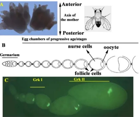
Each female fly has a pair of ovaries within which the process of oogenesis occurs. Each ovary is composed of independent chains of structures called ovarioles (Fig.2). Each ovariole consists of the germarium (Fig.2B&C) at the anterior tip, which houses the germ line and somatic stem cells. The germ line stem cell in the niche, at the anterior tip of the germarium undergoes unequal cleavage followed by mitotic amplification. After mitotic amplification, clusters of 16 interconnected germ cells get ensheathed by a monolayer of somatic follicle cells to form an egg chamber (Morgan and Mahowald, 1996; Ray and Schupbach, 1996). As these cells exit from the germarium as egg chambers, one of the germ line cells has been determined by germ line-somatic communication to be the oocyte and the remaining 15 germ line cells develop as nurse cells. The oocyte nucleus is almost transcriptionally quiescent and therefore is dependent on the polyploid nurse cells, for transport of materials required for its growth. The egg chamber is usually arranged in such a manner that the oocyte is positioned posterior to the nurse cells (Godt and Tepass, 1998; Gonzalez-Reyes and St Johnston, 1994; Gonzalez-Reyes and St Johnston, 1998).
The egg chambers then go through 14 morphologically distinct developmental stages before the egg is laid (King, 1970; Spradling, 1993). Both the germ line cells and the somatic follicle cells undergo a series of differentiation steps during the transit of the egg chamber through the developmental stages.
This initial polarity which is brought about by the oocyte-nurse cell arrangement is finally converted into both the axes of the egg and embryo by reciprocal communication between the oocyte and the overlying follicle cells. This involves two rounds of signaling
by the TGF-α like ligand, Gurken (Grk), emanating from the oocyte and signaling to the Drosophila Epidermal growth factor Receptor (DER/EGFR), Torpedo (top), on the follicle cells (Gonzalez-Reyes et al., 1995; Gonzalez-Reyes and St Johnston, 1994; Roth et al., 1995).
Fig. 2 Drosophila ovary (A) and the organisation of ovarioles which make up the ovary. Each ovariole (B&C) has the germarium which is at the anterior end and consists of the germ line and somatic stem cells. Egg chambers of progressive stages are pushed out of the germarium as the somatic follicle cells ensheath the 16 germ cells. One of the germ line cells is also determined to be the oocyte as the cells exit the germarium. The egg chambers have been categorised into fourteen distinct morphological stages before the egg is laid (King, 1970). Picture C is an RNA in-situ for gurken (grk) indicating both the Grk signals in a fixed tissue (ovariole), the in-situ is visulaised using a fluorescent TSA substrate and HRP reaction.
1.3 First round of Grk Signaling from the oocyte to the overlying follicle cells results in the assumption of posterior identities by these cells
Grk signals at mid oogenesis (stage 6/7) from the oocyte to the abutting follicle cells, where the signal is perceived by the DER. The oocyte nucleus at this stage is located at a symmetric position towards the posterior of the oocyte. The follicular epithelium at this stage is divided into main body and terminal follicle cells. Both populations of the terminal follicle cells, at the anterior and posterior ends of the egg chambers respectively,
Fig. 3 Schematic representation of the two rounds of Grk signaling from the oocyte to the overlying follicle cells. The first round of Grk signaling from the oocyte (yellow shaded) to the follicle cells via DER, resulting in the assumption of posterior fates by the signal receiving cells (pink shaded), which reciprocate the signal by an as yet unknown mechanism back to oocyte. On perception of the cue from the posterior follicle cells (PFC), the oocyte nucleus migrates from its erstwhile symmetric posterior position to a corner of the anterior cortex. From it new residence, the oocyte now starts second round of Grk signaling, thus initiating the dorsal side of the egg (and future embryo). Dorsal egg shell structures are patterned on the dorsal sides by secondary signal refinement in the receiving cells which involves a highly regulated process involving a number of EGF pathway components to pattern the DAs. In the picture, the follicle cells shaded green are the anterior follicle cells, which is the default identity of these cells in the absence of both rounds of Grk signal. Picture modified from (Roth, 2003).
differ from the main body follicle cells, in their competence to respond to the Grk signal.
The two terminal follicle cell populations also contain a symmetric prepattern which is independent of Grk signaling (McGregor et al., 2002; Xi et al., 2003). Grk signals directly to about 200 cells, which extend 10-11 cell in diameter from the posterior pole, via the DER to induce posterior fates in these cells. Both terminal follicle cells can be induced to assume posterior fates by Grk signaling, as has been shown by the assumption of posterior fates by anterior terminal cells in dicephalic mutants where the oocyte position is altered without affecting detectable Grk signaling (Gonzalez-Reyes and St
Johnston, 1998). Further, in the absence of Grk signaling both the populations of terminal follicle cells develop as anterior follicle cells, as has been observed in conditions where Grk signaling is compromised. This property of assumption of the default anterior fates in the absence of Grk signal results in eggs which are symmetrical in appearance and have anterior egg shell structures at the posterior end of the egg i.e., two micropyles on either end. (Fig.8B) (Gonzalez-Reyes et al., 1995; Roth et al., 1995).
The posterior follicle cells then communicate back to the oocyte by an as yet unknown signal (back signal), resulting in a cytoskeletal reorganisation in the oocyte. In early stages of oogenesis the microtubules (MTs) are nucleated from a microtubule organizing centre (MTOC) present at the posterior of the oocyte (Theurkauf et al., 1992). From the posterior MTOC microtubule projections pass through the ring canals into the nurse cells to function in nurse cell to oocyte transport. The signal from the PFC destabilises the posterior MTOC and at the same time a diffuse anterior MTOC emerges, resulting in the reversal of the polarity of the oocyte microtubule network.
Concurrent and/or consequent to these cytoskeletal reorganisation also the oocyte nucleus which was at a symmetrical position at the posterior of the oocyte now appears at a corner at the anterior cortex of the oocyte. The oocyte begins a second round of germ line to follicle cell signaling, which intiates the dorsoventral axis of the egg and embryo.
1.4 A second round of oocyte to follicle cell Grk signaling establishes the DV axis
That the migration of the oocyte nucleus is not just a correlation but indeed establishes DV polarity, has been shown by laser ablation studies of the oocyte nucleus which resulted in the loss of dorsal chorion structures (Montell et al., 1991). In the absence of nuclear migration as in mago nashi (mago) mutants (Micklem et al., 1997) and in cases where Grk signal is delayed (Peri and Roth, 2000), eggs develop that have a normal AP axis but lack the DV axis. The position at the anterior cortex to which the oocyte nucleus migrates is not predetermined by extracellular processes. In alleles of spaghetti squash (sqh A21), which have binucleate oocytes, both nuclei move to the anterior cortex and induce dorsal chorion fates independantly. The choice of the position occupied by each of these oocyte nuclei is random and there has been no statistical correlation observed, with
respect of the position of one with the other (Roth et al., 1999). Although, the anterior asymmetrical positioning of the oocyte nucleus is necessary for the establishment of the DV axis it does not define the dorsal side of the egg. It has been observed that in 21% of the late egg chambers of cornichon (cni) mutants the oocyte nucleus has migrated but the eggs laid are still asymmetric and lacking any sign of DV polarity (Roth et al., 1995).
The oocyte nucleus initiates a second Grk signaling from its new location at the anterior cortex, to initiate the process of DV axis formation of the egg shell and the future embryo. In the dorsal follicle cells the initial paracrine signaling event triggers an autocrine amplification by two other EGF ligands, Spitz (spi) and Vein (vn), resulting in the formation of the twin DAs. This patterning mechanism is initiated by a positive feedback on DER signaling. Grk induces the expression of rho in dorsal follicle cells. Spi only becomes an effective ligand, due to the cleavage of the full length Spi by Rho, which is a seven transmembane Golgi resident serine protease (Urban et al., 2001). The production of activated Spi leads to increased EGFR signaling and thus to an autocrine self-amplification of the signal. Spitz is diffusible and thus this signal amplification has the potential to spread throughout the follicular epithelium. This spreading is prevented in two ways. First, rho can be induced only in anterior follicle cells since its activation requires both TGF-α and TGF-β signaling (Peri and Roth, 2000). The TGF-β ligand, Dpp, is produced from an anterior ring of follicle cells (Twombly et al., 1996) and spreads only upto a certain distance towards the posterior. This sets the field where rho can be activated and in turn, prevents Spi autoactivation from spreading to the posterior pole. The threshold to initiate autoactivation is high and thus remains critically dependent on a certain level of Grk signaling. This is supported by observations from the phenotypic series of grk alleles and from the fact that reducing the dose of grk by half, leads to a fusion of the DAs (Neuman-Silberberg and Schupbach, 1994; Peri et al., 1999). The dependency on Grk signaling restricts Spi autoactivation to the dorsal side of the egg chamber. A consequence of the high-level EGFR activation is the localised expression of the diffusible inhibitor Argos (aos), which alters the profile of signaling. This sequential activation, amplification, and local inhibition of the EGFR forms an autoregulatory cascade that leads to the splitting of an initial single peak of signaling into two, thereby patterning the DAs (Wasserman and Freeman, 1998).
The oocyte nucleus besides migrating to the anterior dorsal cortex of the oocyte has to be maintained in that position for considerable time to bring about DV polarity. In cap’n
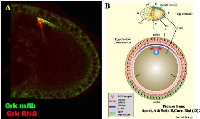
Fig. 4 Second round of Grk signal and its consequences. In A is a picture of a combination of FISH (in red) and immunostaining for the anti-Grk 1D12 (in green) monoclonal antibody to show the tight localization and restriction of the signal. In B is a schematic representation from a review ((Amiri and Stein, 2002) to indicate the restriction of the ventralising signal (green in B), pipe (pip), by the Grk gradient which is intiated from the oocyte.
collar (cnc) alleles, where the oocyte nucleus initially migrates correctly but then slides back to the posterior pole, the eggs exhibit no DV polarity (Guichet et al., 2001).
grk RNA and protein are detectable tightly localised to the oocyte nucleus in wild type and in most conditions where the RNA and/or protein are detectable (see Fig.4A).
See Fig.3, for an overview of these processes.
1.5 Grk signaling initiates the process of the DV axis formation of the embryo
There are at least three types of follicle cells that can be distinguished by their gene expression pattern, by the eggshell structures they secrete and by the function they fulfil
for embryonic axis induction along the DV axis of the egg chamber. These are the dorsal cells that express kekkon (kek1) (Ghiglione et al., 1999) and are responsible for dorsal chorion structures including the dorsal appendages, the ventral cells that express pipe (pip) and contribute the cues for the embryonic DV axis (Sen et al., 1998) (Nilson and Schupbach, 1998) (Fig.4B) and the lateral cells that neither express kek1 nor pip. Grk signaling induces secondary signaling cascades that define these expression domains, as in the patterning of the DAs. Being present in a concentration gradient Grk might act as a morphogen, i.e. kek1 and pip could be direct target genes whose activation or repression depends on particular concentration thresholds of Grk. Genetic mosaic studies support the latter view (James et al., 2002; Pai et al., 2000) (Peri et al., 2002). Analysis of mutant follicle cell clones that are incapable of transmitting the Grk signal (e.g: clones of mutations in downstream components of DER signaling, like Ras or Raf) show ectopic expression of pip in a cell-autonomous manner. Conversely, if the DER pathway is ectopically activated in cell clones, autonomous activation of kek1 as well as autonomous repression of pip is observed.
The expression of pip in the ventral follicle cells is necessary for the establishment of embryonic DV polarity (Nilson and Schupbach, 1998; Sen et al., 1998) (See Fig.4B). pip mutant females produce eggs with normal eggshells, the embryos that develop in these eggs however are completely dorsalized. The embryos are dorsalised because they lack DV polarity during gastrulation and produce dorsalmost cell fates around the entire embryonic circumference. Pip encodes a heparin sulphate 2-O-sulphotransferase and is probably involved in the modification of glycosaminoglycans of the extra cellular matrix (ECM) (Sen et al., 2000). The exact nature of the ECM components modified by Pipe is not yet known, but irrespective of the identity of these components, the pipe expressing cells secretes the activated ECM component into the space between oocyte and follicle cells before the latter starts producing the inner (vitelline) and the outer (chorion) eggshell membranes, respectively. Consequently, after egg deposition the modified ECM components are present on the vitelline membrane facing the embryo or on the surface of the embryo itself. They initiate a proteolytic cascade that comprises a chain of four serine proteases that culminates in the activation of the extracellular ligand Spaetzle (spz) (Morisato and Anderson, 1995; Schneider et al., 1994). Spz has structural similarities to
nerve growth factors (DeLotto and DeLotto, 1998; Mizuguchi et al., 1998; Morisato and Anderson, 1994).
Spz activates the transmembrane receptor Toll which is uniformly expressed on the surface of the embryo (Hashimoto et al., 1988). Activated Toll relays the extracellular signal to the embryonic nuclei by regulating the nuclear import of the NF-kB transcription factor Dorsal (dl).
1.6 The EGF-EGFR repertoire of regulators, their interaction in ligand regulation and role in pattern formation
Grk is one of the four ligands in the fly that is capable of activating the DER. There is only one DER in Drosophila. DER belongs to the EGFR/ErbB family of receptor proteins and is similar to the mammalian family members in overall structure. The extracellular region has the typical four domains, including two cysteine-rich domains, required for ligand binding (Livneh et al., 1985). There are two different splice variants encoding two different protein isoforms and it is not clear whether there is a distinct role to each of the forms (Schejter et al., 1986). The regulation of the receptor is not via differential tissue specific expression as it is broadly detected in diverse tissues during development (Zak et al., 1990). The wide array of roles played by DER during development in different tissues have complicated the identification of its developmental roles by simple analysis of loss of function phenotypes during embryonic or postembryonic stages (Clifford and Schupbach, 1989). The use of dominant negative receptor constructs, temperature sensitive or hypomorphic mutations in the receptor, or mutations in distinct ligands, have been instrumental in identifying discrete roles for DER in different developmental processes. (For a list of all the different developmental processes that DER has been implicated in, see Table 1 of review(Shilo, 2003).
During oogenesis, the removal of DER from the follicle cells using mitotic clones results in eggs laid which lack both AP and DV polarity (Gonzalez-Reyes et al., 1995).
Expression of a constitutively active version of the receptor in the follicle cells using the UAS/Gal4 systems results in dorsalised eggs. Expression all along the follicle cells of the constitutively active construct in a background where the germ line is mutant for the ligand responsible for the initiation of the pathway, Grk, results in eggs which lack all
polarity but have DA material deposited all along the anteroposterior axis of the egg (Queenan et al., 1997). Removal of the ligand from the signal emitting cell (oocyte) during oogenesis results in eggs which lack both DV and AP polarity (Neuman- Silberberg and Schupbach, 1993; Roth et al., 1995).
In order to understand the roles of the DER in initiating the two axes and patterning of the egg shell it is important to know the current state of knowledge as to how the different components interact with each other. Although a lot is known about the activation of the MAPK pathways downstream of the receptor, emphasis will be laid here only on the events upstream of these pathways which are responsible for ligand regulation and the receptor occupation by these ligands.
Picture modified from (Shilo, 2003)
Fig. 5: Overview of the DER pathway. Grk (red) signal from the oocyte to the follicle cells activates the pathway during oogenesis. Secondary signal amplification and refinement of the pattern formed is brought about by the interaction of several DER components, which in clued another ligand Spi (green) which has to be cleaved to form the active moiety sSpi (green), at least three inhibitors Aos, Kek1 and Sprouty (sty) which act at different levels in the pathway. Two proteins, Star (S) and Rhomboid (Rho) are required for the activation of EGF ligands, esp Spi, via the regulated intermebranous proteolysis (Rip) pathway.
Besides Grk, there are three other activating ligands for DER, they are Spi, Vn and Keren (krn). Vn is an atypical EGF ligand because it is more similar to the neuregulin type than to EGF type of ligands. It is also the ligand which shows a weaker activation capacity and
is required in tissues where low level activation is required (Schnepp et al., 1998;
Schnepp et al., 1996).
Spi, Krn and Grk are produced as precursor molecules with transmembrane domains (Fig.6A&B). Processing of these molecules to produce a secreted ligand was shown to be a key regulatory step in DER activation. This mechanism was first established for Spi and subsequently been also applied to the other two ligands (Schweitzer et al., 1995). The spatio-temporal pattern of Spi-induced DER activation is thus dependent upon regulated processing of Spi.
Fig. 6 Comparison of the EGF ligands in Drosophila. (A) Comparison of the protein domains and domain architecture between the different ligands. A red arrow indicates the position where the ligands are cleaved to form the active moiety by Restricted intermembranous proteolysis (RIP) mechanism. Vein the only secreted ligand is also shown. Argos, an inhibitor which functions by a ligand sequestering mechanism, and possess a region which show low level similarity with the EGF domain (pink bar) also is included. (B) Alignment of the EGF (red line below), Juxta membrane domain (JMD) (blue line below) and Transmembrane domain (TMD) of Grk, Spi and Krn indicate that Spi and Krn (65% similar in EGF domain) are more similar to each other than Grk (Grk and Krn, 35% similar).
Spi, rho and Star(S), along with single-minded (sim) and pointed (pnt) belong to the zygotic spitz group of genes due to the similarity in embryonic phenotypes between them.
Spi, rho and S have been genetically shown to be required in the same cell for the production of the active ligand (Mayer and Nusslein-Volhard, 1988). The biochemical nature of the regulation of EGF ligands by S and Rho has been investigated. The first step
of regulation in Spi processing is the trafficking of the protein from the endoplasmic reticulum to the Golgi compartment. This step is carried out by S, a novel type II transmembrane protein (Kolodkin et al., 1994; Pickup and Banerjee, 1999), that serves as a cargo receptor and associates with Spi (Lee et al., 2001; Tsruya et al., 2002). The exact region on Spi that interacts with Star has not been mapped, although a few amino acids immediately after the transmembrane region leads to highest retention in the ER, when removed (Lee et al., 2001).
In the Golgi, the membrane bound ligand is cleaved by a Golgi resident seven membrane pass serine protease, Rho-1 (Bier et al., 1990). rho-1, is one of the four rhomboids found in the fly genome. rho-2 /Brother of Rhomboid (brho) / Stem cell tumour (stet), rho-3/
roughoid (ru) and rho-4 are the other three members (Guichard et al., 2000; Schulz et al., 2002). Rhomboid is essential for Spi cleavage. The catalytic domain of Rho resides within its conserved transmembrane domains and the substrate domain on the ligand also is in the transmebrane domain. Hence Rho gives rise to intramembrane proteolysis of the ligand whose transport to the Golgi is regulated by Star, so this mechanism of regulation of EGF ligand activity has been termed the Regulated Intramembranous Proteolysis (RIP) (Lee et al., 2001; Urban et al., 2001). (See Fig.7 for a schematic representation of the RIP paradigm). spi and S are broadly expressed where as the expression of rho is extremely dynamic (Bier et al., 1990). rho pattern is almost identical to the pattern of DER-induced MAPK activation (Gabay et al., 1997). rho expression is thus one major limiting step in DER activation. Intriguingly, in some tissues DER activation induces rho expression. In tissues where multiple cycles of DER activation are required, the induction of rho expression in the responding cells leads to additional rounds of ligand processing (Sapir et al., 1998; Wasserman and Freeman, 1998).
krn is the last identified of the EGF ligands and although there has been no loss of function analysis done so far, expression studies have shown that Krn goes through S- Rho mediated cleavage/activation and is independent of S-Rho at low level activity/cleavage (Reich and Shilo, 2002).
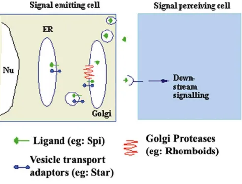
Fig. 7 The Rip pathway of EGF ligand regulation. Star (in blue) functions to ferry the unporocessed Spi (green) from the ER to the Golgi. In the Golgi, it is cleaved in the transmembrane region by Rhomboid (red), a seven membrane pass serine protease, so that active Spi can now be secreted to initiate down stream signaling via DER.
Elucidation of the mechanisms regulating Spi processing has led to insights regarding the other two transmembrane ligands. It was shown that Grk can undergo cleavage that is Rho and S dependent in cell culture. Further, in culture the full length and secreted forms (lacking the TMD and cyotplasmic domains) of Grk have been shown to have differential cell adhesion properties. While cells expressing the full length form adhered together, in case of cells expressing the secreted form no adhesion was observed (Ghiglione et al., 2002). In the ovary, Grk is found predominantly in the cleaved form and is endocytosed by the follicle cells receiving the signal (Peri et al., 1999). A member of the rho family (brho, rho-2 or stet) that is expressed in the oocyte may carry out Grk processing (Guichard et al., 2000; Schulz et al., 2002). Genetic interactions show that Star may also be required (Ghiglione et al., 2002).
However several questions are still not clear regarding EGF ligand production from the germ line. Germ line clones of spi, has absolutely no effect on the egg shells structure. So
though there can be very regulated refinement after DER has been activated, there must be some other mechanisms to initiate the signal.
Cornichon (cni), a small hydrophobic protein, is absolutely required for Grk signaling from the germ line. The role of cni in initiating the Grk signal has not been understood so far.
1.7 Cornichon and initiation of Grk signaling from the germ line
Fig. 8 Cornichon is one of the founding members of a family of small hydrophobic proteins found across a number of species. A: is a dendrogram representing the known cni members from the Swiss- prot data base. Underlined with a red line are the two mebers of cni in flies and the yeast member Erv14p. B: The eggs laid by grk amorphic mothers are identical to the eggs laid by cni.
Cni is a small hydrophobic protein of 144 amino acids and is required in the germ line for the correct DV axis initiation in Drosophila. Cni is absolutely required for the initiation of Grk signaling from the oocyte to the overlying follicle as can be seen by the fact that eggs laid by cni amorphic mother exactly phenocopy the Grk amorphic egg shell phenotype (Fig.8B). The expression levels of cni is low and does not show any specifc mRNA localization patterns (Roth et al., 1995). cni members are present all across the animal kingdom and show varying degrees of similarity with each other (Fig.8A). Cnih, the mouse homologue of Cni shows 93% similarity (68% identity) with the Drosophila
protein (Hwang et al., 1999). Although, Cni homologues from systems as varied as humans (Utku et al., 1999) to ascidians (Davidson and Swalla, 2001) have been reported, very little is understood about the cellular and biochemical roles of these proteins in different systems.
ER-vesicle protein of 14 kD (Erv14p) is the Cni homologue of S. cerevisiae. It is one of the components of the COP-II-coated ER derived transport vesicles in yeasts. COP-II mediated vesicle transport is responsible for the ER to Golgi anterogade transport of proteins in the cell. Erv14p is required for the polarised transport of Axl2p, a protein responsible for selection of axial sites and which normally localises to nascent bud tips or the mother bud neck. In the erv14 mutants Axl2p accumulates in the ER, while other secretory proteins are transported at wild type rates. Haploid cells that lack erv14p are viable but display a modest defect in bud site selection (Powers and Barlowe, 1998). The membrane topology of Erv14p was shown to be three membrane pass structure by factor XA cleavage assays. Erv14p has been proposed to coordinate COP-II vesicle formation with incorporation of specific cargo (Powers and Barlowe, 2002).
In flies cni has been shown to interact with grk and sec23 in yeast two hybrid assays. The interaction with Sec23 was not reproducible with protein-protein interaction assays. Grk and Cni interacts with each in protein-protein interaction assays. The luminal side of Cni interacts with the JMD region of Grk (C. Boekel, Ph.D work) (Fig.9B).
Based on these data the working hypothesis is that this interaction between Cni and Grk specifically enriches Grk into COP-II vesicles on its route of transit from the ER to Golgi (Fig.9A).
CG17262 is an open reading frame in the fly genome which shows highest similarity to cni, hence called cornichon related (cnir). There is no loss of function alleles of cnir and absolutely nothing is known about the cellular function of the protein coded by this ORF.
cni and cnir are synthetically lethal (C. Boekel, Ph.D work). The only other closely related member to cnir is one of the three hundred ORFs of undefined genes expressed in human CD34+ Haematopoietic Stem/Progenitor Cells (HSPC), namely HSPC163 (Zhang et al., 2000).
Fig. 9 Hypothetical model for the interaction between Cni and Grk. A: Shows a model for the interaction between Grk and Cni at the COP-II budding sites of the ER. B: Scheme representing the regions luminal region of Cni (yellow) interacting with the JMD (pink) domain of Grk.
1.8 Cellular Exocytosis, a brief overview
Glycosylation is a characteristic post-translational modification in eukaryotic cells. There are two types of glycosylations namely N-glycosylation and O-glycosylation based on the amino acid side chain on the protein onto which the sugar residues are added. The two types of glycosylations differ from each other with respect to both, the subcellular location where the sugars are added and also the mode in which they are added. As the freshly translated protein leaves the ribosome and enters the ER a multi branched high mannose containing sugar structure is added onto the N-side chains of Asn/Gln acids from dolichol phosphate in a single step reaction. The high mannose branched sugar chain is differentially modified through its exocytic route from the ER to the cis-Golgi structures. Endoglycosidase H (Endo H) is an enzyme which can remove the initial branched high mannose sugar moiety that is added onto the N-amino acid. But as this branched structure matures through its exocytic transit it becomes resistant to Endo H.
Very few proteins retain their Endo-H sensitivity in the mature form that has undergone the transit through the Golgi and hence, Endo-H sensitivity in most cases acts as a litmus test for proteins retained in the ER.
O-glycosylations on the other hand, are the stepwise addition of sugar moieties onto the OH-side chains of Ser/Thr amino acids that takes place in the cis and trans-Golgi
structures. In the case of O-glycosylation reaction it is carried out as a single sugar is added by a single enzyme catalysed reaction, there is no transfer of large branched sugar structures as in the case of N-glycosylation reactions (Lodish et al., 1999).
The ER is the entry point into the secretory pathway for newly synthesized proteins.
Ribosomes dock onto a protein pore in the ER membrane, thereby releasing the nascent polypeptide into the lumen of the ER. The primary role of the ER is to provide a milieu that facilitates protein folding. Besides the addition of N-linked glycan chains, post- translational modification of nascent chains, including addition of and hydroxylation of proline residues, first occurs in the ER. The next region in the exocytotic transit of the protein is the Golgi apparatus, a series of cisternae housing enzymes that function in glycan side chain modification and proteolytic cleavage. Transport of proteins between these various compartments of the secretory pathway occurs via small vesicles that are generated at a donor compartment and fuse with a downstream acceptor compartment.
COP-II coated vesicles are the vesicles that have been associated with ER to Golgi anterogade transport of proteins (Lee et al., 2004). An important aspect of vesicular transport is that only designated cargo proteins are packaged into a nascent vesicle, organelle resident proteins fail to be incorporated (Malkus et al., 2002). The ER is not a homogenous environment, regions dedicated to generating COP-II vesicles have been variously designated as ER exit sites (ERES) or transitional ER (tER). The precise mechanism by which these distinct zones are maintained, the full complement of proteins that mark these sites and the functional significance of these privileged budding sites is not yet fully clarified (Malkus et al., 2004; Pagano et al., 1999).
The ER-Golgi intermediate compartment (ERGIC), also known as vesicular tubular clusters (VTC), is a compartment thought to arise from the homotypic fusion of COP-II vesicles and is the main sorting station for the retrieval of escaped ER resident proteins (Martinez-Menarguez et al., 1999). ERGIC vesicles may represent a functionally distinct budding zone similar to that of the ERES/ tER sites (Lee et al., 2004).
The correctly folded and processed proteins exiting the ER are of two very broad categories:
a) Proteins that are capable of entering transport vesicles at their prevailing concentrations in the ER. These proteins are thought to passively enter the
transport vesicles and this mode of ER exit, believed to be used by protein produced in high quantities is called the “Bulk flow” mode of exocytosis.
b) Proteins that have to enter the transport vesicles at concentrations significantly higher than those in the general ER. Specific enrichment of both membrane and soluble cargo proteins in transport vesicles occur at concentrations ~3-50 fold higher than the bulk flow mode of transport. This enrichment is achieved by interaction of the cytoplasmic coat with distinct sorting signals on cytoplasmic segments of membrane cargo proteins. To be recognized by the coat machinery, these signals must be accessible and in an appropriate confirmation that maybe influenced by a number of factors, including the folding status of the protein, interactions with accessory proteins or transport recepors, and/or oligomerisation state that may influence the presentation of positive sorting signals or masking of retention signals. This mode of exocytosis is termed the “specific enrichment of cargo” mode of transport.
There are several open questions in the regulation of both modes of protein transit (Lee et al., 2004).
1.9 Objectives
The broad objectives of the following study can be listed as follows:
i) Understanding the cellular and biochemical role for Cni in the initiation and subsequent Grk mediated patterning of the two axes in Drosophila.
ii) Identifying a loss of function allele of cnir, which is an ORF similar to cni in the fly genome, to better understand the cell biological role of this family of proteins.
iii) Identify other regions/genes in the genome that have a so far unknown role in axis polarization and/or EGFR signaling using an enhancer suppressor screen.
2a Results - Cornichon (Cni)
2a.1 Sub-cellular localisation of Cni
Erv14p, the yeast homologue of Cni has been shown to be a protein required for ER to Golgi transport of Axl2p, another protein involved in axial budding of yeasts, (Powers and Barlowe, 1998). To investigate a similar cellular function for Cni, we studied the subcellular localization of Cni. A Myc tagged version of Cni was misexpressed in the follicle cells using the UAS/Gal4 system (Fig.10), originally from yeasts. Although cni is expressed and required in the germ line, the reason for using the follicular epithelium as the system of choice for studying co-localisation of Cni-Myc with subcellular markers are two fold. First, the follicular epithelium is a bona-fide polarised epithelium with a basal and an apical side unlike the oocyte in which the subcellular organization is atypical and not pliable with most of the known subcellular markers. Second, pUAST vector based constructs can be driven only in somatic tissue whereas to drive expression in germ line tissue contructs have to be based in pUASp vectors (at present we do not have a UASpcni construct in hand).
A monoclonal antibody, against the immunoglobulin heavy chain binding protein (Bip) was used as a marker for the Endoplasmic Reticulum (ER). Bip is an ER resident protein (Steiner and Smolen, 2002; Vaux et al., 1990). Monoclonal anti-Bip antibodies showed the maximum colocalisation with Anti-Myc polyclonal antibodies, indicating that most of Cni is present in the ER. The distribution of Cni-Myc was very characterstic for an ER localised protein (Fig.11A-C). Besides ER structures which showed colocalisation with both Bip and Myc antibodies, there were also structures solely positive for the individual markers. This indicates not only that Cni is not present in all structures of the ER but also, that it is present in other subcellular structures, besides the ER.
MAC 256 is a rat monoclonal antibody against the KDEL-peptide. We used this as a marker for the Endoplasmic Reticulum Golgi Intermediate Compartment (ERGIC).
MAC256 and Anti-Myc showed some colocalisation indicating that Cni was also present in subpopulations of the ERGIC vesicles (Fig.11D-F). However, the amounts, distribution and colocalisation of MAC256 and Myc was to a lower extent than that seen
in Bip/Myc experiments. This indicates not only that the ERGIC is a much smaller subcellular structure than the ER but also that Cni is more in the ER than in the ERGIC.
Both the ER and the ERGIC colocalisation experiments were done in the Stretched Follicle Epithelium (SFE), which are very flat cells overlying the nurse cells (Fig.11A-F
& Fig.10,yellow arrowheads). A commercially available monoclonal raised against Drosophila embryonic Golgi membranes was used to assay colocalisation of Cni-Myc with Golgi stacks. Experiments for colocalisation with Golgi apparatus marker were done in the Cuboidal Follicular Epithelium (CFE) (Fig.10,red arrowhead), because of difficulties to visualize the Golgi apparatus in the SFE. Cni and the Golgi marker colocalised in a few Golgi vesicles too, although the least amount of Cni appeared to be in the Golgi structures/ vesicles (Fig.11,G-I).
These experiments indicate that Cni can be detected in the ER, ERGIC and Golgi structures of the follicular epithelium, hence validating a ER to Golgi transport function for Cni.
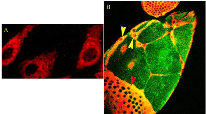
Fig. 10 Distribution of Cni in the follicular epithelium. (A) The distribution of Cni-Myc expressed with follicular drivers and detected using an anti-Myc polyclonal antibody. The picture shows the distribution of Cni in an SFE (indicated with yellow arrowheads in B)
(B) In Green are the nurse cells (germ line) which lie beneath the SFE. Red arrowheads indicate the CFE whereas the yellow arrowheads indicate the SFE.
Fig. 11 Sub-cellular localization of Cni. Distribution of MAC256 in SFE (A), anti-Bip in SFE (D) and anti-Golgi in CFE (G) B, E & H are the distribution of Cni indcated by anti-Myc antibodies. C, F and I are the colocalisation of the respective markers with the anti-Myc antibodies. White arrows in I indicate Golgi vesicles which were found to be real colocalisations by taking optical sections to check whether the apparent localization was due to vesicles with the individual markers lying over each other. The colocalisation was found to be real with the resolution of the confocal microscope we used.
2a.2 Excess Grk signal from the germ line dorsalises the egg shell
The UASp/Gal4 system was used to overexpress Grk in the germ line. The effect of increased Grk was assayed by the dorsalisation of the egg shells. Two Gal4 drivers, namely, TubulinGAL4VP16 (TubGal4VP16) (a tubulin which is restricted to oogenesis) (Source: St.Johnston lab) and Nanos GAL4VP16 (NosGal4VP16) (Source: Ephrussi lab), were used in the study. Both Gal4 drivers, had the ability to increase Grk expression from the germ line, as was evidenced from the resulting, dorsalised egg shells. The dorsalisation of the egg shells in case of both Gal4 drivers was never a single phenotype, rather a spectrum starting from the wild type egg shell with the two dorsal appendages at
the end of the operculum (anterior end). And at the other end of the spectrum was the most dorsalised egg shell, characterised by an enlarged operculum. The characterstic twin dorsal appendages in this case, are replaced by dorsal appendage material distributed all around the cricumference of the enlarged operculum.
The dorsalisation scale is an arbitary scale we devised to assay the spectrum of egg shells that were produced. The basis of this scale is the visual examination of the egg shells.
The spectrum of the egg shell phenotypes can be classified in three broad categories.
a) Wild type to very slight dorsalisation: This corresponds to the first two egg shells from the left in Fig.12a&b. The slightly dorsalised eggs are almost identical to wildtype eggs but the dorsal appendages and operculum are pushed from their normal dorsanterior asymmetric location to a more symmetric anterior location.
b) Intermediate to strong dorsalisation: Eggs in this category, the next two pictures in the Fig.12c&d, are charatcterised by the loss of the asymmetric position of the dorsal appendage material. The dorsal appendages are transformed into a ring of dorsal appendage material, and the operculum circumscribed within the ring, are both pushed to a symmetric anterior position instead of their characteristic dorsal asymmetric location. The eggs with a perfect ring of dorsal appendages (Fig.12c) can be considered to be less dorsalised in comparison to those where the dorsal appendage material protrudes at out at the ventral side of the egg (Fig.12d).
c) Extreme dorsalisation: The last two pictures on the scale represent the maximum dorsalisation observed (Fig.12e&f). The operculum is extremely enlarged and the dorsal appendage material is almost absent in extreme cases but when present is present in patches all along the rim of the enlarged operculum.
Fig. 12 The dorsalisation scale: Egg shell phenotypes representative of the degrees in dorsalisation observed due to overexpression of Grk from the germ line. (a) and (b) indicate the wildtype to weak dorsalisation, (c) and (d) the intermediate to strongly dorsalised egg shells and finally (e) and (f) represent the transition to extreme dorsalisation.
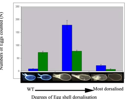
Fig. 13 Excess Grk from the germ line results in the dorsalisation of the resultant egg shells. In both graphs the Y axes represents the number of the eggs that were considered for analysis, the X axes gives the dorsalisation scale. The panel above is the distribution of egg shell phenotypes using TubGal4VP16 driving different UASpGrk constructs (Myc tagged/ non tagged, with bniAR55/CyO or DfH60Scob/CyO on the second chromosome).The panel below indicates similar distribution using NosGal4VP16 as the driver with different UASpGrk versions.
The spectrum of egg shell dorsalisation is not changed irrespective of whether the UASpGrk construct is a C-terminal Myc tagged version or an untagged version (Fig.13).
In general, egg shell dorsalisation due to Grk mis/overexpression using NosGal4VP16 as the driver, was weaker than Tubgal4VP16. This is based on the fact that the dorsalised egg shell spectrum is spread more widely in case of NosGal14VP16, with the distribution frequency peaking at the intermediate to strong range in dorsalisation. The spectrum in case of using TubGal4VP16, on the other hand, is narrower and the peak frequency being pushed further towards the extreme dorsalisation phenotypes.
Fig. 14 Comparison of egg shell dorsalisation using different germ line Gal4 driver lines.
2a.3 Patterning of the embryo affected when Grk overexpressed in DfH60Sco heterozygous background
There is a significant number of apparently wild type egg shells laid when Grk is overexpressed using UASpGrk and using NosGal4VP16 as the driver (46%, N=152). The number of wild type egg shells were considerably less in the case of overexpression using TubGal4VP16 (5%, N=198). In both cases there were no embryonic cuticles visible in dorsalised eggs shells, indicating that these eggs could not even be fertilized. In case of the egg shells, which were wildtype in appearance, from the cross in which Grk was overexpressed using NosGal4VP16 as the driver in a heterozygous amorphic cni background (bcniAR55/CyO), insignificant number with embryonic cuticles observed (5%, N=36) . Intriguingly, when the same experiment was done using the deficiency of the cni (DfH60Scob/CyO) region in heterozygous condition on the second chromosome, significant number of egg shells with embryonic cuticle phenotype were observed (39%, N=57) (Fig.15A). The cuticles phenotypes varied from subtle phenotypes such as the fusion of ventral denticle belts, missing denticle belts and gaps in denticle belts to a strong phenotype such as entire anterior segments being misformed, missing terminal segments and complete absence of dentilce belts (Fig.15B). DfH60Scob, besides
removing cni also removes cactus (cact) and fizzy(fzy). This indicates that the removal of one copy of cact has a small suppression effect, which makes the dominant maternal phenotype of cact visible in these embryos. It is probable that a similar phenomena happens with the TubGal4VP16, Grk overexpression too, but it is difficult to quantify this due the lower numbers of the wild type eggs laid in this case. We did observe a few c very strong cuticle phenotypes in this case, too (Data not shown).
Fig. 15 Egg and cuticles from the wild type egg shell fraction laid by UASpGrk; DfH60Scob/CyO;
NosGal4VP16 mothers. (A) i-vi Apparently wildtype eggshells in which the misinformed embryonic cuticles are visible. (B) i-vi Range of cuticle phenotypes from these eggs.
2a.4 Timing of onset is different between NanosGal4VP16 and TubGal4VP16
TubGal4VP16 and NanosGal4VP16 were the drivers of choice because they were germ line specific Gal4 drivers. To ascertain whether these are strict germ line drivers or they also expressed the overlying somatic follicle cells, we expressed an UASpEGFP construct (UASp constructs should be expressed in the soma and germa) using the two Gal4s as drivers. While the two drivers are strictly germ line restricted as visualised by
the complete absence of GFP from all somatic follicle cells and presence in nurse cells and oocyte. However, there was a dramatic differerence between the two Gal4 drivers as to the stages of the egg chambers where GFP could be first detected. In case of Tubgal4VP16, GFP expression could be detected as early as in stage 4 and stage 5 egg chambers. In these stages the oocyte nucleus is still in a symmetric posterior location.
These are stages before the first round of Grk signaling occurs. In case of the NosGalVP16, GFP could be detected only in stages as late as stage 6 or stage 7 eggchambers. Not a single egg chamber was detected in which the oocyte nucleus was still in the posterior symmetric position and expressed GFP. Hence, NosGal4VP16 is active between the two Grk signals from the germ line (Fig.16).
Fig. 16 Comparison of the expression profile between TubGal4VP16 and NanosGal4VP16, using an UASpGFP reporter. The GFP signal could be detected in much earlier stages of oogenesis (stage 4-5) in case TubGal4VP16 (upper panel) than in case of NanosGal4VP16 (lower panel). Green arrowheads in either panels indicate the earliest time point where GFP was detectable in either of the drivers. Red line, below the two panels, indicate the approximate time window of the two Grk signals. Black line is to indicate the time window of early oogenesis events.
In both the Gal4 drivers no GFP expression was observed in any follicle cell at any stage of oogenesis, ascertaining the fact that the drivers used are both strictly germ line drivers (Fig.16).
Although, from the egg shell dorsalisation assays TubGal4VP16 appeared stronger than NosGal4VP16, this issue could not be clarified by differences in GFP signal. GFP was not detected in early germarium of both drivers, although NosGAL4VP16 is supposed to have early germarium function/expression (P. Filardo, personal communication).
2a.5 Mis/over-expression of Grk by TubGAl4VP16 but not NosGal4VP16 can rescue Cni function
Fig. 17 Egg shells indicating that mis/over expression of Grk with TubGal4VP16 can overcome Cni requirement for realisation of the active Grk ligand. The wild type egg shell (A) has clearly patterned dorsal appendages and anterior and posterior structures, whereas the egg shells laid by cni amorphic mothers (D), is dorsoventrally symmetric with anterior structures on either ends. While both NosGal4VP16 and TubGal4VP16, overexpressing Grk in a cni heterozygous background results in dorsalisation of the egg shells to different degrees (B&C), the same experiment in a cni homozyogous background does not show any rescue of the cni egg shell in case of NosGal4VP16 (E), but both AP and dorsoventral patterning are rescued in case of TubGal4VP16 (F). Appropriate controls of the TubGal4Vp16 with cni homozygous background(G), and the UAS constructs in a cni homozygous background (H&I) all result in cni amorphic egg shells.
If the biological function of Cni is the enrichment and/or giving a subcellular vectoriality to Grk containing vesicles en-route its exocytotic transit from the ER to Golgi, could it be possible to overcome Cni requirement by overloading the system with Grk. From the egg shell dorsalisation assays it is already obvious that higher amounts of Grk from the germ line is produced in case of both TubGal4VP16 and NosGal4VP16. To test whether Grk produced in bulk and passing through the exocytotic route can replace the requirement