Spectroscopic and dynamic properties of electronically excited retinal in
C1C2 channelrhodopsin
Inaugural-Dissertation
zur Erlangung des Doktorgrades der Mathematisch-Naturwissenschaftlichen Fakultät
der Heinrich-Heine-Universität Düsseldorf
vorgelegt von
Irina Dokukina
aus Odessa
Düsseldorf, September 2018
der Heinrich-Heine-Universität Düsseldorf
Gedruckt mit der Genehmigung der
Mathematisch-Naturwissenschaftlichen Fakultät der Heinrich-Heine-Universität Düsseldorf
Berichterstatter:
1. PD Dr. Oliver Weingart
2. Univ.-Prof. Dr. Christel M. Marian
Tag der mündlichen Prüfung: 26.10.2018
Eidesstattliche Erklärung
Ich versichere an Eides Statt, dass die Dissertation von mir selbstständig und ohne unzulässige fremde Hilfe unter Beachtung der Grundsätze zur Sicherung guter wissen- schaftlicher Praxis an der Heinrich-Heine-Universität Düsseldorf erstellt worden ist. Die Dissertation wurde vorher bei keiner anderen Fakultät vorgelegt und es hat keine er- folglosen bzw. erfolgreichen Promotionsversuche gegeben.
Unterschrift:
Datum:
The only way of discovering the limits of the possible is to venture a little way past them into the impossible.
Clarke’s second law
Meiner Familie gewidmet
Danksagung
An dieser Stelle möchte ich mich bei den Menschen bedanken, die mich während meiner Doktorzeit begleitet haben. Ohne sie wäre die vorliegende Doktorarbeit nicht realisier- bar.
In erster Linie möchte ich mich bei meinen Doktoreltern Professor Christel M. Marian und PD Dr. Oliver Weingart herzlich bedanken.
Professor Marian bin ich zunächst für die Aufnahme in den Bereich der Theoretischen Chemie dankbar und für die Möglichkeit das fehlende Hintergrundwissen nachzuholen, um den Aufgaben im Rahmen der Doktorarbeit gewachsen zu sein. Ich bin Ihr für die Übernahme der Erstbetreuung beim größten Teil meiner Promotion und für Ihr En- gagement dankbar. Ich möchte mich bei Ihr auch für bereichernde wissenschaftliche Diskussionen bedanken. Diese gemeinsamen Gespräche hatten stets eine motivierende Wirkung und halfen mir herausfordernde Fragestellungen in einem anderen Licht zu betrachten. Nicht zuletzt bin ich Professor Marian auch für die Ermunterung dankbar, mir meiner Fähigkeiten und Kompetenzen bewusst zu werden.
Ich möchte mich bei PD Dr. Oliver Weingart für ein spannendes und vielseitiges Pro- jekt bedanken, bei dem sich die meisten meiner wissenschaftlichen Interessen vereint haben. Zudem bin ich Ihm für die kontinuierliche fachliche Begleitung meiner Arbeit und für die Übernahme der Erstbetreuung am Ende meiner Promotion dankbar. Seine Hilfsbereitschaft (auch zur späten Stunde) und optimistische Art haben zum Fortschritt meiner Dissertation beigetragen. Er hat immer eine Lösung für manch ein fachliches oder technisches Problem gesehen, bei dem ich des öfteren anfing zu verzweifeln. Ich bin Ihm außerdem für die Möglichkeit dankbar, während der Zusammenarbeit die zahl- reichen „Werkzeuge“ und Tricks kennengelernt zu haben, die einem nicht nur das Ar- beiten vereinfachen, sondern auch für weitere berufliche Perspektiven von Bedeutung sein könnten.
Sowohl Professor Christel M. Marian als auch PD Dr. Oliver Weingart bin ich dafür dankbar, dass ich die Möglichkeit hatte an wissenschaftlichen Konferenzen teilzuneh- men. Dadurch konnte ich mein Wissen erweitern, für mich interessante Bereiche ken- nenlernen und andere Wissenschaftler mit meiner Arbeit in Berührung bringen.
Herrn Professor Wolfgang Gärtner danke ich für das Einverständnis mein Mentor im Rahmen der Promotionsvereinbarung zu sein.
Bei der Arbeitsgruppe von Professor Marco Garavelli (Bologna) und insbesondere bei Dr. Artur Nenov möchte ich mich für Ihre Kooperation bedanken.
Mein herzlicher Dank gilt Jelena Föller, Oliver Schillinger und Fabian Dinkelbach für die entgegengebrachte Geduld bei meinen Fragen (fachlicher und nicht immer fachlicher Natur) und für Teerunden bei denen Angelegenheiten unterschiedlicher Wichtigkeit be- sprochen wurden. Ich bin froh nicht nur von netten Kollegen umgeben gewesen zu sein, sondern auch Freundschaften geknüpft zu haben. An dieser Stelle möchte ich mich auch
ix bei Gudrun Brauwers vielfach bedanken, die mich immer mit einem guten Rat und ei- nem aufmunternden Wort unterstützt hat.
Ich möchte mich auch bei der gesamten Arbeitsgruppe der Theoretischen Chemie (so- wohl bei den ehemaligen als auch bei den aktuellen Mitgliedern) bedanken, die un- terschiedlich zu meinem Promotionsdasein beigetragen haben, aber insgesamt für ein angenehmes Arbeitsklima sorgten und immer bereit waren zu helfen.
Rita Sevcuk und Jelena Föller bin ich zudem für das Korrekturlesen meiner Arbeit und für fachliche und stilistische Anmerkungen dankbar.
Ein besonderer Dank gilt meiner Familie und meinem Freund Jakob Schlegel. Meinen Eltern (Elena und Gennadij Dokukin) und Großeltern (Johanna und Yevgen Gorbulski) bin ich für eine bedingungslose Unterstützung während meines Werdegangs noch lange vor der Promotion dankbar. Sie haben zu meiner Affinität für die Naturwissenschaften beigetragen und mit Ihrer Hilfe konnte ich meinen Interessen nachgehen. Jakob Schlegel bin ich dafür dankbar, dass er schon nicht die erste Abschlussarbeit mit mir durchge- standen hat und mich während dieser Zeit aufgebaut und mir den Rücken freigehalten hat. Auch für seine Geduld beim Zuhören und jeden praktischen Ratschlag bin ich ihm dankbar.
Bei meinen Freunden möchte ich mich für die emotionale Unterstützung bedanken und auch dafür, dass sie geduldig darauf warten bis ich wieder mehr Zeit für sie habe.
Für die Finanzierung meiner Arbeit möchte ich mich bei mehreren Menschen bedanken.
Zum einen danke ich PD Dr. Oliver Weingart für die Gründung des Projekts und für das Erwerben der Förderung durch den Strategischen Forschungsfonds der Heinrich- Heine-Universität Düsseldorf. Bei Professor Christel M. Marian und bei Professor Lutz Schmitt möchte ich mich für das Ermöglichen der Finanzierung in der fortgeschrittenen Phase meiner Promotion bedanken. Anschließend gilt mein Dank Professor Christel M.
Marian für die Finanzierung des letzten Abschnitts meiner Doktorarbeit.
Es war eine nicht immer einfache, aber eine für mich wichtige und lehrreiche Zeit, in der ich mich persönlich und wissenschaftlich weiterentwickelt habe.
xi
Contents
List of publications included in the thesis xiii
Abbreviations xiv
Summary xvii
Zusammenfassung xviii
1 Introduction 1
1.1 Opsin proteins . . . 1
1.2 Optogenetics . . . 2
1.3 Channelrhodopsins . . . 3
1.3.1 General description . . . 3
1.3.2 Crystal structures . . . 4
1.4 Retinal chromophore . . . 5
1.4.1 Polyenes . . . 5
1.4.2 Spectral properties of retinal in the protein environment . . . 8
1.4.3 Quantum yield and reaction time of retinal isomerization . . . . 9
1.5 State of the art . . . 11
2 Motivation and objectives 15 3 Theory and methods 17 3.1 Photochemical reactions . . . 17
3.1.1 Relaxation processes after photoexcitation . . . 17
3.1.2 Conical intersections . . . 17
3.2 Quantum mechanical approach . . . 20
3.2.1 Description of potential energy surfaces . . . 20
3.2.2 Configuration interaction . . . 22
3.2.3 CASSCF . . . 23
3.2.4 CASPT2 . . . 23
3.2.5 DFT and DFT/MRCI . . . 26
3.2.6 Semiempirical methods . . . 28
3.3 Resonance Raman spectra . . . 31
3.4 Molecular mechanical approach . . . 32
3.5 QM/MM methods . . . 34
3.5.1 The QM/MM energy . . . 34
3.5.2 Handling of the QM/MM boundary . . . 36
3.6 Nonadiabatic dynamics . . . 37
3.6.1 Initial conditions . . . 37
3.6.2 Trajectory surface hopping . . . 38
3.6.3 Transition probabilities . . . 40
3.6.4 Kinetic energy redistribution . . . 41
3.6.5 Decoherence correction . . . 41
3.6.6 Integration of Newton’s equations with the velocity Verlet algo- rithm . . . 42
4 Implementations (Paper III) 45 4.1 General description . . . 45
4.2 Active space related problems . . . 49
4.3 Adaptive time step algorithm by Spörkel and Thiel . . . 49
4.4 Corrections introduced into the MNDO interface . . . 49
5 Results 51 5.1 Ab initioand DFT QM/MM study (Paper I) . . . 51
5.1.1 Ground state properties of retinal in C1C2 . . . 51
5.1.2 Absorption properties of retinal in C1C2 . . . 52
5.1.3 Geometry optimizations in the excited state . . . 53
5.1.4 Resonance Raman spectra . . . 54
5.2 Distinct excited state properties of linear polyenes and PSBs (Paper II) 55 5.2.1 The two S1 minima of PSBs . . . 56
5.2.2 OMx/MRCI at a test . . . 58
5.3 Excited state dynamics simulations (Paper IV) . . . 58
5.3.1 Static calculations . . . 58
5.3.2 Photodynamics simulations . . . 60
6 Conclusions and Outlook 67 6.1 Concluding remarks . . . 67
6.2 Outlook . . . 68
Bibliography 71
Publications 85
xiii
List of publications included in the thesis
• PAPER I
Spectral properties and isomerisation path of retinal in C1C2 channelrhodopsin I. Dokukina, O. Weingart, Phys. Chem. Chem. Phys. 2015,17, 25142-25150.
Own contribution: Execution and analysis of all calculations, preparation of sup- plementary material, generation of all figures, co-writing and proofreading of the manuscript.
• PAPER II
New perspectives on an old issue–a comparative MS-CASPT2 and OM2-MRCI study of polyenes and protonated Schiff bases
I. Dokukina, C. M. Marian, O. Weingart, Photochem. Photobiol. 2017, 93, 1345-1355.
Own contribution: Execution and analysis of all calculations, generation of all figures, preparation of the manuscript and supplementary material.
• PAPER III
COBRAMM 2.0 – A software interface for tailoring molecular electronic structure calculations and running nano-scale (QM/MM) simulations
O. Weingart, A. Nenov, P. Altoè, I. Rivalta, J. Segarra-Martí,I. Dokukina, M.
Garavelli,J. Mol. Model. 2018,24, 271-301.
Own contribution: Implementation of an interface for the MNDO program pack- age, proofreading of the manuscript.
• PAPER IV
QM/MM photodynamics of retinal in the channelrhodopsin chimera C1C2 with OM3/MRCI
I. Dokukina, A. Nenov, M. Garavelli, C. M. Marian, O. Weingart, manuscript submitted to: ChemPhotoChem 2018.
Own contribution: Execution and analysis of all calculations except pump-probe spectra, generation of all figures in the main paper and most of the figures for the supplementary section, providing data for calculation of pump-probe spectra, preparation of the manuscript and supplementary information.
Abbreviations
BO Born-Oppenheimer
CASPT2 Complete Active Space Second-Order Perturbation Theory CASSCF Complete Active Space Self Consistent Field
CT Charge Transfer
ChR Channelrhodopsin
CoIn Conical Intersection
DFT Density Functional Theory
FC Franck-Condon
GS Ground State
HF Hartree-Fock
HOMO Highest Occupied Molecular Orbital
HOOP Hydrogen Out-Of-Plane
LUMO Lowest Unoccupied Molecular Orbital
MD Molecular Dynamics
MM Molecular Mechanics
MO Molecular Orbital
MRCI Multi Reference Configuration Interaction
MS Multi State
OMx Orthogonalization-corrected Methods
PSB Protonated Schiff Base
QM Quantum Mechanics
RPSB Retinal Protonated Schiff Base
RR Resonance Raman
SA State Averaged
SE Schrödinger Equation
TFS Tully’s Fewest Switches
TSH Trajectory Surface Hopping
xv One letter amino acid code
One letter code Amino acid
A Alanine
C Cysteine
D Aspartic acid
E Glutamic acid
F Phenylalanine
G Glycine
H Histidine
I Isoleucine
K Lysine
L Leucine
M Methionine
N Asparagine
P Proline
Q Glutamine
R Arginine
S Serine
T Threonine
V Valine
W Tryptophan
Y Tyrosine
xvii
Summary
Channelrhodopsins (ChRs) are light-gated ion channels that play an important role in optogenetics. Their application ranges from basic neurobiological research to the development of therapies against severe degenerative diseases. The opening of an ion channel is triggered by all-transto 13-cisisomerization of a co-factor, retinal, covalently bound to the protein. A detailed understanding of photoisomerization is particularly important for optimization of absorption and kinetic properties in ChRs. The research conducted within my thesis focused on the investigation of retinal isomerization in the chimeric channelrhodopsin C1C2. For this purpose, a combined quantum mechanical molecular mechanical (QM/MM) approach was applied. The current work comprises static calculations in ground and excited states of retinal as well as nonadiabatic ex- cited state surface hopping dynamics. For the QM part, ab initio (HF, CASSCF, MS-CASPT2) and semiempirical strategies (DFT, DFT/MRCI, OMx/MRCI) were used. The MM part was described by the Amber force field. All QM/MM calcu- lations were performed by using the in-house software package COBRAMM, which is developed in Bologna, Düsseldorf and Lyon. Static calculations provided first in- sight into the photoreaction and document the influence of the environment on the absorption properties of retinal. These calculations showed that highly demanding multireference strategies are necessary to properly describe the electronic properties of retinal in C1C2. I decided to use the semiempirical OMx/MRCI approach for my cal- culations because it provides qualitatively accurate results at low computational cost.
To use OMx/MRCI with COBRAMM, I implemented an interface to the semiempirical program package MNDO developed by the group of Prof. Walter Thiel. Validation of the new implementation was performed on small linear polyenes and correspond- ing protonated Schiff base models. This study did not only confirm the applicability of the OMx/MRCI methodology for dynamics studies, but also provided new insights into the excited state properties ofπ-conjugated systems. As a final step, I performed OM3/MRCI dynamics of retinal within C1C2, which delivered quantum yields and photoproduct distributions along with time constants of the excited state processes.
These calculations also provided details on the possible isomerization mechanisms. An intriguing finding concerns the relationship between the structure of retinal and the speed and efficiency of the photoisomerization. An additional twist in the chromophore structure leads to an increase in photoreaction speed. Accelerated photoreaction in turn resulted in a highly selective photoproduct formation and high quantum yield of the 13-cis retinal. This relationship might be used as a design strategy for novel ChR variants.
Zusammenfassung
Channelrhodopsine (ChR) sind lichtgesteuerte Ionenkanäle, die eine besondere Rolle in der Optogenetik spielen. Deren Anwendung reicht von neurobiologischer Grundla- genforschung bis zur Entwicklung von Therapiemethoden gegen degenerative Erkran- kungen. Die Öffnung des Ionenkanals wird durch lichtinduzierte all-trans zu 13-cis Isomerisierung des kovalent an das Protein gebundenen Co-Faktors Retinal ausgelöst.
Ein grundlegendes Verständnis der Photoreaktion ist besonders für die Optimierung der Absorptions- und der Kinetikeigenschaften der ChR von Bedeutung. Im Fokus meiner Dissertation lag die Untersuchung der Retinalisomerisierung im chimären ChR C1C2 mit kombinierten quantenmechanischen und molekülmechanischen Methoden (QM/MM). Die von mir durchgeführte Arbeit beschäftigt sich sowohl mit der stati- schen Beschreibung des Grundzustands und der angeregten Zustände des Retinals als auch mit der Simulation der nicht-adiabatischenSurface-HoppingDynamik im angereg- ten Zustand. Das Spektrum der QM-Methoden umfassteab initio(HF, CASSCF, MS- CASPT2) und semiempirische (DFT, DFT/MRCI, OMx/MRCI) Ansätze. Die MM- Beschreibung erfolgte mit dem Amber-Kraftfeld. Für die Ausführung der QM/MM Berechnungen wurde das Programmpaket COBRAMM verwendet, welches in Bologna, Düsseldorf und Lyon entwickelt wurde. Statische Berechnungen lieferten einen ersten Einblick in den möglichen Verlauf der Isomerisierung. Außerdem konnte der Einfluss der Umgebung auf die Absorptionseigenschaften des Retinals erfasst werden. Die wäh- rend der statischen Studie durchgeführte Methodenvalidierung hat die Notwendigkeit der Multireferenzbeschreibung für das Retinal in C1C2 aufgezeigt. Im Hinblick auf die anspruchsvolle Modellierung der Dynamik bot sich der Einsatz semiempirischer OMx/MRCI Methoden an. Diese liefern qualitativ gute Ergebnisse bei geringem Re- chenaufwand. Um den OMx/MRCI Ansatz verwenden zu können, habe ich das Pro- grammpaket COBRAMM um eine Schnittstelle zum semiempirischen Programmpaket MNDO (entwickelt in der Gruppe von Prof. Walter Thiel) erweitert. Die Validierung der OMx-Methoden vor der Dynamik-Studie erfolgte an kleinen symmetrischen Polyenen und entsprechenden Modellen für protonierte Schiff Basen. In dieser Studie konnte nicht nur die Eignung der OMx-Methoden für die Dynamik gezeigt werden, sondern es wur- den auch neue Einblicke in die Eigenschaften vonπ-konjugierten Systemen gewonnen.
Die Untersuchung der Isomerisierung wurde durch OM3/MRCI Dynamikberechnun- gen vervollständigt. Diese lieferten die Quantenausbeuten der Photoprodukte sowie deren Verteilung und entsprechende Reaktionskonstanten. Außerdem konnten mögli- che Reaktionsmechanismen beschrieben werden. Zusätzlich wurde ein Zusammenhang zwischen der Retinalstruktur und der Geschwindigkeit und Effizienz der Photoreaktion festgestellt. Daraus ergibt sich, dass eine Vorverdrillung der Chromophorstruktur zur Beschleunigung der Photoreaktion führen kann. Eine beschleunigte Photoreaktion wie- derum führte in meinen Berechnungen zu einer erhöhten Photoproduktselektivität und
xix einer hohen Ausbeute des 13-cis Isomers. Dieser Zusammenhang bietet neue Ansätze für die weitere Optimierung der Channelrhodopsine.
1
Chapter 1
Introduction
1.1 Opsin proteins
The ability to capture and respond to light has manifested itself in all domains of life.
For this purpose, numerous organisms use organic chromophores as cofactors, which are covalently or non-covalently bound to a protein. [1, 2] In photosystems, pigments like chlorophyll, pheophytin and carotenoids are non-covalently bound to protein subunits enabling photosynthesis. [3] Retinal (vitamin A aldehyde) triggers the photoactivity of retinylidene proteins e.g. visual rhodopsins, channelrhodopsins or bacteriorhodopsin, all being members of the largest photosensitive protein family of opsin proteins, also generally referred to as rhodopsins. [4]
Opsins can be categorized into two subfamilies: microbial (type I) opsins and animal (type II) opsins. Both families originate from distinct genetic sequences. They, how- ever, share a common scaffold: 7 transmembrane helices embedding a retinal cofactor.
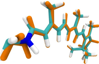
Retinal is covalently bound via a protonated Schiff base linkage (retinal protonated Schiff base, RPSB1, Fig. 1.1) to a lysine side chain in one of the helices. [1, 4, 5] The initial retinal conformation is all-trans in microbial and 11-cis in animal rhodopsins.
14 13
NH
all-trans retinal
lys
hν 14
13
NH
13-cis retinal lys
(a) all-trans→13-cis isomerization in microbial rhodopsins
12 11
NH
11-cis retinal
lys
12 11
NH
all-trans retinal
lys
hν
(b) 11-cis→all-trans isomerization in animal rhodopsins
Figure 1.1: Retinal isomerization in type I (microbial) and type II (animal) opsins.
1Within the current work RPSB is also termed just retinal.
Light induced isomerization of retinal (all-trans/13-cis in microbial [1] and 11-cis/all- transin animal rhodopsins [6, 7] as shown in Figure 1.1) subsequently leads to protein activation. Animal rhodopsins are part of a G protein–coupled receptor superfamily.
Thus,cis-transisomerization is coupled to a G protein mediated signalling cascade. Af- ter isomerization, all-trans retinal disconnects from the protein and has to be replaced by a new 11-cis molecule. In contrast to animal rhodopsins, the initial all-trans retinal conformation of type I opsins is restored after isomerization during a cycle of thermal events. [4]
Considering their function, type I opsins can be subdivided into ion pumps, ion chan- nels and transducer coupled light sensors (Fig. 1.2). [1, 5] Subsequent to retinal iso-
NH
lys N
H N lys
H
lys N
H
lys N
H lys
bR HR ChR SR
H+ Cl- H+/Na+/K+/Ca2+ P
His kinase
Htr extracellular space
cytoplasm
all-trans retinal
Figure 1.2: Proteins of type I opsin family: bacteriorhodopsin (bR), halorhodopsin (HR), channelrhodopsin (ChR) and sensory rhodopsin (SR) with indicated all-trans retinal chro- mophore. Image reproduced close to Ref. [5].
merization, ion pumps relocate ions across the membrane against the electrochemical gradient, while no intermediary proteins (like in animal rhodopsins) are involved. This way, they build up a membrane potential, which is used for ATP-synthesis. Thus, ion pumps serve as light-energy converters. The first discovered microbial opsins were the ion pumps bacteriorhodopsin (bR) [8] and halorhodopsin (HR) [9] fromHalobacterium salinarium. Two more photosensitive proteins from Halobacterium salinarium are the sensory rhodopsins SRI and SRII. [10] Photoisomerization in SRs leads to activation of an associated transducer molecule Htr. Activated Htr initiates a phosphoryllation cascade, which subsequently controlls flagellar motion of the bacterium and affects its phototaxis (light-dependent motion). [5, 10] Light-gated ion channels termed channel- rhodopsins (ChRs) form a pore in the cell membrane and conduct ions according to the electrochemical gradient. [11, 12] The first discovered ChRs were responsible for cation influx, thus leading to cell membrane depolarization.
1.2 Optogenetics
Ion conducting properties of light-sensitive pumps and channels lead to a change of membrane polarization and thus are closely related to neuronal signalling processes.
1.3. Channelrhodopsins 3 A logical consequence of such similarity was to introduce microbial rhodopsins into neurons enabling light mediated control of neural firing. Remote control of neurons by light was already attempted earlier by application of spatially guided lasers and multicomponent biological affectors. [13, 14] Nevertheless, a real success was achieved with microbial rhodopsins. It resulted in the development of optogenetics, a technology that genetically introduces photosensitive proteins into cells enabling their control by light. [15–17] The advantages of microbial rhodopsins in optogenetics are their one- gene-one-protein encoding (compared to multicomponent systems which are difficult to express in cells) and fast response to light. Before the discovery of ChRs, bR and HR were applied for selective silencing of neuronal signals due to their hyperpolariz- ing properties. [15, 16, 18] The discovery of ChRs not only extended the optogenetical toolbox, but also revolutionized the entire field by providing an activation tool due to their depolarization properties. Neurons equipped with ChRs can be fast and precisely switched on by light, enabling neuroscientists to study neuron communication and, e.g., understand brain activity. [19–22] A highly promising application of ChRs is the development of new therapies against, e.g. Parkinsons disease, heart diseases or even blindness. [18, 22–25] Application of ChRs is also highly desirable for neuron deacti- vation as an alternative to the ion pumps. The biggest advantage of ChRs compared to ion pumps is the stochiometry of the ion conductance. While ion pumps transport less than one ion per activating photon, channels let a swarm of ions flow into the cell providing faster response to the light stimulus. [26] In recent years, several anion ChRs were discovered (see next section for details). A more detailed description of ChR properties will follow below.
1.3 Channelrhodopsins
1.3.1 General description
The roots of ChRs lead to the green algaChlamydomonas rheinhardtii, a unicellular or- ganism inhabiting freshwater. Already in the early 20th century a connection between light perception of the eyespot and flagellar movement of algae was mentioned. [27]
Several decades later (2002-2003), two proteins, Channelrhodopsin-1 (ChR1) [12] and Channelrhodopsin-2 (ChR2) [11], responsible for this behavior, were discovered by Nagel and co-workers. They revealed a new class of rhodopsins – light gated ion channels. ChR1 and ChR2 are permeable for positive ions (H+, Na+, K+, Ca2+).
Light-induced all-trans to 13-cis retinal isomerization (Fig. 1.1(a)) leads to structural changes within the protein resulting in channel opening. Subsequently, cations flow into the cell and depolarize it. Due to its high expression level, ChR2 became one of the most used ChRs in optogenetics. Later on (2009), two further ChR species,Volvox channelrhodopsin-1 (VChR1) andVolvox channelrhodopsin-2 VChR2 [28, 29] from the multicellular green algaVolvox carteri were described. While VChR2 shares similarity
with ChR2, VChR1 shows a more red-shifted absorption. Meanwhile, numerous ChR variants were discovered or identified via targeted genetic screening. Moreover, new variants with desired properties are steadily designed. Until recently, all discovered ChRs were conducting cations. In 2014, C1C2, a chimera protein of ChR1 and ChR2, was modified to form a Cl−selective channel (iC1C2), providing a tool for inhibition of neuronal signals. [30] Further mutagenesis engineering of C1C2 and ChR2 resulted in new highly selective Cl− channels (iChloC [31] and iC++ [32]). Later on, a naturally occuring anion conducting ChR (AChR) was reported. [13, 33] So far, light absorption of ChRs was mostly restricted to wavelengths in the blue-green spectral range. Nowa- days, many red-shifted opsins exist including C1V1 (a chimera of ChR1 and VChR1), ReaChR (engineered red-activatable channelrhodopsin with VChR1 as a template) as well as MChR1 (Mesostigma viride ChR1), Chrimson (nicknamed due to its spectral sensitivity) and bReaCHES (ReaChR with mutations based on an accelerated ChR2 variant ChETA). (Ref. [13] and citations therein)
1.3.2 Crystal structures C1C2
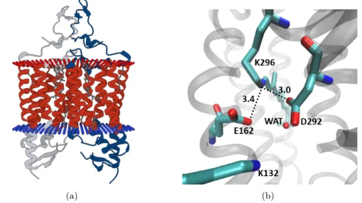
One of the biggest challenges in studying proteins is to obtain a crystal structure in high resolution. Structural information provides insight into the functionality of the partic- ular protein and species related to it (homologues). In 2012, Katoet al. presented a crystal structure of a chimeric channelrhodopsin termed C1C2 at 2.3 Å resolution. [34]
Until lately, it was the first and the only known crystal structure for channelrhodopsins.
C1C2 comprises five helices of ChR1 and two helices of ChR2. In the membrane it forms a homo-dimer (Fig. 1.3(a)). As in all rhodopsin proteins, retinal is covalently bound to a lysine (K296), forming a protonated positively charged Schiff base. The protonated Schiff base terminus (NH+) is exposed to a hydrophilic environment, while the polyene chain and the β-ionone ring experience influence of the hydrophobic residues. In the vicinity of the NH+-terminus two negatively charged counterions D292, E162, a pos- itively charged lysine residue (K132) and a water molecule are located. The distance between the RPSB nitrogen and the counterions E162 and D292 is 3.4 Å and 3.0 Å, respectively (Fig. 1.3(b)). A similar arrangement is known from bacteriorhodopsin.
In bR, a water molecule acts as a proton shuttle during deprotonation. It receives a proton from the Schiff base terminus and passes it to the counterion D85. However, in C1C2 the counterions are closer to the NH+-terminus than the water molecule, sug- gesting one of the counterions as a proton acceptor rather than the water molecule.
Furthermore, Katoet al. conclude that D292 has the highest probability to accept the proton, while E162 is protonated from the beginning. They justify their hypothesis by the closer distance of D292 compared to E162 and also by results of a mutation study.
While D292A mutation causes lack of photocurrent, substitution of E162 by alanine leads to a moderately decreased current. The arrangement of the negatively charged
1.4. Retinal chromophore 5
(a) (b)
Figure 1.3: (a) C1C2 dimer in a membrane. Figure adopted from pdb.org (b) Residues close to the RPSB terminus in the C1C2 crystal structure.
residues close to the positive charge of the RPSB is suggested to be responsible for the blue-shift of the maximum absorption in ChRs compared to bR. In bR, negatively charged amino acids are located further away. [34]
ChR2
The investigation of ChRs is a fast developing research area. Recently, a crystal struc- ture of ChR2 was obtained at a resolution of 2.39 Å. [35] Its general structure is similar to that of C1C2 and comprises a dimer consisting of two protomers. However, the authors note differences in length and helix orientation and in the number of water molecules compared to the C1C2 crystal structure. Furthermore, the presence of two retinal conformations, all-trans,15-anti and 13-cis,15-synin the ground state (GS) was suggested. The distance between the RPSB nitrogen and the counterions E123 and D253 is 2.8 Å and 3.2 Å respectively, which is closer than in C1C2. Another difference refers to the number of gates and the gating mechanism in both proteins, which is not discussed in the current work. The differences between the two crystal structures are supported by recent electrophysiological and Fourier-transform infrared spectroscopy (FTIR) measurements, where C1C2 exhibited light-induced responses different from ChR2. [36]
1.4 Retinal chromophore
1.4.1 Polyenes
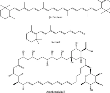
Linearπ-conjugated systems (polyenes) have been a matter of research through decades involving theoretical and experimental studies. Naturally, they occur as cofactors in
photosensitive biological systems: β-carotene in light harvesting complexes of photo- systems or retinal in animal and microbial rhodopsins. Moreover, polyene macrolide antibiotics are known for their antifugal properties. [37]. Figure 1.4 illustrates some nat- urally occurring polyenes. Within the scope of the current work, photochemical prop-
O
Retinal β-Carotene
O O
O O O
O
NH2 OH
OH
OH OH
OH
OH OH
OH
OH OH
H
Amphotericin B
Figure 1.4: Different polyenic systems. β-carotene, retinal and the polyenic antibioticum amphotericin B.
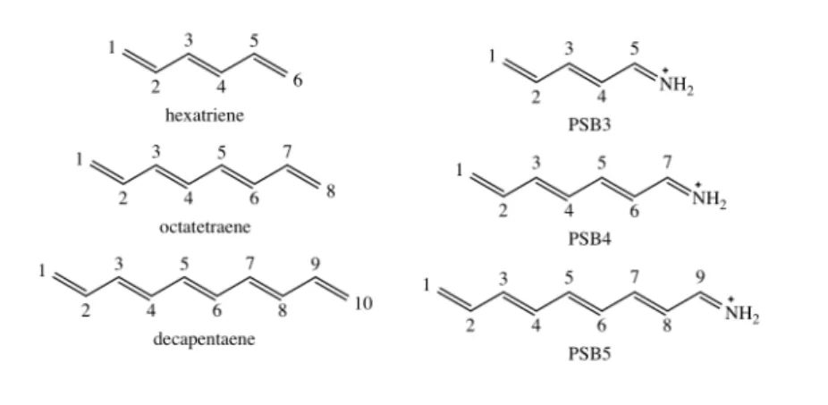
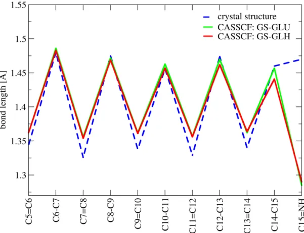
erties of the retinal protonated Schiff base are of particular interest. Protonated Schiff bases (PSBs) are structurally related to symmetric linear polyenes (e.g. β-carotene or simple polyenes like butadiene, hexatriene etc.). However, their electronic properties differ in some aspects due to alteration of theπ-conjugated system by an NH+terminal group. One of my studies included in the present work [38] compares symmetric linear polyenes (termed linear polyenes hereafter) and corresponding PSB models in vacuum (Fig. 5.5). The aim of the current section is to provide a general description of linear polyenes and PSBs relevant for understanding of the properties of retinal in a protein environment.
In the ground state (GS), the conjugatedπ-system leads to a geometric structure char- acterized by alternating double and single bonds. Primarily, the excitation wavelength depends on the length of the conjugated chain: The longer the chain the less energy is required for the excitation. With regard to retinal isomerization, the character of low-lying excited states is of particular interest. In all linear π-conjugated systems, one-photon absorption results from π-π∗ excitation, which is mainly governed by the transition of one electron from the highest occupied molecular orbital (HOMO) to the lowest unoccupied molecular orbital (LUMO). [39, 40]
In linear polyenes, the HOMO–LUMO state (also referred to as symmetry allowed) has 1Bu symmetry. According to valence bond theory (VB), the nature of the 11Bu
state is ionic. [41] This gives rise to a large transition dipole moment responsible for
1.4. Retinal chromophore 7 strong photoabsorbance of this state. The inner bond lengths of the 11Bu geome- try are equalized compared to an alternating character of the GS structure (11Ag) as shown in Figure 1.5. [39, 42, 43] Close to the singly excited HOMO–LUMO state lies
11Bu 21Ag
11Ag
Figure 1.5: Schematic representation of bond length patterns in decapentaene. Relative to the ground state (11Ag), the 21Agstate is characterized by inverted bonds and the 11Bu state by an equalization of inner bonds.
an optically forbidden 21Ag state termed covalent by VB definition. [41] It entails a HOMO2–LUMO2 double excitation augmented by HOMO-1 to LUMO and HOMO to LUMO+1 single excitations. The geometry of the 21Ag state is characterized by re- versing of the bond length pattern compared to the 11Ag state, thus double bonds gain single bond character and vice versa. [39, 44]
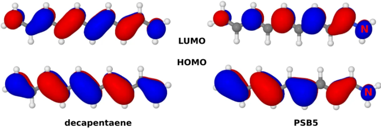
In Schiff bases, introduction of a positive charge through protonation leads to a red- shift of the HOMO–LUMO excitation compared to linear polyenes of the same length or equivalent unprotonated Schiff bases. [45] Due to this shift, absorption of RPSB emerges in the biologically relevant spectral range. Another consequence of the posi- tive charge is a localization of the π-orbitals. In the HOMO of PSBs, more electron
decapentaene PSB5
N
N
HOMO LUMO
Figure 1.6: Highest occupied (HOMO) and lowest unoccupied (LUMO) molecular orbitals in decapentaene and the corresponding protonated Schiff base model PSB5.
density is distributed at the opposite side of the positively charged N-terminus and the opposite trend is found in the LUMO (Fig. 1.6). Thus, HOMO–LUMO excitation involves relocation of the positive charge from the nitrogen to the opposite end of theπ- system, a so called charge transfer (CT) process. In contrast to linear polyenes, the ionic HOMO–LUMO state of PSBs is usually characterized by bond length inversion [46–48]
more similar to the 21Ag state of linear polyenes. I described this property in my sec- ond publication. [38] On the other hand, the doubly excited covalent HOMO2–LUMO2 state is characterized by an equalization of the bond lengths similar to the 11Bu state in linear polyenes, a finding also documented in my second publication. [38] Moreover, in PSBs the interaction of HOMO–LUMO singly and doubly excited states is symmetry allowed, resulting in a non-zero absorbance of the latter.
1.4.2 Spectral properties of retinal in the protein environment
As already mentioned before, the spectrally visible absorption of retinal comes from HOMO–LUMO single excitation, which usually occurs in the first excited S1 state.
Primary factors determining the absorption of the RPSB are the length of the polyene chain and the protonation state of the Schiff base terminus (Sec. 1.4.1). The absorption spectra of rhodopsin proteins cover the entire visible spectral region. For example, the maximum absorption wavelength of C1C2 is ca. 470 nm [34, 49], while bR absorbs at ca. 570 nm [8]. This effect emerges from the influence of the protein environment, which can be electrostatic or steric in nature.
Electrostatic interaction of RPSB and the environment arises from the CT character of the HOMO–LUMO state (Sec. 1.4.1). In the ground state, the positive charge is located at the NH+-terminus. Upon excitation, it moves toward the β-ionone ring (Fig. 1.7). Thus, charges placed in the vicinity of the NH+-terminus (e.g. counterions)
Figure 1.7: Influence of external charges on vertical excitation energies of retinal caused by a CT process assigned to the S1 state. Left: Charges placed at NH+-terminus. Right: Charges placed atβ-ionone ring. Blue arrows denote an increase of the excitation energies (blue-shift) and red arrows denote a decrease (red-shift) of the excitation energies relative to the neutral environment.
1.4. Retinal chromophore 9 or near the β-ionone ring affect the CT process and retinal absorption compared to a neutral environment. [1, 50–52] Negative charges placed in the vicinity of the RPSB terminus stabilize the ground and the first excited state. In the GS, the distance be- tween the positive NH+-terminus and surrounding negative charges is shorter than in the S1 state. Consequently, stabilization of this state is stronger compared to the S1
state. As a result, the energy amount required for the absorption of a photon increases compared to the non-charged surrounding. This effect is known as hypsochromic- or blue-shift. Red- or bathochromic-shift occurs when positive charge is located near the NH+-terminus. In this arrangement, stronger destabilization of the GS with regard to the S1 state leads to a decrease of the absorption energy relative to a neutral environ- ment. [53, 54] I noted these effects in my first publication. [55] If charges are placed near the β-ionone ring, the S1 energy gets more affected relative to the ground state.
Consequently, negative charges lower the S1 energy stronger than the S0energy, causing a bathochromic-shift. In turn, positive external charges near the β-ionone ring cause the opposite effect, resulting in a hypsochromic-shift. [1]
Besides electrostatic interactions, the protein pocket can influence the absorption by affecting the geometry of the chromophore. Planar retinal achieves maximum conju- gation and thus maximum length of theπ-system. Such a structure would exhibit the maximum possible absorption wavelength. A twisted geometry may shorten the π- system, which would cause a blue-shift compared to the fully conjugated chromophore.
This effect was already exploited for the design of blue-shifted ChR species. [56]
Electrostatic and steric interactions between retinal and its protein environment open numerous possibilites for color tuning of the chromophore. However, these effects occur simultaneously and their mutual influence has to be taken into account. For example, a change in the electrostatic environment in combination with some unfavourable ge- ometry may not lead to the desirable result. In this case, detailed knowledge of the electronic and geometric structure is required, which must be provided through com- putational studies.
1.4.3 Quantum yield and reaction time of retinal isomerization
Retinal isomerization occurs in the excited state and is one of the fastest processes in photobiology (fs-timescale). In microbial rhodopsins it involves torsion about C13=C14 double bond. In the ground state, this reaction coordinate is characterized by a barrier too high for a thermal process. An ultrafast reaction is possible due to a conical in- tersection (CoIn) connecting the excited and ground states. Similar to the absorption efficiency, the speed of the reaction is influenced by the interaction of retinal with the protein environment. A pre-twist of the chromophore in the direction of the photoprod- uct may speed up the reaction. [57] Charges in the vicinity of retinal could influence the location of the CoIn or even cause it to vanish. [50]
Immediately after excitation, retinal leaves the Franck-Condon (FC) region following
two reaction coordinates: one involves bond length alternation and the second one a twist about the C13=C14 bond. Along the latter coordinate, retinal arrives at the S1/S0 CoIn. [51, 58, 59] From there, the reaction can proceed to the 13-cis photo- product or return to the starting material. The exact bond length pattern strongly depends on the electrostatic interactions of retinal with its environment (this effect is detailed in my studies [38, 55]). Irrespective of bond length inversion or bond length equalization, the C13=C14 bond would be elongated in the excited state facilitating the isomerization. Furthermore, efficient photoisomerization requires a charged counterion complex. [1, 60, 61]
Several computational studies in bovine rhodopsin and small retinal models illustrated the influence of a hydrogen out-of-plane (HOOP) motion on the outcome of the isomer- ization. [62–65] In particular, a correlation between the number of surface approaches prior to the hop event and photoproduct formation was reported. At the first sur- face approach, the HOOP-motion (Fig. 1.8(a)) and the reaction coordinate defined by skeletal carbon motion (Fig. 1.8(b)) are mostly in phase. Incis-PSBs, HOOP-torsion increases at this point. If the conical intersection is passed at the first surface approach,
(a) (b)
(c)
Figure 1.8: (a) HOOP and (b) reaction coordinates in 11-cisretinal denoted in magenta. (c) Decreasing HOOP torsion leading tocis-cis and increasing HOOP torsion leading tocis-trans reactions in PSB3. Image from Ref. [62].
positive HOOP-torsion is maintained in the ground state resulting in the preferred for- mation of the trans-photoproduct. At later surface approaches, HOOP and reaction coordinate torsions begin to dephase and the HOOP-torsion decreases as shown in Fig- ure 1.8(c). Complete dephasing leads to equal probabilities for the formation of cis- andtrans-products. I described this relationship for RPSB in C1C2. [66]
1.5. State of the art 11
1.5 State of the art
The most important studies concerning the chimera protein C1C2 are chronologically summarized in this section. The interaction of retinal with the surrounding residues (E162, D292, K132) and the electronic properties of retinal are especially important in this context. Furthermore, protonation states of E162 and D292 are of particular interest with regard to electrostatic interactions between the positively charged retinal and the counterions. Moreover, recently reported findings about the isomerization of retinal in C1C2 are briefly reviewed.
2012-2013
Kamiya et al. [67] conducted molecular mechanical (MM) molecular dynamics (MD) simulations and computed QM/MM excitation energies using the newly published crys- tal structure of C1C2. In their study, the authors aimed to examine structural proper- ties of the chimera protein and analyzed the electrostatic contributions of the surround- ing residues to the spectral shift of retinal. The performed MD simulations revealed a hydrogen bond between the retinal NH+-terminus and the counterion E162. Analysis of electrostatic contributions has shown that major influence on the absorption comes from charged residues in the vicinity of retinal. The negatively charged E162, D292, and S295 provided a strong blue-shift and the positively charged K132 led to a strong red-shift of the maximum absorption. However, also charged residues at a larger dis- tance to retinal influence its absorption, although at a lower rate. Based on their study, the authors suggested mutation strategies for engineering of new ChR color variants.
Sneskov and co-workers [68] performed polarizable embedding QM/MM mutation stud- ies based on the C1C2 crystal structure. Similar to Kamiyaet al., the authors reported a significant blue-shift of the retinal absorption (0.3 eV) arising from the charged counteri- ons (E162 and D292) compared to E162G and D292G neutral mutations. Furthermore, they noted that the same residues have major influence on the two-photon absorption properties of retinal, thus two-photon absorption might be an alternative to red-shifted ChR mutants. According to this study, application of polarizable embedding results in a ca. 0.21 eV lowering of the excitation energy.
2014
Itoet al. [69] investigated the hydrogen bonding network in the protein pocket of C1C2 by low-temperature (77K) FTIR spectroscopy. The FTIR measurements of C1C2 in- dicated that the counterions E162 and D292 are deprotonated. Absorption and FTIR measurements involving E162Q and D292N mutants confirmed the ionic nature of the counterions. Furthermore, FTIR results imply a direct hydrogen bond between NH+
and E162. Another conclusion of the FTIR study is related to a water molecule in the proximity of the NH+-terminus. The obtained results suggest that this water molecule bridges D292 to the neighboring positively charged K132.
Pescitelliet al. [49] have reported experimental and calculated circular dichroism (CD) spectra based on the C1C2 monomer and dimer structures. The obtained spectra show an exciton coupling between retinal chromophores in the dimer. The authors furthermore provide time dependent density functional calculations (TDDFT) using three chromophore models. Their models refer to (1) RPSB in vacuum, (2) RPSB and counterions E162 and D292 in vacuum and (3) QM/MM calculations with RPSB and counterions in the QM-region and the MM-region containing residues within 10 Å from the atoms of the ion pair. The authors obtained an S0/S1 transition dominated by HOMO–LUMO π-π∗ excitation for model (1), while for the models including counte- rions in the QM-region excitation is denoted as π-π∗ like. Further observation refers toπ and π∗ orbitals partially localized on E162. The energy of the (crude) QM/MM model is blue-shifted compared to the experiment. Moreover, the authors mention a relation between the low polarity of the protein pocket and the vibronic fine structure of the spectrum.
2015
Takemoto and co-workers [70] investigated the channel functionality in C1C2 by elec- trophysiological experiments and MM/MD simulations. The authors also modelled C1C2 with a 13-cis retinal based on the bacteriorhodopsin 13-cis retinal structure to analyze conformational changes of the protein upon isomerization.
Kato et al. [56] designed blue-shifted variants of ChRs and archaerhodopsin-3 (AR3) employing theoretical and computational methods. The design of the ChR mutants was based on the crystal structure of C1C2. Kato and co-workers introduced mutations to enforce a twist about the C6-C7 bond in retinal. Such twist leads to a shortening of theπ-system and results in a blue-shift of the absorption.
Inaguma et al. [36] discovered through difference FTIR measurements that C1C2 un- dergoes light induced conformational changes distinct from ChR2. Furthermore, the authors noted a transient protonation band of a carboxylate group, which may be assigned to either E162 or D292. This finding suggests both counterions as possible candidates for the role of a proton acceptor.
1.5. State of the art 13 2017
VanGordon and colleagues [71] investigated intramolecular interactions in the closed state of C1C2 by MM/MD simulations. The authors paid particular attention to the gating region of the conducting pore. This study suggested D292 as a primary proton acceptor, but it also advises to consider E162 for this role.
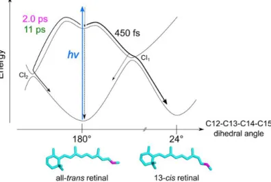
Hontaniet al. provided details of the retinal photoisomerization in C1C2 in their exper- imental study combined with QM/MM calculations (Fig. 1.9). [72] QM/MM computa-
Figure 1.9: Model of excited state reaction dynamics of retinal in C1C2 suggested by Hontani et al. from Ref. [72].
tions were employed for geometry optimization, calculation of relaxed torsion scans and one trajectory without initial velocities (termed 0K trajectory). For the calculations, two initial models were used: one with NH+ hydrogen bonding with the counterion E162 and a second one with a hydrogen bond between NH+ and a water molecule.
The authors reported three time constants (450 fs, 2 ps and 11 ps) for the photoreac- tion. They furthermore suggested relaxation to the ground state through two conical intersections. One conical intersection (termed CI1) is suggested to result in the 13-cis retinal as photoproduct upon clockwise torsion, while the second CoIn (termed CI2) is unproductive and leads back to all-transretinal involving counter-clockwise torsion. A 450 fs time constant was assigned to the clockwise channel that yielded 50% of 13-cis and 50% of all-trans retinal. The slower 2 ps and 11 ps time components describe the return to all-trans retinal via CI2. The overall quantum yield estimated for the 13-cis photoproduct with regard to all reaction channels comprises 30%.
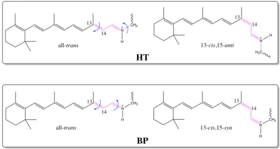
Photoisomerization in microbial rhodopsins initiates a cycle of thermal reactions. Dur- ing this photocycle, a series of retinal de- and reprotonation events take place, while stable photointermediates are formed. Hontani and co-authors suggested models for the C1C2 photocycle (Fig. 1.10(a)). An important aspect in the photocycle refers
to the isomeric composition of the ground state. In ChR2, the dark state is entirely composed of the all-trans,15-anti isomer. [73] However, there is evidence from Raman and nuclear magnetic resonance (NMR) measurements that upon minor illumination a second 13-cis,15-syn configurated GS isomer emerges. The occurence of two ground state isomers consequently leads to a branched photocycle. [74] The all-trans,15-anti isomer converts to 13-cis,15-anti upon excitation, while 13-cis,15-syntransforms to the all-trans,15-anti configuration (Fig. 1.10(b)). Similar findings were reported earlier for the bR photocycle. [1] For C1C2, no evidence for a second ground state isomer was
(a) (b)
Figure 1.10: (a) Photocycle model proposed for the retinal in C1C2 at pH8 from Ref. [72].
(b) Configurations of RPSB in ChR2 reproduced from Ref. [13].
provided yet. Hontaniet al. report only the all-trans,15-anti isomer as initial structure appearing in the ground state at their conditions.
2018
Recently, Adam and Bondar [75] published results of their QM/MM study based on the C1C2 crystal structure. The authors investigated proton transfer possibilities prior to photoisomerization. In particular, the main object of the study was to understand how the protein environment stabilizes the protonated state of the retinal Schiff base in the resting state and how it avoids early deprotonation by nearby charged counterions.
The authors attribute an essential role to the the neighboring lysine (K132). This residue showed a complex hydrogen bonding dynamics, which is reflected in a hydrogen bonding between K132 and water, K132 and E136, K132 and counterions (E162 and D292). Such interactions might have an impact on the protonation energetics resulting in unfavourable protonation of E162 and D292 prior to photoisomerization. In general, both counterions are considered for the role of the possible proton acceptor.
15
Chapter 2
Motivation and objectives
Channelrhodopsins are among the most important tools in optogenetics. Since their discovery, numerous studies were conducted to unravel structural-functional relation- ships within the channels and to apply this knowledge for efficient design of novel ChR variants. In this sense, theoretical atomistic studies play a crucial role. They provide
"live" monitoring of the investigated system and also allow to introduce modifications in a time- and cost-saving manner (compared to laboratory studies). Furthermore, molecular simulations can provide predictions that may help to understand experimen- tal results (e.g. spectra, kinetics) or to conceive new experiments.
A primary step in ChR activation and also one of the key targets for the design of new properties is retinal photoisomerization. For its control, a detailed understanding of spectral features (characterization of involved electronic states and geometries) and reaction parameters (speed, efficiency, possible photoproducts) is inevitable. A first insight into the photoreaction can be provided by static calculations. A quantitative characterization is, however, only possible through simulation of its excited-state dy- namics. Such studies should preferably include a molecular ensemble, i.e. a relevant number of molecules to deliver information about photoproducts, their quantum yields, time constants, reaction channels and reaction mechanisms. Statistical analysis of such an ensemble delivers information about these quantities. Furthermore, dynamical sim- ulations provide a basis for the investigation of factors influencing speed and efficiency of the reaction.
The major objective of my thesis was the detailed understanding of the retinal photoi- somerization mechanism in C1C2. For the description of the chromophore in a protein environment, a combined QM/MM approach was chosen. QM/MM MD simulations in the microcanonical ensemble are known to be a valuable tool for understanding light- induced processes in photosensitive proteins like bR or visual rhodopsin. [76–78] Until yet, no such studies were reported for ChRs. An important step prior to dynamics studies is the choice of a QM method, which should provide a cost efficient, yet ac- curate description of electronic properties and potential energy surfaces. In this step, a detailed characterization of the structural and electronic properties in the ground and low-lying excited states is performed. Furthermore, the influence of the protein environment in the vicinity of retinal on these properties is investigated. The obtained
results are then compared to available experimental data (e.g. IR, Raman and ab- sorption spectra) and other computational studies. Knowledge gained through static calculations is then applied to study retinal dynamics.
I started my thesis with static calculations on retinal within C1C2, which provided a first insight into its photoreaction and allowed for method validation with regard to further dynamical investigations. The results of this study are included in the first publication (Sec. 5.1 and Ref. [55]). The calculations therein refer to ground- and excited-state geometry optimizations, calculation of vertical excitation energies and screening of residues influencing the absorption properties. Results from the investi- gation of static properties and method validation have demonstrated the demand for multireference methodologies to properly describe the chromophore in C1C2. Further- more, this study has shown the need for revisiting the protonated Schiff base properties with regard to structurally related symmetric linear polyenes. To include a multirefer- ence method feasible with regard to dynamics simulations, the COBRAMM software package was extended by a QM interface to the semiempirical methods provided in the MNDO program package. This implementation is documented in the third publication (Chap. 4 and Ref. [79]). To address the issue related to the difference between PSBs and linear polyenes and to test the semiempirical approach, a new study was conducted, which is included in the second publication (Sec. 5.2 and Ref. [38]). Finally, equipped with findings from my previous studies a simulation of retinal excited state dynamics in C1C2 was performed for the first time. The results of this study are captured in my final publication (Sec. 5.3 and Ref. [66]).
17
Chapter 3
Theory and methods
3.1 Photochemical reactions
In a photochemical sense, light provides energy to overcome activation barriers for which thermal energy in the ground state is not enough. Every photochemical reaction starts with the absorption of a photon and subsequent excitation to a higher lying electronic and vibronic level. Immediately after excitation, the excess energy is dissipated via different mechanisms that will be described below.
3.1.1 Relaxation processes after photoexcitation
In a static picture, the main events occuring upon excitation and relaxation to the ground state can be summarized in a Jablonski scheme. [80] The first process is vi- bronic relaxation (VR), which promotes the molecule rapidly and radiationlessly from the higher vibronic level to the lowest one within the same electronic state. From there, deactivation involves different electronic states and can be non-radiative or radiative.
Non-radiative transitions within the same spin multiplicity proceed via internal con- version (IC), while radiative decay results in fluorescence. Non-radiative transitions between states of different multiplicities (e.g. singlet-triplet) are possible via intersys- tem crossing (ISC). If a triplet state is populated, it can relax non-radiatively via ISC, IC, VR or through photon emission via phosphorescence from the T1 state.
At certain geometries potential energy surfaces may intersect, enabling radiationless transition (IC or ISC). A special case of such crossings are conical intersections (CoIns). [81] They are of immense importance in the photochemistry of ultrafast pro- cesses (e.g. photoisomerization), which are ubiquitious in photochemistry. [82]
3.1.2 Conical intersections
Usually, CoIns are pictured as funnels connecting ground and excited states that guide a molecule radiationless and fast between potential energy surfaces. In practice, the funnel is only one part of a high-dimensional picture. In diatomic molecules, states of the same symmetry are not allowed to be degenerate (noncrossing rule). [83] States of different spatial symmetry can, however, cross. In polyatomic molecules, states of same
as well as of different symmetries can intersect. [84, 85] States of different spatial sym- metries cross in an (m-1)-dimensional space, while states of the same symmetry lead to a CoIn inm-2 dimensions, where m describes the number of degrees of freedom in the particular molecule. [86, 87] In multidimensional space, CoIns can be described as points on a (m-2)-dimensional hyperline, referred to as a crossing seam. To derive the crossing rules, two electronic states representing potential energy curves that may cross are considered. They are described by two arbitrary functionsφ1 and φ2 that are or- thogonal and build an orthonormal basis with already known solutions of the electronic Schrödinger equations. [84] Consequently, each of the two unknown eigenfunctions can be expressed as a linear combination ofφ1 andφ2:
ψ=c1φ1+c2φ2 (3.1)
To obtain energy eigenvalues, a secular equation is used:
"
H11−E H12 H21 H22−E
# "
c1 c2
#
= 0 (3.2)
whereHij=hφi|H|φji. The energy eigenvalues are evaluated as following:
E1,2= H11+H22
2 ±
s
H11−H22 2
2
+H21H12 (3.3)
Equation (3.3) allows to derive the conditions for a crossing of the two states. At the crossing point, the energies of the involved states are equal, i.e. E1=E2, which happens when two conditions from the Equation (3.3) are simultaneously fulfilled:
H11=H22∧H12= 0 =H21 (3.4)
Equation (3.4) requires at least two independent molecular degrees of freedom x1 and x2. In diatomic molecules, only one degree of freedom (the interatomic distance) exists.
Consequently, states of the same symmetry would never cross leading to the noncrossing rule. This rule does not apply to the case of different symmetries, because H12 is already equal to zero and only one condition has to be met. In polyatomic molecules, it is generally possible to find two independent degrees of freedom which satisfy the conditions from Equation (3.4). In the case of different symmetries, the intersection space is (m-1)-dimensional for the same reason as for diatomic molecules. For states of the same symmetry it leads to a crossing in the (m-2)-dimensional space. In the (m-2)-dimensional intersection space, degeneracy is preserved even if coordinates are varied. [85] The remaining degrees of freedom x1 and x2 span the so-called branching plane. Motion in this plane lifts the degeneracy. Plotting the potential energy against x1 and x2 leads to the well known double cone shape of the crossing potential energy
3.1. Photochemical reactions 19 surfaces (PESs) (Fig. 3.1(a)). [81, 82] In the beginning of this section, I mentioned that CoIns are points along the crossing seam. A relaxation of a molecule through a conical intersection occurs in thex1/x2plane when the reaction path of the investigated system crosses the seam as shown in Figure 3.1(b). The branching plane is associated with
(a) (b)
Figure 3.1: Different representations of conical intersections: (a) representation as a double cone; (b) conical intersection in a space spanned by the reaction coordinate X3 and branching coordinate X1X2. Figures from Ref. [82].
the molecular motion after exiting the degeneracy region. Consequently, the outcome of the photochemical reaction is influenced by the branching vectors. The branching space coordinatesx1 andx2 can be described as gradient difference vector and gradient of the interstate coupling vector. [86, 88] Thex1 direction is referred to as the gradient difference vector defined as
x1 = δ(E1−E2)
δR (3.5)
It is also called tuning mode, since it adjusts the energy gap between the interacting states. Thex2 direction is given as the gradient of the interstate coupling vector
x2 =ψi δH
δR
ψj
(3.6)
whereψi and ψj are electronic wave functions,H is the electronic Hamiltonian andR denotes a nuclear configuration vector. The vectorx2 is also referred to as a coupling mode, since it describes coupling of the interacting states. Furthermore,x2is parallel to the nonadiabatic coupling vectorfij. [86] The relation between fij andx2 is expressed as:
fij =ψi
δψj δR
=
ψi
δH δR
ψj
E1−E2 (3.7)
where E1 and E2 are the energy eigenvalues. Equation (3.7) shows that nonadiabatic coupling (also denoted as derivative coupling) becomes important when the interacting
states become close in energy. The derivative coupling is required to describe nonadi- abatic nuclear dynamics (Sec. 3.6). [89–91]
3.2 Quantum mechanical approach
3.2.1 Description of potential energy surfaces Born-Oppenheimer Approximation
The time-independent Schrödinger equation (SE) can be written as
H(r, R)Ψ(r, R) =EΨ(r, R) (3.8) wherer corresponds to electronic and R to nuclear coordinates. H is the total nonrel- ativistic Hamiltonian of the system, which can be described as
H(r, R) =Tnuc+He(r;R) (3.9) where Tnuc denotes the nuclear kinetic energy operator and He(r;R) corresponds to the electronic Hamiltonian that depends parametrically onR:
He(r;R) =Te+Vee+Ven+Vnn (3.10) The electronic Hamiltonian includes electronic kinetic energy (Te) and Coulomb inter- actions (electron-electron repulsion (Vee), nucleus-nucleus repulsion (Vnn) and electron- nucleus interaction (Ven)). [92] Analytic solution of the SE for molecular systems with more than two particles is not feasible. The Born-Oppenheimer (BO) approximation is used to simplify this task. [93] It is based on the fact that electrons are much lighter than nuclei and thus both move on different time scales. Particularly, electrons move much faster than nuclei. The coupling between nuclear and electronic degrees of free- dom is ignored. Consequently, the total wavefunction is approximated as a product of nuclearχ(R) and electronicψ(r;R) wavefunctions:
Ψ(r, R) =ψ(r;R)χ(R) (3.11)
Hence, the movement of both particles can be handled separately. Using this assump- tion, the total SE is separated into electronic and nuclear problems. From the electrons
"point of view" nuclei move so slow that they can be assumed fixed. In conjunction with this assumption, the kinetic energy of the nuclei is neglected (Tnuc = 0) and the repulsion between the nuclei (Vnn) is considered constant. This approximation leads to the formulation of the electronic Schrödinger equation:
He(r;R)ψi(r;R) =Eie(R)ψi(r;R) (3.12)