Relaxation Processes in Electronically Excited States: Blue-Light Receptors and
Related Compounds
Inaugural-Dissertation
zur
Erlangung des Doktorgrades der
Mathematisch-Naturwissenschaftlichen Fakult¨ at der Heinreich-Heine-Universit¨ at D¨ usseldorf
vorgelegt von Susanne Salzmann
aus M¨ unchen
D¨ usseldorf 2009
Aus dem Institut f¨ ur Theoretische Chemie und Computerchemie der Heinreich-Heine-Universit¨ at D¨ usseldorf
Gedruckt mit Genehmigung der
Referent:
Korreferent:
Prof. Dr. Christel M. Marian Prof. Dr. Walter Thiel
Tag der m¨ undlichen Pr¨ ufung: 24.07.2009
Dans la vie rien n’est ´a craindre, tout est ´a comprendre.
Marie Curie
Hiermit versichere ich, die hier vorgelegte Arbeit eigenst¨andig und ohne unerlaubte Hilfe angefertigt zu haben. Die Dissertation wurde in der vorgelegten oder in ¨ahnlicher Form noch bei keiner Institution eingereicht. Ich habe keine erfolglosen Promotionsver- suche unternommen.
D¨usseldorf, den
(Susanne Salzmann)
• Paper I
The photophysics of flavins: What makes the difference between gas phase and aqueous solution?
Susanne Salzmann, J¨org Tatchen, and Christel M. Marian, J. Photochem.
Photobiol. A, 198 (2008) 221-231.
After an initial introduction to the calculations of the ISC rate constants by J. Tatchen, I contributed all calculations and major parts of the discussion and conclusion.
• Paper II
Effects of protonation and deprotonation on the excitation energies of lumiflavin Susanne Salzmann and Christel M. Marian, Chem. Phys. Lett., 463, (2008) 400-404.
All calculations and most of the discussion and conclusion were provided by me.
• Paper III
Photophysical properties of structurally and electronically modified flavin deriva- tives determined by spectroscopy and theoretical calculations
Susanne Salzmann, V´ıctor Martinez-Junza, Bj¨orn Zorn, Silvia Braslavsky, Madina Mansurova, Christel M. Marian, and Wolfgang G¨artner,J. Phys. Chem., submitted.
For this combined experimental and theoretical work, I contributed all calculations as well as major parts of the discussion and conclusion.
• Paper IV
The photophysics of alloxazines: A quantum chemical investigation in vacuum and solution
Susanne Salzmann and Christel M. Marian, Photochem. Photobiol. Sci. sub- mitted.
For this paper, I provided all calculations and most of the discussion and conclusion.
• Paper V
Influence of the LOV domain on low-lying excited states of flavin: A combined quantum-mechanics / molecular-mechanics investigation
Susanne Salzmann, Mario R. Silva-Junior, Walter Thiel, and Christel M. Mar- ian, unpublished manuscript.
The setup of YtvA-LOV was taken from M. Silva-Junior, who also contributed an initial introduction to and further supervision of the QM/MM calculations. My con- tribution comprises conception and performance of the calculations, as well as major parts of the discussion and conclusion.
• Paper VI
Excited states of thiophene: Ring opening as deactivation mechanism
Susanne Salzmann, Martin Kleinschmidt, J¨org Tatchen, Rainer Weinkauf, and Christel M. Marian, Phys. Chem. Chem. Phys., 10 (2008) 380-392.
The majority of the calculations for this publication have been carried out within the
framework of my diploma thesis. During my time as a PhD student I continued with additional calculations (i.e. CASPT2 results) and contributed parts to the paper text.
• Paper VII
Ultrafast dynamics in thiophene investigated by femtosecond pump probe photo- electron spectroscopy and theory
Rainer Weinkauf, Leo Lehr, Edward W. Schlag,Susanne Salzmann, and Chris- tel M. Marian, Phys. Chem. Chem. Phys., 10 (2008) 393-404.
The focus of this paper clearly lies on the experimental work. My contribution com- prises the calculation and evaluation of the vertical ionization energies at different geometries in order to support the interpretation of the fs pump-probe experiment.
• Paper VIII
Deactivation via ring opening: A quantum chemical study of the excited states of furan and comparison to thiophene
Nemanja Gavrilov,Susanne Salzmann, and Christel M. Marian,Chem. Phys., 349 (2008) 269-277.
This publication was done together with Nemanja Gavrilov a summer intern from Bel- grade, who performed the calculations under my supervision. In addition I contributed all presented calculations on thiophene as well as a good part of the text.
• Paper IX
UV excitation and radiationless deactivation of imidazole
Mario Barbatti, Hans Lischka, Susanne Salzmann, and Christel M. MarianJ.
Chem. Phys., 130 (2009) 034305 (8 pages).
In this paper, my contribution comprises the calculation and analysis of the DFT/MRCI energy profiles of the four reaction paths.
List of related papers not included in the thesis
• Paper X
Electronically excited states of tryptamine and its microhydrated complex
Michael Schmitt, Robert Brause, Christel M. Marian, Susanne Salzmann, and W. Leo Meerts, J. Chem. Phys. 125 (2006) 124309 (10 pages).
• Paper XI
Photophysics of phenalenone: Quantum-mechanical investigation of singlet-trip- let intersystem crossing
Martha C. Daza, Markus Doerr, Susanne Salzmann, Christel M. Marian, and Walter Thiel Phys. Chem. Chem. Phys., 11 (2009) 1688-1696.
Zusammenfassung
Die vorliegende Arbeit besch¨aftigt sich mit der quantenchemischen Modellierung von Relaxationsprozessen elektronisch angeregter Flavine, welche als Pigment/Kofaktor nat¨urlicher Blaulichtsensoren vorkommen. Im Zuge dieser Doktorarbeit wurde hierbei besonderes Augenmerk auf den ersten Schritt des blaulichtinduzierten Reaktionszyk- lus sogenannter LOV (light, oxygen, and voltage sensitive) Dom¨anen des Phototropins gelegt, einer Interkombination (intersystem crossing, ISC) vom urspr¨unglich angeregten Singulettzustand des Kofaktors in die Triplettmannigfaltigkeit desselben.
AbbildungStrukturformel von Flavin und zwei untersuchter Derivate
Um die photophysikalischen Vorg¨ange, welche direkt nach Absorption eines Photons des blauen Spektralbereichs in der LOV Dom¨ane auftreten, zu verstehen, wurden in einer ersten Studie der optisch aktive Teil des freien Kofaktors (siehe Abbildung) im Vakuum und, mit Hilfe einer Kombination aus Mikrohydrierung und Kontinuumsmo- dell, in w¨assriger L¨osung untersucht. Es stellte sich heraus, dass nicht nur die Form der Absorptionsspektren, sondern auch der Bildungsmechanismus der Triplettspezies in diesen beiden Medien unterschiedlich ist. Im Vakuum wird ein effizienter, nach El Sayed erlaubter, direkter ISC-Mechanismus gefunden. In w¨assriger L¨osung ist dieser Kanal durch L¨osungsmittelverschiebung jedoch nicht mehr zug¨anglich. Stattdessen wird durch diese Verschiebung ein anderer ISC-Kanal erreichbar, der hingegen nach El Sayeds Regel verboten ist. Erst durch die Ber¨ucksichtigung vibronischer Spin-Bahn- Kopplung, einem Mechanismus, der in heterozyklischen Systemen weit verbreitet zu sein scheint, wird das ISC erheblich beschleunigt, wodurch die experimentellen Be- funde erkl¨art werden k¨onnen. Desweiteren wurden in Kooperation mit Experimenta- toren mehrere Flavinderivate, welche als k¨unstliche Pigmente in nat¨urlichen Blaulicht- sensoren Anwendung finden k¨onnten, in Vakuum und w¨assriger L¨osung untersucht.
Hierbei weisen insbesondere die zwei Deazaverbindungen (siehe Abbildung) ver¨anderte spektroskopische Eigenschaften auf. F¨ur das 1-Deazaflavin konnte kein effizienter ISC- Kanal identifiziert werden, w¨ahrend die Population der Triplettmannigfaltigkeit von
In einem zweiten Schritt wurde das Flavin mit Hilfe einer Kombination aus Quan- tenmechanik und Molek¨ulmechanik in der LOV Dom¨ane des Photosensors YtvA aus Bacillus subtilis untersucht. Schwerpunkt dieser Untersuchung war es, zum Einen, den Einfluss von vier polaren Aminos¨auren in der unmittelbaren Umgebung des Flavins, und zum Anderen, den Einfluss der Konformation des f¨ur den Reaktionszyklus essen- tiellen Cysteins auf die Photophysik des Kofaktors zu untersuchen. Hierbei zeigte sich, dass der Einfluss der Proteinumgebung zustandsabh¨angig ist. W¨ahrend die energeti- sche Lage der optisch aktiven Zust¨ande den Verh¨altnissen in Wasser sehr ¨ahnlich ist, zeigt sich, dass die energetische Position relevanter, optisch inaktiver Zust¨ande der Si- tuation im Vakuum sehr nahe kommt. Als Resultat zeichnet sich ab, dass sowohl der direkte ISC-Kanal als auch der durch vibronische Spin-Bahn-Kopplung beschleunigte Kanal erreichbar sind. Zudem zeigt die Untersuchung eindeutige Indizien f¨ur einen externen Schweratomeffekt, welcher durch das Schwefelatom des Cysteins verursacht wird und f¨ur den experimentelle Anhaltspunkte gegeben sind.
In dieser Arbeit wird nicht nur die energetische Lage optisch aktiver Zust¨ande von Flavinen in den verschiedenen Umgebungen in sehr guter ¨Ubereinstimmung mit ex- perimentellen Daten beschrieben, sondern auch die Position dunkler, experimentell schwer zug¨anglicher Zust¨ande wird vorausgesagt, welche ebenfalls eine Rolle f¨ur die Photophysik dieser Verbindungen spielen.
Summary
The present work is concerned with the quantum chemical modeling of relaxation processes in electronically excited flavins, which are found as chromophores/cofactors in natural blue-light sensors. In the course of this dissertation the focus was put on the first step of the blue-light induced reaction cycle of so-called LOV (light, oxygen and voltage sensitive) domains of phototropin: an intersystem crossing (ISC) of the initially excited singlet state to the triplet manifold of the cofactor.
Figure Structure of flavin and two investigated derivatives
In order to understand the photophysical events that take place immediately after ab- sorption of a photon from the blue spectral region, the optically active part of the free chromophore (see Figure) was investigated in a first study in vacuum and, by means of a combination of microhydration and continuum model, in aqueous solution. As it turned out, not only the form of the absorption spectra, but also the mechanism for the generation of the triplet species is different in these environments. In vacuum an efficient El Sayed allowed direct ISC mechanism is found. In aqueous solution this channel is not available due to solvent shifts. Instead, this shift causes another ISC channel to be accessible. As this channel is, however, forbidden according to El Sayeds rule, vibronic spin-orbit coupling, a mechanism that seems to be widely common in heterocyclic systems, has to be taken into account in order to find ISC rates comparable to experiment. In addition, several flavin derivatives that are anticipated to serve as artificial pigments in natural blue-light sensors, have been investigated in close coop- eration with experiment. At this, the two deaza compounds (see Figure) in particular show altered spectroscopical properties. For 1-deazaflavin, no efficient ISC channel was found, while in 5-deazaflavin in aqueous solution the population of the triplet manifold follows the respective mechanism of the parent compound.
In a second step, the flavin has been investigated in the LOV domain of the photosen- sor YtvA of Bacillus subtilis by means of a combination of quantum mechanics and
the photophysics of the cofactor of a) the four polar amino acids in the vicinity of the flavin and b) conformational effects of the cysteine that is essential for the photocycle essential. In this connection it could be shown that the influence of the protein envi- ronment is state dependent. While the energetic position of the optically active states is very similar to the circumstances in water, it turns out that in relevant optically inactive states the situation in vacuum is closely resembled. As a result it emerges that the direct ISC channel as well as the channel enhanced by vibronic spin-orbit coupling are available in LOV domains. In addition, clear evidence for an external heavy-atom effect, caused by the sulfur center of the reactive cysteine, are found.
In the present work not only the energetic position of the optically active states of flavins in different environments are reproduced in very good agreement to experi- ment, but also the position of dark states is predicted, which are difficult to capture by means of experimental techniques, but nevertheless participate in the photophysics of these compounds.
Contents
Motivation 1
I. Introduction 3
1. Flavins 5
1.1. Flavins in solution . . . 5
1.2. Flavins in blue-light photosensors . . . 7
1.2.1. Cryptochrome . . . 7
1.2.2. Flavin in the BLUF domain . . . 7
1.2.3. Flavin in the LOV domain . . . 8
2. Project definition and embedding into SFB 663 13 2.1. Open questions concering LOV domains . . . 13
2.2. Contributions within the framework of SFB 663 . . . 13
2.2.1. Modified flavins in biomimetric environment . . . 14
2.2.2. Photochemical reactions of FMN in LOV domains . . . 14
2.3. Own contributions . . . 15
II. Theory and methods 17
1. Theoretical background 19 1.1. The Born-Oppenheimer approximation and the Franck-Condon principle 19 1.2. Photophysical relaxation processes: Molecular response to electronic ex- citation . . . 231.2.1. Luminescence . . . 23
1.2.2. Non-radiative processes . . . 24
1.2.3. Which process prevails? . . . 27
2. Methods 29 2.1. Modeling electronic excited states . . . 29
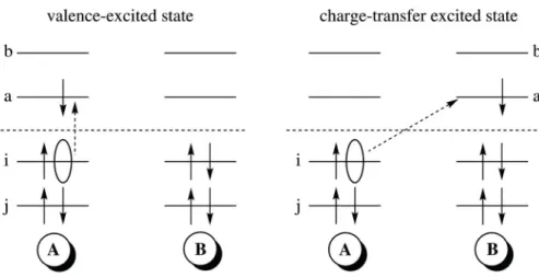
2.1.1. TDDFT and CT states . . . 29
2.1.2. The DFT/MRCI Method . . . 31
2.2. Modeling of solvent effects . . . 34
2.2.1. Aqueous solution . . . 35
2.2.2. LOV domains: The combined quantum-mechanics/ molecular- mechanics approach . . . 37
III. Results and discussion 41
1. Free flavins 43
1.1. Absorption spectra . . . 43
1.1.1. Vacuum . . . 44
1.1.2. Changes in solution . . . 47
1.1.3. Vertical excitation energies versus band maxima . . . 50
1.2. Triplet generation . . . 50
1.2.1. ISC in flavins: The difference between vacuum and aqueous so- lution . . . 50
1.2.2. ISC in flavin related compounds . . . 51
1.3. Competing relaxation mechanisms for the depopulation of the 1ππ∗ state 51 1.3.1. Relaxation processes in LF, MIA and 5DLF . . . 51
1.3.2. Internal conversion in alloxazines . . . 53
1.3.3. Nonradiative decay of 1DRF: What causes the difference? . . . 55
2. Flavin in the LOV domain of YtvA 57 2.1. Initial considerations . . . 57
2.1.1. The binding pocket of LOV . . . 57
2.1.2. Earlier theoretical investigations . . . 58
2.1.3. Interlude: How to accout for the protein? . . . 59
2.2. How YtvA-LOV influences low-lying excited states of its flavin cofactor 61 2.3. ISC in YtvA-LOV . . . 63
3. (De)Protonated flavins 67 3.1. Motivation . . . 67
3.2. Vertical excitation energies . . . 67
3.3. Protonation energies . . . 68
3.4. Future prospects . . . 69
4. Deactivation in aromatic fivemembered heterocycles 71 4.1. Foreword . . . 71
4.2. Ultrafast relaxation in photo-excited thiophene . . . 71
4.2.1. Experimental findings . . . 71
4.2.2. Electronic structure of excited states . . . 72
4.2.3. Relaxation pathways . . . 73
4.3. Relevance to other five membered heterocycles . . . 76
4.3.1. Furan . . . 76
4.3.2. Pyrrole and imidazole . . . 76
4.3.3. More complex systems . . . 78
IV. Conclusion and outlook 79
1. Concluding remarks 81
2. What is still missing? 83
List of Tables 85
List of Figures 91
References 100
Acknowledgement 101
Abbreviations 103
Motivation
The sun is the source of energy for a vast majority of biomass on this planet. Au- totrophic creatures like plants use that energy directly and are able to transform it into a form that can be used by heterotrophic creatures, like us. Apart from the ben- eficial effects, sunlight holds danger. On a molecular basis, the interaction of light and matter results in electronically excited molecules. Hereby, electronic excitation triggers a manifold of mechanisms that participate in the dissipation of the excess energy. Those dissipation mechanisms range from physical processes to chemical reac- tions. While the photophysical processes preserve the chemical identity of the molecule and do not pose an immediate threat, photochemical reactions possess the danger of permanent alteration to the molecule.
One of the largest biomolecules in organisms is DNA. In plants, but also in other living creatures, DNA is exposed to sun light on a daily basis. This exposure contains the risk of various photochemical reactions. Although ultrafast decay of the UV-induced excited-state population to the electronic ground state is an intrinsic photophysical property of all nucleobases (see Ref. [1]), sites with two or more pyrimidine bases are mutational hotspots. [2] These photo lesions alter the genetic information and possibly render the same useless. Over the eons, nature developed elaborate detection and repair mechanisms for the various photo damages. These mechanisms are able to identify such a mutation and, in the ideal case, repair the DNA or at least initiate programmed cell death (apoptosis). Otherwise, the mutated cell bears the risk of unfavorable biological responses, e.g. cancer.
An even better strategy than detection and elimination, is to prevent photo damage in the first place. There are various examples for photophobic reactions of bacteria (mostly unicellular organisms) upon exposure to (intense) blue light, presumably in
FigureLeft: X-ray structure of the light, oxygen, and voltage sensitive (LOV) domain of the photosensor YtvA in Bacillus subtilis. [3] Middle: Schematic illustration of the blue-light triggered photocycle in the LOV domains of photopropin and related photosensors. Right: Phototropism, the ordered movement in response to light, one of the processes triggered by blue light and mediated by phototropin.
order to avoid the damaging effects of UV light. [4] While this and other responses have been known for many years, they have been widely anticipated to be a reaction to photosynthetic electron flow or reactive oxygen species. It took some time to real- ize that most of these responses are actually mediated by a variety of photosensors, even though this concept has emerged quite early in evolution, as the discovery of a photosensor in cyanobacteria demonstrates. [5, 6]
The sensing of quality, quantity, duration and direction of light is, of course, also vital with regard to the optimization of photosynthesis. Higher plants possess various photoreceptors and are able to respond to blue-light radiation with a multitude of processes. [7] The most commonly observed in everyday life is the ordered movement towards light (phototropism, see Figure). This photophysiological process is mediated by the protein phototropin. [8] As genome sequencing and annotation projects have shown, similar structures are also found in some prokaryotes (e.g. Bacillus subtilis[9]).
[7] The process of light sensing hereby utilizes fully oxidized flavins, vitamin B2 related molecules, as cofactors in so-called light, oxygen, and voltage (LOV) domains. (See Figure (left) for an X-ray structure of such a LOV domain.) Blue-light absorption triggers a series of fully reversible photophysical and photochemical reactions of the chromophore schematically shown in Figure (middle).
Until the start of this thesis, the processes that lead to signalling were not completely understood. Apart from some exceptions little was known about intermediate transient states or the influence of the LOV domain on the low-lying electronically excited states of the cofactor. This is where the contribution of this work lies. As modern quantum chemical methods nowadays are able to predict the structure and energetic position of relevant electronic states in a satisfying manner, I have added to the understanding of the blue-light induced photophysical processes from a quantum chemists point of view.
Part I.
Introduction
1. Flavins - Sensing the blue
’ There was no moon, and although the night was perfectly clear and the stars shone brightly, the lustre of the heavens was fairly eclipsed by that of the sea. The unbroken part of the surface appeared pitch black, but wherever there was the least ripple the whole line broke into a brilliant crest of clear white light. Near the ship the black inter- spaces predominated, but as the distance increased the glittering ridges looked closer, until toward the horizon, as far as the eye could reach, they seemed to run together and melt into one continuous sea of light.’
C. W. Thompson (1877)a
Since their first description in the year 1879 by the English chemist A. W. Blyth, [11]
who isolated the bright yellow pigment (riboflavin) from cow milk, flavins have been the object of many investigations. This interest is well founded due to the chemical versatility of flavins. Next to their role as a constituent of the vitamin B complex (riboflavin is vitamin B2), flavin-binding proteins are involved in a variety of biological processes mostly in the context of redox reactions. (For a detailed review see Ref [10].)
Figure 1.1.: Left: Structure and IUPAC labeling of flavins.(R=R’=R”=H: Isoallox- azine (IA); R=Me, R’=R”=H: 10-Methylisoalloxazine (MIA,DMLF); R=R’=R”=Me:
Lumiflavin (LF)) Right: Three biologically relevant flavins, riboflavin (RF, vitamin B2), flavin mononucleotide (FMN), and flavin adenine dinucleotide (FAD).
1.1. Flavins in solution
The photophysical and photochemical behavior of flavins is dominated by the isoallox- azine (benzol[g]pteridine-2,4,(3H10H)-dione) core ring, shown in Figure 1.1. The exper- imental absorption and emission properties of the 7,8-dimethylisoalloxazines lumiflavin (LF), RF and flavin mononucleotide (FMN) are very similar. [12–14] Flavins absorb in
aExcerpt from the Voyage of the Challenger, vol. 2, p 85, Macmillan & Co., London. Described was the bioluminescence of marine bacteria originating from the enzyme luciferase, acting on reduced flavin and oxygen. [10]
Figure 1.2.:Absorption spectrum of riboflavin in neutral aqueous solution in the energy range below 5 eV. Given is the energy [eV] against the intensity of the absorption.
the visible and ultraviolet regions (200 - 800 nm) of the spectrum. In the energy regime up to 5 eV (500 - 250 nm) the absorption spectrum of neutral, oxidized isoalloxazines is dominated by three bands as can be seen in Figure 1.2, which shows the absorption spectrum of RF in aqueous solution. [12, 14–17] The first band is located around 2.8 eV (446 nm, blue light). In water this band is structureless, but in less polar solvents such as ethanol, (EtOH) acetonitrile (AcCN) and 2-methyltetrahydrofuran (MTHF) vibronic structure with peaks at 2.63 eV, 2.79 eV and 2.99 eV (LF in EtOH; [14]
2.72 eV, 2.87 eV and 3.03 eV for MIA in MTHF, [18]) can be observed. Apart from this lack of vibrational structure, the position of this band is hardly affected by the environment. In variance to that, the second absorption band shows a pronounced solvatochromism. [15, 17, 19] Whereas in nonpolar solvents like dioxane (DX) [15] and MTHF [18] it is located around 3.7-3.8 eV (330 nm), a noticeable red shift of that band to 3.3 eV (375 nm) [15, 17, 20] is found in water. Experiments hereby have shown that the influence of the polarity on the position of the second band is rather low and that the ability of the solvent to form hydrogen bonds plays a more decisive role. [15, 19]
The third band, which is found to be the most intense band in the absorption spec- trum below 5 eV, is located around 4.6 - 4.7 eV (270 - 260 nm). As for the first absorption band the influence of the environment on its position is weak. [17] Previous CASPT2, [21] SAC-CI, [22] and TDDFT [12,23–26] studies assign the three absorption bands to (π →π∗) transitions.
In aqueous solution flavins exhibit a bright yellow fluorescence from which their name is derived (Latin flavus=yellow). In LF the emission band is centered around 2.33 eV (530 nm) and its position is mostly unaffected by the surrounding solvent. The quantum yield of flavin fluorescence, however, is dependent on the solvent and rises in nonpolar, aprotic solvents. [15, 17] In water the experimentally determined quantum yield of LF fluorescence ranges between 0.14 and 0.29 depending on the pH. (Bowd et al. ΦF=0.16 and pH 2.2, [13] Sikorska et al. ΦF=0.14 and pH ≈6, [15], Visser et al. ΦF=0.25 and pH 7, [17], Bowd et al. ΦF=0.29 and pH 7, [13]. For RF and
1.2. Flavins in blue-light photosensors FMN similar fluorescence properties are found. [13]) For LF, RF and FMN in neutral aqueous solution, the strongest competition for the spin-allowed radiative deactivation is triplet formation. Experimentally the triplet quantum yield ΦT is found to range between 0.4 [27, 28] and 0.6 [29, 30]. The quantum yield of photosensitized formation of singlet oxygen is found to be around 0.5. [31, 32] Although O2(1Δ) can be formed from a singlet excited state, too, this hints for ΦT <0.5. [7]
1.2. Flavins in blue-light photosensors
In the last 15 years flavins, in the form of FMN and flavin adenine dinucleotide (FAD), have gained further interest due to their role as chromophores in blue-light photosen- sors. In connection with their discovery it became evident that a change in configu- ration of the chromophore, via an E/Z isomerization as it occurs in all photoreceptor protein families known so far (rhodopsins, phytochromes and xanthopsins), does not cover the full richness of nature.
Until today, three different kinds of flavin-based photoreceptor families are known, namely, cryptochromes (cry), BLUF (Blue Light sensing Using FAD) containing pro- teins and phototropins (phot). (On this see Ref. [7].) Those blue-light photosensors show a great variety of functions in lower and higher eukaryotes (including mammals, insects, plants, fungi, and algae) and prokaryotes (bacteria). [33]
1.2.1. Cryptochrome
Cryptochromes, which received their name from the long-hidden nature of their chro- mophore, are the oldest family of flavin-containing photoreceptors. They are involved in a variety of processes triggered by blue light, but the most prominent application is the synchronization of the circadian clock in animals including mammals. [33–35] It it worth mentioning that the amino acid sequence of cryptochromes is highly homologous to DNA photolyases, although the two flavoproteins accomplish completely different tasks in the cell. [36, 37]
Cryptochromes are two-chromophore proteins, containing either a pterin or a deaza- flavin and a FAD. Hereby, the pterin/deazaflavin merely acts as an antenna for the reduced flavin. For the signaling, electron transfer mechanisms are under discus- sion. [7, 33, 38] However, cryptochromes exhibit significant mechanistic and functional differences in comparison to the other two flavin-binding blue-light photosensors.
1.2.2. Flavin in the BLUF domain
In BLUF proteins, the flavin photosensor last being identified, the signaling state, commonly denoted BLUFRed, is formed from the excited singlet state of the FAD chromophore. [40, 41] In comparison to the dark-adapted absorption spectrum, the two spectral features undergo a red shift in BLUFRed of about 0.1 eV and 0.05 eV, respectively. [7] The formation of the BLUFRed state happens within 1 ns, whereas the amount of triplet state formed is insignificant (ΦT=0.09). [40] In the dark, the signaling
Figure 1.3.:Scheme for the possible orientations of glutamine (Q63) and its hydrogen- bonding interactions with the flavin chromophore and the nearby tyrosine (Y21) residue in the BLUF domain (AppA-BLUF, 1YRX [39]). Reproduced from Ref. [7].
state decays within minutes. Two amino acids (tyrosine (Y) and glutamine (Q), see Figure 1.3) are known to be crucial for the BLUF photocycle. Even with the exact orientation of the glutamine under question, the mechanistic details of signaling are a matter of discussion. However, it is widely accepted that the light-induced red shift in the absorption spectrum is caused by hydrogen-bond rearrangement. (See [7, 42] and references within.)
1.2.3. Flavin in the LOV domain
Phototropin is a plasma membrane-associated protein. In plants, it mediates pho- totropism, chloroplast relocation, stomata opening and leaf expansion (see Figure 1.4). [43] It comprises of two LOV (light, oxygen, and voltage sensitive) domains, LOV1 and LOV2, each binding FMN as a chromophore and a serine/threonine kinase.
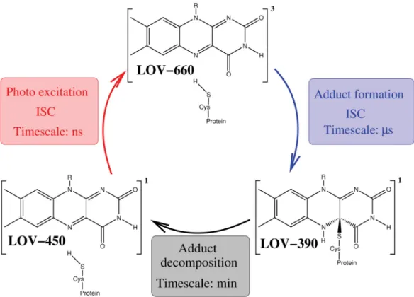
In the dark, the FMN is bound non-covalently. Upon illumination with blue light, a photocycle (see Figure 1.5) is triggered that leads to photobleaching. Schematically this photocycle is shown in Figure 1.5.
Figure 1.4.: Four processes in plants triggered by blue light and regulated by pho- totropin: Stomata and leaf opening, chloroplast relocation and phototropism (left to right).
1.2. Flavins in blue-light photosensors
Figure 1.5.:Photocycle of the LOV domains. In the darkness the chromophore FMN is bound noncovalently in the LOV domain. (LOV-447) Upon photoexcitation the lowest- lying triplet state (LOV-660) is formed via intersystem crossing (ISC). Accompanied by a second ISC an adduct (LOV-390) between the chromophore and a nearby cysteine is formed. For possible reaction pathways see Figure 1.6. LOV-390 usually decomposes on the timescale of minutes and the photocycle can start again.
The primary step after blue-light absorption hereby involves a rapid decay of the ex- cited singlet state population to the lowest excited triplet state on the nanosecond timescale. In the second step an adduct is formed between the C(4a) atom of the isoal- loxazine ring and the sulfur atom of a nearby cysteine residue. This metastable adduct usually decomposes on the timescale of minutes and the photocycle can start again.
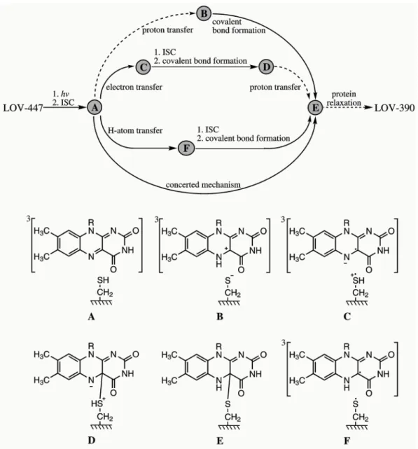
Experimentally the quantum yields for formation of triplet state ΦT and formation of adduct ΦAd are found to depend on the respective LOV domain (for a detailed com- pilation of spectroscopic data see [7] and citations therein). As I will present results of flavin in the LOV domain of the photosensor YtvA from B. subtilis in section 2, I will limit my description to quantum yields of this system. In YtvA the formation of triplet state is measured to be 0.62 and the quantum yield of adduct formation is found to be 0.49. [9] Although time and effort have been directed into the elucidation of the adduct formation, the mechanistic details are still a matter of debate. [43] It has been shown that in the dark the cysteine is protonated. [44] This gives rise to the four possible reaction pathways shown in Figure 1.6.
Figure 1.6.: Suggested reaction pathways (top) and intermediates (bottom) of the pho- toadduct (LOV-390) formation in wild-type LOV domains. A (bottom) is the reactive triplet state LOV-660. Reaction steps that are inhibited at T ≤ 80 K are shown with dashed arrows. Reproduced from Schleicher et al.. [45]
1. As a possible reaction mechanism an initial proton transfer from the cysteine to the N(5) atom of the chromophore converts the S-H to the S− in LOV-660 (Figure 1.6,B). In a second step, the thiolate attacks at the C(4a) atom of FMN and after an ISC the covalent bond is formed. [30]
2. As second reaction mechanism an initial electron transfer from the cysteine to the FMN produces a radical pair consisting of the anionic FMN and the cationic cysteine radicals (Figure 1.6,C). After ISC radical recombination leads to cova- lent bond formation (Figure 1.6, D) and in a final step the proton is transferred to the N(5) atom. [45]
1.2. Flavins in blue-light photosensors 3. In an alternative reaction pathway hydrogen transfer yields a neutral radical (Figure 1.6, F) After ISC radical recombination leads to the formation of the covalent bond. [46]
4. Finally, a concerted mechanism remains as a fourth possibility.
Transient EPR experiments, however, show that at a temperature of 77 K proton transfer to the 3FMN does not occur. [45, 47] Therefore, at a temperature of 77 K reaction pathway 1 can be ruled out which, of course, is different at room temperature.
In addition, FTIR investigations find the designated proton-donor cysteine protonated in the transient triplet state, which rules out pathway 1 and 3. [48, 49] However, the latter reaction mechanism is favored by recent quantum chemical [25,50] and combined quantum mechanics / molecular mechanics (QM/MM) [51] studies. This reflects that, as to date, the protonation state of the transient triplet and its immediate influence on the adduct formation is under discussion.
The absorption spectra of the initial, dark-adapted state of FMN in the LOV domain are very similar for the LOV domains. Besides, absorption spectra of the dark-adapted state of FMN in LOV are almost identical to those of free FMN in water. The first absorption band is centered around 2.76 eV (450 nm), showing a well resolved vibronic structure with peaks at 2.62-2.63 eV (474-471 nm), 2.77-2.79 eV (447-445 nm), and 2.94-2.95 eV (422-420 nm). The second absorption band is centered around 3.31-3.44 eV (375-360 nm), reflecting the proximity of polar amino acids to the heteroatom moiety of the flavin core ring. [9, 30, 52, 53]
2. Project definition and embedding into SFB 663
2.1. Open questions concering LOV domains
In the previous chapter I outlined open questions with regard to the signaling mecha- nism of two flavin conatining photosensors. At the beginning of my work (September 2005) relatively little was known about the blue-light triggered processes in BLUF do- mains, while the photo-cycle of LOV domains (see Figure 1.5) was quite established.
Therefore, the present work focuses on light-induced processes in LOV domains. The following list contains the main questions that were still open at the time:
1. How does the triplet formation occur in flavins and what is the influence of the environment on this process?
2. According to which mechanism does the adduct form?
3. How can cofactor and protein be modified in order to manipulate the photocycle with respect to possible applications?
2.2. Contributions within the framework of SFB 663
Thematically, this work is embedded into the Sonderforschungsbereich (SFB, Col- laborative research center) 663 “Molekulare Antwort nach elektronischer Anregung (Molecular response to electronic excitation)”. Within this collaborative research cen- ter three further research groups are working on flavins, namely the groups of Prof. W.
G¨artner, Prof. Heberle, and Prof. W. Thiel (see below). Additionally, the Deutsche Forschungsgemeinschaft (DFG, German research foundation) funds a Forschergruppe (FOR, Research unit) 526 “Blaulicht-sensitive Photorezeptoren (Blue-light Sensitive Photoreceptors)”, concerned with the understanding of the primary processes of three principle blue-light sensitive photoreceptor domains LOV, Cryptochrome and BLUF.
Since 2002 this research unit has been contributing results in various fields, includ- ing structural determination, spectroscopic characterization, site-directed mutagene- sis and elucidation of biophysical processes (For a complete list of publications see http://www.bluelightphotoreceptors.de). Since it would go beyond the scope of this work to completely summarize the studies of FOR 526, I restrict myself to including only those findings directly related to my work.
2.2.1. Modified flavins in biomimetric environment
The emphasis of project B3 (G¨artner and Heberle) lies on the synthesis and spec- troscopical characterization of structurally and electronically modified flavins (corre- sponding to item 3 in the list above). During the time of my PhD (September 2005 to Mai 2009) the workgroup synthesized the four modified flavins shown in Figure 2.1: 1-deaza-RF (1DRF), 5-deaza-RF (5DRF), 7,8-didemethyl-RF (DMRF) and 8- isopropyl-RF (iprRF). The photochemistry of these compounds was investigated by spectroscopical means (absorption and fluorescence spectra, fluorescence lifetimes, flu- orescence quantum yields, transient triplet-triplet absorption spectra, transient triplet lifetimes and quantum yields of triplet generation) in aqueous solution. In addition, the incooperation of a modified flavin (5DRF) into a recombinant, flavin binding protein (LOV domain of the bacterial photosensor YvtA) was started and yielded promising results. [54] As reported by Hecht et al., [55] with 5DRF as cofactor the LOV pho- tocycle stopped after the adduct was formed. During the experiment the 5DRF-LOV adduct remaind stable for several days and was transferred to the initial state by means of light exposure. The impact of this finding lies in the identification of a dual photo switch with a broad range of potential biological and biotechnological applications.
Figure 2.1.: Chemical structure of the four modified riboflavins synthesized by Mansurova et. al. [56]
2.2.2. Photochemical reactions of FMN in LOV domains
The goal of project C4 (Thiel) is to determine the mechanism of adduct formation (and adduct decomposition) by means of a combined quantum mechanics / molecular mechanics (QM/MM) treatment. In this connection, the relative stability of occuring transition states and the thioadduct, the influence of nearby residues on the mechanism, plus the spectroscopical properties of intermediate species are of interest. The QM/MM investigations have been carried out for phot1-LOV1 of the wild-type protein (PBD codes 1N9L and 1N9N) and the two mutants C57S and C57M, where the reactive cysteine residue is replaced with serine (replacement of SH against OH) and methionine (replacement of SH against CH2-S-CH3), respectively.
2.3. Own contributions
2.3. Own contributions
This work is concerned with understanding the first step in the photocycle of LOV domains, the formation of the reactive flavin triplet species. In order to understand the ISC mechanism in the protein, it is important to obtain a comprehensive knowledge about the relaxation processes that are triggered by blue-light absorption in flavins.
As a starting point the following issues were investigated for free flavins (see Chapter 1, Paper I, Paper II and Paper III):
A What is the nature of low-lying excited states in flavins?
B What is the ISC mechanism?
C Which other relaxation processes compete?
D How are A-C affected when solvent (water) is taken into account?
E How does (de)protonation influence spectroscopical properties?
With this background the investigation was expanded to flavin in the LOV domain.
This was done by means of QM/MM calculations with help from Prof. Thiel and his coworker M. R. Silva-Junior, who provided the setup of the protein, force-field parameters for the FMN and technial support. With respect to the work of Prof.
G¨artner the LOV domain of the bacterial photosensor YvtA was chosen as working system. This investigation laid its main focus on the influence of the LOV domain on the low-lying excited states in flavins and discussed the ISC mechanism of FMN in YtvA-LOV in a qualitative manner (see Chapter 2 and Paper IX).
Along with flavins, I investigated photophysical relaxation processes in various flavin related compounds. The experimental investigation of the electronically modified flavin compounds 1DRF and 5DRF (Prof. G¨artner see 2.2.1) was accompanied by quantum chemical calculations. Additionally, alloxazines which differ from flavins in the ordering of the low-lying excited states, have been studied.
Part II.
Theory and methods
1. Theoretical background
1.1. The Born-Oppenheimer approximation and the Franck-Condon principle
aBorn-Oppenheimer approximation When a molecule absorbs light, the transition of the electron from the initial state to the final state occurs on an ultrafast timescale (≈ 10−15−10−16 s−1). In comparison to the timescale of nuclear motions (≈10−12−10−13 s−1), an electronic transition can be considered to take placeinstantaneously. Hence, in a simple picture, when electrons move the nuclei can be seen as stationary and, on the other hand, when the nuclei rearrange the motion of the electrons can be regarded as simultaneous. This is the basic thought behind the Born-Oppenheimer (BO) approxi- mation, which separates the Schr¨odinger equation of the electrons and the nuclei. With the neglect of nonadiabatic coupling terms, the dependence of the electronic problem on the position of the nuclei is reduced to parametric. This approximation introduces the potent paradigm of potential energy hypersurfaces (PEH). However, as we shall see later, when two PEHs come very close this approximation breaks down.
Franck-Condon principle The Franck-Condon (FC) principle is based on the sepa- ration of nuclear and electronic degrees of freedom as introduced by the BO approxi- mation. In this case, electronic transition between the initial state and the final state is vertical. The probability of a transition between the two states is proportional to the square of the corresponding electronic transition moment matrix element b. The latter is given as:
R=i|M|f (1.1)
where M is a vector, here given in the dipole approximation, which can be resolved into one part depending on the electrons and one depending on the nuclei.
M =Me+Mn (1.2)
The total eigenfunctions of the initial and final state, i| and |f, respectively, can be put (neglecting rotations)
i|=iein|, |f=|fefn. (1.3) Substituting expressions 1.2 and 1.3 into 1.1 leads to:
aThis chapter keeps close to Ref. [57–59].
bThe following formulation keeps very close to the explanations in ’Molecular Spectra and Molecular Structure’ from G. Herzberg. [57]
R=iein|Me|fefn+iein|Mn|fefn (1.4) Mn does not depend on the coordinates of the electrons and the second term in this expression can be written as:
in|Mn|fnie|fe. (1.5) However, as the electronic eigenfunctions, which belong to different electronic states, are orthogonal to one another, Rn is trivially zero due to the following relation:
ie|fe=δif. (1.6)
Therefore, expression 1.4 is reduced to the first term which, taking the implications of the BO approximation into account, can be obtained to:
R=ie|Me|fein|fn. (1.7) The first term in this expression is the electronic transition moment (also denoted μel(i, f) in section 1.2.1). Due to the BO approximation, the electronic eigenfunction and, as a result, this matrix element do not depend directly on the position of the nu- clei. However, a parametric dependence of the electronic eigenfunctions on the nuclear coordinates remains. If the electronic transition takes place instantaneously, i.e. the nuclei do not move during the time of the transition and, if only one nuclear arrange- ment is considered, this matrix element will not change and can be treated as constant.
(In section 1.2.2 we shall see that for intersystem crossing in some cases the description of the transition matrix element has to be extended beyond this zeroth-order term.) With the transition probability and therefore the intensity being proportional to the square of expression 1.7 and in the end to the square of the integral over the product of the vibrational eigenfunctions of the initial and the final state, the so-called Franck- Condon factor (FCF).
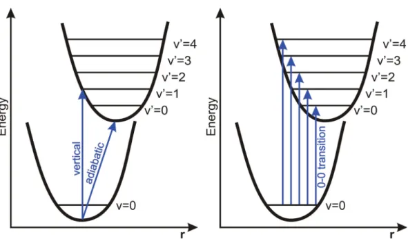
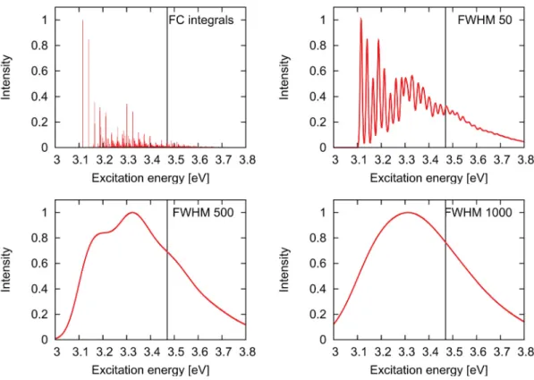
I ≈R2 ≈c· |in|fn|2 (1.8) Observed intensity distributions in absorption bands The FC principle is a pow- erful means in order to understand the shape and intensity distributions of an ab- sorption (emission) band. A schematic illustration of this principle is shown in Figure 1.1. In a simple picture, the transition occurs vertically between the zeroth vibra- tional level of the initial state (Ψi,v = 0) and the vibrational levels of the final state (Ψf,v ={0,1,2, ...}). (See dashed line in Figure 1.1.) In the lowest vibrational level of a given electronic state the probability density distribution for r is large only at the equilibrium nuclear arrangement of this state. In excited vibrational levels this proba- bility distribution becomes larger near the classical turning points until, for high-lying vibrational states, a quasi-classical situation is reached. For the low-lying vibrational states of the final state this situation is illustrated schematically in Figure 1.1.
The shape of the absorption band not only depends on the form of PEHs of initial and final state and the vibrational wave functions, but also on the displacement between
1.1. The Born-Oppenheimer approximation and the Franck-Condon principle
Figure 1.1: Schematic illustration of the Franck-Condon (FC) prin- ciple for the absorption of light.
The electronic transition is repre- sented by the dashed vertical line.
The absorption intensity is pro- portional to the square of the over- lap of the two vibrational wave functions (Franck-Condon factors, see text).
the geometries of the two states. The latter is illustrated in Figure 1.2, where three cases are distinguished.
• A:Equilibrium nuclear arrangements of initial and final state are (almost) iden- tical
In this case the by far largest FCF is obtained for the 0-0 transition and transi- tions to higher-lying vibrational levels are nearly forbidden. In an ideal system, where the nuclear coordinates of initial and final state are exactly identical no other transitions are observable, due to the orthogonality of the vibrational eigen- functions.
• B: Equilibrium nuclear arrangements of initial and final state are similar
In this case the 0-0 transition is not longer the most probable, but still possesses a sizable FCF. Instead, the highest overlap for the eigenfunctions occurs between v = 0 and a low-lying vibrational level (v = 2 in Figure 1.2). In addition, it can be seen that in this case the largest probability distribution does not coincide with the classical turning point.
• C: Equilibrium nuclear arrangements of initial and final state are (very) different In this case the FCF of the 0-0 transition is very small and the most probable transition occurs between v = 0 and a high-lying vibrational level (v = 6 in Figure 1.2). The shape of the resulting absorption band is wide.
Figure 1.2.:Potential curves explaining the intensity distribution in absorption spectra according to the FC principle for three different cases. (The electronic transition is represented by the dashed vertical line.) In addition, the intensity distributions and resulting shape of the absorption band are given. In caseAthe equilibrium geometries of initial and final states are the same. For case B the geometry difference between the two states is small and for case Cit is large. (See text for further details.)
1.2. Photophysical relaxation processes: Molecular response to electronic excitation
1.2. Photophysical relaxation processes: Molecular response to electronic excitation
In comparison to the electronic ground state, the lifetime of electronically excited states is rather short. In general, the absorption of light triggers a multitude of processes that compete for the dissipation of the excess energy in the excited states. One generally dis- tinguishes between photochemical processes, where the electronically excited molecule undergoes a chemical reaction in order to form a new species, and photophysical pro- cesses, which lead to alternative states of the same species, although it is not always straight forward to make such a distinction. As will be seen later, the major part of my work is concerned with photophysical deactivation processes. Therefore, this section outlines the most important photophysical processes and gives an overview over the dominance and relevance of these processes.
It is common practice to outline the competing photophysical processes that occur after light absorption in a so-called Jablonski diagram (also: state energy diagram) as it is depicted in Figure 1.3. In principle, there are three different ways for a molecule to get rid of the excess energy: (1) emission of light, (2) conversion of electronic energy into heat, and (3) dispension of energy to the environment. While in condensed phase all three possibilities are available, isolated molecules in the gas phase, of course, cannot decay via the last process. In the following, processes that belong to 1 and 2 are discussed in more detail.
1.2.1. Luminescence
One way for a molecule to get rid of the excess excitation energy is the emission of light (luminescence). In the Jablonski diagram (Figure 1.3) these processes are indicated as straight arrows. Depending on the initial state, luminescence may correspond to spin- allowed (fluorescence) and spin-forbidden (phosphorescence) transitions, respectively.
Originally the distinction between the two processes was defined on the basis of the
Figure 1.3: Jablonski diagram: Absorp- tion (A) and emission processes are in- dicated by straight arrows (F: fluores- cence, P: phosphorescence). Radiation- less processes are symbolized by wavy arrows (IC: internal conversion, ISC:
intersystem crossing, VR: vibrational relaxation, IVR: intramolecular vibra- tional energy relaxation (not shown)).
lifetime of the radiation. In fluorescence, the radiation ceased as soon as the exciting radiation was removed. In phosphorescence, however, the luminescence continues for a short time. [60] Although this paradigm is out-dated, it throws some light on the long lifetime and the associated reactivity of the first excited triplet state.
The radiative rate constant depends on the transition dipole momentμel(i, f) between the initial state i and the final state f and the vertical energy gap ΔEi,f between the two states. It can be estimated with the following expression [61] c
krad(i→f) = 4e2
3c34 ·ΔEi,f3 ·μel(i, f)2 (1.9) For fluorescence, the determination of the transition dipole moment is accessible in the framework of non-relativistic theory. For phosphorescence this is different. In a non- relativistic treatment, these transitions are spin-forbidden. In a relativistic treatment, however, it is not so. In a simple picture, spin-orbit coupling mixes singlet and triplet states, which in turn results in non-vanishing transition dipole moment between singlet and triplet states.
Figure 1.4: Schematic illustration of the Stokes shift. If the 0-0 tran- sition of absorption (A) and emis- sion (F, fluorescence) do not coin- cide, this is referred to as anoma- lous Stokes’ shift. (See text)
The shape of the emission bands are determined in the same way as those of the absorption bands. For most molecules the equilibrium geometry of the ground state and the excited state are different. This results in a red shift of the emission maximum with respect to the absorption maximum, which in case of fluorescence, is denoted as Stokes’ shift. Schematically this is shown in Figure 1.4. In condensed phase, various intermolecular interactions in the ground and excited states cause a separation of the 0- 0 transitions of absorption and emission. This phenomenon is referred to asanomalous Stokes’ shift and can be seen as a measure of the interaction of solvent and molecule.
1.2.2. Non-radiative processes
In the Jablonski diagram (Figure 1.3) the radiationless processes are symbolized by wavy arrows. In processes, such as internal conversion (IC) and intersystem crossing (ISC), the transition between the two states occurs isoenergetically, resulting in a vibrationally excited (hot) final state. While IC is a transition between states of the same multiplicity, ISC occurs between states of different multiplicity.
The rate of radiationless transitions between different states is particularly large, when the adiabatic energy separation between the two involved states is rather small. This
cExpressing the rate constants in s−1, ΔEi,fin cm−1, andμel(i, f) in atomic units (ea0) the numerical value of the prefactor becomes 2.0261×10⁻⁶.
1.2. Photophysical relaxation processes: Molecular response to electronic excitation finding holds importance especially for the rate of IC processes. Usually, Sn S1 and Tn T1 ICs are substantially faster than S1 S0 IC, since the adiabatic energy differences among the electronically excited states are much smaller than between the S1 and the S0 state. Experimental observations show an exponential dependence of the S1 S0 IC rate constant on the energy gap between the two states. Commonly, this relation is referred to as the energy-gap law. [62] One consequence of the fast Sn S1 and Tn T1 ICs in the vast majority of molecules is that luminescence is observed only from the lowest excited-state PEH. (Kasha’s rule)
Within the framework of the Born-Oppenheimer approximation, radiationless transi- tions between two PEHs are impossible. In order to describe those transitions it is nec- essary to go beyond the Born-Oppenheimer approximation. The interaction between different electronic states of the molecule arises through vibrational motion and has to be included in order to be able to describe those transitions. Using time-dependent per- turbation theory, the rate constant kN R for a radiationless transition starting from an initial single level|i toward a manifold of final levels{|f}is given by the well-known Fermi Golden Rule expression [63]
kN R(|i{|f}) = 2π
{|f}
|i|Hˆ|f|2δ(Ei0−Ef0). (1.10)
Hˆ is the perturbation operator. In case of internal conversion this operator is the kinetic energy operator of the nuclei. In case of intersystem crossing, spin-orbit inter- action couples the two states.
The consequences of direct and vibronic spin-orbit coupling on intersystem cross- ing dIn order to obtain an ISC rate constant from Fermi’s Golden Rule approximation several modifications have to be done. According to Toniolo and Persico [65, 66], it is possible to approximate expression 1.10 by a summation over rates of transitions in an energy interval of width 2η around the energy of the zeroth level of the initial state (Ei,v=0). (Here, the vectors v and v represent sets of vibrational quantum numbers in all normal modes of the initial (i) and final (f) electronic state, respectively.) If we denote the coupling matrix elements driving the radiationless transition by HvSO=0,v, the rate constant is obtained as
kISC(if) = 2π η
|Ef,v−Ei,v=0|<η
HvSO=0,v2 . (1.11)
HvSO=0,v can be expanded in a Taylor series in the variables{qκ}, the normal coordinates, around some reference pointq0[67] which we have chosen to coincide with the minimum
dThis brief overview is kept very close to the work of Tatchenet al.[64]
of the initial state.
HvSO=0,v =
iHˆSOf
q0=0v=0|v
+
κ
∂
∂qκ i|HˆSO|f q0=0
v=0|qκ|v
+O
|q|2 (1.12)
The first term on the right-hand side of equation (1.12) is a purely electronic matrix element and is denominated direct spin-orbit coupling or Condon approximation in the following, whereas the term in the second line of equation (1.12) represents the first- order derivative coupling and is named vibronic spin-orbit coupling or Herzberg-Teller type coupling. (For more background on this deviation see [64] and [68].) In section 1.1 we have approximated the transition matrix element ie|Me|fe to be constant, which is equivalent with the Condon approximation. However, this approximation is only justified, when the change of the matrix element is negligible with respect to the variation of the nuclear coordinates. In cases, where this prerequisite is not fulfilled the Condon approximation is not sufficient and the contribution of the first order term of expression 1.12 will be substantial.
The implications of direct spin-orbit coupling are also known as El-Sayed’s rule. [69]
According to this rule of thumb, spin-orbit coupling matrix elements (SOME) between (ππ∗) and (nπ∗) states are in general much larger than SOME between two (ππ∗) states.
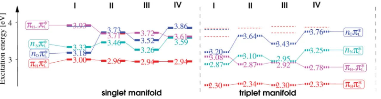
Thus, when only direct spin-orbit coupling is considered (Condon approximation), ISC rate constants are much slower for (ππ∗) (ππ∗) processes than for (ππ∗) (nπ∗) processes. In case of vibronic spin-orbit coupling, out-of-plane distortions along the vibrational normal modes yield an interaction and thus a mixing of (ππ∗) and (nπ∗) states. Qualitatively the resulting enhancement of the ISC rate constants between two (ππ∗) states can be seen as an intensity borrowing from the much faster (ππ∗) (nπ∗)/(nπ∗) (ππ∗) processes. As recent studies have shown, this ISC mechanism is common in various chromophores, including flavins (See Paper I and Paper III for details).e [64, 70]
Figure 1.5: Conical intersection of two potential energy hypersur- faces. Left: The dependence of the two PEHs on two nuclear de- grees of freedom. Right: Mag- nification of the conical intersec- tion. (Source: J¨org Tatchen.)
eHow the choice of the parameters, e.g. number of accepting and coupling modes, width of the search interval,... influences the ISC rate constants is explaind in Paper I, Paper III, and Paper IV and the respective Supplementary information.
1.2. Photophysical relaxation processes: Molecular response to electronic excitation Conical intersections In the last ten to twenty years, the so-called conical intersec- tions (ConIn) have been accepted to be efficient funnels for the decay of electronically excited molecules.f At a ConIn, two PEHs are degenerate, which is illustrated in figure 1.5 for two reaction coordinates. In general, such a ConIn point is part of a subspace of ConIn points, usually denoted intersection space or seam of the dimension 3N-8 (N is the number of atoms of the system). In order to provide an ultrafast deexcitation of the excited state population, not only the presence, but also the energetic accessibility of a low-lying ConIn from the FC region is crucial.
IC, ISC and absorption of energy (i.e. a photon) create vibrationally hot states of a molecule. In condensed phase, the excess vibrational energy can be transferred to surrounding solvent molecules in order to reach the vibrational ground state. g This process is called vibrational relaxation (VR) and is, of course, not available in vacuum.
A process that is able to relax a vibrationally hot state even in isolated molecules is intramolecular vibrational energy relaxation (IVR). This process redistributes the en- ergy initially localized in a given vibrational mode of the molecule among the remaining vibrational modes. At least 5-10 atoms are required for IVR to occur. [72]
1.2.3. Which process prevails?
The question which process dominates the deexcitation of the molecules, depends pre- dominantly on the electronic structure of the chromophore. A useful measure for the contribution of photophysical and photochemical processes to the depopulation of the excited state is the quantum yield Φ. The quantum yield of a process is defined as the ratio between the number of molecules that undergo this process and the number of light quanta absorbed. If the rate constants of all competing relaxation processes are known, the quantum yield of a process P can be calculated according to:
ΦP =ηabs· kP
k (1.13)
withηabsbeing the efficiency of the absorption. In addition, when the quantum yield of a process is known it is possible to obtain the naturallifetime from the experimentally accessible observed lifetime of a process with the following relation.
τobserved = Φ·τnatural (1.14)
In the absence of competing deactivation processesnaturalandobserved lifetime are the same, of course. The rate constant of a given process P is the reciprocal of the natural lifetime and compares to theoretically determined rate constants. However, when a process is not the dominant deexcitation mechanism, the natural and the observed lifetime can differ by several orders of magnitude. A very prominent example for this variance is the fluorescence in the nucleobases. Although the natural fluorescence
fFor an elaborate introduction to conical intersections see Ref. [71]. The part about conical inter- section shown here is very close to this reference.
gNote: More accurately, thermal equilibrium is reached, since the occupation of the vibrational levels is temperature dependent according to Boltzmann’s law.
lifetime in DNA and RNA bases is several nanoseconds, the low fluorescence quantum yield results in an observed fluorescence lifetime (often denoted as S1 lifetime) ≤ 1 ps. [1]

![Figure 1.3.: Scheme for the possible orientations of glutamine (Q63) and its hydrogen- hydrogen-bonding interactions with the flavin chromophore and the nearby tyrosine (Y21) residue in the BLUF domain (AppA-BLUF, 1YRX [39])](https://thumb-eu.123doks.com/thumbv2/1library_info/4533130.1596403/26.892.118.759.126.402/possible-orientations-glutamine-hydrogen-hydrogen-interactions-chromophore-tyrosine.webp)