Synthesis and Development of a Mucin Glycopeptide Microarray System for Evaluation of Protein-
Interactions
D
ISSERTATIONZur Erlangung des akademischen Grades eines Doktors der Naturwissenschaften
(Dr. rer. nat.)
der Fakultät Chemie und Chemische Biologie der Technischen Universität Dortmund.
Vorgelegt von Diplom Chemiker
CHRISTIAN PETT
aus Mainz.
Dortmund 2016
Die vorliegende Arbeit entstand im Zeitraum von Oktober 2009 bis September 2015 unter Anleitung von Dr. Ulrika Westerlind an der Fakultät Chemie und Chemische Biologie der Technischen Universität Dortmund und dem Leibniz-Institut für analytische Wissenschaften ISAS -e.V-, Dortmund.
Dekanin: Prof. Dr. Insa Melle
Erster Gutachter: Prof. Dr. Herbert Waldmann Zweiter Gutachter: Prof. Dr. Christian Hedberg
Teile dieser Arbeit wurden bereits in folgenden Beiträgen veröffentlicht:
A Unified Strategy for the Synthesis of Mucin Cores 1-4 and the Assembled Multivalent Glycopeptides
C. Pett, M. Schorlemer, U. Westerlind, Chem. Eur. J. 2013, 19, 17001-17010.
A convergent Strategy for the Synthesis of Type-1 Elongated Mucin Cores 1-3 and the Corresponding Glycopeptides
C. Pett, U. Westerlind, Chem Eur. J. 2014, 20, 7287-7299 (inkl. Back Cover der Ausgabe).
Weitere Beteiligung an folgenden Beiträgen:
Assignment of Saccharide Identities through Analysis of Oxonium Ion Fragmentation Profiles in LC-MS/MS of Glycopeptides
A. Halim, U. Westerlind, C. Pett, M. Schorlemer, U. Rüetschi, G. Brinkmalm, C. Sihlbom, J.
Lengquist, G. Larson, J. Nilsson, J. Proteome Res. 2014, 13, 6024-6032.
Distinctive MS/MS Fragmentation Pathway of Glycopeptide Generated Oxonium Ions Provide Evidence of the Glycan Structure
J. Yu, M. Schorlemer, A. Gomez Toledo, C. Pett, C. Sihlbom, G. Larson, U. Westerlind, J.
Nilsson, Chem. Eur. J. 2015, 22, 1114-1124.
FÜR MARTHA
1 Introduction ... 1
1.1 The mucin glycoprotein family ... 1
1.2 Abnormal glycosylation and the role of MUC1 in cancer ... 5
1.3 Galectins and the role of galectin-3 in cancer ... 8
1.4 MUC1 carbohydrate vaccines ...11
1.5 Mucins in airway diseases ...15
2 Motivation ... 16
3 Synthetic strategies ... 20
3.1 Glycopeptide synthesis strategies ...20
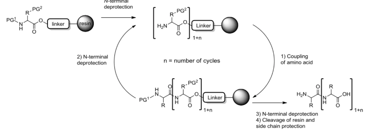
3.1.1 Glycopeptide synthesis on solid phase ...21
3.1.2 Side reactions in solid phase glycopeptide synthesis ...26
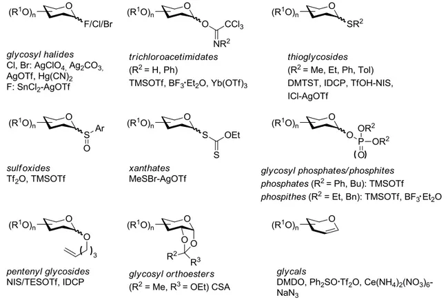
3.2 Strategies in carbohydrate synthesis ...29
3.2.1 Glycosylation methods ...29
3.2.2 Protecting group chemistry ...31
4 Results and discussion ... 34
4.1 Part 1 – Synthesis of core 1, 2 and 3 glycosylated amino acids ...34
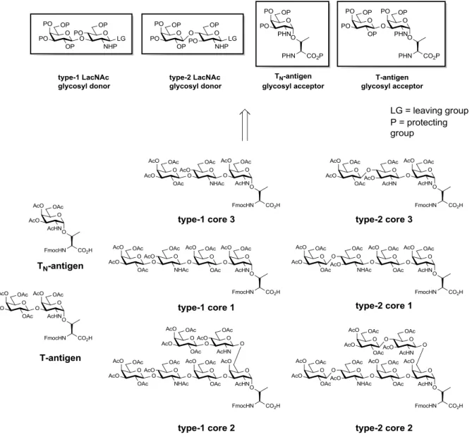
4.1.1 Retrosynthetic considerations for glycosyl amino acid building block synthesis .. ...34
4.1.2 Examples for syntheses of glycosylated amino acids/glycopeptides in the field and comparison to the strategy of this work ...37
4.1.3 Synthesis of the TN-antigen glycosyl acceptor ...38
4.1.4 Synthesis of the T-antigen amino acid ...41
4.1.5 Synthesis of the type-1 N-acetyllactosamine glycosyl donor ...42
4.1.6 Synthesis of the type-2 N-acetyllactosamine glycosyl donor ...48
4.1.7 Synthesis of the galactose extended core 3 amino acids ...49
4.1.8 Synthesis of the extended core 1 tetrasaccharide amino acids ...53
4.1.9 Synthesis of the extended core 2 hexasaccharide amino acids ...57
4.2 Part 2 - Syntheses of mucin glycopeptide libraries ...59
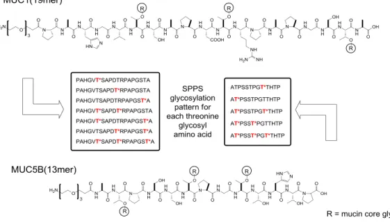
4.2.1 Synthesis plan for MUC1 and MUC5B glycopeptide library build-up ...59
4.2.2 Synthesis of a triethlyene glycol linker ...60
4.2.3 Synthesis of a MUC1 glycopeptide library ...61
4.2.4 Synthesis of a MUC5B glycopeptide library ...70
4.2.5 Synthesis of sialylated MUC1 glycopeptides ...74
4.2.6 Tandem MS of sialylated mucin O-glycopeptides - oxonium ion pattern analysis ...82
4.3 Part 3 – Microarray studies with glycopeptide binding proteins ...91
4.3.1 Introduction on microarray development ...91
4.3.2 Immobilization methods for glycopeptide microarrays ...92
4.3.3 Principle of serum screening by glycopeptide microarrays ...94
4.3.4 Evaluation of mucin anti-tumor vaccines by glycopeptide microarray analysis of induced antibody responses ...95
4.3.5 Vaccine candidates for induction of immune response and antisera generation in mice...97
4.3.6 Utilized microarray platforms for antisera, plant lectin and galectin-3 screening . ...99
4.3.7 Antibody titer determination of antisera induced by MUC1 vaccine candidates .. ... 100
4.3.8 Elucidation of antibody specificity from vaccination experiments using the MUC1 glycopeptide microarray ... 102
4.3.9 Elucidation of antibody binding specificity towards sialylated MUC1 glycopeptides on a microarray ... 126
4.3.10 Discussion of MUC1 serum antibody specificity elucidation ... 128
4.3.11 Binding of plant lectins to MUC1 glycopeptides on a microarray ... 136
4.3.12 Binding of galectin-3 to MUC1 glycopeptides on a microarray ... 141
5 Summary ... 151
6 Zusammenfassung ... 164
7 Experimental ... 178
7.1 Syntheses of chapter 4.1 ... 178
7.1.1 General ... 178
7.1.2 Synthesis of the TN-antigen glycosyl acceptor ... 180
7.1.3 Synthesis of the T-antigen amino acid ... 187
7.1.4 Syntheses of the type-1 N-acetyllactosamine glycosyl donor ... 191
7.1.5 Syntheses of the type-2 N-acetyllactosamine glycosyl donor ... 200
7.1.6 Synthesis of the extended type-2 core 3 amino acid... 203
7.1.7 Synthesis of the extended type-1 core 3 amino acid... 209
7.1.8 Synthesis of the T-antigen glycosyl acceptor building block ... 215
7.1.9 Synthesis of the extended type-1 core 1 amino acid... 220
7.1.10 Synthesis of the extended type-2 core 1 amino ... 227
7.1.11 Synthesis of the extended type-1 core 2 amino acid... 235
7.1.12 Synthesis of the extended type-2core 2 amino acid ... 241
7.2 Syntheses of chapter 4.2 ... 248
7.2.1 General ... 248
7.2.2 Synthesis of the triethylene glycol spacer amino acid ... 248
7.2.3 General protocol for MUC1 and MUC5B solid phase glycopeptide synthesis ... ... 252
7.2.4 Synthesis of MUC1 glycopeptides ... 253
7.2.5 Synthesis of MUC5B glycopeptides ... 266
7.2.6 Synthesis of α2,3- and α2,6-sialylated MUC1 glycopeptides ... 274
7.3 Microarray experiments with immobilized MUC1 glycopeptides... 283
7.3.1 General ... 283
7.3.2 Nexterion slide H microarray loading capacity ... 284
7.3.3 General spotting conditions ... 285
7.3.4 Spotting of MUC1 microarrays MA1, MA2, MA3, MA4, MA5 ... 286
7.3.5 General protocol for microarray assays with antiserum or lectin samples ... 293
8 References ... Fehler! Textmarke nicht definiert. 9 Appendix ... 316
9.1 Spectroscopic data ... 316
9.2 HPLC chromatograms ... 334
9.2.1 MUC1 glycopeptides ... 334
9.2.2 MUC5B glycopeptides ... 338
9.2.3 Sialylated MUC1 glycopeptides ... 341
9.3 Microarray data ... 344
9.3.1 Microarray format 1 (MA1)... 344
9.3.2 Microarray format 2 (MA2)... 345
9.3.3 Microarray format 3 (MA3)... 353
9.3.4 Microarray format 4 (MA4)... 355
9.3.5 Microarray format 5 (MA5)... 357
Acknowledgements ... 359
ABBREVIATIONS
aa amino acid
AAA amino acid analysis
Ac acetyl
APC antigen presenting cell arom aromatic
AuNP gold nanoparticle
AW acid washed
BCR B-cell receptor Boc tert-butyloxycarbonyl BSA bovine serum albumin
Bu butyl
Bn benzyl
Bz benzoyl
c concentration
calc. calculated cat. catalytic
CD cluster of differentiation
CHex CID CMP
cyclohexane
collision induced dissociation cytidine-5´-monophospho- CFG Consortium for Functional
Glycomics
Cosmc core 1 β3-Gal-T-specific molecular chaperone COSY correlated spectroscopy CRD carbohydrate recognition
domain
CTL cytotoxic T-lymphocyte
d duplet or day
DBU 1,8-diazabicylo[5.4.0]undec-7- ene
DC dendritic cell
DCC N,N'-dicyclohexylcarbodiimide DCM dichloromethane
dd duplet of duplet
dhb 2,5-dihydroxy benzoic acid DIC N,N'-diisopropylcarbodiimide DIPEA diisopropylethylamine
DMAP 4-(dimethylamino)pyridine DMF dimethylformamide
DMSO dimethyl sulfoxide
DMTST dimethyl(methylthio)sulfonium trifluoromethanesulfate DNA deoxyribonucleic acid DPS diphenyl sulfoxide
EDC N-ethyl-Nꞌ-(dimethyaminopropyl)- carbodiimide
EGRF epidermal growth factor receptor ELISA enzyme-linked immunosorbent
assay
Em emission
eq equivalents
ESI electrospray ionization
Et ethyl
Ex excitation
FA formic acid
FAC frontal affinity chromatography Fmoc N-(9H-fluoren-9-yl)-
methoxycarbonyl Fuc L-fucose
Gal D-galactose
GalNAc N-acetyl-D-galactosamine
gg gauche-gauche
Glc D-glucose
GlcNAc N-acetyl-D-glucosamine grad gradient
gt gauche-trans
GTP guanosine-5'-triphosphate
h hour
HATU O-(1H-7-azabenzotriazol-1-yl)- 1,1,3,3-tetramethylunronium hexafluorophosphate
HBTU
HCD
O-(1H-benzotriazol-1-yl)-1,1,3,3- tetramethylunronium
hexafluorophosphate
Higher energy C-trap dissociation HER2 =
ERBB2
human epidermal growth factor receptor 2
HMBC heteronuclear multiple bond correlation
HMFG human milk fat globule
HOAt 7-aza-1-hydroxybenzotriazole
VI ABBREVIATIONS
HOBt 1-hydroxybenzotriazole HPLC high performance liquid
chromatography HR high resolution
HSQC heteronuclear single quantum coherence
Hz hertz
IDCP iodonium dicollidine perchlorate Ig immunoglobulin
IL interleukin iPr isopropyl
ITC isothermal titration calorimetry J coupling constant
KLH Keyhole Limpet Hemocyanin
Lac lactose
LacNAc N-acetyllactosamine Lex/Lea Lewis x/Lewis a
LG leaving group
m multiplet
M molarity or mega
mAb monoclonal antibody
MALDI matrix assisted laser desorption ionization
Man D-mannose
mbar millibar
MD molecular dynamics
Me methyl
MGL macrophage c-type lectin MHC major histocompatibility
complex
min minutes
miRNA micro ribonucleic acid MPL monophosphoryl lipid A mRNA messenger ribonucleic acid MS molecular sieve or mass
spectrometry NBS
NCE
N-bromosuccinimide normalized collision energy Neu5Ac N-acetylneuraminic acid NHS N-hydroxysuccinimide NIS N-iodosuccinimide NMP N-methylpyrrolidone
NMR nuclear magnetic resonance NSCLC non-small cell lung cancer
OTf trifluoromethanesulfonate (triflate)
p para
Pam3CSK4 N-palmitoyl-s-[2,3-
bis(palmitoyloxy)-(2RS)-propyl- [R]-cysteinyl-[S]-seryl-[S]-lysyl-[S]- lysyl-[S]-lysyl-[S]-lysyl-
Pbf 2,2,4,6,7-
pentamethyldihydrobenzofuran-5- sulfonyl
PBS/PBST phosphate buffered saline/PBS+Tween-20 PCR polymerase chain reaction PEG polyethylene glycol
PG protecting group
Ph phenyl
pH potentia hydrogenii Phth phthalimido
PMP para-methoxy phenyl
pos positive
PNGase Peptide N-glycosidase ppGalNAcT polypeptide N-
acetylgalactosamine transferase ppm parts per million
PS polystyrene
PSGL-1 P-selectin glycoprotein ligand-1 p-TsOH para-toluenesulfonic acid PyBOP (benzotriazol-1-
yloxy)tris(pyrrolidino)phosphonium hexafluorophoshate
q quartet
quart quaternary
R residue
Ras rat sarcoma Rf retention factor RNA ribonucleic acid
RP reverse phase
Rt retention time
RTK receptor tyrosine kinase
s singlet
SEA sea urchin sperm protein, enterokinase and agrin sLex/sLea sialyl-Lewis x/sialyl-Lewis a SNP single nucleotide polymorphism SPE solid phase extraction
SPPS solid phase peptide synthesis
ST sialyl-Thomsen-Friedenreich antigen
STD saturation transfer difference STN sialyl-Thomsen Nouveau antigen
Su succinimide
t triplet
T/TF Thomsen-Friedenreich antigen TACA tumor associated carbohydrate
antigen
TBAF tert-butylammonium fluoride TBS tert-butyldimethylsilyl
TBTU O-(1H-benzotriazol-1-yl)-1,1,3,3- tetramethylunronium
tetrafluoroborate TCR T-cell receptor TEG triethylene glycol tert tertiary
TES triethylsilyl
TFA trifluoroacetic acid
TfOH Trifluoromethanesulfonic acid
tg trans-gauche
THF tetrahydrofuran TIPS triisopropylsilane
TLC thin layer chromatography TLR Toll-like receptor
TMS tetramethylsilyl
TN Thomsen-Nouveau antigen TOCSY total correlation spectroscopy TOF time of flight
Troc trichloroethoxycarbonyl Trt trityl
TT tetanus toxoid UV ultraviolet
VEGF vascular endothelial growth factor VNTR variable number of tandem
repeats
α specific optical rotation
δ chemical shift
λ wavelength
Amino acid codes Ala, A Alanine Arg, R Arginine Asn, N Asparagine Asp, D Aspartate Cys, C Cysteine Gln, Q Glutamine Glu, E Glutamate Gly, G Glycine His, H Histidine Ile, I Isoleucine Leu, L Leucine Lys, K Lysine Met, M Methionine Phe, F Phenylalanine Pro, P Proline Ser, S Serine Thr, T Threonine Trp, W Tryptophan Tyr, Y Tyrosine Val, V Valine
1 INTRODUCTION
1.1 The mucin glycoprotein family
Mucins belong to a family of highly glycosylated proteins expressed by epithelial cells.1 Up to now, 21 members of this family have been identified. The carbohydrate-content of the mucins is usually higher than 50 wt% of the protein. These glycoproteins are a major part of the innate immune system. Secreted mucins form the mucus layer that protects the epithelial tissues of the gastrointestinal and respiratory tract and the ductal surfaces of breast, pancreas and kidney tissue against invading pathogens and chemical or physical stress factors.2 While most mucins are restricted to specific tissues, the MUC1 glycoprotein is ubiquitously distributed.3 MUC1 was first identified by murine monoclonal antibodies raised against human milk fat globule (HMFG).4 Mucins can be subdivided into two major classes, the secreted or gel-forming mucins and the membrane-bound mucins. The ubiquitous MUC1 belongs to the class of the membrane tethered mucins. It is embedded into the membrane lipid bilayer by a single trans-membrane domain. The N-terminal ectodomain has a rod-like structure and stretches out 200-500 nm into the extracellular space, significantly surpassing the thickness of the glycocalyx (ca. 10 nm).5 The short transmembrane domain connects to the 72 amino acids cytoplasmatic domain, which is involved in cell signaling processes.6,7 The MUC5B glycoprotein is belongs to the class of the secreted mucins and is mainly expressed in the respiratory system. Together with MUC5AC, it is the major airway mucin.
Most secreted mucins have cysteine-rich regions, enabling formation of disulfide bridges and thus protein networks.8 Almost all mucins have an extracellular domain rich in proline, serine and threonine and arranged in domains called VNTR-regions (variable number of tandem repeats). In MUC1, a VNTR of 20 amino acids with the sequence PAHGVTSAPDTRPSAPGSTAP is repeated 20-125 times, in MUC5B a VNTR region consisting of 29 amino acids with the sequence ATGSTATPSSTPGTTHTPPVLTTTRTTPT is repeated in total 11 times.9,10 The overall number of VNTRs in MUC1 depends on genetic polymorphism and is different in each individual. A MUC1 tandem repeat contains five glycosylation sites (3x threonine, 2x serine) and the high proline content disturbs the formation of secondary structures, giving MUC1 a stretched, rod-like appearance.11,12 MUC1 is expressed as a single precursor protein with a proteolytic cleavage site within a SEA (sea urchin sperm protein, enterokinase and agrin) domain between the ecto- and transmembrane
2 1INTRODUCTION
domain. This site is auto-catalytically cleaved during posttranslational processing and the extracellular domain is then non-covalently associated with the transmembrane domain.13 All O-glycosylated proteins are posttranslational modified during the passage through the golgi complex. Here, the carbohydrate decoration is added by the sequential addition of monosaccharides which requires an ensemble of various different glycosyltransferases. After the exocytotic transport to the cell membrane, MUC1 is internalized and transported back to the trans-golgi with subsequent re-localization to the cell membrane. During this cyclic process, the glycosylation pattern is further matured.14
Mucin-like O-glycosylation is commonly found on threonine and serine amino acid residues.
Recently, tyrosine was identified as a mucin-type glycosylation site, however, only a few examples of glycosylated proteins with this new posttranslational modification were found until now.15,16 Mucin-type glycosylation is characterized by eight core structures, namely core 1 to core 8.17 All cores structures have in common, that the first added carbohydrate is N-acetylgalactosamine in α-configuration, which is attached by a family of polypeptide N- acetylgalactosaminyltransferases (ppGalNAcT). This structure alone is known as the TN- antigen (Thomsen-Nouveau antigen). Further elongation by the action of a β1,3-galactose transferase (C1GalT, T-synthase), results in the basic core 1 motif, also named T-antigen (or TF for Thomsen-Friedenreich antigen). Instead of a galactose, a glucosamine can be attached by the core 3 N-acetylglucosaminyltransferase (C3GnT) resulting in the core 3 glycan. Core 3 structures are specifically found on mucins of the gastrointestinal tract.18 Elongation on the 6-position of the first N-acetylgalactosamine of core 1 or core 3 by different members of the core 2 N-acetylglucosaminyltransferase family (C2GnT) results in the core 2 and core 4 glycans, respectively. Core 1 to core 4 are the most abundant core motifs, while core 5 to 8 are very rare and only found in a few mucins. The genes encoding for the glycosyltransferases involved in core 7 and core 8 biosynthesis have not been identified yet (figure 1.1).
Figure 1.1: Structures of the common O-glycan core motifs.
The O-glycan core structures can be elongated by the alternating action of a β1,3-GlcNAc- transferase (β1,3GlcNAcT) and by either a β1,3- or β1,4-Gal-transferase (β1,3GalT, β1,4GalT) (figure 1.2).
4 1INTRODUCTION
Figure 1.2: Biosynthesis of mucin type O-glycosylation.
Like this, the core structures are elongated either by a Galβ1,3-GlcNAcβ (type-1 N- acetyllactosamine, LacNAc) or by a Galβ1,4-GlcNAcβ (type-2 LacNAc) disaccharide unit (figure 1.2).17 Chain termination may occur by the performance of a family of six α2,3- sialyltransferases (ST3Gal-I to -VI), which attach a sialic acid (e.g. N-acetylneuraminic acid, Neu5Ac) to the terminal galactose of the carbohydrate chains. Sialylation may also occur by a α2,6-linkage on a terminal Gal or the initial αGalNAc of the TN- or T-antigen. Terminal α2,6- sialylation on galactose is mainly performed on N-glycans by a family of α2,6-galactose sialyltransferases (ST6Gal-I and -II), while O-glycans are mainly sialylated on the initial αGalNAc by a family of α2,6-N-acetylgalactosamine sialyltransferases (ST6GalNAc-I to -VI).17,19 Many glycosyltransferases do not recognize sialylated glycans, and therefore further
chain growth is prevented. An exception is the α1,3/4-fucosyltransferases (FUT3) which adds a fucose monosaccharide, either to the 3- or the 4-position of the GlcNAc of the corresponding type-1 and type-2 LacNAc disaccharide units, even when sialylated. This results in the formation of the Lewisx (Lex) and the Lewisa (Lea) antigens or the corresponding sialyl-Lewisx (sLex) and sialyl-Lewisa (sLea) glycans. Sialylated Le antigens are important motifs, involved in leukocyte recruitment and emigration during inflammations.20 The carbohydrates may also be terminated by sulfation (figure 1.2). The number of carbohydrates per O-glycan chain on mucins on healthy cells, usually varies between 2 and 20 monosaccharides.21
1.2 Abnormal glycosylation and the role of MUC1 in cancer
In epithelial carcinomas, expression of MUC1 undergoes a number of fundamental changes.
The expression of the MUC1 glycoprotein is dramatically increased and the epithelial cell polarity is lost.22 Usually, MUC1 and other membrane-bound mucins are expressed only on the apical site of the cells, which is exposed to the ductal environment. However, in carcinoma cells, MUC1 is found also on the lateral and basal sites of the cells. In cases of cellular stress, e.g. in acute or chronic inflammatory insults, the cell polarity may further be lost. Cells can perform a repair program to regain polarity as soon as the stress response is terminated.23 In cancer however, the stress response is irreversible. During these processes, the cytoplasmatic domain of the dislocated MUC1 can now interact with receptor tyrosine kinases (RTKs) that are located on the basal cell membranes. For instance, MUC1 interacts with the RTK ERBB2 (HER2) and its activation is associated with the loss of tight junctions24 between the cells.25 Also, MUC1 binds β-catenin, which is usually bridging between actin filaments and the cell-cell-adherence molecule E-cadherin. As a result, adherence junctions between the cells are lost.26 Overexpression of the rod-shaped MUC1 extracellular domain also masks the cell surface, resulting in destabilization of integrin-mediated adhesion of the cell with the extracellular matrix.27,28 These factors promote a separation of tumor cells from normal cell clusters and trigger metastasis.
Additionally to overexpression of the MUC1 glycoprotein, the glycan structures attached to the mucin polypeptide tandem repeats are altered due to changes in the expression levels of various glycosyltransferases. These aberrantly structured glycans are referred to as TACAs (tumor associated carbohydrate antigens). In mammary cancer, the levels of C2GnT1 are downregulated or even completely obliterated.29 The branching to core 2 glycan motifs is therefore inhibited and core 1 structure formation is favored instead. Concomitantly, an
6 1INTRODUCTION
overexpression of sialyltransferases results in premature sialylation of the truncated carbohydrates.30,31,32,33 After sialylation further glycosylation is prevented. As a result, tumor cell glycosylation consists of short and often sialylated glycans. TN- and T-antigens and the corresponding sialylated STN- and ST-antigens are the most prominent glycans on cancer- associated MUC1 (figure 1.3).
Figure 1.3: Structures of tumor-associated carbohydrate antigens (TACAs) on mucins.
TACAs were found on several human and murine breast cancer cell lines, on tissue samples of breast cancer patients and as well as on shed or secreted MUC1 in the serum of advanced breast cancer patients.30,34 Interestingly, expression of sLex epitopes, which are a ligands for E-selectins, also occurs in breast cancer.35 For normal O-glycosylation in the gastric, colorectal, pancreatic and hepatic tissue, the expression of core 3 β1,3- N-acetylglucosaminyltransferase (C3GnT) is pivotal. Disturbed expression of this enzyme consequently results in higher levels of TN-antigen instead, which is correlated to cancer growth and susceptibility for metastasis.36,37,38
The reasons for the aberrant levels of glycosyltransferase expression are not fully understood. The increased levels of TN- and STN-antigen are related to a genetic mutation in the gene Cosmc (core 1 β3-Gal-T-specific molecular chaperone).39,40,41 This gene encodes for the chaperone Cosmc, required for the correct folding of the C1GnT (β1,3GalT, T- synthase). A lack of this chaperone leads to C1GnT aggregation and subsequent degradation in the proteasome. Consequently, appearance of TN-antigens increases because of faulty core 1 glycan elongation.40 Increased levels of ST3Gal-I in cancer cells could be related to hypoxia, which also influences expression levels of various other glycosyltransferases.42 In colon cancers, increased T-antigen and sLex and sLea glycosylation is not only related to glycosyltransferase expression rates. Rather higher transcription of the UDP-Gal transporter gene leads to increased amounts of galactose-donor in Golgi network.43
As a consequence, aberrant glycosylation results in a higher content of short and truncated carbohydrates. The tumor associated TN- and T-antigen have been identified in about 90% of all carcinomas.44,45 Because of the smaller glycans, the protein backbone is not effectively covered any more. Peptide epitopes are exposed, which in normal mucins would be masked by extensive glycosylation. These distinct differences of healthy and malignant cells, permits the humoral immune reactions to unique protein epitopes in cancer.
The PDTR peptide domain within the MUC1 tandem repeat sequence has been identified as an immune-relevant peptide epitope, which is recognized by many MUC1 antibodies.46,47,48,49
A secondary structure in this peptide sequence was found and identified as either a type I or II β-turn.50,51,52 Later NMR studies confirmed a type I turn,53,54,55 but also consecutive γ-turns leading to s-shaped structures were proposed.56,57 The reported variations might be referred to different experimental conditions and the interplay between the different analyzed peptide backbones and attached glycoforms, resulting in conformational variations. However, the secondary turn structure in the PDTR motif is usually regarded as the reason for its pronounced immune reactivity as it forms a “knob-like” structure that projects out of the straight protein. The GSTA domain of the tandem repeat was later identified as a novel cancer specific epitope, by the generation of MUC1 mAbs.58 In further experiments, a MUC1 glycopeptide microarray system was presented for the screening of serum antibodies.
Former breast cancer patients who were classified as disease-free were treated with a MUC1 triple tandem repeat sequence, perglycosylated with TN-antigen (25 x TN) and conjugated to KLH (keyhole limpet hemocyanin) as carrier protein. Antisera were extracted before and after administration. The sera of 18 out of 20 patients showed recognition of glycopeptides with double TN-antigen glycosylation in the GSTA domain after the injection.59,60 Further tests with sera from large cohorts of breast-, ovary- and prostate cancer patients, revealed auto-antibodies directed against the GSTA epitope.61,62 Of the reported monoclonal antibodies against MUC1 tandem repeats, only a few were directed against the VTSA peptide domain, however, this region seems to be less immunogenic.63,64 According to the site-specificities of the relevant ppGalNAcTs, involved in MUC1 glycosylation, the VTSA domain is likely to be glycosylated on the endogenous protein. However, in between the VT/SA motif is a cleavage site (/) for the protease cathepsin L, which is involved in antigen processing in the endosomes in mammals. Glycosylation in VTSA, whether on threonine or serine, obstructs the action of the protease. The resulting peptide fragments may not be in the optimal size range for MHC II (major histocompatibility complex II) protein presentation to the T-cells. This might be one reason for the low immunogenicity of this motif.65,66 In many cases not only the peptide sequence is part of the epitope. A carbohydrate hapten may be included in the antigenic determinant. Several MUC1 antibodies that have been raised, show
8 1INTRODUCTION
better reactivity, when the peptide/protein contains glycosylation and many are exclusively reactive on glycosylated MUC1.46
1.3 Galectins and the role of galectin-3 in cancer
Galectins are a family of lectins with conserved C-terminal carbohydrate recognition domains (CDRs) for recognition of β-galactosides. The 15 found members of the family of mammal galectins are divided into three main classes: 1) The prototype galectins (galectin-1, -2, -5, -7, -10, -11, -13, -14, -15) consisting of two dimerized CDR chains, 2) the tandem repeat type galectins (galectin-4, -6, -8, -9, -12) consisting of a single chain with a CRD at each end and 3) chimera type galectin-3 with one CRD, connected to a tandem repeat of a collagen-like sequence, which enables oligomerization to pentamers. Although all members of the galectin family have a general affinity for β-galactosides, each galectin has a fine specificity regarding the type of galactosyl-oligosaccharide.67 In contrast to other common mammalian lectins, such as C-type lectins, selectins and siglecs, galectins are not membrane-bound. They appear on the intracellular side in the cytosol, in the nucleus and are also secreted into the extracellular space. Accordingly, galectins are involved in a plethora of different physiological cell functions, such as involvement in cell-cell junctions, adhesion to extracellular matrix, signaling pathways, cell cycle progression, apoptosis, regulation of RNA splicing, modulation of signal transduction and cell migration.68
Galectin-3 is one of the most studied galectins and shows increased expression in various carcinomas. Further, galectin-3 plays several roles in tumorgenesis and metastasis.69 For example, cytosolic galectin-3 binds selectively to the oncogene K-Ras but not to the isoforms H- and N-Ras.70 The galectin-3 interaction with K-Ras increases the amount of activated GTP-K-Ras, leading to enhanced activation of downstream effectors, preventing apoptosis or promoting cell proliferation.71 Further, upon an exogenous apoptotic stimuli, cytosolic galectin-3 is translocated to the mitochondrial membrane where it prevents release of cytochrome C, thereby preventing apoptosis.72 The mechanism might be similar to the anti- apoptotic properties of Bcl-2 (B-cell lymphoma 2), which shows significant sequence homology to galectin-3. Tumor promotion may further be executed by galectin-3 involvement in the Wnt/β-catenin pathway. Galectin-3 prevents phosphorylation of β-catenin and thereby degradation in the proteasome. As a result β-catenin translocates into the nucleus, accumulates and activates gene expression of the cell cycle progressor cyclin D or the proto- oncogene c-Myc.73,74 These examples show the various tumor-promoting effects of intracellular galectin-3. Galectin-3 was co-immunoprecipitated with several proteins involved
in the mentioned pathways, such as K-Ras70 or β-catenin75, indicating a recognition mode independent of glycosylation. Many more examples for intracellular, carbohydrate- independent interactions are reported.76 In general, intracellular galectin-3 mediates cancer promoting effects by preventing apoptosis and providing tumor cell survival.
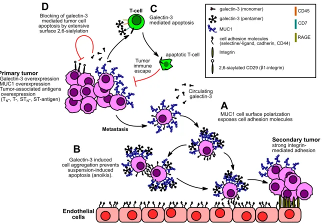
Extracellular galectin-3 is able to bind galactosides on glycoproteins presented on the cell surface. For instance, mucin glycoproteins on different epithelial carcinoma cells are primary binding partners for galectin-3.77,78 Furthermore, co-immunoprecipitation from colon cancer cell lines showed that cancer associated MUC1 is a natural target for galectin-3 and binding seems to be mediated by glycosylation77 with the tumor-associated T-antigen.79 MUC1 proteins with T-antigen presentation on cancer cells are proposed to permit adhesion to endothelial cells in the presence of galectin-3.80,81,82 Binding was increased when the cells were pretreated with sialidase and depletion of binding occurred by O-glycanase treatment (T-antigen specific endoglycosidase from Streptococcus pneumoniae). MUC1-transfected melanoma cells with T-antigen glycosylation exhibited stronger endothelial cell binding when treated with galectin-3, while no effect was visible on the MUC1 negative revertants.83 It was therefore proposed that galectin-3 captures overexpressed MUC1 on the surface of circulating tumor cells and through clustering of MUC1, several shorter adhesion molecules, such as E-cadherin, CD44 and E-selectin ligands are revealed which are then exposed on the cell surface and are able to interact with their corresponding binding partners on the endothelial side (figure 1.4, mechanism A).79 By this mechanism, galectin-3 supports settling of metastasizing tumor cells. Also, adhesion of the circulating tumor cells to each other is increased through mucin/galectin-3 interaction, leading to aggregation of tumor cells (figure 1.4, mechanism B).84 The cell adhesion prevents apoptosis of the tumor cells, induced by the loss of cell-cell or cell-matrix interactions (anoikis).85,86 By recognizing tumor related glycosylation on overexpressed MUC1 and following mediated adhesion to epithelial tissue, galectin-3 promotes installation of secondary tumors. Furthermore, extracellular galectin-3 has been shown to be immunosuppressive. T- and B-leukocytes, which were exposed to galectin-3, became apoptotic (figure 1.4, mechanism C).87,88,89,90,91 As molecular receptors on the leukocytes CD7, CD29 (β1-integrin), CD45 (protein tyrosine phosphatase) and RAGE were identified.87,88,92,93,94 All interactions are mediated by protein N- and O-glycosylation with varying contributions. For example, CD45 contains N- and O-glycans, but the splice isoform CD45RO, which lacks the heavily O-glycosylated protein domain, shows almost no galectin-3 binding, highlighting the significance of O-glycosylation for galectin-3 induced apoptosis.94 Overexpressed galectin-3 in circulation and membrane-associated to the tumor cells is therefore suspected to be beneficial for the tumor cells and helps to escape immune surveillance. Apart from apoptosis of immune cells, the human colon adenocarcinoma cell line SW48 was also shown to enter apoptosis after removal of N-acetylneuraminic acid by
10 1INTRODUCTION
sialidase treatment and contact with galectin-3.93 CD7 and CD29 were identified as galectin-3 receptors. CD29 was found to be α2,6-oversialylated on this carcinoma cell line, thus hindering galectin-3 interaction. It was therefore proposed, that overexpression of galectin-3 and oversialylation is advantageous for cancer progression in several ways. The cancer cells benefit from the intracellular anti-apoptotic effects and are at the same time protected from apoptotic effects of extracellular galectin-3 by extensive α2,6-sialylation of the galectin-3 receptor CD29 (figure 1.4, mechanism D).
Figure 1.4: Proposed effects of extracellular galectin-3 on tumor metastasis and immune evasion. A: Clustering
of MUC1 by galectin-3 exposes cell adhesion molecules. B: Tumor cell aggregation through exposed cell adhesion molecules prevents anoikis. C: Galectin-3 mediated apoptosis of T-cells results in tumor immune escape. D: Extensive α2,6-sialylation blocks galectin-3 mediated apoptosis of tumor cells.
Other members of the galectin family were found to share the same metastasis promoting features.95 MUC1 and galectin-3 act in a tumor-promoting interplay. The N-glycosylated (Asn- 36) intracellular, C-terminal part of MUC1 further signals an increase of galectin-3 mRNA translation by stabilizing the transcript through decrease of the inherent, galectin-3 suppressing miRNA-322. Tumor-related MUC1 overexpression is therefore correlated with increase in galectin-3 expression. In return, galectin-3 binds the MUC1 N-glycosylation at Asn-36 and bridges to ERBB1, integrating MUC1 into EGRF signaling.96
Galectin-3 may be involved in tumor progression in several ways. In general, intracellular galectin-3 acts anti-apoptotic and therefore promotes cancer development via carbohydrate- independent protein-protein interactions. Extracellular galectin-3 promotes metastasis and regulates cell apoptosis via carbohydrate-dependent mechanisms. Through direct contact with cell surface glycans, for example the tumor-associated T-antigen, it is probably involved in installment of daughter tumors, which makes it an interesting subject for further carbohydrate-lectin binding studies.
1.4 MUC1 carbohydrate vaccines
The fact that MUC1 is overexpressed and aberrantly glycosylated made it a prime target in immunotherapy and immunodiagnostics of carcinomas, including breast, ovarian, lung, colon and pancreatic cancers. Anti-MUC1 auto-antibodies are found in serum of cancer patients and their identification is in general associated with a better prognosis.11,61,97 Treatment of cancer relies on the removal of the tumor burden by chemotherapy, radiation or surgery.
Hence, immunotherapeutic methods are a valuable asset to established tumor therapy.
Passive immunization with monoclonal antibodies is a successful and relatively safe approach in immunotherapy. Several cancer target reactive monoclonal antibody drugs have already entered the market and second generation approaches, such as engineered antibody fragments or antibody-drug-conjugates (ADCs), are in the focus of contemporary pharmaceutical research and clinical studies, with first products already launched.98 However, the protection by passive immunization with monoclonal antibodies is not long- lasting, most available therapeutic antibodies on the market and in late stage clinical trials are chimeric or humanized antibody drugs,99 which can still provoke an unwanted immune response100 and full human sequence antibody production by in vitro affinity maturation through biological display systems or in vivo expression in transgenic animals is complex.
Active immunization with an anti-tumor vaccine on the other hand, could induce inherent antibody production against tumor related tissue. Especially against metastasizing tumors, an augmented immune surveillance would be helpful. Although auto-antibodies are found in the serum of patients, the induced immune reaction by the inherent tumor-associated antigens is obviously not strong enough to eradicate the tumor burden. The reason is, that the tumor-associated antigens are often seen as self-antigens and are only poorly immunogenic, although being abnormal.101
An organism has a natural tolerance for antigens regarded as “own”. All lymphocytes are generated in the bone morrow. To avoid autoimmunity, lymphocytes are selected for
12 1INTRODUCTION
tolerance against endogenous tissues. Cells that recognize self-antigens are selected for cell lysis. For B-cells, this education happens directly in the bone morrow and for the T-cells in the thymus. Dendritic cells, macrophages and B-cells are antigen-presenting cells (APCs) and can detect and internalize exogenic pathogens. After digestions, parts of the pathogens are linked to major histocompatibility complex II (MHC II) proteins, which are presented at the surface of the APCs to CD4+ T-cells.102 If a presented antigen is of endogenous nature, the T-cell receptor (TCR) will only bind weakly to the MHC-antigen-complex, according to maturation in the thymus. Important costimulatory signals cannot emerge and the T-cell remains inactive. If the APC is a B-cell, it can proliferate into a plasma cell and start to secret antibodies in a T-cell independent reaction. These antibodies are of the IgM-type and are short-lived and of low affinity (figure 1.5, A). In general, sole carbohydrates are T-cell- independent antigens.103 Tumor related MUC1 IgM antibodies are found in cancer patient sera but immune response is too weak to successfully generate immunity. On the other hand, if an APC presents an unknown antigen to the T-cell, then the MHC-II-antigen-complex is bound with high affinity. Costimulation takes place and the T-cell proliferates into a T- helper (Th) cell and secretes cytokines which dock to cytokine receptors at the B-cell to induce proliferation into an antibody-producing plasma cell (figure 1.5, B). The antibodies generated by the T-cell-dependent pathway are long-lived, high affinity IgG antibodies.
Furthermore, B-cells may also proliferate into memory B-cells. This class change from IgM to IgG antibodies characterizes an effective humoral immune response.
Figure 1.5: A: T-cell independent response by carbohydrate antigens elicits IgM antibody production. B:
Costimulatory signals enables proliferation of B-cells into plasma cells and IgG antibody production.
At any time, the immune effector cells patrol for unusual structures and start an immune reaction if necessary. However, tumor cells also develop mechanisms to escape the permanent immune surveillance. For instance, the ability to express MHC I molecules may be gradually lost during increasing stages of tumorgenesis.104,105 Some solid tumors also start to overexpress cytokines with immunosuppressive functions, such as IL-10, IL-4 or vascular endothelial growth factor (VEGF).106 It has been found, that MUC1 itself is an immunosuppressive factor. Shed and secreted MUC1 in tumor tissue arrests T-lymphocytes in their cell cycle and proliferation of naïve T-cells is blocked.107,108
A suitable vaccine must therefore be able to overcome natural tolerance and effectively augment the immune response. Commonly, a chemically synthesized antigenic B-cell epitope is conjugated to an immunostimulant. A common strategy is the conjugation to a carrier protein. Carbohydrate antigens may be directly linked to a carrier, such as keyhole limpet hemocyanin (KLH), bovine serum albumin (BSA) or tetanus toxoid. Immunological studies in mice revealed, that the induced immune reactions by such a conjugate elicits modest responses, often significantly of the IgM-type, indicating insufficient antibody class change.109,110,111,112,113 Variants of using unglycosylated peptide epitopes, in form of multi tandem repeat peptides or as carrier protein conjugates have been tested. Most of the formulations used with unglycosylated peptides suffer from low humoral response or lack of class switching to IgG isoforms.114,115,116,117,118 The reasons might be related to missing conformational effects of the carbohydrates on the peptide backbone. Tecemotide (L-BLP25, Stimuvax®, Merck KGaA) is a liposomal formulation of an unglycosylated MUC1 tandem repeat peptide (25mer), conjugated to monophosphoryl lipid A (MPL). The compound went into clinical trials with application in NSCLC (non-small cell lung cancer). After not meeting the primary end point in an initial phase III clinical trial,119 the formulation was withdrawn in 2014, shortly after the beginning of a second phase III trial and is discontinued.120
Vaccine designs based on glycopeptides seem to be more promising.103 In comparison, glycosylated peptide vaccine candidates usually elicit stronger immune responses than the unglycosylated counterpart.121 An early study already showed that serum antibodies of breast cancer patients react stronger with glycosylated MUC1 tandem repeats than with the unglycosylated tandem repeats.47 Commonly, MUC1 glycopeptides are also conjugated to immunostimulants to enhance immune response, to overcome tolerance and ensure antibody class switching. Several glycopeptide vaccine conjugate designs have been reported in the literature. A MUC1 glycopeptide may be attached to a carrier protein, for instance bovine serum albumin (BSA),122,123 keyhole limpet hemocyanin (KLH)124,125 or tetanus toxoid (TT) (figure 1.6, A).126,127,128 Carrier proteins fortify immune responses by presenting T-cell-epitopes for T-cell-dependent immune reactions and multivalent
14 1INTRODUCTION
presentation of the B-cell epitopes enhances the tendency for receptor clustering on the antigen presenting cells (APC), such as dendritic cells (DCs), B-cells or macrophages.
Exogenous proteins also provoke a response against carrier epitopes, which sustains the risk of overriding the immune reaction against the selected B-cell-epitope.129,130,115
Alternatively, short peptide sequences may be used as T-cell epitopes (figure 1.6, B).
Examples are peptides coming from ovalbumin,131 the polio virus132 or rationally designed peptides like the PADRE-sequence.133 The P30-sequence, a 30mer peptide derived from tetanus toxoid, has recently been reported to give robust antibody titers. Also, this peptide may function as a built-in adjuvant, making the use of additional adjuvants in immunotherapy obsolete.134,135 Other common immunostimulants are Toll-like-receptor (TLR) ligands, like the lipopeptide Pam3CSK4.136 As a mitogen, the lipopeptide is not immunogenic but stimulates TLR-1/2,137 which initiates induction of costimulatory proteins in B- and T-cells. Conjugation of these components, with spatial separation via a non-immunogenic spacer, results in two- and three component vaccines.
Figure 1.6: Design examples for potential glycopeptide vaccine conjugates: A: Two-component peptide-protein vaccine. B: Two-component peptide-peptide vaccine. C: Three-component peptide vaccine with TLR-lipopeptide.
In addition to a humoral antibody response, it would be beneficial if the vaccine could induce cytotoxic T-cell responses. In that way, malignant tumor cells would also be opposed to cytotoxic T-lymphocytes (CTLs), which could directly lyse the cells or induce apoptosis.
Exogenous substrates, such as the potential vaccine, are endocytotically internalized, processed by digestion and the resulting (glyco-)peptides loaded onto MHC II molecules.
The loaded antigens are then exclusively presented to CD4+-T-cells and differentiation into T- helper cells proceeds. CTL activation, on the other hand, relies on antigen presentation by MHC I to CD8+ T-cells. Nevertheless, CTLs with specificity against the MHC-unrestricted epitope localized to the DTR domain have been isolated from tumor patients.138,139 Since then, MHC I restricted peptide sequences within or outside the MUC1 tandem repeat were also reported and specific CTL-responses could be induced in mouse models.140,141,142,143,144
Moreover, it was documented that glycosylation is able to stabilize the binding to MHC I.142,144 It is not clear though, how the tandem repeat sequences get access to the MHC I pathway.
This would involve mechanisms of cross-presentation. The antigen could for example escape into the cytosol and be processed by the proteasome,145,146 or be digested by cathepsins to yield 8-11mer peptides which would fit into the MHC I binding groove.147 It was also shown, that the C-type lectin MGL (macrophage galactose c-type lectin) can mediate internalization of glycopeptides into APCs upon which the glycopeptides are directed into MHC II as well as into MHC I compartments.148,149 The uncertainties regarding efficient MHC I presentation makes addressing cytotoxic immune responses by vaccines difficult and so far no example of a CTL-response, being able to successfully neutralize human cancer cells is reported (for example in transgenic mouse models).143
1.5 Mucins in airway diseases
The glycans on mucin glycoproteins are covering the epithelial cell-surface and determine physical and biological properties of the mucus. The glycans are effecting the conformation of the protein and the hydrophilic glycans are preventing dehydration, thus influencing the mucus flow properties. The different carbohydrate structures also function as ligands to different pathogens, which during normal flow properties to a large extend are cleared by the mucus.150 This process is an important part of the innate immune system. In airway diseases, the binding of pathogens to glycan ligands results in inflammatory-infective responses in the respiratory tract contributing to the mucin overproduction and causing an aberrant flow of the mucus, which further is an important factor in the morbidity and mortality of COPD, CF and asthma patients. Changes in terminal mucin glycosylation has been found in these patient groups, e.g. altered levels of sialylation, sulfation and fucosylation.151 These alterations of the O-glycosylation influence both biophysical and biological (binding to bacteria, viruses and cells) properties of specific mucins. The changes in glycosylation and mucin production in COPD, CF and Asthma are probably one of the major factors involved in the non-optimal transport properties of the mucus and rather than protecting the host, the gel instead provides an environment where the pathogens thrive and contribute even more to inflammation and disease.152