Immunology of a cancer mucin:
Proteasomal processing and presentation of human MUC1 tandem repeat glycopeptides
in the MHC class I pathway
INAUGURAL-DISSERTATION zur
Erlangung des Doktorgrades
der Mathematisch-Naturwissenschaftlichen Fakultät der Universität zu Köln
vorgelegt von
Tanja Ninković
aus Belgrad, Serbien 2006
Referees/Berichterstatter Prof. Dr. Franz-Georg Hanisch
Prof. Dr. Dietmar Schomburg
Date of oral examination/ 26.10.2006 Tag der mündlichen Prüfung
The present research work was carried out under the supervision and the direction of Prof. Dr.
Franz-Georg Hanisch in the Institute of Biochemistry II, Medical Faculty, University of Cologne, Cologne, Germany, from February 2003 to October 2006.
Diese Arbeit wurde von Februar 2003 bis Oktober 2006 am Institut für Biochemie II der
Medizinischen Fakultät der Universität zu Köln unter der Leitung und der Betreuung von
Prof. Dr. Franz-Georg Hanisch durchgeführt.
To my parents
Silent gratitude isn't much use to anyone.
~ Gladys Browyn Stern
Therefore,
I would like to thank to my PhD supervisor Prof Hanisch for excellent mentorship and advices, endless patience and for teaching me to think critically. I appreciate very much that he gave me freedom to develop in my own pace and to find my own ways and interests. His support for independent thinking and crisp analysis led me to the path of being a scientist.
Above all, I am happy that I had a supervisor that true all the years of his experience managed to keep passion for science and flexible and open intellect.
Also, I would cordially like to thank my Prof Dimitrijević, supervisor during my master studies who made me fell in love in cancer research and to my biology teacher Jelisavka Luković who “infected” me with the “biology virus”. My success I also owe to Nasta Dedović and Nikola Tanić who taught me how important understandment of experimental techniques can be.
The great gratitude I owe to Mirko, my soul mate, who shared with me sunshine and fog of scientific work and always took my hand to help me come to the solution and provided calm space to think.
Many, many thanks to my dear parents and my darling brother Ivan who supported all my decisions and helped me deal with all problems and challenges. They taught me to trust myself and all my self-confidence I owe to them.
Here I would also like to thank my friends in “exile”, Dragana, Mirjana, Natasa, Marija, Aneta, Marko and Hilal who made my stay in Germany easier. And many thanks to my Belgrade friends who always wait for me at home with a smile.
Many thanks to my colleagues in the lab and institute for exchanging knowledge and offering help, especially to Hanieh, Katja, Marcus, Regine, Isabelle, Sebastian, Tilo, Yeliz, Uschi and Nefres.
Also, I would like to thank Prof Paulsson for offering me the possibility to do my PhD at the excellently organized Institute for Biochemistry and many thanks to Frau Pelzer and people from the Service lab, with the special gratitude to Stefan Müller and Udo Roth.
This work would also not have been possible without help of Aleksej Popov and Kondo Eisei
from the Molecular Tumor Biology and Immunology and kind cooperation of their supervisor
Prof Joachim Schultze.
ABREVIATIONS ... 1
1 INTRODUCTION ... 2
1.1 BREAST CANCER ... 3
1.2 MUC1 AND MUCINS ... 4
1.2.1 MUC1 ... 6
1.2.2 Antigen presenting cells ... 18
1.2.3 Proteasome ... 18
1.2.4 MHC molecules ... 21
1.3 AIM OF THE STUDY ... 23
2 MATERIALS AND METHODS ... 24
MATERIAL ... 25
2.1 CELL-BIOLOGICAL METHODS ... 28
2.1.1 Culture of mouse dendritic cell line ... 28
2.1.2 Culture of T2 cell line ... 28
2.1.3 Culture of human mammary carcinoma cell lines ... 28
2.1.4 Long time storage of cells ... 28
2.2 BIOCHEMICAL METHODS ... 29
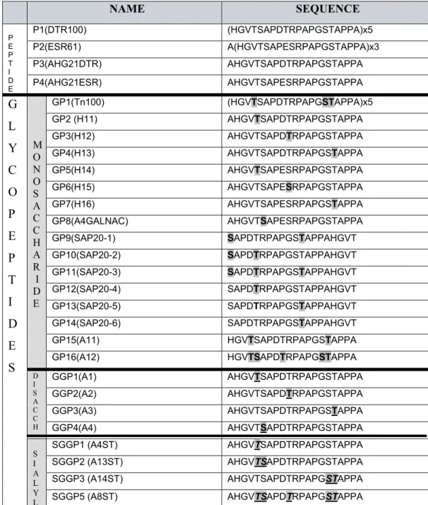
2.2.1 MUC1 derived peptides and glycopeptides ... 29
2.2.2 Enzymatic glycosylation of peptides in vitro ... 30
2.2.3 Proteasomal processing of MUC1 peptides and glycopeptides ... 31
2.2.4 Mass spectrometric analyses of processed peptides ... 32
2.2.5 Amino acid sequence analysis of generated fragments ... 32
2.3 MATHEMATICAL ANALYSES ... 33
2.3.1 Quantification of generated peptide fragments ... 33
2.3.2 Calculation of the frequency of digestion between two amino acids ... 34
2.3.3 Calculation of average fragment length ... 34
2.3.4 Calculation of peptide length distribution ... 34
2.3.5 Calculation of relative amounts of short 8 to11-meric peptides ... 35
2.3.6 Calculation of relative amounts of glycopeptides within 8 to 11-mers ... 35
2.3.7 Analyses of the origin region of 8 to 11mers ... 35
2.3.8 Algorithm prediction of peptide ligation strength to MHC molecule ... 35
2.4 IMMUNOLOGICAL METHODS ... 36
2.4.1 Selection of short peptides ... 36
2.4.2 MHC stabilization assay ... 36
2.4.3 Cross presentation ... 37
2.4.4 Isolation of MHC bound peptides from breast cancer tumor cells ... 41
3 RESULTS ... 43
3.1 BIOCHEMICAL STUDIES ... 44
3.1.1 Influence of buffer composition on processing efficiency ... 44
3.1.2 Control of proteasome purity ... 45
3.1.3 Influence of glycosylation on proteasomal processing efficiency ... 46
3.1.4 Influence of glycosylation on processing pattern of MUC1 VNTR sequence ... 51
3.1.5 Analyses of generated short fragments ... 57
3.2 CELLULAR ASSAYS ... 65
3.2.1 Cross presentation ... 65
3.2.2 Analyses of MHC bound MUC1 glycopeptides on breast cancer cell lines ... 67
4 DISCUSSION ... 69
4.1 TUMOR ANTIGEN MUC1 AND THE IMMUNE SYSTEM ... 70
4.2 PROCESSING OF NONGLYCOSYLATED MUC1 SEQUENCE BY IMMUNOPROTEASOMES ... 71
4.3 PROCESSING OF GLYCOSYLATED MUC1 BY IMMUNOPROTEASOMES ... 73
4.4 INFLUENCE OF PEPTIDE STARTING MOTIF ... 75
4.5 THE BUFFER INFLUENCE ... 76
4.6 LOW IMMUNITY OF CANCER RELATED MUC1 GLYCOFORMS ... 76
4.6.1 Sterical influence of glycans on processing outcome ... 77
4.7 IDENTIFICATION OF THE BEST GLYCOPEPTIDE FOR MHC CLASS I LIGANDS GENERATION ... 78
4.7.1 Identification of MHC class I binding MUC1 glyco-epitope ... 80
4.8 CROSS PRESENTATION ... 81
4.9 MHC CLASS I BOUND PEPTIDES OF A BREAST CANCER CELL LINE ... 83
4.10 PREVIOUS IMMUNOLOGICAL STUDIES ... 84
4.11 MUC1 BREAST CANCER VACCINE - OUTLOOK ... 85
4.11.1 Peptide-based vaccination strategy ... 85
4.11.2 Glycopeptide-based vaccination strategies ... 86
4.11.3 Stimulation of MHC-unrestricted MUC1-specific recognition of tumor cells ... 87
4.12 CONCLUDING REMARKS ... 87
SUMMARY ... 88
BIBLIOGRAPHY ... 90
ERKLÄRUNG ... 103
LEBENSLAUF/CURRICULUM VITAE ... 104
ABREVIATIONS
MUC1 mucine 1 RNA ribonucleic acid
VNTR variable number of tandem repeats GalNAc N-acetylgalactosamine
GlcNac N-acetylglucosamine
MHC Major histocompatibility complex HLA-A,B,C human leukocyte antigen
H2K
bmouse MHC class I allele CTL cytotoxic T lymphocytes
IgG, IgM Immunoglobuline G, Immunoglobuline M APC antigen presenting cells
DC dendritic cells IPs immunoproteasomes INF γ Interferon gamma IL-4 Interleukin-4
TNF-α Tumor Necrosis Factor-alpha
GM-CSF Granulocyte-Macrophage Colony Stimulating Factor ER Endoplasmatic Reticulum
ERAD Endoplasmatic Reticulum Associated Degradation Tg mice transgenic mice
HPLC high pressure liquid chromatography
Units of Measure and Prefixes
°C degree Celsius
Da Dalton
g gram
h hour
L litre
m meter
min minute
s sec
V volt
MW molecular weight Symbol Prefix (Factor)
k kilo (103)
c centi (10-2)
m milli (10-3)
µ micro (10-6)
n nano (10-9)
p pico (10-12)
Introduction
1 INTRODUCTION
Introduction
1.1 Breast cancer
Breast cancer is the primary cause of death of women in the western countries, accounting 1 out of 10 women. Just over 1 million of people per year get diagnosed with breast cancer and around 400.000 die. Despite a 60% survival rate 1100 persons die daily due to breast cancer (Parkin DM et al.,2001).
The small size of most cancer nodes (below two centimetres) results in late detection, and localisation in the proximate neighbourhood of lymph nodes increases the metastatic probability. The best prognosis for breast cancer patients is still dependent on an early detection. Although many efforts have been made to develop new diagnostic and therapeutic tools, surgery with chemotherapy and radiation are still the methods of choice for breast cancer treatment.
Breast cancer is actually a common name for different malignant incidences which can be classified in ductal, lobular, medular, tubular and mucinous carcinoma or as inflammatory breast cancer, Paget’s disease of the nipple and phyllodes tumor (Berg JW et al., 1995).
Ductal carcinoma originates in the milk ductus and after conveying metastatic potential, it can spread through the body.
Around 80% of all breast cancers are invasive ductal carcinoma.
Lobular carcinoma originates in the milk producing glands. In invasive form it spreads to surrounding fatty tissue and can metastasize. 10-15% of all breast cancers are of invasive lobular type.
Other types of breast cancer are rare and represent between 1 and 5% of all breast cancer cases.
A large number of tumor markers in the sera of patients indicate breast cancer. These include MUC1 (e.g. CA15-3), CEA, HER-2oncogene, p53 tumor-suppressor, glycolytic enzymes and cytokeratins. Compared to healthy individuals and patients with benign breast diseases, patients with breast cancer have elevated levels of breast antigens circulating in their plasma and/or serum.
Figure 1: Brest anatomy showing ducti and lobule.
From the web site
http://www.breastbiopsy.com/bi oresults_typesofbreastcancer2 .jsp
Introduction
1.2 MUC1 and mucins
Mucins are high-molecular weight glycoproteins, representing the main constituents of mucus and the glycocalix of epithelial and some non-epithelial cells. Common functions of mucins are lubrication and protection. The family of mucin proteins consists of up to now 19 defined proteins (Entrez-Protein database, Table 1), MUC1-MUC20. Their MW of 100.000 till 1.000.000 is by 50-90% contributed by carbohydrates.
Mucin- Name
Secretory Membraneous
Chromosomal
localisation Expressing tissue Literature Mucin 1 m,s 1q21-q24 Ubiquitous Gendler et al., 1990 Mucin 2 s 11p15 GI tract, respiratory tract Gum et al., 1994 Mucin 3 m,s 7q22 GI tract van Klinken et al., 1997 Mucin 4 m,s 3q29 GI tract Nollet et al., 1998
Mucin 5AC s 11p15 Respiratory tract Guyonnet Duperat et al., 1995 Mucin 5B s 11p15 Respiratory tract, Stomach Meerzaman et al., 1994 Mucin 6 s 11p15.5-15.4 Bile duct Toribara et al., 1993 Mucin 7 s 4q13-q21 GI tract Bobek et al., 1993 Mucin 8 / 12q24.3 Saliva gland Shankar et al., 1994 Mucin 9 / 1p13 Trachea Lapensee et al., 1997 Mucin 11 / 7q22 Oviduct Williams et al., 1999 Mucin 12 m,s 7q22 GI tract, Uro-genital tract Williams et al., 1999 Mucin 13 m 3q13.3 GI tract, Uro-genital tract Williams et al., 2001 Mucin 15 / 11p14.3 GI tract Pallesen et al., 2002 Mucin 16 m 19p13.3 Genital tract, GI tract,
lymph organs
Yin et al., 2001
Mucin 17 m 7q22 Ovaries Gum et al., 2002 Mucin 19 / 12q12 Trachea, Saliva gland Chen et al., 2003 Mucin 20 / 3q29 Kidneys, Placenta, Lungs,
Prostate, Liver
Higuchi et al., 2004
Table 1. The list of currently identified mucins, with chromosomal localisation, tissue expression
Introduction
The currently known MUC species can be subdivided into two groups based on their structural aspects and biosynthetic routes: membrane bound and secretory mucins.
Membrane bound mucins (MUC1, MUC3, MUC4, MUC12) are essential components of the glycocalix. Their hydrophobic transmembrane domains are anchoring the protein within the lipid bilayer with C-terminus oriented toward the cytosol (Table 1). Most of the membrane bound mucins show tissue-specific expression patterns (Hanisch FG et al., 2000).
Secretory mucins are the main constituents of the visco-elastic mucus gel and are mainly coded by a gene cluster on chromosome 11p15.5. All secretory mucins (MUC2, MUC5AC, MUC5B, MUC6) except MUC7 possess one or several von Willebrandt factor-like D domains (Hanisch FG et al., 2000), cysteine-rich peptides, which function in the oligomerization of mucin monomers and in the packaging into secretory vesicles (Perez-Vilar J et al., 1999).
Other mucin genes are randomly scattered over the genome.
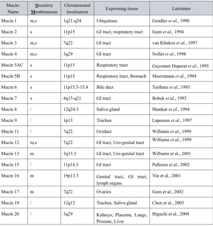
The special characteristic of all mucins is the presence of a “tandem repeats domain”, several tens till hundreds of repeats of a peptide (Fowler J et al., 2001). The number of tandem repeats varies due to the diversity of mRNA length from 0,4 to 20kb transcribed from the single gene. Tandem repeats are heavily O-glycosylated by long glycan chains (up to 20 monosaccharides per chain) on numerous serines and threonines. At terminal position negatively charged sugars, like sialic acid or sulphated sugars, are often found. The sugar chain composition and length are tissue specific. Because of the high proline content, the complete molecule is extended and protruding from the cell surface (Figure 2).
Figure 2. Model of mucins and other cell surface glycoproteins/proteoglycans. The figure schematically portrays structures of four size classes of mucins as well as consensus structures for syndecans and integrins. All extracellular portions of these molecules are roughly drawn to scale. Blue squiggles represent O-linked oligosaccharides while red squiggles represent glycosaminoglycans.
Integrins, syndecans and most other surface receptors do not extend beyond 50 nm from the cell surface; however, with the exception of MUC13, mucins extend much further due to the extended structures contributed by the proline-rich heavily O-glycosylated tandem repeat domains. MUC1 and MUC4 are the largest transmembrane mucins, extending >200 nm from the cell surface. The ectodomain structures of MUC16 and MUC17 are considerably shorter than those of MUC1 and MUC4, but still much larger than those of other surface glycoproteins. Soluble mucins, such as MUC2, 5AC, 5B and 19 are even larger, reading 500–100 nm in length (Brayman M et al., 2004).
Introduction
The functions of mucins are mainly influenced by sugar presence. The major common function of mucins is protection of the cellular surface of mechanical injuries and micro- organisms, prevention of dehydration as well as lubrication, especially in the gastrointestinal, respiratory or reproductive tracts (Perez-Vilar et al., 1999). These functions are contributed mainly by secretory forms of mucins, which reside close to the apical surface of epithelia cells in the form of oligomers connected by disulfide bridges. Membrane bound mucins are as monomers attached to the cell membrane, where beside protective functions they influence cell-cell interactions. The long, protruding structure of mucins (Bramwell ME et al.,1986) introduces anti-adhesive effects and prevents cell-cell and cell-extracellular matrix contacts (Ligtenberg MJ et al., 1992), thereby taking part in epithelial morphogenesis (Hilkens J et al., 1992), migration, proliferation of cells, carcinogenesis and metastasis. Specific interactions with E-selectine and integrins involve mucins in inflammation, development and lymphocytes homing (Walz et al., 1990; Yuen CT et al.,1992; Schimizu Y et al.,1993).
1.2.1 MUC1
The first mucin characterized during the 80ies was MUC1, a large glycoprotein existing in both secretory and transmembrane forms. It was described under numerous names:
Polymorphic epithelial mucin, PEM, PEMT, Episialin, Tumor-associated mucin, Carcinoma- associated mucin, Tumor-associated epithelial membrane antigen, EMA, H23AG, Peanut- reactive urinary mucin, PUM, Breast carcinoma-associated antigen DF3, CD227 antigen.
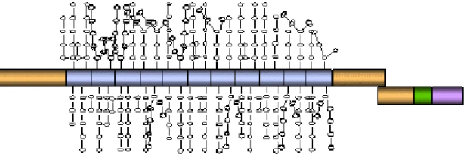
Finally it was systematically designated as MUC1 (Figure 3).
Figure 3. The structure of MUC1. The 72 amino acids cytosolic domain is shown in purple. The 28 amino acids domain is shown in green. The VNTR region is shown in blue while the rest of the protein is shown in tan. The extensive O-linked glycosylation in the VNTR region is indicated with the stick figures.
Introduction
1.2.1.1 MUC1 localisation
MUC1 exhibits a ubiquitous organ distribution (Fig. 4), (Zotter et al., 1988) including mammary gland acini and ducts, salivary gland ducts and serous acini, but not mucinous acini;
squamous epithelium of the esophagus; parietal cells, canaliculi and peptic cells of the stomach; acini and ducts of the pancreas; bile ducts in the liver; enterocytes of the duodenum, but not the large intestine; respiratory and ciliated epithelium of the lungs, serous bronchial glands, but not mucinous ones; distal tubules of the kidney and collecting ducts, but not proximal tubules; bladder urothelium; prostate gland epithelium; resting endometrium of the uterus; rete testis; (activated) mesothelium. Important negative tissues are the skin epithelium and all kinds of mesenchymal tissues. Beside epithelial tissues, MUC1 was found on lymphoid cells and lymphomas, especially on plasma cells and myelomas (Zotter et al., 1988;
Treon et al., 1999) as well as on erythroblasts. MUC1 presence is moreover reported for activated T cell and mature dendritic cells, but not on resting T cells and immature dendritic cells.(Clossen S et al.,2004; Agrawal et al., 1998)
Figure 4. Structure of MUC1 in the transmembrane form and the list of MUC1 positive tissues
Introduction
1.2.1.2 MUC1 Gene
MUC1 is coded by a gene localised on the long arm of chromosome 1 in the 1q21-q24 region.
The gene is composed of seven exons that together comprise 4,2 to 7,0 kb of genomic DNA.
The alternative splicing of mRNA gives rise to transcripts of 3.7 to 6.4 kb coding for the different isoforms of the MUC1 protein, transmembrane, secretory, and MUC1 X/Y/Z isoforms.
1.2.1.3 MUC1 protein structure
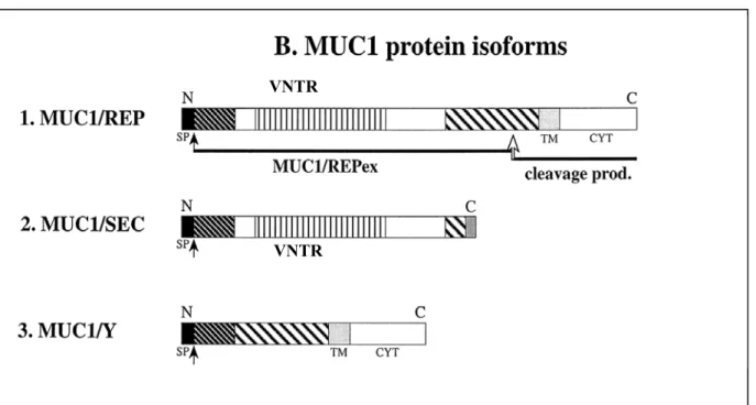
The alternative splicing results in the generation of several isoforms of MUC1 protein, and therefore we speak of MUC1 polymorphism. Several forms of MUC1 are formed:
1. MUC1/REP: heterodimeric complex of a large tandem repeat ectodomain component and a small transmembrane component (Ligtenberg MJ et al., JBC 1992)
2. MUC1/SEC that contains tandem repeat but no transmembrane domain (Smorodinsky N et al., 1996)
3. Short MUC1/X/Y/Z membrane anchored isoforms generated by alternative splicing and devoid of tandem repeats. (Zrihan-Licht S et al., 1994; Baruch A et al., 1997;
Oosterkamp HM et al., 1997)
Figure 5. MUC1 protein isoforms. Domains re-occurring in various MUC1 isoforms are marked with the pattern. N and C represent amino and carboxy termini of the protein, TM – transmembrane domain; CYT-cytoplasmic domain, SP – signal peptide. The proteolytic cleavage site in the
Wreschner DH VNTR
VNTR VNTR
Introduction
The best characterized MUC1 gene product is a long membrane associated protein, named also a MUC1/REP. It is a type 1 transmembrane mucin-like protein (Fig. 5) consisting of two sub-domains: large extracellular domain and short cytoplasmic tail. After a translation of MUC1 mRNA, the MUC1/REP protein is autolytically cleaved in the endoplasmatic reticulum. This cleavage generates two peptide chains that get noncovalently re-associated in the proximity of the cleavage site (Ligtenberg MJL et al., 1992). The larger of the cleavage products is the ectodomain component, mainly built by the variable number of tandem repeats (VNTR), flanked by short degenerate sequences and non-repetitive sequences at both sides of the protein (Wreschner DH et al., 1990; Ligtenberg MJL et al., 1990; Gendler SJ et al., 1990;
Abe M et al., 1993). The VNTR region contains 20-125 repeats of the 20-meric amino acid sequence PAPGSTAPPAHGVTSAPDTR repeat which represents 50-80% of the peptide mass (reviewed in Gendler SJ et al., 1995; Lagow E et al., 1999). MUC1 protein is highly polymorphic in the population with respect to the number of tandem repeats as well as the presence of sequence variations within the tandem repeats (major being DT>ES). Shorter cleavage product carries the cytoplasmic and transmembrane domain.
The MUC1/REP extracellular domain can be shed from the cell by a yet not completely understood mechanism and found in the sera of breast cancer patients at increased levels (Baruch A et al., 1999). During its biosynthesis, the MUC1/REP protein is extensively modified by O- and N-glycosylation. O-glycosylation takes mainly place within tandem repeats of the VNTR region. Further on, MUC1/REP proteins can be subdivided into four variants A-D, which arise from the single nucleotide polymorphisms.
The MUC1/SEC protein contains full length sequence corresponding to the extracellular domain of MUC1/REP, but lacks the membrane anchoring hydrophobic domain as well as the intracellular domains. (Fig. 5)
In contrast to MUC1/SEC, short MUC1/X/Y/Z splice isoforms contains a transmembrane and cytoplasmic domains identical to the MUC1/REP, but their extracellular part contains neither the repeat array domain nor its flanking region and, hence, are devoid of the hallmark mucin- like features (Fig. 5). (Baruch A et al., 1999)
Due to the abundant glycosylation of the VNTR region with frequent negatively charged
terminal sugars and because of the high number of proline residues, MUC1 has an extended,
rigid form protruding 200-500 nm from the cell surface into the lumen. (Fig.2 Brayman et al.,
2004).
Introduction
1.2.1.4 MUC1 functions
Beside functions common to all glycocalix-building proteins, like lubrication, hydration, and protection from microbes, MUC1 fulfils several other more complex roles. So is MUC1 reported to take part in signal transduction, binding of actin cytoskeleton and it influences cell-cell and cell-matrix contacts and metastases.
It has been shown that MUC1 mucin undergoes phosphorylation on both tyrosine and serine residues of the cytosolic domain and, as a consequence, can potentially bind second messenger or adaptor proteins such as Grb2 (Wreschner DH et al., 1994, Zrihan-Licht S et al., 1994). Kufe D has reported the participation of MUC1 in the Grb2/Sos/ras signalling pathway, whereas interaction of MUC1 with erbB1 during lactation was reported by Gendler SJ (Pandey P et al., 1995; Schroeder JA et al., 2001). Moreover, high levels of MUC1 in mammary gland promote MAPK (ERK1/2) signal transduction (Schreder JA et al., 2001).
Also, the cytoplasmic domain of the MUC1 protein interacts directly with ß-catenin and therefore with the actin cytoskeleton (Yamamoto M et al., 1997). Besides this, the secretory MUC1 can interact with MUC1/Y and trigger Ser/Thr phosphorylation of its cytoplasmic part which involves MUC1 in signal transduction (Baruch A et al., 1999). These results suggest that MUC1 protein participates in signal transduction and may well play an active role in the oncogenic process.
The tandem repeat domain is reported to interact with ICAM-1 of endothelial and epithelial cells (Hayashi T. et al., 2001), and therefore to participate in cell migration and metastasis.
MUC1 has also influence on the cell-cell and cell-ECM contacts by sterically introducing anti-adhesive effects (Hilkens J et al., 1992, Wesseling J et al., 1995) or promoting adhesion by specific interaction of carbohydrate moieties with adhesion molecules like selectines (Zhangh K et al., 1996).
1.2.1.4.1 MUC1 glycosylation
MUC1 is post-translationally modified by both N- and O- type of glycosylation.
MUC1 has five potential N-glycosylation sites, three of which are located in the C terminal
transmembrane component of MUC1, and two proximate to the VNTR domain. However, the
major type of MUC1 glycosylation is O-glycosylation on numerous Ser and Thr residues
Introduction
H G V T S A P D T R PA P G S T A P PA
Potential O-glycosylation sites
Figure 6. MUC1 tandem repeat amino acid sequence with label potential O-glycosylation sites
The average glycosylation density and structure vary in vivo and are cell and tissue specific.
O-glycosylation is performed by the ordered action of glycosyltransferases adding carbohydrates to the protein backbone. The reaction initiates with addition of N-acetylgalactosamine on side chains of hydroxyl-amino acids Ser or Thr by one isoform of the 20 different polypeptide N-acetylgalactosaminyltransferases. The product, peptide linked GalNAc, is named Tn antigen and presents a starting point for the generation of more complex glycan chains. The elongation of the carbohydrate chain is performed by one of the numerous glycosyltransferases isoforms (Varki A). Based on the structure of two or three sugars attached to the Tn antigen eight basic core structures can be defined. Core structures are further elongated by sialylation, acetylation, sulphatation and (poly)lactosamine extension. The membrane attached protein appears as incompletely glycosylated form on the membrane and is repetitively re-internalised for additional glycosylations probably in the trans Golgi network.
The mature form of the protein can remain attached to the membrane or become shed without the short, membrane attached subunit. The secretory form of MUC1 is not re-internalised and reaches the extracellular space via secretory vesicles with only one passage of the Golgi.
The subcellular location where O-glycosylation initiates is the Golgi, since the up to 20 polypeptide GalNAc-transferases are localized throughout the cis to trans compartments. The O-glycosylation position within the polypeptide chain is not easily predictable and depends on the availability of GalNAc-transferases, the amino acids sequences around the putative glycosylation site and already existing glycosylation on proximal Ser/Thr residues.
(Elhammer AP et al., 1999). Up to now, at least 14 polypeptide GalNAc-transferases potentially involved in glycosylation of MUC1 are cloned and available for in vitro glycosylation (Clausen H et al., 1996; Bennett EP et al., 1998; Ten Hagen KG et al., 1999).
Each of these enzymes has a specific site preference, but some overlapping between them exists.
The complexity of glycan chains is tissue specific, and characteristic patterns were described
for a number of organ sites and tissue specific secretions. So the predominant presence of
core2-based linear or branched glycan chains was detected in lactation-associated MUC1
compared to short glycans, mainly of core 1 type, found in urine associated MUC1.
Introduction
1.2.1.5 MUC1 as a tumor antigen
MUC1, as well as ovarian tumor antigen CA-125, HER2neu and Carcinoembrynic Antigen (CEA), are glycosylated tumor antigens with proven potential in clinical diagnostics or in immunotherapy.
Tumor antigens are those antigens that are produced predominantly or exclusively by tumor cells and are recognised by the immune system. When presented only by tumor cells, but never by normal cells, they are called tumor-specific antigens and typically result from a tumor specific mutation. More common are antigens presented by both tumor and normal cells, named tumor-associated antigens. Cytotoxic T lymphocytes that recognize these antigens may be able to destroy the tumor cells before they proliferate or metastasize.
Two research groups showed by using specific monoclonal antibodies that sera from tumor patients were positive for MUC1/SEC and MUC1/REP (Smorodinsky N et al., Hilkens J et al., 1986). Thus, both the MUC1/SEC protein and the MUC1/REP extracellular cleavage product can be secreted or shed from breast cancer cells and used for monitoring during the patients recovery. Beside this, it was shown that the MUC1/Y protein is expressed by various human secretory epithelial tumors, but is not detectable in the adjacent normal tissue (Zrichan- Licht S et al., 1994; Hartman M et al., 1999; Baruch A et al., 1997)
In the mid 80ies it was shown that the tumor-associated MUC1 is over-expressed in a non- polarised manner and shed into the circulation in increased amounts (Hilkens J et al., 1986;
Wesseling J et al., 1996). Due to pericellular expression of the several hundred nanometre long ectodomain, the MUC1 on carcinoma cells represents a primary immune target in anti- cancer strategies.
During the 90ies the MUC1-specific immune response in human sera was detected. Natural B cell responses were detected in breast cancer patients as well as in pregnant women or in women after passage of one lactation phase. These weak humoral responses were shown to provide a certain degree of survival benefit for breast cancer patients (Kotera Y et al., 1994).
More specific, cellular responses of MUC1 specific T lymphocytes were detected in the
cancer patients. These T-cells were able to lyse the cancer cell, but in an MHC unrestricted
manner (Jerome KR et al., 1991). Due to their low efficiency, these detected immune
responses to MUC1 are not sufficiently strong to eradicate the growing tumor.
Introduction
Both humoral and cellular immune responses found in cancer patients were directed toward the tandem repeat domain of MUC1, mainly to its PDTR motif. Over 50 monoclonal antibodies were generated in mice and more than half of these were directed to this immunodominant motif (von Mensdorff-Pouilly S et al., 2000). Initially regarded as non- glycosylated, the PDTR peptide motif turned out to be O-glycosylated on the tumor- associated MUC1 (Muller S et al., 1997). Many of the peptide-specific antibodies bind with higher affinity to the glycosylated PDTR motif (Karsten U et al., 1998, 2004), indicating a glycosylation-induced stabilisation of the epitope.
The antibodies and T lymphocytes detected in cancer patients were specific for the cancer associated MUC1. Cancer associated MUC1 differs from lactation-associated MUC1 on the level of O-glycosylation (Muller S et al., 1999). The aberrant glycosylation is the structural difference that characterises the cancer-associated MUC1 as a tumor-specific antigen (Fig 7).
It is visible in the form of three structural changes:
Glycans of the tumor-cell associated MUC1 are shorter than the polylactosamine-type chains found on lactation-associated MUC1. They are predominantly core 1-based, with numerous Tn (core1-GalNAc) or T antigen (core1-disaccharide Gal-GalNAc) and in particular their sialylated derivatives. The percentage of sialylated glycans is higher in cancer- than in lactation-associated glycoform.
This tumor-associated truncation could be caused by reduced levels of core2-specific enzymes, ß-N-acetylgalactosaminyltransferase or by the increased sialylation (over-expression of 3GalST) (Dalziel M et al., 2001; Brockhausen I et al., 1995), which presents the stop signal for further addition of sugars (Whitehouse C et al., 1997).
The third remarkable difference in MUC1 glycosylation occurring on tumor cells is the increased glycosylation density. The density of glycosylation is significantly higher on tumor- associated MUC1 than on lactation-.associated MUC1. The number of glycosylated Ser/Thr in the tumor MUC1 VNTR domain is remarkably increased with 4,8 glycan chains per repeat found in breast carcinoma cell lines compared to 2,6 glycan chains per repeat in lactation-associated MUC1 (Muller S et al., 1997,1999). The shortening of sugar chains directly correlated with the density of glycosylation in the different breast cancer cell lines.
Together with increased and unpolarized expression, this deviation unmasks the core peptide
epitopes and exposes otherwise (sterically) hidden glycan epitopes. (Karsten U et al., 2005).
Introduction
Figure 7. Glycosylation of lactation- and tumor-associated MUC1. Glycosylation positions within a tandem repeat are bolded. Glycan chains attached to Tn antigen characteristic for lactation- or tumor- associated MUC1 are depicted on opposite sites of the figure.
1.2.1.5.1 MUC1 functions in tumor cells
The aberrant expression and glycosylation levels of MUC1 have consequences for the functions and interactions of the protein.
The pericellular expression of tumor-associated MUC1 protein plays a distinctive role in sterically reducing cell-cell and cell-extracellular matrix interactions. It has been postulated that MUC1 may be involved in the metastatic spread of cancer cells from the initial tumor site (Ligtenberg MJ et al., 1992; Hartman M et al., 1992; Wesseling J et al., 1995). It was demonstrated that MUC1 expression positively correlates with tumor formation and progression in knock out mice (Spicer W et al., 1995). Also the short form of MUC1 protein, MUC1/Y is reported to stimulate oncogenesis and tumor progression (Baruch A et al., 1997).
The negative charge of abundant sialyl groups present on the terminal ends of MUC1 sugar chains facilitates the interaction of tumor cells with E-and P-selectine, promoting the adhesion to endothelial cells and extravasation into the surrounding tissue during metastasis. Moreover, the negative charge is supporting spreading and dissemination of tumor cells by introducing
GalNAc
NeuAc
NeuAcα2-3 Galß1-3GalNAc
NeuAcα 2-6
GalNAc NeuAcα 2-6
GalNAc Galß1-3
α 2-6
GalNAc
α2-3Galß1-3 NeuAc
α3ST
αa 6ST6ST
Lactation-associated glycoform
GalNAc Galß1-3
Galß1-3GlcNAcß1-3Galß1-4GlcNAcß1-6 Galß1-4GlcNAcß1-3Galß1-4GlcNAcß1-3
- 3 Fuc α1
Galß1-4GlcNAcß1-3Galß1-4GlcNAcß1-6
Galß1-4GlcNAcß1- Galß1-
Galß1-3GalNAc ß6GnT
GlcNAc ß1-6
GalNAc Galß1-
AHGVTSAPDTRPAPGSTAPP
Tumor-associated glycoform
GalNAc
Introduction
The over-expressed MUC1 on tumor cells shields the cell from immune recognition by the cellular part of the immune system, and thus favours tumor cell survival and metastasis.
Therefore role played by MUC1 in cancer progression represents two sides of one coin: On the one end, loss of polarity and over-expression of MUC1 in cancer cells interferes with cell- adhesion and shields the tumor cell from immune recognition by the cellular arm of the immune system, thus favouring metastases. On the other hand, MUC1, in essence a self- antigen, is displaced and altered in malignancy and induces immune responses to it. Tumor- associated MUC1 has short carbohydrate side-chains and exposed epitopes on its peptide core; it gains access to the circulation and comes in contact with the immune system provoking humoral and cellular immune responses to it.
1.2.1.6 Immunity to MUC1
Natural antibodies to MUC1 are present in the circulation of cancer patients, both as free antibodies and as immune complexes, bound to MUC1. Both IgM and IgG antibodies were detected in breast cancer patients and their early appearance is connected with good prognosis (Fig 8).
Natural MUC1 antibodies from breast cancer patients reacted more strongly to the MUC1 peptides with cancer-specific glycosylation (GalNAc) than to the naked VNTR sequence.
Therefore, as the naturally existing tumor-related MUC1 is glycosylated, the immune response against breast cancer should be directed toward glycosylated MUC1 epitopes.
Initially, the glycosylation was considered to prevent antibody binding to the peptide backbone. Besides this, it was shown that circulating tumor MUC1 antigen found in tumor patients can not be processed by dendritic cells and does not elicit either MHC class II- (Hiltbold EM et al., 2000) nor CTL response (Hiltbold EM et al., 1999).
These findings were shown to be related to heavy glycosylation (Hiltbold et al., 2000) and not to a tolerance to MUC1 (Hiltbold et al., 1998).
Figure 8. MUC1 VNTR specific antibodies.
Introduction
The high avidity interaction of MUC1 glycan chains with the mannose receptor residing in early endosomes prevents the protein to traffic to late endosomes after endocytosis where processing occurs (Hiltbold EM et al., 2000).
Additionally, the digestion of densely glycosylated MUC1 by the enzyme Cathepsin L, involved in MHC class II epitope generation is prevented or restricted by site-specific O- glycosylation (Hanisch FG et al., 2003). Consequently, under-glycosylation was regarded as the prerequisite for an effective immune response against tumor MUC1. In particular, it was postulated that the immunodominant domain within tandem repeats, the PDTR sequence, was not glycosylated. As a consequence, cancer vaccines were developed based on unglycosylated MUC1 (Karsten U et al., 2005).
MUC1 positive tumors were targeted in a series of vaccination attempts in mouse models and in clinical trials. The clinical trials comprised
•
gene-based vaccine approaches, where MUC1 is encoded by a highly attenuated vaccinia virus vector (Scholl SM et al., 2000),
•
antibody-based vaccines (Heuser C et al., 2003, Wilkinson RW 2000 ),
•
peptide-based vaccination (Ramanathan RK et al., 2005; Wierecky Y et al., 2006;
Yamamoto K et al., 2005; Kohlgraf KG et al., 2004 ; Plamer M et al., 2001),
•
MUC1/interleukins synthetic chimera (Snyder LA et al., 2006),
•
fusion of tumor and dendritic cell (Tanaka Y et al., 2005) or
•
vaccination based on autologous, peptide-pulsed dendritic cells (Loveland BE et al., 2006; Wierecky Y et al., 2006).
The peptide-based strategies stimulated strong humoral IgM and IgG responses, but with no or with weak cellular immune response (Soares MM et al., 2001; Stepensky D et al., 2006).
The developed IgM and IgG reactivities were peptide-specific and therefore failed to recognize glycosylated tumor related MUC1. The approach using autologous dendritic cells loaded with nonglycosylated-MUC1 stimulated peptide-specific CTL in 50% of the patients with metastatic renal cell carcinoma (Wierecky Y et al., 2006).
Such a moderate success of peptide vaccines can presumably be ascribed to the structural
Introduction
antigen of targeted cancer cells. As glycosylation, albeit aberrant, is still present in the tumor- associated mucin the optimal MUC1 vaccine is not a peptide, but a glycopeptide.
1.2.1.7 Immunity to tumor and MUC1 tumor vaccines
The theory of immune surveillance claims that the immune system continually recognizes and eliminates tumor cells. Immune surveillance is most likely to be successful against virus- induced tumors, which express foreign antigens. But even the tumors with antigens which can be recognized by the host immune system can evade immune elimination. There are numerous mechanisms used by tumor cells to evade immune elimination: (1) expression of anti-apoptotic genes blocking the activity of CTLs and NK cells as well the effects of chemotherapy, (2) secretion of inhibitory cytokines TGFβ, IL-10, VEGF which prevent maturation of dendritic cells and recruit regulatory T lymphocytes inhibiting activation of helper and cytotoxic T cells that arrived at the tumor site. (3) reduction / lack of MHC expression, (4) lack of co-stimulation, (5) antigen shedding, (6) sequestration of tissue from surveillance , (7) lymphocyte killing…
To overcome these sabotaging mechanisms of the tumor, efforts are made to develop tumor vaccines for the induction of a stronger and more specific anti-cancer cell response. Tumor vaccines should utilize tumor specific antigens and stimulate dendritic cell maturation and T lymphocyte activation outside of the tumor micro-surrounding and thereby overcome some of the possible evasion mechanisms. Therefore, for the design of anti-breast cancer vaccine the best MUC1-derived antigens and glycosylation forms have to be known and interaction of glycopeptides with the immune machinery has to be elucidated.
Processing of glycosyalted MUC1 antigen is one of the crucial steps in generation of MHC
epitopes and stimulation of MUC1 specific T-cells. Previously, Vlad et al (2002) showed that
O-linked glycans are not removed from MUC1 during processing in the MHC class II
pathway in dendritic cell. The glycans control the extent of MUC1 proteolysis in endosomes
and site specificity of Cathepsin L, the major endosomal protease involved in MHC class II
antigen processing (Hanisch FG et al., 2003). T-helper cell responses specific for MUC1
glyco-peptides were reported by Vlad et al (2002). A MUC1 glycopeptide was also shown to
bind to MHC class I and to induce activation of CTLs which was not glycopeptide specific
(Apostolopoulos V et al., 2003).
Introduction
1.2.2 Antigen presenting cells
The most potent antigen-presenting cells (APCs) involved in the tumor defence are dendritic cells. Their role is to internalise circulating antigens and present them on MHC class II and I molecules. During the process of internalisation and processing of antigen, immature dendritic cells undergo maturation, expressing high amounts of MHC and co-stimulatory molecules.
Co-stimulatory molecules are necessary for the activation of naïve T lymphocytes, in the T cell areas of lymph nodes.
Dendritic cells are able to present endocytosed antigens in MHC class II and class I fashion.
This process of presentation of extracellular antigens in the MHC class I pathway is called cross-presentation of antigen or cross-priming of APCs. It refers to two possible situations of processing of endocytosed extracellular antigen: (1) antigen can escape from endosomes and enter the cytosol where it reaches proteasomes and other cytosolic enzymes (Hearn AR et al., 2004; Seifert U et al., 2003) or (2) antigen gets processed within endosomes by cathepsins residing there and loaded onto recycled MHC class I molecules (Chen L et al., 2004, Shen l 2004 immunity). Both of these pathways can generate 8 to 11-meric peptides potentially fitting to the groove of MHC class I molecules.
1.2.3 Proteasome
Proteasomes are multi-enzyme complexes that digest proteins. They were discovered by Alfred Goldberg and Martin Rechtsteiner in 1980s. The average human cell contains 20.000 to 30.000 proteasomes present in both cytoplasmic and nuclear compartments, which in 90%
degrade missfolded proteins, but also proteins with high turnover rates are degraded by IPs.
By degrading antigen, proteasomes create immune epitopes, which is their second function.
The general structure of a proteasome involves three
subunits: two regulatory 19S particles and catalytic
20S core particle (Fig. 9). At both ends of the 20S
core are 19S caps, also known as regulatory units
since they recognise proteins to be degraded by the
attached polyubiquitin chain and unfold the proteins
Introduction
The dimensions of the proteasome cavity (148 Å) and entrance (131 Å) are such that only unfolded proteins can enter (Unno M et al., 2002
). The unfolding of polypeptide chains of native proteins by cap is ATP driven.
The basic 20 S core is evolutionary conserved from archebacteria through yeast down to humans, whereas the associated ubiquitin system is found only in eukaryotes. The 20S core consists of a barrel shaped cylinder made of four annular rings of protein subunits: the end ones known as alpha rings sandwich the two beta rings (Fig 10). Beta rings contain proteolytic enzymes on the inner side of the cylinder; the alpha rings have no known enzyme activity.
Figure 10. Structure of the Saccharomyces cerevisiae 20S proteasome
A. Slightly tilt side view in space-filling mode showing the overall subunit arrangement, which results in a C2 symmetry of the particle. All seven α- and β-type subunits are marked and shown in different colors.
B. Surface view of the proteasome molecule clipped along the cylinder axis shows the central active chamber (formed by two face to face-oriented β-rings) which is separated from two antechambers (formed by the β- and α-ring) by narrow constrictions. The catalytic Thr1 residues of active subunits in the central active chamber have calpain inhibitor molecules bound (shown as space-filling models in yellow).
There are two kinds of proteasomes existing: constitutive and immuno-proteasomes.
Constitute proteasomes are expressed in each cell type. Immunoproteasomes are found mainly in antigen-presenting cells after stimulation of these by INF-γ. Human immunoproteasomes represent a subset of inducible 20S proteasomes, occurring by substitution of three proteolytically active subunits LMP2, LMP7 and MECL-1 upon interferon-γ induction.
Due to their cytosolic and nuclear localisation, immuno-proteasomes are digesting mainly
non-glycosylated proteins and the only glycosylation form they come into contact with is the
O-linked GlcNAc, a substoichiometric modification at the monosaccharide level that is
involved in the regulation of protein function, similar to, but mostly antagonistic to
Introduction
phosphorylation. It was demonstrated that proteasome can process O-GlcNAc modified proteins. Although no data showed that proteasomes can deal with longer glycan chains, this information indicated that proteasomes have some sort of tolerance to at least short glycan moieties. As the cancer related MUC1 is glycosylated with short glycan chains, there is a possibility that MUC1 glycopeptides or complete glycoprotein can be substrates of immunoproteasomes.
1.2.3.1 The proteasome specificity
The two central β rings of proteasomes have six threonine protease catalytic active sites. Two of these active sites have a chymotrypsin like activity and digest the peptide after hydrophobic amino acids, two sites have trypsin like activity digesting after basic amino acid residues, and the residual two sites cleave behind acidic amino acid residues similar to caspase activity. The active sites take part in antigen degradation and production of immune-epitopes. The efficiency of immune-epitope generation is rather low, and requires digestion of 1000 molecules for production of a single MHC binding epitope (Boes M et al., 2004). Short 8 to 11-meric peptides leave proteasome and with the help of TAP protein (transporter associated with antigen processing) enter ER where they are loaded onto empty MHC class I proteins.
The proteasome is a threonine protease (Seemuller E et al.,1995). Its active centre is build by
an N-terminally located threonine, one lysine, two aspartic acids and two serines. The two
serines and one aspartic acid carry electronegative oxygen atoms that are attracting hydrogen
of the amino group of the terminal threonine. The lysine with its positive charge makes a salt
bridge with second aspartic acid, and this positive charge lowers the pKa value of the
hydroxyl group of terminal Thr (Chen P et al., 1996; Schmidtke G et al., 1996; Seemüller E et
al., 1996; Heinemeyer Wet al., 1997). The hydrogen of the Thr-OH group is transferred to its
amino group, probably with the help of water molecule, and a reactive oxygen can
nucleophilically attack the peptide bond of the substrate. The cleavage is terminated by
degradation of generated acyl-ester intermediate mediated by water molecules. Thereby is
proteasomal threonine released for the next catalytic cycle.
Introduction
1.2.4 MHC molecules
The Major Histocompatibility Complex (MHC) is a set of molecules displayed on cell surfaces of all nucleated cells. Their main function is a presentation of “self” and “non-self”
antigens and activation of T lymphocytes. There are app. 50,000 - 100,000 MHC molecules on a typical cell and most of them (class I) are occupied by self peptides.
MHC molecules can be divided in two classes: MHC class I and class II. MHC class I molecules are present on all nucleated cells, whereas MHC class II are restricted to antigen- presenting cells. Each class of human MHC is represented by three gene loci that are called
“HLA” for Human Leukocyte Antigen. The class I loci are HLA-A,-B and -C and the class II loci HLA-DR, -DQ and -DP. In the human genome more than 200 MHC alleles that spread over HLA loci are described. This polymorphism allows a variety of peptide antigens to bind to MHC molecules. The population of app. 100.000 MHC class I molecules on a somatic cell displays >1000 different peptides. One single peptide-MHC complexe can be represented in widely different amounts from 1 - 5000 molecules/cell (mean~100).
The allelic variation of MHC molecules is functionally reflected in the selection of peptides, which can bind; each allelic product has a unique set of peptides, which it can bind with high affinity (though rarely particular peptides may bind to more than one MHC allele).
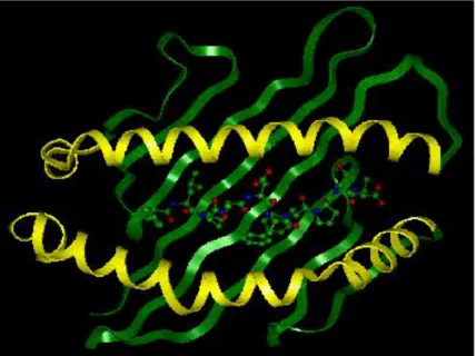
Figure 11. MHC class I molecule from top with peptide bound to the protein groove. Yellow spirals present two alpha helixes, green bands – beta-plated sheet. Between two alpha helixes is green/blue/red peptide chain (antigen).
Introduction
MHC Class I is a heterodimeric membrane molecule consisting of a heavy alpha chain and a light beta-microglobulin chain (Fig. 11). The alpha chain can be divided into three extracellular domains, alpha1, alpha2 and alpha3, in addition to the transmembranous and cytoplasmic domains. Between the alpha-1 and alpha-2 domains a peptide binding groove is located that is formed by beta-pleated sheet on the bottom and two alpha helices on the sides.
This region has several pockets and is capable of binding a small peptide via non-covalent interactions. The beta subunit, a beta-Microglobulin, associates primarily with the alpha-3 domain and is necessary for MHC stability.
Class I molecules usually bind nonapeptides, and less frequently octapeptides or decapeptides.
The peptides are anchored within "pockets" in the peptide-binding groove of MHC class I only at a few positions of the peptides, which are called "anchor residues". Other side chains of the bound peptide also make contact with the groove, but apparently not constrained by any specific pocket. Nonapeptides bind in an extended conformation with a kink near P4 (fourth amino acid of the peptide). Octapeptides bind with less acute kink, whilst decapeptides and longer peptides bind with more pronounced kinks or “zigzags” in the extended conformation.
The MHC/peptide complexes play a fundamental role in regulation of immune responses. T-
cells can recognize antigens normaly as a complex with MHC molecules. Rare MHC-
independent activation of T-cells was also reported. T-cells vary in the need for number of
specific MHC/peptide complex from a few complexes per cell to a few thousand, depending
on the affinity or activation state of the T-cell and on the antigen presenting cell. Recognition
of MHC/peptide complex by T-cells requires antigen to be processed by unfolding and
proteasomal digestion before it complexes with the MHC molecule. Once formed the complex
of antigenic peptide and MHC is generally very stable (half life ~ 24h).
Introduction
1.3 Aim of the study
Breast cancer patients often develop weak but helpful immune response directed toward the tumor antigen MUC1. This protein is in cancer cells expressed in increased and unpolarized manner. Moreover, it is aberrantly O-glycosylated with shorter, denser and more often sialylated sugar chains than those of normal epithelial cells. These structural changes make MUC1 a tumor antigen with diagnostic and therapeutic potential. Several studies showed that the most immunodominat part of the protein is exactly this aberrantly O-glycosylated domain with tandem repeats of a 20-meric peptide sequence. The stimulation of immune responses against the cancer-related form of MUC1 is rationalized to have therapeutic effects in breast cancer patients. For the development of a breast cancer vaccine, those glycoforms should be identified, which are able to induce the strongest cytotoxic and humoral response to the natural target. Hence, we attempted in this study to:
• Identify the influence of glycan structure and position on antigen processing by immunoproteasomes of antigen-presenting cells
• Identify the structure of MUC1 derived glycopeptides that are the best substrate for immunoproteasomes
• Define those MUC1 glycopeptides which are processed to MHC class I binding glyco- epitopes
• Identify glycosylated peptides with high binding capacity to the most frequent MHC class I allele in the human population (HLA-A0201)
• Understand the weak immunogenicity of natural glycoforms of tumor-associated
MUC1.
Materials and Methods
2 MATERIALS AND METHODS
Materials and Methods
MATERIAL
o
2,5-Dihydroxybenzoic acid, DHB, Bruker Daltonics
o
2-mercaptoethanol 50mM, Gibco, UK
o
Accutase PAA Laboratories GmbH,
Germany
o
Acetic acid, Fluka Chemie, Germany
o
Acetone HPLC grade, JT Baker, Germany
o
Acetonitrile Chromasolv for HPLC, Sigma-Aldrich, Germany
o
Alpha-dithiotreithol, DTT, minimum 98%, Sigma-Aldrich, Germany
o
Beta-2 micro globulin from human urine, 90%,
Sigma-Aldrich, Germany
o
BSA Bovine serum albumin fraction V, Carl Roth GmbH, Germany
o
Cacodylic acid sodium salt, 98%, Sigma Chemical
o
Chloroform, anhydrous, 99+%, Aldrich, Germany
o
Cholesterol, min 99%, Sigma-Aldrich, Germany
o
Citric acid monohydrate, KMF optichem, Laborchemie Handels
o
Dimethyl sulphoxide, DMSO, hybrid-max, Sigma, Germany
o
Ethanol, Absolut Chromasolv, Sigma-Aldrich, Germany
o
Ethylendiaminetetraacetic acid, EDTA, disodium salt dihydrat,
Fluka Chemie, Germany
o
FITC Fluorescein isothiocyanate isomer I, Sigma-Aldrich, Germany
o
Foetal Bovine Serum, FCS, heat inactivated,
Gibco, Invitrogen Corporation
o
Formaldehyde solution, min 35%, extra pure,
Merck, Germany
o
HEPES pufferan, >99,5%, Roth, Germany
o
Hydrochloric acid, HCl, 1 mol/l, volumetric solution,
Sigma-Aldrich, Germany
o
Magnesium chloride, MgCl
2, 99%, Sigma-Aldrich, Germany Manganese (II) chloride dehydrate, MnCl
2, Merck, Germany
o
Methanol dried, Sigma-Aldrich, Germany
o
Sodium hydroxide solution, NaOH Fluka
o
Nonessential amino acids, 100X, Biochrom AG
o
Nonidet-P40, Fluka BioChemika
o
PBS Dulbeco, Biochrom AG
o
PEG-PE 2000 1,2-Dipalmitoyl-sn-Glycero-3-Phosphoethanolamine-N- [Methoxy(Polyethylene glycol)-2000], sodium salt,
Avanti Polar Lipids, USA
o
Penicillin-Streptomycin solution, Sigma Aldrich, Germany
o
Phosphoric acid, orto-phosphoric acid 85%, Merck, Germany, KGaA
o
Poly-lactic acid Fluka Chemie GmbH
o
Polyvinyl alcohol Sigma Chemicals
o
POPC, 1-Palmitoyl-2-Oleoyl-sn-Glycero-3-Phosphocholine,
Avanti Polar Lipids, USA
o
Potasium chloride, KCl, >99,5%, Roth, Germany
o
Potasium hydroxide, KOH, Carl Roth GmbH
o
Primulin, Sigma, Germany
o
Protein A Sepharose 4 Fast Flow Amersham Pharmacia Biotech, Sweden
o
Sodium azide, Fluka Chemica
Materials and Methods
o
Sodium chloride, NaCl, KMF Laborchemie Handels GmbH, Germany
o
Sodium hydrogen phosphate, dehydrate, Merck
o
Trifluoroacetic acid, TFA Fluka Chemie
o
Tris-(hydroxymethyl)-aminomethan, Merck
o
Trypsin/EDTA solution, 0,5%/0,2% (w/v) in PBS (10x),
Biochrom AG SPECIFIC CHEMICALS
o
20S immunoproteasomes, Immatics;Germany
o
Albumin from hen egg white, Fluka Biochemika
o
Ficoll-Paque PLUS, Amersham Bioscience, Sweden
o
Fluorescent mounting medium, Dako Cytomation;Danemark
o
GM-CSF, Recombinant human, BD Bioscience, Pharmingen, Germany
o
IL-4, recombinant human, Cell Genix Tecnologie Transfer GmbH Germany
o
Peptides, Mimotopes/Perbio
o
Protease inhibitor cocktail tablets, Complete Mini,
Roche Diagnostics GmbH
o
Proteasome inhibitor Ada(Ahx)3-(Leu)3-vinyl sulphone,
Biomol
o
Sepharose 4b, Pharmacia Bioscience
o
TNF alpha, Tumor necrosis Factor-alpha, Recombinant,
Sigma-Aldrich, Germany
SOFTWARE
o
Confocal Microscope software, Leica confocal software
o
ESI/MS/MS software MassLynx 4.0 Waters
o
FACS software, Expo 32 ADC XL4 Colour/Analysis,
Bruker Daltonics
o
Find Pep http://expasy.org/tools/findpept.html
o
HPLC software Gold chromatography data software,
Beckman Coulter
o
MALDI software Flex Control /Analysis, Bruker Daltonics
o
NetChop http://www.CBS.dtu.dk/services/NetChop/
o
Syfpeithi http://www.syfpeithi.de
PARTICULARS
o
Cell culture plates: 6 well, 48 well, 150cm², 300cm²,
TPP Technoplastic Products AG,
Switzerland
o
Chromabond HR-P glass column, Carl Roth, Germany
o
Cryo-tube vials, NUNC
o
Glass coverslides, microscope cover glasses, Marienfeld, Germany
o
Glass slides, microscope slides, precleaned polysine
tm,
Menzel GmbH, Germany
o
Maldi plate MTP 384 massive Au coated, Bruker Daltonics
Materials and Methods
o
Nanospray needle, spray capillaries, Medium, for Micromass Qtof,
Poxeon Biosystems
o
Nanospray source, Qtof II, Micromass
o
Previal C18 5u rpHPLC column, 150mm x 10mm,
Alltech, Munich, Germany
o
Solid phase extraction backerbond C18 SPE 200mg/3ml,
JT Baker
o
Syringe sterile filter 0,22µm fast flow PES membrane,
TPP Technoplastic Products AG,
Switzerland
o
Thin layer chromatography plates HPTLC aluminium sheets silica gel 60 20x20cm, MERCK, Germany
o
Ultrafiltration tubes, Centriprep centrifugal filer devices,
Millipore Corporation, USA
o
Ultrasphere HPLC ODS column, 2 mm x 15 cm,
Beckman Coulter MASHINES
o
Centrifuge Z323K Hermle
o
Confocal microscope, Leica CTR MIC, Leica Microsystems GmbH
o
ESI/MS/MS Qtof II, Micromass
o
Extruder, Lipofast with stabilisator, Avestin Inc
o
FACS- flow cytometer EPICS XL4C, Beckman Coulter
o
HPLC system, Beckman system gold, Beckman Coulter
o
Incubators- cell culture, Heraeus 6000 Heraeus Instruments
o
Incubators- dry , Function line, Heraeus Instruments
o
MALDI-TOF system, Reflex IV, Bruker Daltonics
o
Peristaltic pump P-1, Pharmacia, Biotech
o
pH-Meter pH 538 Multical Wisseschaflich-Technische Werkstätten GmbH, Germany
o
Sonicator, Brandsonic
o
Ultracentrifuge Optima-L70 Beckman Coulter
o
UV lamp UVIS multipurpose equipment for UV254/366 nm
Desaga, Heidelberg,Germany
o
Vacuum centrifuge, DNA plus, Heto
CELL CULTURE MEDIA
o
CellGrow DC medium, Cell Genix Technologie Transfer GmbH, Germany
o
DMEM Dulbeco’s MEM with stabile glutamine,
Biochrom AG
o
IMDM, basal Iscove medium with stabile glutamine,
Biochrom AG
o
Phenol-red free RPMI 1640 medium, Biochrom AG
o
RPMI 1640 medium, Biochrom AG
ANTIBODY
o
Alexa Fluor 488, goat anti-mouse IgG, Molecular Probes
o
Anti-CD14 microbeads, human, Miltenyi Biotech, Germany
o