• Keine Ergebnisse gefunden

Signaling networks involved in patterning dorsal chorion structures in Drosophila

N/A
N/A
Protected

Academic year: 2021

Aktie "Signaling networks involved in patterning dorsal chorion structures in Drosophila"

Copied!
148
0
0

Wird geladen.... (Jetzt Volltext ansehen)

Volltext

(1)
(2)

Signaling networks involved in patterning dorsal chorion structures in Drosophila

Inaugural-Dissertation zur

Erlangung des Doktorgrades

der Mathematisch-Naturwissenschaftlichen Fakultät der Universität zu Köln

vorgelegt von Bhupendra Shravage

aus Pune, Indien

Köln, 2005

(3)

Bericherstatter:

Prof. Dr. Siegfried Roth PD Dr. Thomas Klein

Tag der muendlichen Pruefung: 8 February 2006

(4)

Acknowledgements

First and foremost I thank Siegfried Roth for his thorough guidance, help, criticisms and allround support. I learnt a lot from him.

I thank all the Roth lab members, past and present for a wonderful co-operative atmosphere, especially Claudia Wunderlich and Martin Technau. I am thankful to Maurijn for his comments on my thesis. Oliver Karst assisted me in creating transgenic flies. I am thankful to Dr. Linne von Berg for helping me with Scanning Electron Microscopy.

I thank Professors of my graduate programme, especially Late Jose’Campos- Ortega, Maria Leptin, Mats Paulsson and group leaders Thomas Klein, Frank Sprenger and Veit Reichmann for their guidance and help. I am thankful to Eberhard Rudloff, Kristina Rafinski, Sebastian Granderath and Brigitte Wilcken-Bergmann for administrative help.

Financial help from Graduate School (IGSGFG) and SFB572 is gratefully acknowledged.

Special thanks to Nelly for giving me a wonderful company and for being there at times when it really mattered. And last but not the least, I would like to express a deep sense of gratitude to my parents and family for their constant support and encouragement.

Bhupendra Shravage

(5)

(To my parents)

(6)

CONTENTS

ACKNOWLEDGEMENTS ABBREVIATIONS

1. INTRODUCTION...1

1.1 Patterning of a developmental field ... 1

1.2 Competence of cells ... 2

1.3 Drosophila oogenesis ... 3

1.3.1 Egg chamber formation ...3

1.4 Patterning of the follicular epithelium... 7

1.5 Genes affecting patterning of the follicular epithelium... 9

1.6 EGF signaling cascade ... 12

1.7 Model for patterning of dorsal appendages ... 16

1.8 TGF−β−β−β signaling cascade ... 18−β 1.8.1 Regulation of TGF- β signaling...22

1.8.2 Ski and Sno oncoproteins ...23

1.8.3 Mechanism of Ski/SnoN action ...25

1.8.4 Role of Ski and SnoN in specification and differentiation...26

2. AIM OF THE RESEARCH WORK ...29

3. RESULTS ...30

3.1 Dpp forms a gradient along the AP axis in the follicular epithelium... 30

3.2 Misexpression of dpp in the follicle cells expands dorsal cell fates along the AP axis... 33

3.3 Loss of Dpp signaling in the follicular epithelium renders them unresponsive to Grk signaling ... 37

(7)

3.4 Misexpression of Grk from the germline dorsalizes the eggshell ... 44

3.5 Combined misexpression of Grk and Dpp leads to novel eggshell phenotypes ... 48

3.6 Cloning of SnoN and its expression of during oogenesis ... 51

3.7 snoN is expressed as a dorsolateral stripe in the follicular epithelium during oogenesis. ... 54

3.8 snoN expression in the follicular epithelium depends on Grk and Dpp... 59

3.9 Generation of a snoN mutant ... 62

3.9.1 sno N-/- is a molecular null allele. ...63

3.9.2 SnoN is required for formation of operculum and dorsal appendages...64

3.9.3 SnoN inhibits Dpp signaling in ovary and in wing ...68

3.10 brk and sog are expressed in distinct domains in the follicular epithelium ... 69

3.10.1 Loss of Sog in follicle cells leads to induction of ectopic dorsal appendage material ...75

3.10.2 Loss of Brk in follicle cells leads to expansion of operculum fate ...77

3.11 Dpp inihibitors function redundantly in the follicular epithelium to specify operculum and dorsal appendages ... 79

3.12 EGF targets rho, aos and kek are regulated by Dpp signaling ... 82

3.13 Rho and Aos function are not essential for specification of dorsal midline... 85

4. DISCUSSION ...88

4.1 The Dpp and Grk function in follicular epithelium... 88

4.1.1 The Dpp gradient and prepatterning of follicular epithelium. ...88

4.1.2 Dpp acts as a competence factor in the follicular epithelium ...90

4.1.3 Dpp promotes at least two different cell fates in the main body follicle cells ...93

4.1.4 Distinct levels of Grk signaling specify operculum and dorsal appendage cell fates ...95

4.1.5 Combinatorial signaling by Dpp and Grk specify operculum and dorsal appendage cell fates 96 4.1.5.1 Model for patterning of dorsal chorion structures...97

4.2 Regulation of Dpp gradient activity... 100

4.2.1 SnoN acts as a repressor of Dpp target genes. ...100

4.2.2 Brk acts as a transcriptional repressor of operculum fate genes ...102

4.2.3 Sog controls diffusion of Dpp in the follicular epithelium ...103

4.2.4 Cooperative roles of Dpp inhibitors in patterning of dorsal chorion structures. ...104

(8)

4.3.1 Rho and Aos function is essential for maintaining dorsal midline fate...106

5. SUMMARY ...109

6. ZUSAMMENFASSUNG ...111

7. MATERIALS AND METHODS ...113

7.1 Fly stocks and genetics ... 113

7.1.1 Breeding of Drosophila melanogaster...115

7.2 Preparation of egg shell and embryonic cuticle ... 115

7.3 Immunohistochemistry and in situ hybridization... 116

7.3.1 Fixation of ovaries for immunostainings ...116

7.3.2 Antibody staining of ovaries...116

7.3.3 Fixation of ovaries for in situ...116

7.3.4 In situ hybridisation of ovaries ...116

7.3.5 Mounting the stained embryos and ovaries...117

7.4 Molecular Cloning ... 118

7.4.1 Cloning of snoN/ski ...118

7.4.2 DNA work and germline transformation ...118

7.4.3 Production of antibody against SnoN ...119

7.5 Induction of Mitotic clones ... 120

7.6 Western blotting ... 120

7.7 Scanning Electron microscopy (SEM) ... 120
8. BIBLIOGRAPHY ...121

ERKLAERUNG...136

LEBENSLAUF...137

(9)

Abbreviations

TGF-β Transforming Growth Factor β

aa Amino acid

Alp Activin like Protein Anti β gal Anti β galactosidase

Aos Argos

AP Anterior-Posterior

Babo Baboon

BMP Bone Morphogenic Protein

bp base pairs

BR-C Broad-Complex

Brk Brinker

cAMP cyclic Adenosine mono-phosphate CBP cAMP binding protein

CMFCs Centripetally Migrating Follicle Cells

Cni Cornichon

Co-Smad Common Smad

CREB cAMP response element binding protein

Cys Cysteine

Dac Dachshund

Dad Daughters against Dpp

dl Dorsal

Dpp Decapentaplegic

Drk Downstream of Receptor Kinase

DV Dorsal-Ventral

E2F Elongation Factor 2 E2F-4 E2F transcription factor 4 EGF Epidermal Growth Factor

EGFR Epidermal Growth Factor Receptor

ETS E-twenty six

Fas III Fasciclin III

FGF Fibroblast Growth Factor Gbb Glass bottom boat

GDP Guanine Di-Phosphate GFP Green Fluorescent Protein

Grk Gurken

GS Glycins and Serine rich GTP Guanine Tri-Phosphate HDAC Histone deacetylase complex

His Histidine

hop hopscotch

Iro-C Iroquious-C I-Smads Inhibitory Smads JAK Janus activated kinase

kb kilo bases

kDa Kilo Dalton

Kek Kekkon

(10)

Krn Keren

LRR Leucine Rich Repeat Mad Mothers against Dpp

MAPK Mitogen Activated Protein Kinase

MAPKK Mitogen Activated Protein Kinase Kinase

Mav Maverick

Med Medea

MH1 Mad homology domain 1

MH2 Mad homology domain 2

Mirr Mirror

Myo Myoglianin

ng Nanogram

Omb Optomotor Blind

ORF Open Readinfg Frame

pMad Phophorylated Mad

Pnt Pointed

Put Punt

Rho Rhomboid

R-Smad Receptor regulated Smad

Sal Spalt

Sara Smad anchor for receptor activation

Sax Saxophone

SBE Smad Binding Element

Scw Screw

SEM Scanning Electron Microscopy Ski Sloan Kettering virus induced protein

Sl semi-lethal

Sno Ski related novel Sog Short Gastrulation

Sos Son of sevenless

Spi Spitz

Sty Sprouty

TGF-α Transforming Growth Factor α TIE TGF-β inhibitory element

Tkv Thickveins

Tld Tolliod

Tsg Twisted Gastrulation

UAS Upstream Activator Sequence

Upd Unpaired

Vg Vestigial

Vn Vein

Wg Wingless

Wit Wishful thinking

wt Wild type

λtop Lambda-torpedo

(11)

INTRODUCTION

1. Introduction

Signaling between cells is a widely used mechanism by which cell fate and patterning is determined in development. It has emerged in the recent years that only a relatively small number of genetic networks are essential for designing the body plan during development. Signaling molecules are key to such genetic networks.

The work presented in this thesis was inspired by the question “How Transforming Growth Factor−β (Decapentaplegic) signaling pathway and Epidermal Growth Factor (Gurken) signaling pathway collaborate to pattern the follicular epithelium during Drosophila oogenesis? “. Emphasis was laid on elucidating the role of Dpp signaling and its regulation by SnoN, a transcriptional repressor of TGF−β pathway.

1.1 Patterning of a developmental field

Patterning of a developmental field is primarily achieved by two distinct mechanisms involving cell-cell communication: short range signaling and long range signaling. Short range signaling functions across a few cell diameters and specifies distinct cell fates in a developmentally equivalent group of cells. A well known example is the phenomenon of lateral inhibition governed by Notch signaling during neurogenesis (reviewed in Beatus and Lendahl, 1998).

Patterning by long range signaling can occur over several cell diameters and is

achieved by two different mechanisms: Relay signaling and Morphogen

signaling. In relay signaling, different cell identities are designated by a series

of sequential inductive signaling events that are relayed between adjacent cells

in the developmental field. One of the best studied examples of relay signaling

is that of Epidermal Growth Factor Receptor (EGFR) controlled vulval

development in Caenorhabditis elegans (Dutt et al., 2004). LIN-3, one of the

EGF-like ligands in C. elegans, is expressed in the anchor cell of the developing

gonad. It induces primary vulval cell fates in adjacent precursor cells by

(12)

INTRODUCTION

(the Rhomboid homolog in C. elegans) in turn positively amplifies the signal in these cells by activating LIN-3, thus relaying the signal further to additional precursor cells. In contrast, morphogen signaling is executed by secreted ligands (peptides) which have an ability to diffuse away from the source tissue.

Secreted signaling molecules belonging to the TGF−β superfamily, Wingless/Wnt, Hedgehog and Fibroblast Growth Factor (FGF) family have been identified and implicated as organizers of pattern and growth in many developmental contexts both, in vertebrates and in invertebrates (reviewed in Tabata, 2001). These molecules are termed as morphogens (literally “form giving”) because of their ability to specify cell fates (activate genes) in a concentration dependent manner. Thus, by the process of long range signaling distinct cell fates can be induced over several cell diameters in a developmentally competent field.

1.2 Competence of cells

Patterning can be induced only when two different components of the system

are present. First component is the inducing cells (tissue) that produce a

signal(s) and the second component is the responding cells (tissue) which have

an ability to react to the inductive signal(s). Competence is the ability to

respond to specific inductive signal (Waddington, 1940; Stern, 2000- in this

article Waddington’s contributions to developmental biology are described). It

is an essential pre-requisite of the responding cells. Competence is not a passive

state but an actively acquired condition. It may depend on, for example, the

presence of appropriate receptor and transducing mechanism or on the presence

of particular transcription factor(s) needed for gene activation. For example, in

the developing mammalian eye, Pax6 protein appears to be important in making

the ectoderm competent to respond to the inductive signal from the optic

vesicle. Pax6 is expressed only in the head ectoderm which can respond to

signals from the optic vesicle by inducing the lens (Li et al., 1994). The final

proof for Pax6 acting as a competence factor was demonstrated by

recombination experiments using embryonic rat eye tissue. When the head

(13)

INTRODUCTION

ectoderm from Pax-6 mutant rat embryos was combined with wild type optic vesicle no lenses were formed, however lens tissue was formed when the head ectoderm was derived from wild type embryos even though optic vesicles were obtained from Pax6 mutant embryos (Fujiwara et al., 1994).

1.3 Drosophila oogenesis

Drosophila oogenesis involves co-coordinated development of the germ cells and the overlying follicular epithelium. The follicle cells provide a genetically tractable system to investigate the process of patterning and morphogenesis.

Both long range and short range cell-cell communications pattern the domains of follicle cells that will create specific eggshell structures. Hence, the Drosophila ovary provides an excellent system for the study of epithelial patterning.

1.3.1 Egg chamber formation

The mature Drosophila egg is an amazingly complex cell. It is over 0.5 mm long, contains patterning information to establish the anterior-posterior (AP) and dorsal-ventral (DV) axes of the embryo and bears a highly complex eggshell that facilitates embryonic development in harsh external environment.

This highly sophisticated egg develops from a discrete structure called the “egg

chamber” (King, 1970; Spradling, 1993). Egg chambers are formed at the

anterior tip of the ovary and move posteriorly as they develop. They are

connected via stalk cells to form a linear array called the “ovariole”. Each ovary

contains on an average 16-17 ovarioles. The ovariole is the functional unit of

the ovary. The whole process of oogenesis takes about 79 hours at 25

0

C and is

divided into various stages based on morphological details (Figure 1A, B).

(14)

INTRODUCTION

Figure 1

Stages of Drosophila oogenesis

(A) The anterior tip of a dissected ovariole showing the germarium (G) with young egg chambers (st. 1-4). (B) Dissected egg chambers from stage 8 to 14. Anterior in all figures is the left and dorsal to the top except in stage 14 where it is facing the viewer.

The developing oocyte can be seen here as an opaque structure (the posterior half) at

stage 9 onwards. Follicular epithelium surrounds the ooctye and can be seen as a

transperant layer around the oocyte. Adapted from King (1970).

(15)

INTRODUCTION

The egg chamber forms from two distinct cell types: germ cells and somatic cells. The germ cells form the developing oocyte and 15 nurse cells while the somatic cells form a monolayer epithelium surrounding the germ cells. The germarium, present at the anterior tip of the ovariole, is an assembly line in which new egg chambers are produced from progeny of germline and somatic stem cells. Based on morphological differentiation, the germarium is subdivided into three regions: region 1, 2 and 3 (Spradling, 1993). The process of formation of the egg chamber starts in region 1 of the germarium where each stem cell divides to form a new stem cell and a cystoblast. The cystoblast undergoes four incomplete divisions to form a syncytial cyst containing 16 cells. These cells are interconnected by cytoplasmic bridges called the “ring canals”. As the cyst moves posteriorly in the germarium, one of the cells is selected as an oocyte while the remaining 15 cells become nurse cells. The follicle stem cells reside in region 2a of the germarium and encase the cyst in region 2b to form a monolayer epithelium (Margolis and Spradling, 1995).

As the newly formed egg chamber moves from region 2b to region 3 of the germarium, the developing oocyte acquires an asymmetric localization within the cyst and its localization provides first co-ordinates for the anterior-posterior axis. Thus, the oocyte acquires posterior position in the newly formed egg chamber (Spradling, 1993; Huynh and St Johnston, 2004). The synthetically active nurse cells continuously transfer their contents via the ring canals to the transcriptionally silent oocyte during oogenesis. At the end of oogenesis, the nurse cells rapidly transfer their contents into the oocyte in a process known as

“nurse cell dumping”. As a result, the nurse cells are reduced to a small group of apoptotic nuclei at the anterior end of a large oocyte.

The follicle cells proliferate untill stage 6 of oogenesis to a number of around

1000 and stop dividing. At this stage of oogenesis, the follicle cells start

migrating towards the posterior of the egg chamber. This leaves the anterior end

of the egg chamber with very few follicle cells which become stretched to cover

the nurse cells, while the follicle cells covering the oocyte, the “main body

follicle cells”, become columnar in shape. At the same time a small group of

follicle cells, called the “border cells”, migrate from the anterior tip of the egg

(16)

INTRODUCTION

chamber to the anterior tip of the growing oocyte. At the end of oogenesis these cells form the channel for sperm entry called the “micropyle”. Finally, at stage 10 of oogenesis a group of follicle cells migrate anteriorly in between the oocyte and the nurse cells called the “centripetally migrating follicle cells”

(CMFCs).

During stage 7 till 10, the follicle cells undergo endoreduplication and become polyploid and it is at this time transcription of genes for forming the chorion starts. Two large groups of follicle cells lay down respiratory appendages, called “the dorsal appendages”, which are positioned at the dorsal anterior of the egg. Each dorsal filament is formed by a group of follicle cells (50-55 cells;

Roth, 1999; James and Berg, 2003) that migrate over the anterior part of the oocyte at stage 11. Imprints of the follicle cells are visible on the mature egg.

Several structures on the chorion allow orientation of the axes of the egg. The

anterior end bears the micropyle and operculum while the posterior end is

marked by an aeropyle. Figure 2 shows wild type (wt) Drosophila egg.

(17)

INTRODUCTION

Figure 2

The Drosophila egg

A Scanning Electron Microscope (SEM) image of a wildtype Drosophila egg (dorsal view). The anteriorly located micropyle, operculum and dorsal appendages can clearly be seen. The dorsal boundary of the operculum is limited by a collar.

1.4 Patterning of the follicular epithelium

In Drosophila, patterning of the follicular epithelium covering the developing oocyte is achieved by inductive signaling. Two major signal pathways converge to induce a subpopulation of dorsal anterior follicle cells to adopt cell fates which give rise to the dorsal appendages and the operculum (Dobens and Raftery, 2000; Peri and Roth, 2000; Roth, 2003; Berg, 2005; Horne-Badovinac and Bilder, 2005). One of the signals is initiated by the EGF signaling pathway.

The EGF/TGF-α (Transforming Growth Factor-α) like ligand Gurken signals

twice from the developing oocyte to the overlying follicle cells at stage 5-6 in

order to specify first, posterior cell fates and subsequently dorsal cell fates at

stage 9-10 (Gonzalez-Reyes et al., 1995; Roth et al., 1995; Nilson and

(18)

INTRODUCTION

Schupbach, 1999; Roth, 2003). Following the first Grk signal, a cytoskeletal rearrangement triggers the migration of the oocyte nucleus towards the anterior of the oocyte, where it lies at an asymmetric position (Koch and Spitzer, 1983;

Peri and Roth, 2000). Grk, which is tightly associated with the oocyte nucleus, signals to the adjacent follicle cells inducing them to take on dorsal fates (Schupbach, 1987). The doralizing Grk signal has two effects on the follicle cells. First, it restricts pipe to a ventral stripe in the follicular epithelium (Sen et al., 1998; Peri et al., 2002). This event is essential for DV patterning of the embryo (Nilson and Schupbach, 1999; van Eeden and St Johnston, 1999; Peri et al., 2002; Roth, 2003). Second, it induces dorsal anterior follicle cells to produce operculum and dorsal appendages. Dorsal appendages are derived from two dorsal patches of Broad-Complex (BR-C) expressing cells (Deng and Bownes, 1997). The characteristic pattern and placement of dorsal appendages along the DV axis is suggested to be established by an intricate mechanism involving signal amplification by two genes: rhomboid (rho) and spitz (spi) and feedback inhibition by argos (aos) (Wasserman and Freeman, 1998;

Shvartsman et al., 2002). This aspect is discussed in detail in later sections.

The restriction of pipe to the ventral follicle cells occurs along the entire AP axis. However, the operculum and dorsal appendages form only at the anterior.

In addition, Queenan et al. (1997) also found that the expression of the dorsal genes: rho and aos is still confined to the anterior half when an activated form of the EGFR (λtop) was misexpressed in the whole follicular epithelium. How is the response to the second Grk signal localized? It is suggested that formation of the operculum and dorsal appendages require a combination of both dorsalizing Grk and a second anterior signal provided by Dpp, one of the TGF-β family member in Drosophila (Deng and Bownes, 1997; Queenan et al., 1997;

Peri and Roth, 2000). dpp expression can be first detected in the follicle cells

present at the anteriormost tip of the egg chamber at stage 8 of oogenesis

(Twombly et al., 1996; Dobens and Raftery, 2000; Peri and Roth, 2000). These

cells start migration at stage 9 of oogenesis towards the posterior to cover the

developing oocyte. In late oogenesis, dpp is associated with the stretched

follicle cells and the CMFCs. Partial loss of function alleles of dpp and/or its

components lead to production of eggs which exhibit extremely reduced

(19)

INTRODUCTION

opercula and dorsal appendages (Twombly et al., 1996). Conversely, eggs produced by females ubiquitously expressing dpp display expanded anterior eggshell structures (Twombly et al., 1996; Dobens et al., 1997; Dobens and Raftery, 2000; Gupta and Schupbach, 2003). Based on these findings, Dpp is suggested to function in two different ways. First, it is suggested to form a morphogen gradient in the follicular epithelium along the AP axis (Twombly et al., 1996; Peri and Roth, 2000). Second, Dpp is suggested to induce competence in the anterior subpopulation of main body follicle cells so that they are able to respond to the dorsalizing Grk signal (Peri and Roth, 2000). In addtition, Dpp is suggested to define the boundary between operculum and dorsal appendages (Twombly et al., 1996; Dobens et al., 1997). We would like to address these questions and define the molecular role of Dpp in the follicular epithelium.

Peri and Roth (2000) showed that the combined activation of EGFR and Dpp signaling cascades in a posterior subpopulation of main body follicle cells leads to induction of dorsal appendages at the posterior of the egg. Moreover, induction of rho and BR-C was observed at the posterior in such egg chambers.

This proved that in addition to Grk, rho needs Dpp for its expression (Peri and Roth, 2000). Thus, how the Dpp and EGFR activities specify domains (distinct cell populations) along the AP and DV axis in the follicular epithelium needs to be clarified.

1.5 Genes affecting patterning of the follicular epithelium

The specification of dorsal appendages and their subsequent morphogenesis

requires induction and interaction of several different signaling pathways within

the follicular epithelium. Thus, several genes control the specification of DV

pattern in the follicle cells (Spradling, 1993). Many genetic screens have

identified mutants which affect the patterning process. Indeed, over five-dozen

genes which affect the dorsal appendage patterning have been identified so far

(Table 1; reviewed in Berg, 2005).

(20)

INTRODUCTION

Dorsoventral patterning mutations are essentially of three general types – ventralizing (e.g. grk), dorsalizing (e.g. K10) and midline-minus (e.g. pointed).

All genes in these three classes are required to establish correct cell fate and/or placement of the dorsal appendages. Loss of Grk signal ventralizes the egg, producing elongated eggs lacking dorsal structures. Null alleles of grk produce eggs which have a micropyle at both the termini indicating that Grk signaling is needed for establishing posterior cell fates (Gonzalez-Reyes et al., 1995; Roth et al., 1995). In fact, the correct dosage of Grk is needed to induce two dorsal appendages. Hypomorphic alleles of grk lead to production of eggs with single thin dorsal appendage (Schupbach and Wieschaus, 1991; Neuman-Silberberg and Schupbach, 1994). The dorsalized egg phenotype can result from mutations in genes affecting several aspects of Grk signaling i.e. mislocalization of grk mRNA during oogenesis or loss of inhibitory control on the signaling cascade.

K10 encodes for DNA binding protein and has been shown to restrict localization of grk to the dorsal anterior corner of the oocyte, close to the oocyte nucleus (Prost et al., 1988). Midline minus mutation(s) produces eggs with single broad dorsal appendage. This phenotype results from defects in genes required for modulating Grk signaling such as spitz or pointed (Golembo et al., 1996b; Wasserman and Freeman, 1998).

The genes listed in Table 1 belong to and/or are regulated by several signaling cascades including TGF-β, EGF and Notch signaling pathways. The exact mechanisms that achieve the patterning, the identity of all molecules and the role that each gene has in patterning remain unclear. Knowledge of their molecular role and their interactions with each other would not only help us understand the patterning of follicular epithelium but also on larger scale aid us in understanding their roles in relation to one another in development of several model organisms.

A detailed description and illustration of Dpp and Grk pathways in Drosophila

is described in later sections.

(21)

INTRODUCTION

Table 1

Genes involved in dorsal appendage patterning

Genes marked with an asterick (*) were used for mutant studies and/or for expression

analysis in this work. Table Adapted and modified from Berg, CA (2005).

(22)

INTRODUCTION

1.6 EGF signaling cascade

The evolutionary conserved receptor tyrosine kinase (RTK) pathway is extensively used intercellular signal transduction mechanism that regulates cell fate specification, proliferation, migration and survival (Moghal and Sternberg, 1999). RTKs comprise a large family of integral membrane proteins with highly divergent extracellular domains coupled to a conserved intracellular protein tyrosine kinase motif (Freeman, 1998; Schlessinger, 2000). RTKs can be activated by several classes of ligands one of them being EGF. Ligand binding induces dimerization of RTKs which results in both auto and trans phosphorylation of specific tyrosine residues on the C-terminal cytoplasmic tail of the receptor (Yarden and Ullrich, 1988; Schlessinger, 2000). This modification generates docking site for phosphotyrosine binding adaptor proteins (PTB) known as (DRK ) Downstream of Receptor Kinase (vertebrate homologue is designated as Grb; Lowenstein et al., 1992; Olivier et al., 1993;

Simon et al., 1993). DRK recruits the guanidine nucleotide exchange factor which activates the GTPase-Ras by catalyzing the exchange of GDP for GTP.

Once Ras is active, it recruits and activates the serine/theronine kinase Raf and many other cytoplasmic components (Kolch, 2000; Avruch et al., 2001).

Activated Raf phosphorylates and activates MEK, a dual specificity kinase which phosphorylates tyrosine and serine/threonine residues (also called Mitogen Activated Protein Kinase Kinase-MAPKK). Activated MAPKK in turn phosphorylates and activates MAPK which translocates to the nucleus where it phosphorylates and modulates the activity of specific transcription factors (Marshall, 1994; Kolch, 2000).

The Drosophila EGF signaling cascade is depicted in Figure 3. In Drosophila,

only a single gene has been found to encode a homologue of the vertebrate EGF

receptor called “EGFR” (Wadsworth et al., 1985; Schejter et al., 1986). EGFR

is used several times during Drosophila development and has separate roles in

oogenesis, embryogenesis and imaginal disc development (Schweitzer and

Shilo, 1997). Drosophila EGFR can be activated by four different ligands Spi,

Keren (Krn), Grk and Vein (Vn) (Shilo, 2003). Three of the ligands namely Spi,

Krn and Grk, belonging to EGF/TGF-α family, are produced as transmembrane

(23)

INTRODUCTION

precursors. The primary activating ligand Spi is responsible for EGFR activation in most tissues (Rutledge et al., 1992). A ligand structurally related to Spi has been recently identified and termed as Krn (Reich and Shilo, 2002;

Urban et al., 2002). Krn can substitute Spi in most tissues during development.

Grk, a third TGF-α homologue, is restricted to the activation of EGFR in the follicle cells of the ovary (Neuman-Silberberg and Schupbach, 1993; Gonzalez- Reyes et al., 1995; Roth et al., 1995). Vn, a secreted ligand similar to neuregulins, is used in tissues where low activation of EGF pathway is needed (Schnepp et al., 1996). It functions as a main ligand in some tissues. For example, Vn induces several distal cell fates in the leg imaginal discs (Schnepp et al., 1998). Spi, Krn and Grk have to be processed to an active form by proteolysis. A clue to the mechanism regulating Spi processing emerged from the identification of mutations in two genes, Star and rho, that gave rise to phenotypes surprisingly similar to spi (Mayer and Nusslein-Volhard, 1988).

Star, a novel transmembrane protein was shown to act as a cargo receptor in trafficking Spi from the endoplasmic reticulum to Golgi (Lee et al., 2001). Once reaching the Golgi, Spi is proteolytically cleaved by Rho. This releases Spi from the membrane and thus is able to activate the EGFR once it reaches the plasma membrane. Spi and Star are broadly expressed while the expression of Rho is tightly regulated and is dynamic. It thus appears that expression of Rho is the limiting step in EGFR activation. This was demonstrated experimentally.

Ectopically expressing rho in diverse tissues and contexts is sufficient to increase the level of EGFR activation (Golembo et al., 1996a). Similarly, increased levels of Grk lead to increase in the level of EGFR activation (Ghiglione et al., 2002).

EGFR signaling can occur over several cell diameters due to secretion of

diffusible ligands. This has been very well demonstrated in case of patterning of

the embryonic ventral ectoderm by Spi and induction of the leg segments by Vn

(Schweitzer et al., 1995b; Golembo et al., 1996a; Campbell, 2002). In both

these cases the ligands are believed to function as morphogens inducing more

than one cell fates depending upon the concentration of the ligand.

(24)

INTRODUCTION

EGFR activation is regulated at the extracellular, the receptor and the intracellular level by various inhibitors and repressors which constitute negative feedback loops (for a recent review see Shilo, 2005). At the extracellular level, Aos is the primary inhibitor and is expressed at highest levels of EGF signaling.

Aos was believed to function by competing with the activating ligands to bind

to the EGF receptor (Freeman et al., 1992; Schweitzer et al., 1995a; Jin et al.,

2000). However, Klein and colleagues (2004) proved that Aos inhibits EGFR

signaling without interacting with the receptor directly but instead by

sequestering the EGFR activating ligand Spi. Aos binds tightly to the EGF

motif of Spi and forms (1:1) Spi: Aos complex that does not bind to the EGFR

in vitro or at the cell surface (Klein et al., 2004). Aos is a secreted protein and

hence can inhibit EGFR signaling several diameters away from its site of

synthesis. Kekkon (Kek) is a transmembrane protein that binds the EGFR

extracellular domain and attenuates receptor dimerizaton (Ghiglione et al.,

1999). Another inhibitor Sprouty (Sty), an intracellular protein, acts by

intercepting essential elements of the Ras/MAPK cascade through diverse

mechanisms (Casci et al., 1999; Kim and Bar-Sagi, 2004). Both Kek and Sty act

cell autonomously and in a context dependent way. At the transcriptional level,

the EGFR target genes can be repressed in the nucleus by Pointed (Pnt), an

ETS-domain transcriptional repressor (Gabay et al., 1996; Morimoto et al.,

1996). Expression of D-Cbl, an E3 ubiquitin ligase, was shown to be critical for

attenuating EGF signaling in the follicle cells. D-Cbl ubiquitinates the activated

EGFR and targets it to the proteosome for subsequent degradation (Pai et al.,

2000).

(25)

INTRODUCTION

Figure 3

The EGF signaling pathway in Drosophila

Binding of the any of the four ligands- Spi, Grk, Vn, Krn to the EGFR induces Drk binding to the phosphorylated receptor and to Sos (Son of Sevenless). Sos stimulates GTP/GDP exchange on the G protein Ras. Activated Ras interacts with the effector protein Raf which phosphorylates MAP-kinase-kinase (MAPKK, MEK; D-sor in Drosophila). As a result, MAPKK activates MAPK (ERK, rolled in Drosophila);

MAPK can phosphorylate several other molecules in the nucleus which control gene activation or repression. Rho is expressed in response to EGFR activation. Rho processes Spi in the Golgi (shown in green) leading to positive feedback loop on pathway activation. Induction of Aos in turn downregulates the signal thus forming a negative feedback loop. D-Cbl targets the phosphorylated EGFR to the proteosome. In addition, Sty and Pnt (in the nucleus) can inhibit EGFR signaling. Positive inputs are represented with arrows while inhibitory inputs are depicted by a bar. Nucleus is depicted as a oval shaped structure (dotted black oval). P denotes phosphorylation.

Black arrow in the nucleus represents the direction of transcription.

(26)

INTRODUCTION

1.7 Model for patterning of dorsal appendages

Based on the sequence of EGFR mediated signaling events an elegant model for patterning of the dorsal appendages was proposed by Wasserman and Freeman in 1998 (Figure 4). The model is described as following. At stage 8/9 Grk, localized close to the oocyte nucleus, initiates signaling to the overlying follicle cells. Grk signal induces expression of rho in a dorsoanterior patch of follicle cells where it activates Spi by proteolytic processing (Urban et al., 2001).

Cleavage of Spi leads to an increase in EGFR signaling visualized by increase in MAP kinase activation in width and amplitude. At stage 10, the vitelline membrane is secreted around the developing oocyte and therefore may prevent further signaling from the oocyte to the follicle cells. Vn also contributes to this autocrine amplification of EGFR signaling. The Rho/Spi/Vn amplification induces Aos expression in dorsal midline cells at stage 10, leading to local inhibition of signaling at the dorsal midline. The resulting signaling profile has twin peaks that eventually specify the position of the dorsal appendages.

Although the model integrates both positive and negative feedback loops

involved in patterning, there are several drawbacks. First, the clonal analysis of

genes involved in patterning namely rho, spi and aos was performed in

unmarked clones. Only the resulting eggshell phenotype was observed as

readout of the patterning processes (Wasserman and Freeman, 1998). Second,

there is a disparity between the results obtained by Wasserman and Freeman

(1998) who propose that dorsal appendages are formed at highest levels of

MAPK signaling and Peri et al. (1999) who suggest that dorsal appendages are

formed at intermediate levels of MAPK signaling. Finally, recent biochemical

studies have revealed that Aos associates predominantly with Spi, to form

nonfunctional heterodimers (Klein et al., 2004). Therefore, Aos might allow

other EGF ligands namely Vn to diffuse readily and allow a lower level of

MAPK activation. We have addressed these aspects to better define the role of

Rho and Aos mediated feedback loops on patterning aspects

(27)

INTRODUCTION

Figure 4

Dorsal patterning of the egg is a three stage process

Left panel shows representative cross section through developing oocyte at different

developmental stages. Dorsal side is up. Right panel represents the net EGFR signaling

(MAP kinase activity) at the respective stages. See text for more details. Adapted from

Wasserman and Freeman (1998).

(28)

INTRODUCTION

1.8 TGF−β −β −β signaling cascade −β

The TGF−β signaling cascade regulates numerous cellular responses such as proliferation, differentiation, migration and apoptosis (reviewed in Massague, 1998; Massague et al., 2000). The basic molecular mechanism of TGF−β signaling pathway has been unravelled (reviewed in Attisano and Wrana, 1998;

Derynck and Zhang, 2003; Shi and Massague, 2003). TGF−β related ligands bind to a heteromeric complex of type I and type II transmembrane serine threonine kinases. Ligand binding triggers association between the receptors, enabling the constitutively active type II kinase to phosphorylate the type I receptor within a specific domain termed the GS (glycine and serine rich) domain. The activated type I receptor initiates pathway-specific (BMP specific or TGF-−β/Activin specific) signaling by phosphorylating pathway-specific receptor-regulated Smad (R-Smad) family of cytoplasmic signal transduction protein. Activated R-Smads associate with Smad4, a common Smad (Co-Smad) shared by both the pathways. The heteromeric R-Smad/Co-Smad complex translocates to the nucleus and associates with DNA−binding cofactors to directly activate or repress target gene expression. Smad4 is obligately required for most cellular responses; however, in few cases signaling has been reported to occur in absence of Co-Smad (Wisotzkey et al., 1998; Sirard et al., 2000).

Both R-Smads and Co-Smads contain highly conserved Mad homology domain (MH1 and MH2) domain separated by a linker region. The MH2 domain of R- Smads has an additional SSXS motif which is substrate for phosphorylation by the type I receptor kinase (Wrana, 2000).

The Drosophila TGF−β cascade is depicted in Figure 5. Seven TGF−β related

ligands have been identified in Drosophila through a combination of molecular

genetics and genome annotation techniques (Raftery et al., 1995; Raftery and

Sutherland, 1999). Three of the ligands belong to the BMP subfamily while one

belongs to the TGF−β/Activin subfamily. The molecular details of ligands

Maverick (Mav), Activin like protein (Alp) and Myoglianin (Myo) are still

unknown and therefore they are designated as orphan ligands (Raftery and

Sutherland, 1999; Parker et al., 2004). Dpp represents the BMP2/4 ortholog in

(29)

INTRODUCTION

flies, while Screw (Scw) and Glass bottom boat (Gbb/60A) are related to BMP5/6/7/8 (reviewed in Parker et al., 2004).

Besides its role in specification of embryonic axes and patterning of imaginal discs, Dpp signaling is also essential for Drosophila oogenesis (Spencer et al., 1982; Padgett et al., 1987; Twombly et al., 1996). One of the striking properties of Dpp is its ability to function as a morphogen thereby inducing multiple cell fates in a concentration dependent manner (Tabata, 2001; Raftery and Sutherland, 2003). How is a stable gradient of Dpp established and interpreted is one of the key issues in Drosophila development? Dpp is suggested to form gradient by at least two different mechanisms in Drosophila. One is dependent on long range diffusion of Dpp from its site of synthesis, while the second involves diffusion of extracellular inhibitor, called Short gastrulation (Sog), into the domain of Dpp expression thus resulting in a gradient of signaling activity (Srinivasan et al., 2002; Raftery and Sutherland, 2003). The role of Sog in gradient formation is described in later sections.

Dpp acts as a long range morphogen in the wing imaginal disc (Tabata, 2001).

dpp is transcribed in a narrow stripe of cells at the AP compartment boundary of the wing disc where it can induce transcription of target genes over 20 cell diameters. The Dpp-responsive genes vestigial (vg), optomotor blind (omb) and spalt (sal) are activated at different signaling thresholds which subsequently subdivide the wing disc into distinct regions (Nellen et al., 1996). Biologically active GFP-tagged Dpp has been visualized to form a gradient in either direction from its site of synthesis in the wing disc (Entchev et al., 2000;

Teleman and Cohen, 2000). In the embryo, Dpp is required to induce different cell fates in the dorsal region. High levels of Dpp specify the amnioserosa while lower levels specify the dorsal ectoderm (Raftery and Sutherland, 2003).

In Drosophila, only two type II receptors, Punt (Put) and Wishful thinking

(Wit) and three type I receptors Thickveins (Tkv), Saxophone (Sax) and Baboon

(Babo) have been described (Brummel et al., 1994; Penton et al., 1994; Xie et

al., 1994; Letsou et al., 1995; Aberle et al., 2002; Parker et al., 2004). In

vertebrates, so far a dozen receptors (I and II combined) and up to 29 ligands

(30)

INTRODUCTION

have been identified (for a recent review see Feng and Derynck, 2005). Ligand selectively is determined by the type I receptor in BMP pathway. In contrast, in TGF−β/Activin pathways, ligand selectively is directed primarily by the type II receptor (Parker et al., 2004; Feng and Derynck, 2005). Moreover, R-Smads, the substrates for type I receptor, show pathway specificity. Smads 1, 5 and 8 are specific for BMP signaling while Smad 2 and Smad 3 show specificity towards Activins/TGF−βs mediated signaling (Miyazawa et al., 2002). In Drosophila, Mothers against Dpp (Mad) is the only Dpp/BMP-specific R-Smad (Sekelsky et al., 1995) while Smox/dSmad2 (Smad on ‘X’ chromosome) has been reported to be specific to Activin signaling (Henderson and Andrew, 1998). Only one member of Co-Smad has been identified in each model system, namely Smad4 (also called DPC4) in vertebrates (Lagna et al., 1996), Medea in Drosophila (Raftery et al., 1995; Das et al., 1998; Wisotzkey et al., 1998) and sma-4 in C. elegans (Savage et al., 1996). Co-Smads are therefore shared by all R-Smads and are required for the formation of functional transcriptional complexes (Parker et al., 2004).

Once translocated to the nucleus, the Smad complex modulates TGF−β target

gene expression by binding to Smad binding elements (SBE) via their MH1

domain (Feng and Derynck, 2005). Smad3 MH1 domain binds 5’-GTCT

sequence with a higher affinity while Smad1 (Mad) binds to 5’-GCCG (Kim et

al., 1997; Shi et al., 1998; Korchynskyi and ten Dijke, 2002). Several target

genes of TGF- β have been shown to harbor SBEs in their 5’ promoter regions

(Jonk et al., 1998; Attisano and Wrana, 2000).

(31)

INTRODUCTION

Figure 5

The BMP and Activin signaling pathway in Drosophila

The BMP ligands Dpp, Gbb and Scw, act through the type I receptors Tkv and Sax, resulting in phosphorylation of Mad, its association with the co-Smad Med, translocation of the complex into the nucleus and regulation of target gene expression.

The type II receptors Put and Wit display dual specificity and function in both BMP and Activin pathways. Activin pathway that involves the type I receptor Babo and dSmad2 has been identified in Drosophila. Only one ligand, dActivin, has been shown to signal through Baboon (Babo) in Drosophila. It is unknown if Mav, Alp and Myo activate BMP or the Activin pathway. dSmurf, FKBP12, dSara function intracellularly;

here they are depicted extracellularly due to space constraints. Positive inputs are represented with arrows while inhibitory inputs are depicted by a red bar. Nucleus is depicted as a oval shaped structure (dotted black oval). P denotes phosphorylation.

Black arrow in the nucleus represents the direction of transcription.

(32)

INTRODUCTION

1.8.1 Regulation of TGF- β β β β signaling

TGF−β signaling is modulated by several molecules at the extracellular level.

These molecules are primarily secreted proteins which result in alteration of ligand-receptor binding, ligand diffusion and/or the local ligand concentration.

In Drosophila, the function of several proteins has been identified i.e Sog, Tsg (Twisted gastrulation) and Tolloid (tld). Sog, Tsg and Tld function by a unique mechanism to promote Dpp signaling in the dorsal most region of the embryo (Shimell et al., 1991; Francois et al., 1994; Mason et al., 1994). Sog forms a ventral to dorsal gradient in the embryo (Ashe, 2002; Srinivasan et al., 2002).

Sog binds Dpp thus inhibiting its function and simultaneously transports it to the dorsalmost region of the embryo alone or together with Tsg (Mason et al., 1994; Srinivasan et al., 2002). Dpp is released in the dorsal region where Tld, a metalloprotease, cleaves Sog from the Dpp-Sog-Tsg complex (Shimell et al., 1991). Thus, the Dpp gradient is formed post-transcriptionally through modulation of ligand distribution. Activins/TGF−βs are proposed to be regulated by follistatins at the extracellular level which function by masking the ability of ligands to associate with the receptor (Keutmann et al., 2004).

sog has a dynamic expression pattern in the follicle cells during oogenesis (Araujo and Bier, 2000). At stage 10 of oogenesis sog is expressed in the CMFCs. It is not known if Sog has a function during oogenesis and regulates Dpp activity by a similar mechanism as described above.

At the receptor level, several proteins have been demonstrated to regulate TGF−β signaling. Smad anchor for receptor activation (Sara) also known as dSara in Drosophila, is a membrane associated protein that regulates R-Smad recruitment to the receptor (Bennett and Alphey, 2002). A third class of Smads called I-Smads (I for inhibitory) have been shown to inhibit Dpp signaling in the wing imaginal discs (Tsuneizumi et al., 1997). Daughters against Dpp (Dad) is the only known I-Smad in flies and functions by either occupying the Mad binding site at the type I receptor or by competing with Med for oligomerization with R-Smad (Hayashi et al., 1997; Tsuneizumi et al., 1997; Hata et al., 2000).

The TGF−β pathway is also regulated by the ubiquitin mediated degradation.

(33)

INTRODUCTION

Two E3 ubiquitin ligases Smurf1 and Smurf2 have been shown to target R- Smads, the type I receptor and SnoN, a transcriptional repressor, for ubiquitination and subsequent degradation in the proteosome (Zhu et al., 1999;

Bonni et al., 2001; Zhang et al., 2002).

Smads bind to DNA with weak affinity and hence they associate with other DNA−binding proteins for pathway specific gene activation or repression (Massague and Wotton, 2000). Smads can activate target genes by recruiting coactivators of the p300/CBP families to the promoter (Miyazono, 2000). In Drosophila, similar activation has been shown between Mad and CBP homolog, Nejire (Takaesu et al., 2002). Smads are also known to associate with several repressors and corepressors. Smad3/4 in combination with E2F-4 and p107 has been demonstrated to bind to a novel sequence element called TGF-β inhibitory element (TIE) in the promoter of c-myc gene in vertebrates. This binding represses c-myc expression in response to TGF-β (Chen et al., 2002; Frederick et al., 2004). Brinker (Brk) is a nuclear protein that functions as a sequence specific transcriptional repressor of Dpp target genes during embryogenesis and wing imginal disc development (Campbell and Tomlinson, 1999; Jazwinska et al., 1999a; Jazwinska et al., 1999b). brk is expressed during oogenesis in dorsal anterior follicle cells (Peri et al. unpublished). However, its role in regulation Dpp target genes in the follicular epithelium is not known. One of the best characterized corepressors belongs to the Ski/SnoN oncogene family (Liu et al., 2001). A great deal of knowledge has been gathered about the mechanism of Ski/SnoN action from vertebrate model systems. Ski/SnoN homologues are present in Drosophila but they have not been characterized either at molecular or functional level.

1.8.2 Ski and Sno oncoproteins

The v-ski (viral ski) oncogene was isolated from Sloan-Kettering virus (also

known as Avian Leukosis Virus) transformed chicken fibroblasts cells by Ed

Stavnezer and named for Sloan Kettering Institute (Stavnezer et al., 1981; Li et

al., 1986; Stavnezer et al., 1986). It was the founding member protein of Ski

(34)

INTRODUCTION

superfamily of oncoproteins. c-ski (cellular ski) proto-oncogene was first isolated from a chicken cDNA library and later found in other vertebrates including Mouse, Xenopus, Zebrafish, Tilapia (Liu et al., 2001). Recently, a Ski homolog has also been identified in C. elegans and termed as Daf-5 (da Graca et al., 2004). The transforming activity of c-Ski proto-oncoprotein was shown to be associated with its nuclear localization (Stavnezer et al., 1989; Sutrave and Hughes, 1989).

A 100 amino acid domain was found conserved in all the Ski proteins identified across different phyla and named as Ski/Sno domain (Figure 6).

Recently the crystal structure of the Smad4−binding domain of human c-Ski in complex with the MH2 domain of Smad4 has been solved (Wu et al., 2002).

The Smad4−binding fragment of Ski represents a novel class of Cys

2

-His

2

type zinc binding module in which these four residues, co-ordinate a bound zinc atom. This structure is highly homologous to the SAND domain named after Sp100, AIRE-1, NucP41/75 and DEAF-1 proteins (Bottomley et al., 2001).

SAND domain proteins are nuclear proteins that are involved in chromatin dependent transcriptional regulation (Bottomley et al., 2001; Wu et al., 2002).

A related human gene sno (for Ski related novel) was cloned by low

stringency hybridization of the chicken v-ski probe to human cDNA libraries

(Nomura et al., 1989; Pearson-White, 1993). The sno gene, like ski, has also

been found in vertebrates like chicken, quail, zebrafish and Xenopus (Pearson-

White, 1993). Sno has also been shown to both transform cells and promote

differentiation. However, Sno needed to be expressed at higher levels as

compared to Ski (Boyer et al., 1993). Both Sno and Ski share a high homology

throughout their entire length of protein sequence. Unlike Ski, Sno is known to

form 4 different isoforms by alternative splicing in humans. These isoforms

differ by inclusion of different mutually exclusively spliced alternative exon(s)

and are named as SnoA (Alu-containing) or SnoN (non Alu-containing) and

SnoI (insertion). Functional characterization of SnoA and SnoI has not been

performed in vertebrates. Interestingly, Ski is known to form homodimers but

preferentially forms heterodimers with SnoN (Cohen et al., 1999). This

indicates that they might function together to repress TGF-β target genes.

(35)

INTRODUCTION

Figure 6

Domain structure of Ski family oncoproteins.

The number of amino acids is indicated to the right. The highly conserved Ski homology region is highlighted in red (including the SAND domain). This region is necessary and sufficient for the transforming and differentiation activities of Ski and SnoN. The C-terminal dimerization domain (highlighted in blue) mediates homo and hetero-dimerization between Ski and SnoN. Adapted from Luo, K (2004)

1.8.3 Mechanism of Ski/SnoN action

Ski and SnoN do not contain any intrinsic catalytic activity and therefore must function through interaction with other cellular proteins (Stavnezer et al., 1989).

The mechanism of Ski/Sno action was clarified recently. They have been shown to be important negative regulators of signaling elicited by TGF-βs and BMPs via their interaction with Smad proteins (Luo et al., 1999; Stroschein et al., 1999; Luo, 2004). Ski and SnoN can physically associate with Smad2, Smad3, Smad4 and are recruited to the Smad Binding Element (SBE) present in many TGF−β responsive promoters in a ligand dependent manner (Jonk et al., 1998).

Ski uses I-loop in its SAND domain to interact with L3 loop region of Smad4.

SAND domain proteins bind DNA through an interaction loop called I-loop (Bottomley et al., 2001). Several amino acid residues in the I-loop of Ski proteins are also conserved in SnoN. Interestingly, the Ski binding surface on Smad4 significantly overlaps with the surface required for interaction with phosphorylated R-Smads (Wu et al., 2002). Thus, by binding to Smad4, Ski and SnoN interfere with formation of active functional R-Smad-Co-Smad complex.

Thus, Ski and SnoN function as co-repressors of Smads.

(36)

INTRODUCTION

Figure 7

Mechanism of repression of the Smad proteins by Ski and SnoN.

Ski and SnoN can recruit histone deacetylase complex (HDAC) directly or indirectly to DNA via bridging molecules including N-CoR, mSin3 and MeCP2. They induce deacetylation of core histones and thereby repress transcription. Adapted from Luo, K (2004)

Another mechanism by which Ski and SnoN can repress TGF−β signaling is by interacting directly with various transcriptional co-repressors like N-CoR, mSin3A, HIPK2 and methyl-CpG−βinding protein called MeCP2 (Nomura et al., 1999; Tokitou et al., 1999; Kokura et al., 2001; Harada et al., 2003). These co-repressors are found associated with the Histone deactylase complex (HDAC). Thus, by binding to Smads, Ski and SnoN can recruit the HDAC complex to TGF−β target promoters to repress transcription of the target genes (Miyazono, 2000; Luo, 2004). A proposed model for Ski and SnoN action is depicted in Figure 7.

1.8.4 Role of Ski and SnoN in specification and differentiation

The involment of Ski in development was first shown in avian cells, where non-

myogenic cells were converted to the myogenic lineage by transfecting chicken

fibroblasts with v-ski or c-ski (Colmenares and Stavnezer, 1989). Following ski

transfection, induction of muscle specific genes including myoD and myogenin

was observed in the undifferentiated cells. This and other experiments proved

that ski is a “unique” proto-oncogene which has an ability to increase cell

proliferation and simultaneously promoting terminal differentiation (de la

(37)

INTRODUCTION

Brousse and Emerson, 1990; Sutrave et al., 1990; Colmenares et al., 1991;

Pownall and Emerson, 1992). In, addition, ski has an essential role in the development of neuronal and muscle cell lineages. For example, ectopic expression of Ski in Xenopus leads to cell-autonomous induction of a secondary neural axis (Amaravadi et al., 1997). In mouse, ski is expressed in all normal adult tissues at a low level (Lyons et al., 1994). ski-null mice are embryonic lethal (Berk et al., 1997). The embryos show a marked decrease in skeletal muscle mass while transgenic mice overexpressing ski display type II skeletal muscle hypertrophy thus confirming its crucial role in proliferation and survival of the muscle and neural crest cell lineages (Sutrave et al., 1990; Berk et al., 1997).

In humans, a genetic syndrome called the “1p36 deletion syndrome” is associated with deletion in the short arm of chromosome 1 at region 1p36 (Shapira et al., 1997; Colmenares et al., 2002). Individuals suffering from this syndrome display craniofacial phenotypes similar to the ones observed in case of ski-null mice. The phenotypes include broad forehead, widely separated eyes and ventral midline fusion of the forebrain. Interstingly, ski has been mapped to 1p36 region of short arm of chromosome 1 (Colmenares et al., 2002).

SnoN has a very different role inspite of sharing high homology with Ski.

snoN null mice have been described from two different labs. Shinagawa et al.

(2000) showed that replacing snoN exon 1 with a neomycin cassette generated a snoN null situation. These snoN-/- mice die as embryos (Shinagawa et al., 2000). In addition, the authors show that mice heterozygous for snoN (+/-) show increased susceptibility to tumors. Another group Pearson-White and McDuffie in 2003 reported two different snoN mutant lines of mice. One snoN-/- line had a deletion of 5’ region of snoN which removes snoN regulatory sequences.

SnoN protein was still detected in this line albeit at a lower level. While the

second snoN-/- line had a deletion of exon 1 thus generating a molecular null

situation. SnoN protein was absent in this snoN-/- line. Interestingly, the authors

found out to their surprise that snoN null line was homozygous viable (Pearson-

White and McDuffie, 2003). One of the prominent defects in this knockout line

was a defect in T-cell activation (Pearson-White and McDuffie, 2003). Thus, it

(38)

INTRODUCTION

is still unclear whether SnoN has an important role in early development in mice. In adult mice, SnoN has been proved to have a vital role in liver regeneration (Macias-Silva et al., 2002). And recently, SnoN has been shown to couple TGF−β signaling to gene expression in the lung epithelial cells (Sarker et al., 2005).

Expression of SnoN, but not Ski is tightly controlled by the TGF−β at the level of both protein stability and transcriptional activation (Stroschein et al., 1999). Immediately after TGF−β stimulation, SnoN is rapidly degraded by the ubiquitin dependent proteosmal pathway (Bonni et al., 2001). Thus, SnoN appears to be specific for TGF-β induced signaling while Ski has been reported to regulate BMP targets in Xenopus (Wang et al., 2000).

Ski/Sno domain proteins are largely uncharacterized in Drosophila. So far

only one gene named dachshund (dac) has been reported to have Ski/Sno

domain, however, it shows poor homology outside the Ski/Sno domain at the

amino acid level to c-Ski and c-SnoN (Mardon et al., 1994). Therefore, it is

necessary to analyze the Drosophila genome for the prescence of additional

Ski/Sno genes and characterize their role in development. Interstingly, Dac

function is needed in proper cell fate specification in the developing eye and the

leg (Miguel-Aliaga et al., 2004). In addition, Dac has been shown to have an

important role in axonal guiding (Martini and Davis, 2005).

(39)

AIM

2. Aim of the research work

The major objectives of this research work were as follows:

I. To elucidate the molecular role of Dpp signaling in patterning of the follicular epithelium.

II. To investigate the interaction between Dpp and Grk signaling in induction of dorsal chorion structures.

III. To decipher the role of Ski family proteins in regulation of Dpp signaling in the follicular epithelium.

IV. To dissect the function and interactions of Dpp inhibitors Sog, Brk, Dad and Ski proteins

V. To understand the role of Rho and Aos in specification of dorsal midline

fate.

(40)

RESULTS

3. Results

3.1 Dpp forms a gradient along the AP axis in the follicular epithelium

Dpp is known to form a gradient in the wing epithelium and in the embryo. It specifies distinct cell fates in a concentration dependent manner in the embryo and in the developing wing (Wharton et al., 1993; Entchev et al., 2000; Teleman and Cohen, 2000; Wharton et al., 2004). We asked whether Dpp could also form a gradient in the follicular epithelium and therefore function as a patterning molecule. To answer this question it would be necessary to have antibody against Dpp or alternatively to have a tagged version of Dpp transgene (e.g DppGFP) driven by its own promoter. But lack of these tools prevented us from visualizing Dpp gradient directly. Therefore, we decided to follow expression of two components of the Dpp pathway, Med the common signal transducer of the TGF- β cascade in Drosophila and dad which is a direct target gene of the Dpp cascade (Tsuneizumi et al., 1997; Wisotzkey et al., 1998).

An anti β-galactosidase (anti β-gal) staining was performed on ovaries dissected from females bearing dadLacZ transgene shown in Figure 8A-C.

Strikingly, dadLacZ is expressed in form of a gradient in the follicular

epithelium along the AP axis. dadLacZ expression was detected first at stage 8

of oogenesis where it is expressed in the follicle cells at the anterior tip of the

egg chamber (Figure 8A). The highest intensity of anti β-gal signal was

observed in these anterior cells. Towards the posterior the intensity of the

staining progressively gets weaker (Figure 8A). At stage 9, dadLacZ expression

was associated with the posteriorly migrating follicle cells and the stretched

follicle cell nuclei (Figure 8B). At stage 10B, dadLacZ expression was strongest

in the CMFCs and weak expression was observed in two to four follicle cell

rows posteriorly (Figure 8C). dppLacZ at these stages is expressed only in a

subset of follicle cells expressing dadLacZ implying that dadLacZ is a sensitive

marker of Dpp activity in the follicle cells (Twombly et al., 1996). Thus,

dadLacZ is expressed even at moderate levels of Dpp signaling (Twombly et

al., 1996; Dobens and Raftery, 2000; Casanueva and Ferguson, 2004). Taken

Abbildung

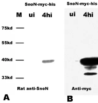
Figure legend). SnoN fusion protein was detected in an independent experiment  using anti-myc antibodies which served as an important control (Figure 25B)

Referenzen

ÄHNLICHE DOKUMENTE

Diese Annahme wird unterstützt durch Experimente, in denen beispielsweise die Steigerung der Expression von VEGF auf m-RNA-Ebene unter erniedrigtem

Für die Immunhistochemie kamen zwei verschiedende, kommerziell erhältliche Antikörpern, Clone 31G7 (Zymed Laboratories, Inc.) und EGFR pharmDX (DakoCytomation) zum

Therefore, in contrast to the idea that irradiation always induces pEGFR signaling (Schmidt- Ulrich et al., 1997), NCI-H460 and Calu-6 might be cell lines in which EGFR

Auch für Weichteilsarkome konnten eine EGFR-Überexpressionen nachgewiesen werden, insbesondere für folgende Tumore: Synovialsarkomen, endometrialen Stromasarkomen des Uterus,

The discovery that mutations in the gene coding for the bone morphogenetic protein receptor type 2 (bmpr2) as well as for the activin receptor-like kinase 1 (alk1), both members

Discussion 4.1 Expression of TGFβ during embryogenesis and in the adult mouse 4.2 TEC respond to TGFβ in vitro 4.3 Elucidating the role of TGFβ-signaling in TEC in vivo

Our results from transplantation of Smad4 Δ / Δ bone marrow into wild type recipients and deletion of Smad4 selectively in hematopoietic cells in VavCre;Smad4 fl/fl

Es zeigte sich somit ein Überlebensvorteil für die Patienten mit Exon 19-Mutation im Vergleich zu Patienten mit Exon 21-Mutation an, sowie ein besseres Ansprechen auf die Therapie