BMP signaling in the dorsal-ventral patterning system of the milkweed bug Oncopeltus fasciatus
I n a u g u r a l - D i s s e r t a t i o n zur
Erlangung des Doktorgrades
der Mathematisch-Naturwissenschaftlichen Fakultät der Universität zu Köln
vorgelegt von
Lena Mareike Sachs aus Dachau
Köln
2014
1
Berichterstatter: Prof. Dr. Siegfried Roth Prof. Dr. Günter Plickert Vorsitzende der Prüfungskommission: Prof. Dr. Ute Höcker
Tag der mündlichen Prüfung: 1.7.2014
2
Table of Contents
Zusammenfassung ...6
Abstract ...8
1. Introduction ...9
1.1 The milkweed bug, Oncopeltus fasciatus...9
1.1.1 Formation of the blastoderm ... 10
1.1.2 Gastrulation and embryonic movements ... 11
1.1.3 Segmentation ... 13
1.1.4 Preliminary fate map of Oncopeltus fasciatus ... 13
1.2 BMP and Toll signaling ... 15
1.2.1 Toll signaling pathway... 15
1.2.2 BMP signaling pathway ... 16
1.3 DV patterning in Drosophila melanogaster ... 17
1.4 Evolution of DV patterning ... 18
1.5 Aim of this study ... 19
2. Material and Methods ... 20
2.1 Material ... 20
2.1.1 Chemicals & Enzymes ... 20
2.1.2 Reagent Kits ... 20
2.1.3 Buffers and Solutions ... 20
2.1.4 Antibodies ... 25
2.1.5 Oligonucleotides and PCR programs ... 25
2.2 Methods ... 28
2.2.1 Oncopeltus husbandry ... 28
2.2.2 RNA interference (RNAi) ... 29
2.2.2.1 Synthesis of dsRNA ... 29
2.2.2.2 Phenol chloroform extraction and 2-propanol precipitation of dsRNA ... 30
2.2.2.3 Injection of dsRNA into adult females ... 30
2.2.3 In situ hybridization (ISH) ... 30
2.2.3.1 Embryo fixation ... 30
2.2.3.2 Synthesis of RNA probes ... 31
2.2.3.3 Preparing of embryos for the hybridization ... 31
2.2.3.4 Hybridization ... 31
2.2.3.5 Removal of excessive probe ... 32
2.2.3.6 Detection ... 32
3
2.2.4 Fuchsin staining ... 32
2.2.5 Antibody staining... 33
2.2.5.1 Standard Antibody staining ... 33
2.2.5.2 Antibody staining using the PerkinElmer TSA PLUS DNP HRP enhancer Kit ... 33
2.2.7 RNA isolation and cDNA synthesis ... 34
2.2.7.1 RNA isolation ... 34
2.2.7.2 cDNA synthesis ... 34
2.2.8 Identification of transcripts ... 35
2.2.9 Cloning of PCR products ... 35
2.2.9.1 Electroshock Transformation ... 35
2.2.9.2 Mini Prep ... 35
2.2.9.3 Restriction of the extracted plasmids ... 36
3. Results ... 37
3.1 Disrupting BMP signaling severely impairs morphogenesis of O. fasciatus ... 37
3.1.1 Disrupting BMP signaling leads to a failure in katatrepsis ... 37
3.1.2 Knockdown of decapentaplegic impairs germ band formation ... 40
3.1.3 Defective germ band elongation upon impaired BMP signaling ... 41
3.2 BMP signaling appears not to be involved in body segmentation ... 42
3.3 BMP signaling plays a major role in DV patterning of Oncopeltus fasciatus ... 45
3.3.1 BMP signaling and DV marker expression in wild type embryos ... 45
3.3.1.1 BMP signaling activity is dynamic in Oncopeltus fasciatus blastoderm stage embryos ... 45
3.3.1.2 Expression patterns of BMP signaling components in Oncopeltus fasciatus... 47
3.3.1.3 Expression patterns of DV marker genes in Oncopeltus fasciatus ... 49
3.3.2 BMP ligands have distinct roles in DV patterning of Oncopeltus fasciatus... 52
3.3.2.1 Depletion of the BMP ligand Decapentaplegic leads to strongly ventralized embryos ... 52
3.3.2.2 The anterior is more sensitive to dpp knockdown than the posterior ... 58
3.3.2.3 Knockdown of glass bottom boat leads to lateralization ... 59
3.3.2.4 The consequences of gbb knockdown differ along the AP axis ... 65
3.3.3 BMP receptors are required for a proper BMP signal transduction ... 68
3.3.3.1 Knockdown of punt causes a dpp knockdown-resembling DV phenotype ... 68
3.3.3.2 Knockdown of thickveins causes ventralization ... 69
3.3.3.3 Knockdown of saxophone causes ventralization ... 70
3.3.3.4 Partial recovery of DV marker expression in sax-RNAi germ band stage embryos ... 71
3.3.4 Extracellular modulators strongly influence BMP signaling activity during DV patterning ... 73
4
3.3.4.1 Ventral fates require protection from BMP signaling ... 73
by Twisted gastrulation and Short gastrulation... 73
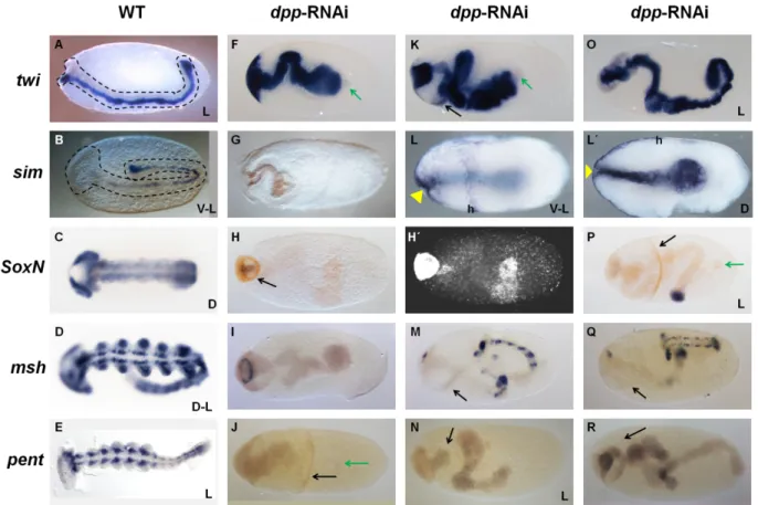
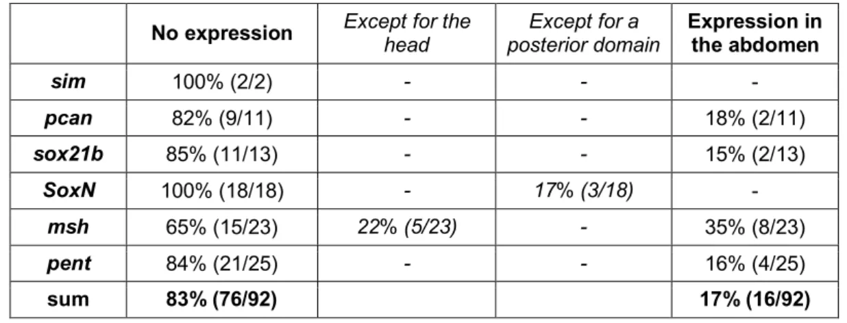
3.3.4.2 DV patterning of sog knockdown embryos is less impaired in the abdomen compared to the head and thorax ... 81
3.3.4.3 dpp is epistatic to sog ... 81
3.3.4.4 Sterility effects caused by loss of dpp and tsg are cumulative ... 82
3.3.4.5 Tolloid is essential for BMP signaling activity ... 84
3.3.5 Toll signaling is essential for DV polarity formation ... 85
3.3.5.1 The DV phenotype of dpp is epistatic to Toll... 85
3.3.5.2 Toll is not required for the early activation of sog ... 88
4. Discussion ... 91
4.1 BMP signaling seems to be required for extraembryonic membrane function ... 91
4.2 Excursus: Crosstalk between DV and AP patterning during head specification ... 95
4.3 BMP gradient formation requires Tsg and Sog ... 97
4.4 The main DV patterning function of Tsg is dependent on Sog ... 98
4.5 Growth zone patterning might dependent on posterior signaling centers established at the onset of gastrulation ... 100
4.6 BMP heterodimers might be required for shaping of the BMP signaling activity gradient ... 104
4.7 BMP heterodimers might contribute to robust patterning ... 105
4.8 BMP signaling restricts mesoderm formation ... 106
4.9 BMP signaling represses sog ... 107
4.10 The evolution of Toll signaling in DV patterning: From a trigger to a ruler ... 108
4.11 Evolution of DV patterning ... 111
5. Outlook ... 115
5.1 The future of the system Oncopeltus fasciatus ... 115
5.2 Towards a broader perspective ... 117
6. References ... 118
7. Supplementary Information ... 132
7. 1 Cell distributions differ between dpp-RNAi and Toll-dpp-RNAi embryos at the onset of gastrulation ... 132
7.2 tsg expression is not lost in Toll-RNAi embryos ... 133
7.3 Transcript sequences ... 134
Acknowledgments/Danksagungen ... 136
Erklärung ... 138
Lebenslauf ... 139
5
6
Zusammenfassung
Der BMP Signalweg spielt in der gesamten Tierwelt eine konservierte und wichtige Rolle während der Musterbildung entlang der dorsoventralen (DV) Achse. In Insekten übt allerdings auch der Toll Signalweg einen bemerkenswerten Einfluss auf die DV
Musterbildung aus und scheint in abgeleiteteren Insekten einige Aufgaben, die ursprünglich dem BMP Signalweg unterlagen, übernommen zu haben. Um diese Annahme zu überprüfen wurde der relative Beitrag beider Signalwege zur DV Musterbilung in verschiedenen Insekten verglichen. Dabei hat sich herausgestellt, dass das DV Musterbildungsystem der sehr
abgeleiteten Taufliege Drosophila melanogaster extrem abhängig vom Toll Signalweg ist, welcher sogar die räumliche Verteilung des BMP Signals weitgehend festlegt. Die Bedeutung des Toll Signalwegs für die DV Musterbildung scheint in basaler abzweigenden Insekten abzunehmen, während das Gegenteil auf den BMP Signalweg zutrifft. In der Wespe Nasonia vitripennis, welche zu den basalsten holometabolen Insekten, den Hymenopteren, zählt, wird der Toll Signalweg zur Mesoderm Induktion verwendet. In dieser Studie wurde zum ersten mal das DV Musterbildungssystem eines hemimetabolen Insektes, der Baumwollwanze Oncopeltus fasciatus, analysiert. O. fasciatus ist ein Kurzkeim Insekt, d.h. seine posterioren Segmente werden, nach Beginn der Gastrulation, sukzessive von einer posterior gelegenen Wachstumzone gebildet. Seine anterioren Segmente werden dagegen während des Blastoderm Stadiums etabliert. Unterschiede in der Enstehung anteriorer und posteriorer Segmente
könnten sich möglicherweise auch in der DV Musterbildung wiederspiegeln. Um das DV Musterbildungssystem von O. fasciatus zu verstehen wurde die Aktivität von Kandidaten- Genen mit Hilfe von parentaler RNA Interfernz (pRNAi) herunterreguliert. Die daraus resultierenden Phänotypen wurden auf molekulare und morphologische Abweichungen vom Wildtyp hin untersucht. Zu diesem Zweck wurden Kernfärbungen, in situ Hybridisierungen (ISH) sowie Antikörperfärbungen unternommen. Es stellte sich heraus, dass der BMP Signalweg das ventralste Schicksal, das Mesoderm, limitiert und sogar vollständig
unterdrücken kann. Desweitern reprimiert der BMP Signalweg auch short gastrulation (sog),
einen Inhibitor von BMP Liganden. Der Verlust der DV Polarität als Folge des Fehlens der
BMP Inhibitoren Twisted gastrulation (Tsg) und Sog war ein weiter Hinweis auf den enormen
Einfluss des BMP Signalwegs auf die DV Musterbildung von O. fasciatus. Das Fehlen DV
Asymmetrie in der Abwesenheit von Toll schien durch den Verlust späterer, nicht jedoch
initialer sog Expression begründet zu sein.
7
Diese Ergebnisse führen zur Annahme, dass die DV Musterbildung in O. fasciatus ein selbst regulierendes System darstellt, das von BMP und seinen extrazellulären Modulatoren
dominiert und durch den Toll Signalweg lediglich polarisiert wird. Außerdem deuten
Unterschiede in den RNAi Phänotypen von Blastoderm- und Keimstreifembryonen daraufhin,
dass das DV Musterbildungsystem für den Übergang vom Blastoderm zur Wachstumszone
die Etablierung zweier gegensätzlich wirkender Signalzentren am posterioren Pol benötigt.
8
Abstract
BMP signaling plays an essential role in dorsal-ventral (DV) axis patterning throughout the animal kingdom. However, in insects Toll signaling also has a remarkable influence on DV patterning and, in higher branching lineages, fulfills some functions, which depend on BMP signaling in more basally branching lineages. Thus, the DV patterning system of the highly derived fruit fly Drosophila melanogaster is extremely dependent on Toll, which determines also the pattern of dorsal cell fates by specifying the polarity of BMP signaling. However, the wasp Nasonia vitripennis, which belongs to the most basally branching holometabolous lineage, the hymenopterans, uses Toll signaling only as mesoderm inductor. In this study for the first time the DV patterning system of a hemimetabolous insect, the milkweed bug Oncopeltus fasciatus was analyzed. O. fasciatus is a short germ insect, i.e. its posterior segments successively arise from a posterior growth zone, after the onset of gastrulation. In contrast, the anterior segments are synchronously established during the blastoderm stage. A different emergence of anterior and posterior segments might also be reflected in the DV patterning system. To understand the DV patterning system of O. fasciatus candidate genes were knocked down via parental RNA interference (pRNAi) and the resulting phenotypes were investigated for morphological as well as molecular deviations from wild type embryos.
Nuclear staining techniques, in situ hybridization (ISH) and antibody staining were performed for this purpose. BMP signaling was found to be able to completely repress mesodermal fates and to be required to restrict it to the ventral side. Furthermore the repression of the BMP inhibitor short gastrulation (sog) seemed also to be mediated by BMP signaling. The lack of DV polarity upon depletion of the extracellular BMP inhibitors Sog and Twisted gastrulation provided further evidence for the high impact of BMP signaling on the O. fasciatus DV patterning system. The absence of DV asymmetry upon depletion of Toll was indicated to be due to a loss of later but not initial expression of sog. These results led to the proposal of a highly self-regulating BMP-dependent DV patterning system for O. fasciatus, which is only polarized by Toll signaling. In addition, differences between the early blastoderm and the later germ band DV pattern in knockdown embryos suggested that the transition of the two
dimensional blastoderm DV patterning system into the three dimensional growth zone DV
patterning system requires the establishment of two opposing signaling center located close to
the posterior pole at the onset of gastrulation.
9
1. Introduction
The diversity of body plans, sizes and shapes among animals is incredibly amazing. However, despite their morphological variety the vast majority of animals possess two major body axes, the anterior posterior (AP) and the dorsal-ventral (DV) axis, which need to be established during embryogenesis. Remarkable similarities in setting up these axes are found on the molecular level. It appears that the gene regulatory networks (GRN) used for axis formation are partially conserved (Petersen & Reddien 2009; Little & Mullins 2006). For example most bilaterians, analyzed so far, use Wnt signaling to set up the AP axis (Petersen & Reddien 2009). Despite of such commonalities, GRNs underlying axial patterning also underwent major changes in the course of evolution. Our research group attempts to reconstruct the evolution of the GRN guiding DV patterning in arthropods, especially in insects.
Insects are appropriate to study evolution as they have a small body size, often exhibit a short life cycle, and many species produce a large number of offspring relative to other animals, e.g. mammals. For these reasons a lot of insects are easy and inexpensive to culture and especially suitable for studying embryonic development. In addition, insects are the most diverse class of animals, so it is possible to examine many different sampling points (Nentwig et al. 2007).
1.1 The milkweed bug, Oncopeltus fasciatus
This study focuses on the analysis of the DV patterning network with special regard to Bone morphogenetic protein (BMP) signaling using the milkweed bug Oncopeltus fasciatus as sampling point for investigation.
O. fasciatus is a true bug (order: Hemiptera, suborder: Heteroptera), with widespread distribution in the new world (Butt 1949). Its phylogenetic position within the group of winged insects (Pterygota) is shown in Figure 1-1. The duration of its embryogenesis is dependent on temperature and humidity. With conditions in our laboratory (25°C,
approximately constant humidity), hatching from the approximately 1.2 mm long egg occurs
after seven days.
10
Figure 1-1: Phylogenetic tree of Pterygota
The tree was drawn after information from the publications Lynch & Roth 2011; Roth 2004. The clade indicated with green branches is the Holometabola; the assemblage indicated with lilac branches are hemimetabolous (paraphyletic with respect to the Holometabola). Orders and superorders are written on top or below the respective branches, orders in non-bold fonts, superorders in bold font. Genera are written in italic font; dorsal- ventral patterning mechanisms of species with genera highlighted in yellow are discussed later.
1.1.1 Formation of the blastoderm
Insects typically form a superficial, mono-layered "blastoderm" embryo during early embryogenesis (Roth 2004; Counce & Waddington 1973). The blastoderm is established during the first 15 hours of embryogenesis in O. fasciatus. After fertilization and egg activation (concomitant with oviposition), the zygotic nucleus starts to divide. Most of the emerging nuclei migrate as energids (the nucleus itself and an associated island of cytoplasm) towards the periphery of the egg. Some nuclei remain in the yolk and are referred to as vitellophages. The nuclei located close to the egg cortex undergo several rounds of
synchronous division. Finally, cellularization is initiated, i.e. the syncytial uniform blastoderm
is transformed into a cellularized, uniform blastoderm (Figure 1-2 A; Butt 1949).
11
Figure 1-2: Early embryogenesis of Oncopeltus
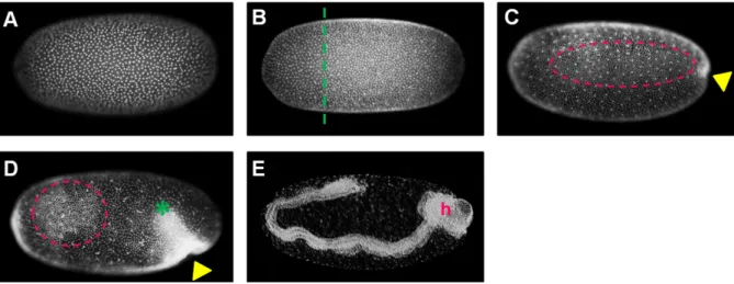
The nuclei were stained with SYTOX-green (A-D) or with fuchsin (E). In all images the anterior of the egg is oriented to the left. All embryos are shown in a lateral view with egg-dorsal to the top (hence the embryo in (E) has embryo-ventral oriented to the top). Image (A) shows a uniform, cellularized blastoderm stage embryo (~20 hours post fertilization (hpf)). (B) pictures an embryo at the late uniform1 blastoderm stage (~28 hpf). The dashed green line approximately separates an anterior region of low nuclear density from the remaining blastoderm. The embryo in C (~30 hpf) is at the differentiated blastoderm stage with a yellow arrowhead pointing towards the (future) invagination site. Laterally condensed cells are highlighted by a pink oval. The embryo in (D) is at the anatrepsis stage (~40 hpf). The green asterisk marks the posterior end of the germ rudiment (which already invaginated into the yolk). The embryo in (E) has finished anatrepsis (1.1.2), and it therefore has an inverse orientation with respect to the egg axes, i.e. its head (marked by an h) is at the egg posterior. Abbreviations: hpf: hours post fertilization, h: head.
1.1.2 Gastrulation and embryonic movements
During the second day of embryogenesis the cells become unevenly distributed within the blastoderm. First they become more widely spaced in the anterior 25% of the embryo (Figure 1-2 B)
1. The serosa is proposed to be located within this region (1.1.4; Figure 1-3 A; Figure 1- 4). The serosa as well as the amnion are extraembryonic tissues covering the complete
embryo and the yolk, or the ventral side of the embryo, respectively, during a certain stage of development (Roth 2004).
Around 30 hours post fertilization (hpf) cells become more widely spaced in the ventral region, while they start to condense in the lateral region and form a slight indentation at the posterior pole (Figure 1-2 C). The formation of the posterior indentation marks the
1 Blastoderm embryos before gastrulation (i.e. before they formed a posterior indentation) are considered as uniform blastoderm embryos, although the cells are not completely equal distributed throughout this stage.
h
12
onset of gastrulation, which is defined as the sum of (morphological) processes that lead to the formation of the three germ layers (Solnica-Krezel & Sepich 2012).
At the end of the second day several patches of highly condensed cells emerge in the lateral region of the germ rudiment (Figure 1-2 D). This process occurs during the complete invagination of the germ rudiment into the yolk, whereby the embryonic orientation becomes inversed relative to the axes of the egg, a movement which is termed anatrepsis (Figure 1-2 D, E; Figure 1-3 B, C; Panfilio et al. 2006; Panfilio 2008). During anatrepsis the mono-layered surface (i.e. blastoderm embryo) becomes transformed into a multi-layered embryo, which is referred to as germ band in insects. This transformation includes internalization of the
mesoderm and is directly followed by elongation of the newly formed germ band (Butt 1949;
Panfilio et al. 2006; Roth 2004).
Around mid-embryogenesis (early on the fourth day) the orientation of the embryo becomes reversed again with respect to the egg axes, during an embryonic movement termed katatrepsis (Panfilio et al. 2006; Panfilio 2008). This event is preceded by the fusion of the amnion and serosa at the posterior pole of the egg (i.e. at the anterior end of the germ band) (Figure 1-3 D). The fused membranes rupture, which permits the contracting serosa to pull the germ rudiment out of the yolk (Figure 1-3 E; Panfilio et al. 2006; Panfilio 2008).
Figure 1-3: Schematic drawing of embryonic movements of Oncopeltus fasciatus
The serosa is depicted in yellow, the amnion in blue, the embryo in gray and the gray thickening indicates the head. Anterior is to the top, ventral to the left, regarding egg axes. The directions of movements are indicated with arrows. The egg and embryonic axes correspond to each other during the blastoderm stage (A). During anatrepsis the whole germ rudiment invaginates into the yolk (B). This leads to the formation of a germ band with inversed axes relative to the egg axes, which is surrounded by yolk and serosa (C). The amnion covers the embryo on the ventral side (C, D). Around mid embryogenesis the amnion and serosa fuse (D). Afterwards the
13
serosa contracts and pulls the germ rudiment out of the yolk (E). In this manner the egg and the embryonic axes become again correlated to each other (F). Abbreviations: h: head. This schematic drawing was based on the publication Panfilio 2008.
1.1.3 Segmentation
Segmentation starts already before germ band elongation, during the blastoderm stage. The head segments and the three thoracic segments are determined at this stage (Birkan et al.
2011; Liu & Kaufman 2004). However, the 11 abdominal segments arise successively from a posterior growth zone, after the onset of gastrulation (Liu & Kaufman 2004). If posterior segments are established after the onset of germ band elongation the mode of development is referred to as extreme short, short or intermediate germ, depending on the number of
segmental primordia present before germ band elongation (Roth 2004). For simplification this study referrers to all of these modes as short germ development.
In contrast to embryos with short germ development, embryos with long germ development establish all segments during the blastoderm stage. Long germ development is exclusively found in a subset of holometabolous insects (Roth 2004).
1.1.4 Preliminary fate map of Oncopeltus fasciatus
Like segmentation, the specification of DV fates starts also already during the blastoderm stage. From 25 hpf onwards several known marker genes are expressed in distinct regions along the DV axis in O. fasciatus (Francois et al. 1994; Jiménez et al. 1995; Kasai et al. 1992;
Leptin 1991; Miller-Bertoglio et al. 1997; van der Zee et al. 2006; Handel et al., 2005;
Sommer & Tautz 1994).
The anterior 25% of the blastoderm is largely excluded from the expression of those markers. This would be consistent with this region having a serosal identity, as the serosa does not contribute to the germ layers (Roth 2004). However, some head patterning genes are expressed in this region, indicating that it contains anterior head anlagen as well (Birkan et al.
2011; Weisbrod et al. 2013). In addition, it was proposed that at least a part of the serosa
anlage is dorsally located within the O. fasciatus blastoderm (Ben-David & Chipman 2010).
14
However, the anterior region of the blastoderm is usually specified as serosa in short germ insects (Roth 2004). The anterior half as well as the dorsal 30% of the anterior quarter of the O. fasciatus blastoderm are not reported to express head patterning genes during specification of head tissue (Birkan et al. 2011; Weisbrod et al. 2013). Therefore, it is proposed in this study that the serosa anlage covers this region (Figure 1-4).
The expression of the highly conserved mesodermal marker twist (twi) was observed in the ventral 20 to 30% of the posterior 75% of the blastoderm (Drechsler 2007; Leptin 2004;
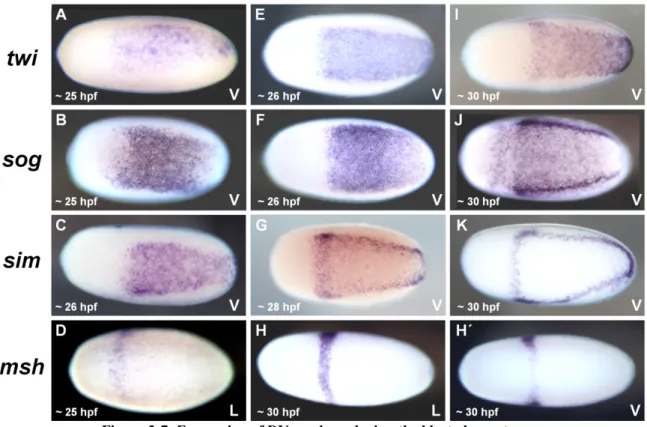
Roth 2004; Sachs 2009; Figure 3-7 A, E, I).This indicates that the ventral-most tissue corresponds to the mesoderm anlage, since twi expression is indicative for mesoderm in all insects analyzed so far (Handel et al. 2005; Lynch & Roth 2011; Roth 2004).
The arrangement of the remaining DV fates seems also to be widely conserved among insects (Lynch & Roth 2011). The position of the future neuroectoderm in the ventral-lateral region was verified by the expression pattern of marker genes like ventral nervous system defective (vnd), short gastrulation (sog) and single minded (sim) (Sachs 2011; Figure 3-7 B, C, F, G, J, K; unpublished data of Yen-Ta Chen). Usually the non-neurogenic, also referred to as dorsal, embryonic ectoderm is located dorsally adjacent to the neuroectoderm, while the amnion (dorsal, extraembryonic ectoderm) is often positioned in the dorsal-most region of insect blastoderm fate maps (Goltsev et al. 2007; Lynch & Roth 2011; Nunes da Fonseca et al.
2008). This arrangement could not be assured by the expression of marker genes, as those dorsal marker genes which are known from holometabolous insects are not expressed in the same manner in O. fasciatus (data not shown).
Figure 1-4: Presumptive blastoderm fate map of Oncopeltus fasciatus
The schematic embryo is orientated in a lateral view with dorsal to the top and anterior to the left. The mesoderm anlage is positioned in the ventral 20-30% of the egg circumference, followed by the neuroectoderm, dorsally to which the dorsal, embryonic ectoderm and amnion are located. The serosa is assumed to be located in the anterior 15% of the egg with a dorsal, posterior pointing protrusion. The position of the borders between the neuroectoderm and the dorsal ectoderm as well as of the serosa is uncertain (dashed lines). Abbreviations: A:
anterior, D: dorsal, P: posterior, V: ventral.
P
V A
D
15
1.2 BMP and Toll signaling
To establish DV fates insects use the BMP and the Toll signaling pathway both of which are conserved among metazoans (Hibino et al. 2006; Hughes & Piontkivska 2008; Satake &
Sasaki 2010; Valanne et al. 2011; Guo & Wang 2009).
1.2.1 Toll signaling pathway
The Toll signaling pathway is involved in various processes, e.g. pathogen recognition, cell adhesion and development (Anderson et al. 1985; Eldon et al. 1994; Hughes & Piontkivska 2008; Tauszig et al. 2000). Toll signaling is especially famous for its conserved role in immunity (Hughes & Piontkivska 2008; Satake & Sasaki 2010; Zhong et al. 2012; Zheng et al. 2012; Pradeep et al. 2013; Yeh et al. 2013; Han-Ching Wang et al. 2010; Satake &
Sekiguchi 2012).
Despite the broad conservation of the pathway’s immune function, some deviations are found with regard to signaling components. Among these are remarkable differences between insect and vertebrate Toll ligands. Insects express an endogenous
2The activated Toll receptors recruit DEATH domain proteins as adaptors, e.g. MyD88, which enable the docking of other DEATH domain proteins, at least one of them containing an additional kinase domain. Examples for the latter are IL-1R associated kinases (IRAKs) or their insect homologs Pelle and Tube or Tube-like kinases (TTLK) (Hughes & Piontkivska 2008; Santegoets et al. 2011; Towb et al. 2009; Valanne et al. 2011). This intracellular
signaling cascade leads finally to the phosphorylation of IκB, in insects referred to as Cactus, thereby initiating its degradation. Cactus prevents the transcription factor Dorsal, in
vertebrates termed Nuclear factor κB (NF-κB), from nuclear translocation by masking its nuclear localization signal. After degradation of Cactus, Dorsal translocates into the nucleus and regulates the transcription of target genes (Moussian & Roth 2005; Valanne et al. 2011).
Toll1-ligand, referred to as Spätzle, which is cleaved and thereby activated (Moussian & Roth 2005). This is in contrast to vertebrates, where Toll-like receptors (TLRs) are activated by direct, or co-receptor- mediated, binding of pathogen associated molecular patterns (PAMPs) or danger associated molecular patterns (DAMPs) (Santegoets et al. 2011).
2 Endogenous ligand is here used as expression for a genome encoded ligand, which does not include DAMPs, e.g. fragments of gene products which are only formed upon an injury.
16
1.2.2 BMP signaling pathway
The BMP signal transduction cascade is known to be involved in several developmental processes; one of them is DV patterning (Guo & Wang 2009; Dutko & Mullins 2011).
In this regard, Decapentaplegic (Dpp) is the most important BMP ligand, which is
homologous to BMP 2/4 in vertebrates (Johnston & Gelbart 1987; Miyazono et al. 2005).
Other ligands of this signaling pathway are, for example, Glass bottom boat (Gbb) and Screw (Scw), which are homologous to the vertebrate BMP5/6/7/8 ligands (Miyazono et al. 2005).
All BMP ligands are secreted proteins, which are synthesized as pro-proteins and require processing by proteases before they can act as ligands. They dimerize through the formation of disulfide bonds and subsequently bind to two type II BMP receptors and two type I BMP receptors (Figure 1-5; Miyazono et al. 2005; Sun & Davies 1995; Constam & Robertson 1999; Cui et al. 1998). Type I receptors, like Saxophone (Sax) or Thickveins (Tkv), are responsible for the ligand specificity (Nguyen et al. 1998; Haerry et al. 1998; ten Dijke et al.
1994). The receptors tetramerize upon ligand binding and BMP type II receptors, e.g. Punt (Put) and Wishful thinking (Wit), which are constitutively active kinases, activate BMP type I receptors via phosphorylation (Figure 1-5; Miyazono et al. 2005; Little & Mullins 2006). The activated type I receptors specifically interact with and phosphorylate receptor-associated Smad proteins (r-Smads), e.g. Mothers against Dpp (Mad) in the case of BMP signaling in insects (Figure 1-5; Feng & Derynck 1997; Little & Mullins 2006; Miyazono et al. 2005).
These form complexes with Medea, in vertebrates referred to as common Smad (co-Smad), which then translocate into the nucleus and regulate transcription (Figure 1-5; Little &
Mullins 2006; Miyazono et al. 2005).
The BMP signaling cascade, or rather its spatial activity, can be influenced by
extracellular BMP modulators. These can facilitate or impede BMP ligand activity (Ben-zvi et al. 2008; De Robertis & Kuroda 2004; Little & Mullins 2006; van der Zee et al. 2006). The best known extracellular modulator is Short gastrulation (Sog), whose vertebrate ortholog is Chordin (Chd). It sequesters BMP dimers and thus prevents binding to their respective receptors (Little & Mullins 2006; Marqués et al. 1997; Piccolo et al. 1996; Ross et al. 2001;
Shimmi & O’Connor 2003; Xie & Fisher 2005). The metalloprotease Tolloid (Tld) is
responsible for the cleavage of Sog and hence for the release of BMP dimers from Sog-BMP ligand complexes (Ben-zvi et al. 2008; Connors et al. 1999; Little & Mullins 2006; Marqués et al. 1997; Shimmi & O’Connor 2003).
Another extracellular modulator is Twisted gastrulation (Tsg), or its homolog
17
Crossveinless 1 (Cv 1). The molecular mechanism as well as the general function of Tsg is not completely understood. Nevertheless, Tsg appears to be conserved in invertebrates and
vertebrates and its functional depletion or over-activation clearly impairs BMP signaling (Ben-zvi et al. 2008; Little & Mullins 2006; Nunes da Fonseca et al. 2010).
Figure 1-5: Scheme of the BMP signal transduction
Red lines with a blunt end indicate inhibition, green arrows activation and black arrows translocation. A detailed description of the signal transduction is provided in the text. Abbreviations: Dpp: Decapentaplegic, Gbb: Glass bottom boat, K: kinase; Mad: Mothers against Dpp, P: phosphate, Scw: Screw, Tld: Tolloid, Tsg: Twisted gastrulation.
1.3 DV patterning in Drosophila melanogaster
The best understood DV patterning system within insects is that of the fruit fly Drosophila melanogaster. In the following the function of Toll and BMP signaling in this system will be elucidated.
The BMP signaling pathway is required to pattern the dorsal 40% of D. melanogaster
embryos. To this end a gradient of nuclear phosphorylated Mad (pMad) is established via the
extracellular modulators Sog, Tsg and Tld. Sog and Tsg form complexes with BMP ligands,
which quickly diffuse away from the source of Sog, i.e. the ventral-lateral region, towards the
dorsal side. At this side of the embryo, BMP dimers bind to their receptors as they are freed
18
from Sog by Tld (Ben-zvi et al. 2008; Connors et al. 1999; Little & Mullins 2006; Marqués et al. 1997; Shimmi & O’Connor 2003).
The ventral side is patterned by the Toll signaling pathway: upon activation of the Toll receptor, a nuclear Dorsal gradient is formed in the ventral 60% of the egg circumference.
Nuclear Dorsal shows peak levels at the ventral side and gradually decreases towards the dorsal side (Moussian & Roth 2005; Roth et al. 1989). This morphogen gradient is sufficient for establishing most cell fates along the DV axis and even regulates BMP signaling in several regards (Roth et al. 1989; Rusch & Levine 1996; Jaźwińska et al. 1999; Reeves &
Stathopoulos 2009).
1.4 Evolution of DV patterning
Although Toll signaling plays such an important role in D. melanogaster DV patterning it was never found to participate in DV axis formation outside of arthropods (Hughes & Piontkivska 2008; Satake & Sasaki 2010).
3In contrast, BMP signaling appears to be involved in DV patterning in almost all bilaterally symmetric animals (Akiyama-Oda & Oda 2006; Ben-zvi et al. 2008; De Robertis
& Kuroda 2004; Kishimoto et al. 1997; Lapraz et al. 2009; Lowe et al. 2006; van der Zee et al. 2006; Kuo et al. 2012; Raftery & Sutherland 2003). This pathway is especially important for the specification of fates on the non-neuronal side (dorsal in insects, ventral in
vertebrates). However, while the BMP pathway is in D. melanogaster largely under the control of Toll signaling and only responsible for patterning the dorsal, embryonic ectoderm as well as the extraembryonic amnioserosa, it has a more fundamental role in establishing fates along the DV axis in many other bilaterians (Akiyama-Oda & Oda 2006; Ben-zvi et al.
2008a; De Robertis & Kuroda 2004; Kishimoto et al. 1997; Lapraz et al. 2009; Lowe et al.
2006). For instance, vertebrate DV patterning is almost completely dependent on BMP signaling (Ben-zvi et al. 2008; De Robertis & Kuroda 2004; Kishimoto et al. 1997).
Comparing the situation in several insects, BMP signaling appears to gain relevance in DV patterning whereas the NF-κB/Toll pathway loses it, towards more basally branching species of the insect phylogeny (Ferguson & Anderson 1992a; Nunes da Fonseca et al. 2008;
van der Zee et al. 2006; Özüak 2014; Buchta 2014). For example, upon a functional
3 There might be one exception: there is vague evidence that TLR signaling might have a role in Xenopus DV patterning (Armstrong et al. 2012; Prothmann et al. 2000; Armstrong et al. 1998).
19
knockdown of sog the neuroectoderm is completely lost in T. castaneum, while it is only reduced in D. melanogaster (Francois et al. 1994; Raftery & Sutherland 2003; van der Zee et al. 2006). Furthermore, the depletion of Dpp causes a stronger ventralization in the wasp Nasonia vitripennis than in D. melanogaster (Ferguson & Anderson 1992a; Özüak 2014;
phylogenetic position is indicated in Figure 1-1).
The conserved contribution of BMP signaling in DV patterning, together with functional data demonstrating its profound impact on this process in most animals, suggests an ancestral involvement of BMP signaling in DV axis establishment.
1.5 Aim of this study
This evolutionary assumption implies that Toll signaling might have been recruited for DV patterning during arthropod evolution. In the course of arthropod evolution the relevance of Toll signaling in establishing cell fates along the DV axis may have successively increased at the expense of the importance of BMP signaling in this regard.
It is not only of interest to confirm this hypothesis, but to find out how, and maybe also why, such a change could have occurred in the course of evolution. Studies in this context will also improve the general understanding of the evolution of complex gene regulatory networks (GRN) and possibly about stimuli for such evolutionary changes.
Therefore, it is valuable to gain knowledge about the evolution of DV axis establishment by analyzing this process in further insects, thereby generating additional sampling points for a comparative analysis. As we already have insights into the DV
patterning systems of several holometabolous insects, it is reasonable to analyze for the first time the DV patterning systems of a hemimetabolous insect.
O. fasciatus is appropriate for such a study for several reasons: its husbandry is
inexpensive with a high, year-round yield of embryonic material (2.2.1). Transcriptome data
are available; blastoderm stages are amenable for in situ hybridization (ISH) and antibody as
well as nuclear staining techniques. It is possible to synchronously knock down maternally
and zygotically transcribed genes via parental RNA interference (pRNAi) (Liu & Kaufman
2004). The above mentioned techniques were used to analyze the function of some BMP
signaling components of O. fasciatus in order to elucidate the mechanism of DV axis
formation in this species.
20
2. Material and Methods 2.1 Material
2.1.1 Chemicals & Enzymes
Chemicals were purchased from BAUER, Merck, PFALTZ, Roche, Roth, Sigma and VWR.
Chemicals ordered from other companies were marked as such. Enzymes were purchased from Ambion, Fermentas, Invitrogen and Roche.
2.1.2 Reagent Kits
Table 2-1: List of used Reagent Kits
Reagent Kit Company
QUIAGEN Plasmid Miniprep Kit Quiagen
TOPO ® TA Cloning (pCR®IIvector) Invitrogen Zymoclean
TMGel DNA Recovery Kit ZYMO RESEARCH
MEGAscript ® RNAi Ambion
MAXIscript ® In vitro Transcription Kit Ambion
High-Fidelity PCR Kit Invitrogen
TSA PLUS DNP HRP enhancer Kit PerkinElmer
SuperScript® VILO™ cDNA Synthesis Kit Invitrogen SMARTer™ RACE cDNA Amplification Kit Clontech
2.1.3 Buffers and Solutions
The pH was adjusted with HCl or NaOH. The solutions were sterilized either by filtration or
by autoclaving. Applied water was either autoclaved or filter-sterilized.
21
For general use:
Phosphate buffered saline (PBS), 1x
NaCl 137 mM
KCl 2.7 M
Na
2HPO
48 mM
KH
2PO
41.7 mM
PBS with Tween (PBT), 1x
PBS, 1x 99.9%
Tween-20 0.1%
Sodium Dodecyl Sulfate (SDS), 10%
C
12H
25SO
4Na 10%
Saline sodium citrate (SSC), 20x, pH 5
NaCl 3 M
Na
3C
6H
5O
7300 mM
Injection buffer (IB)
KCl 5 mM
Na
2HPO
40.1 mM
Tris acetate EDTA (TAE) buffer
Tris 40 mM
acetic acid 20 mM
EDTA 1 mM
Lysogeny broth (LB) medium, pH 7
yeast extract 0.5%
NaCl 1%
tryptone 1%
22
Super-optimal broth with catabolite repression (SOC) medium
bacto-tryptone 2%
bacto-yeast extract 0.5%
NaCl 10 mM
KCl 2.5 mM
MgCl
210 mM
glucose 20 mM
Fuchsin staining solutions:
Alcoholic fuchsin staining solution (stored at room temperature (RT))
Pararosaniline 5 mg/ml
Ethanol 80%
Solution C
Ethanol 50%
Solution D/BBBA 50%
Solution D/BBBA
Benzyl benzoate 80%
Benzyl alcohol 20%
23
In situ Hybridization and Antibody Staining Solutions:
Hybridization buffer (HYBE)
Formamide 50%
SSC, pH 5 5x
Heparin 100 µg/ml
salmon sperm DNA (ssDNA) 100 µg/ml transfer-RNA (t-RNA) 100 µg/ml
SDS 1%
Tween-20 0.1%
Blocking solution
PBS 1x
Bovine serum albumin (BSA) 2 mg/ml Goat serum 5%
Tween-20 0.1%
Alkaline phosphatase (AP) buffer, pH 9.5
Tris, pH 9.5 0.1 M
MgCl
250 mM
NaCl 100 mM
Tween-20 0.05%
Probe resuspension buffer
Formamide 50%
SSC, pH 5 2x
24
Antibody staining solutions required for the PerkinElmer TSA PLUS DNP HRP enhancer Kit:
TNT
Tris-HCl, pH 7.5 0.1 M
NaCl 0.15 M
Tween20 0.05%
TNB
Tris-HCl, pH 7.5 0.1 M
NaCl 0.15 M
Blocking reagent (PerkinElmer) 0.5%
The blocking reagent was added slowly in small amounts to the buffer while stirring.
Afterwards the buffer was gradually heated to 60°C while stirring to completely dissolve the blocking reagent.
DNP Amplification reagent working solution
The DNP Amplification stock (PerkinElmer) was 1:50 diluted with 1x Plus amplification dilutent (PerkinElmer). This solution was always freshly prepared.
DAB staining solution
0.3-0.5 mg
Ammonium nickel sulfate 0.05%
PBS 1x
Tween20 0.05%
25
2.1.4 Antibodies
Table 2-2: Applied antibodies
Antibodies were stored at 4°C or -20°C. Abbreviation: AP: alkaline phosphatase, DNP: dinitrophenol, HRP:
horse radish peroxidase, IgG: heavy chain of Immunoglobulin G.
Order Target, Designation Animal in which it is generated
Coupled substance
Applied dilution
1° Engrailed, 4D9 mouse - 1:250
1° Phospho-Smad1/5
(Ser463/465), 41D10 rabbit - 1:30
2° Rabbit IgG, α-rabbit::HRP goat HRP 1:100
2° Mouse IgG, α-Mouse::AP ? AP 1:400
2° Digoxigenin, α-DIG::AP sheep AP 1:2000
3° DNP, α-DNP::HRP rat HRP 1:100
2.1.5 Oligonucleotides and PCR programs
Table 2-3: Oligonucleotides
All oligonucleotides were purchased from Sigma. The online version of the program Primer3 Ta Chen (indicated in parentheses) and general available oligonucleotides (M13, T7-SP6, T7) were used. T7 adaptor sequences are written in lowercase.
Designation Sequence 5´-> 3´
LS_Of collagenIV m Fwd ggccgcggGCAATTATGCTAGCCGGAAT LS_Of collagenIV m Rev cccggggcATTACAATATAAACAAGAAAGACATTT
LS_Of croc m2 Fwd ggccgcggAGAAGAGGTGTACGCTCAAC LS_Of croc m Fwd ggccgcggGGAGTCACCAGTGTGGATG LS_Of croc m Rev cccggggc GCATCTCCTGCTGCCTCTT
LS_Of gbb Fwd ggccgcggTCAGTGAAACTATTGTTGGAGCA LS_Of gbb Rev cccggggcTTGGAGCACAACAAGGCTTA LS_Of gbb small Fwd ggccgcggTTGAAGTCCGTAACAATGAAGTT LS_Of gbb intern Rev cccggggcTGCTCCAACAATAGTTTCACTGA LS_Of gbb intern Rev cccggggcTGCTCCAACAATAGTTTCACTGA LS_Of dpp intern Fwd ggccgcggTCCTGGTGGAAGTGGCGAG LS_Of dpp intern Rev cccggggcTTGCGGACCTCACATGGCGT
LS_Of dpp mp Fwd ggccgcggACGCGGGGAGTAGCTAGCC
LS_Of dpp mp Rev cccggggcCTTCCTACATCCACAGCCAACC
26
LS_Of dlp m Fwd ggccgcggCTACGAACAGAATGCCTATCTC LS_Of dlp m Rev cccggggcTTTAACAATATCAGCTGACACACG Msh pF (Yen-Ta Chen) ggccgcggACCTGCGAAGCAGAAGC Msh pR (Yen-Ta Chen) cccggggcTCTGATTTGCTATAATAATGAACTGC
M 13 Fwd GTAAAACGACGGCCAG
M13 Rev CAGGAAACAGCTATGAC
LS_Of perlecan m Fwd ggccgcggCAAGGACGACACCAAGGACT LS_Of perlecan m Rev cccggggcATTTGGGACCATCTGGAACA LS_Of pent- Contig7096
Fwd ggccgcggAGGTGATTGCTTGACTGACC LS_Of pent- Contig7096
Rev cccggggcCTAGTTGAAACATCTTGAAAGCA LS_Of punt marg Fwd2 ggccgcggTGTGGACCCTAGTCCCACTC
LS_Of punt intern Rev cccggggcTTGTTGGCACCTCATTGAAA LS_Of punt marg Rev2 cccggggcTCCAGGCGATCAGTAGAGGT
LS_Of punt intern Fwd ggccgcggTTCCTGAGAATTGACATGTATGC LS_Of_qPCR_HMBS
Fwd 1 TGGAACTGTGGACTTGGTTG
LS_Of_qPCR_HMBS
Rev 2 TTTCTTGTCCGCTGTTATTGG
LS_Of_qPCR_atubulin
Fwd 3 CCCTCGTCTGATCTCCTTGA
LS_Of_qPCR_atubulin
Rev 2 AACAGGGAGGTGAATCCAGA
LS_Of-sax-17917 Fwd ggccgcggTGGTACACAGGGAAAACCAAG LS_Of-sax-17917 Rev cccggggcTTGGGCAAAACAGGTCTTCT LS_Of-sax17917 int Rev cccggggcCTTTTGGTTCCCACACGAGTG LS_Of-sax17917 int Fwd ggccgcggATACATGGGTCCAGAGCTCC
sim 28 Fwd
(Yen-Ta Chen) CCTCCATCATCAGACTGACC sim 771R (Yen-Ta Chen) GCGAGAAGTGTATGATGAGAATA
LS_Of slalom m Fwd ggccgcggAAAGACTGGAAATACTAGCCATTCA LS_Of slalom m Rev cccggggcGATGTTGGAGAAAGAACAGTATGC sog0_1F (Yen-Ta Chen) ggccgcggGTTTGGCAAGTCGTTTCGTA
sog1_2328R
(Yen-Ta Chen) cccggggcCCGTCGATTACACTGAGCAA sogF2P_1339F
(Yen-Ta Chen) ggccgcggATTGCAGTTGACCCACAATG sogR2P_1182R (Yen-Ta
Chen) cccggggcTTCTACCAGCCTTGGTGAGG LS_Of sox21 m2 Fwd ggccgcggCATCAAGAGGGAACCACACA LS_Of sox21 m2 Rev cccggggcAGCAAGGACGAGTGGCTTT
SoxN 400 F GGCCGCGGCAGCACCCTCATCAT
27
(Yen-Ta Chen)
SoxN 1338 R
(Yen-Ta Chen) CCCGGGCAGGTTGGCACTGGGA LS_Of syndecan m Fwd ggccgcggTAGGAGGAGCTGTTGTTGGA LS_Of syndecan m Rev cccggggcGGAGAGGAGCGGTCTGATAA LS_Of-tkv-15135 m Fwd ggccgcggGGAACTGGGAGGTATGGTGA LS_Of-tkv-15135 m Rev cccggggcGGAGGTGAGCTAGACCAGAAGA
LS_Of tld nd Fwd ggccgcggCTTGGTTCCTATGTCTGCTGGTGTC LS_Of tld nd Rev cccggggcAGTCTTATTCGGTGGCCTGGAGTAG Of-tsg marginal forward
ggccgcggAAGTGATTCTCTGAAGGAAGTG Of-tsg marginal reverse cccggggcTTACTGGATGAACATATGTCT
LS_Of tsg intern FWD ggccgcggACAAGAAGTTGATCCAGCCAA Of-tsg-probe -reverse cccggggcGTTCTCCAATACACTCACAGC twi_89F (Yen-Ta Chen) ggccgcggCGACTCACTATAGGGCAAGCAG twi-711R (Yen-Ta Chen) cccggggcGGAATAGTAACATTTCTGGCTTGG
T3359 F (Yen-Ta Chen) ggccgcggGGCAGTTCACACTCACATTGAATTACT T3359 R (Yen-Ta Chen) cccggggcGGATATGTGACTGTCAAGCAAA
T7-Sp6
(SP6 is changed to T7) TAATACGACTCACTATAGGATTTAGGTGACACTATAGA T7 5’ universal (fwd) GAGAATTCTAATACGACTCACTATAGGGCCGCGG T7 3’ universal (Rev) AGGGATCCTAATACGACTCACTATAGGGCCCGGGGC
PCR programs:
The reaction volume was always 25 µl. As template served complementary DNA (cDNA) mixtures or plasmids. The reagent mix REDTaq
®ReadyMix
™PCR Reaction Mix (Sigma) and my-Budget 5 x PCR-Mastermix (Bio-Budget) were applied.
standard PCR program 95°C, 5 min 95°C, 30 sec
50 - 60°C, 30 sec 34 x 72°C, 0.5 - 2 min
72°C, 6 min 12°C
For semi-quantitative PCR the standard program was used with a reduced cycle number (25).
alpha-tubulin and HMBS transcripts were amplified as control.
28
colony PCR program
97°C, 10 min 95°C, 30 sec
50 - 60°C, 30 sec 34 x 72°C, 0.5 - 2 min
72°C, 6 min 12°C
touch down PCR program 95°C, 5 min 95°C, 30 sec
50 - 60°C, -0.5°C/cycle 30-90 sec 20 x 72°C, 0.5 - 2 min
95°C, 30 sec
50 - 60°C, 30 sec 34 x 72°C, 0.5 - 2 min
72°C, 6 min 12°C
The touch-down PCR program was applied if it was difficult to amplify the respective sequence with the standard PCR program.
2.2 Methods
2.2.1 Oncopeltus husbandry
The milkweed bugs were fed with sunflower seeds, and wet paper towels served as water source. They were also equipped with pieces of cotton. The eggs were usually deposited into the cotton, from which they could be easily removed with a brush.
Injected females were housed each individually in large petri dishes (Greiner Bio-one
GmbH, size 145/20 mm) together with two un-injected males.
29
2.2.2 RNA interference (RNAi)
This paragraph is based on the review article “A three-dimensional view of the molecular machinery of RNA interference” (Jinek & Doudna 2009).
This gene knockdown method exploits a defense mechanism against viruses that is present in (almost) all multicellular organisms.
Long double stranded RNA molecules (dsRNAs) that often result from viral
transcription are recognized and cleaved by Dicer, a RNAse III enzyme. Therefore arise small dsRNAs (21-25 nucleotides), which are labeled via mono-phosphate groups on the five prime ends and dinucleotide overhangs on the three prime ends. These small dsRNAs are bound by argonoute proteins. This association forms the core complex of the RNA induced silencing complex (RISC). The argonoute proteins are responsible for the cleavage and binding of the target single-stranded RNA (ssRNA).
This mechanism can be experimentally exploited by injection of long dsRNA
molecules (about 200 bp to 1.5 kb) corresponding to an exonic region of the respective target gene. This leads to a knockdown of the target gene.
This method is very successful and easy to apply in Oncopeltus. It is possible to perform a parental RNA interference (pRNAi) knockdown via injection of dsRNA into adult females. Embryos with a loss of function phenotype can be obtained for several weeks (Liu &
Kaufman 2004).
2.2.2.1 Synthesis of dsRNA
In the first PCR target gene-specific primers were used, which were endowed with an
overhang complementary to the end of the T7 Universal Fwd and Rev primers (Table 2-3). In the second PCR the first PCR reaction served as template and T7 Universal primers were applied with a T7 polymerase binding site overhang (Table 2-3). The templates exhibited a size between 200 bp and 1.5 kb.
In vitro transcription was complied with instruction of the MEGAscript protocol, but
the reaction was allowed to proceed over night.
30
2.2.2.2 Phenol chloroform extraction and 2-propanol precipitation of dsRNA
Table 2-4: Pipetting scheme of the RNA extraction reaction
dsRNA synthesis reaction 20 µl Nuclease free water (Ambion) 115 µl
Ammonium acetate 15 µl
Final volume 150 µl
After mixing of the reagents, listed in Table 2-4, 150 µl of phenol/chloroform (1:1) were added. Afterwards the mixture was vortexed for 60 sec and then centrifuged with 5000 rpm for 5 min at RT. The upper aqueous phase was transferred into a new tube. Thereupon 150 µl 2-propanol were added and the tube was inverted. Then the reaction was incubated over night at -20°C. Subsequently centrifugation for 5 min with 13000 rpm at 4 °C followed. The
supernatant was removed, 300 µl of ethanol (100%) were added and the tube was inverted.
Then the reaction was centrifuged for 5 min with 1300 rpm at 4°C. The RNA pellet was freed from the ethanol and dried 5 min at RT. Resuspension of the pellet was done in 50 µl IB.
Afterwards the RNA concentration and length was estimated via gelelectrophoresis.
2.2.2.3 Injection of dsRNA into adult females
Injection was performed using a 10 µl syringe (Hamilton). Each 5 µl of dsRNA were injected into the belly of adult, virgin females. The dsRNA concentration were between 8 µg/µl and 0.5 µg/µl. IB without dsRNA was injected as negative control. The real amount of absorbed dsRNA was not completely clear, because it is insecure how much of the injected liquid is taken up in the animal´s body.
2.2.3 In situ hybridization (ISH)
The Hybridization method was previously described by Liu & Kaufman 2005.
2.2.3.1 Embryo fixation
The applied embryo fixation method is based on a description of Liu & Kaufman 2004.
Between the chorion of O. fasciatus eggs and the embryo is much pressure. In order to
crack the eggshell without damaging the embryo, the egg was initially heat-fixed. Therefore
the eggs were transferred into a screw top tube and covered with distilled H
2O. The tube was
31
submerged into boiling water for 70 sec to 90 sec. Afterwards it was immediately cooled down in ice water. Then the eggs were vortexed in heptane and 1x PBS with 4%
formaldehyde (1:1) for 5 min. The lower aqueous phase was removed. The addition of methanol initiated the cracking of the chorion. The embryos were fixed in 1x PBS with 4%
formaldehyde for 45 min to 2 h. The fixation works via a nucleophile attack of primary amino functional groups, which are mainly found in lysine side chains, to carbonyl functional groups of formaldehyde. This leads to hemiaminals that condensate with further amino functional groups. Thus the proteins become connected to each other. The fixed embryos were stored in methanol at -20°C.
2.2.3.2 Synthesis of RNA probes
Probes were synthesized applying the MAXIscript Kit (Ambion) or T7 polymerase of Roche or Ambion with RNAse protector (Roche) and a digoxigenin (DIG) RNA labeling mix (Roche). The probes were resuspended in 100 µl probe resuspension buffer and stored at -20
°C. Sense probes were synthesized and applied for the negative control samples.
2.2.3.3 Preparing of embryos for the hybridization
The embryos were stepwise conveyed from methanol to 1x PBT and then washed with 1x PBT (2x rinsing, 1x > 30 min). Afterwards they were gradually transferred into HYBE and rinsed twice with HYBE. Then the endogenous alkaline phosphatase was inactivated by incubation of the embryos at 70°C for 30 min. Subsequently the embryos were prehybridized by incubation at 60°C for at least 30 min.
2.2.3.4 Hybridization
1 to 10 µl probe were added in 500 µl HYBE. Hybridization was performed at 60°C for 12 to 18 h.
The success and specificity of a hybridization of nucleic acids mainly depends on an
appropriate concentration of salt, formamide and on a suitable temperature. Salt (SDS, SSC)
facilitates hybridization because the positively charged cations neutralize the negative charge
of the phosphate backbone of nucleic acids. In contrast, formamide is a hydrogen bond-
breaking substance, so it prevents unspecific hybridization. A high temperature also prevents
unspecific hybridization. Additionally, ssDNA also should inhibit unspecific hybridization
(because it binds unspecifically to nucleic acids). t-RNA is added to protect the probe against
32
RNAses (because the concentration of t-RNA is much higher than the probe concentration it is more likely as a substrate for RNAses).
2.2.3.5 Removal of excessive probe
Excessive probe was removed by six washes in HYBE at 60°C (2 x rinsing, 4 x > 20 min) and two subsequent washings at RT (2 x 5 min) first in 5x SSC/ 50% formamide/ 0.1% Tween-20 and second in 2x SSC/ 50% formamide/ 0.1% Tween-20. Afterwards the embryos were gradually transferred into 1x PBT and then washed in 1x PBT (2 x rinsing, 2x > 15 min).
Subsequently the embryos were once rinsed and afterwards blocked for at least 1.5 h at RT in blocking solution.
2.2.3.6 Detection
The α-DIG::AP Antibody (Table 2-2) and the nucleic acid stain SYTOX-green (Nuclear probes) was added in fresh blocking solution. The embryos were incubated overnight at 4°C.
The excessive antibody was removed by several washes in 1x PBT at RT (4 x rinsing, 2 x >
20 min).The embryos were stepwise transferred into AP buffer. This buffer contains ions and has an alkaline pH, so the enzyme can work efficiently. After several washings in AP buffer (2 x rinsing, 1 x > 20 min) nitro blue tetrazolium chloride (NBT)/5brome-4chlor-3indolyl- phosphate (BCIP) solution was added in fresh buffer to initiate the color reaction. Alkaline phosphatase dephosphorylates BCIP which becomes then oxidized by NBT and adds to a blue indigo colorant while NBT is reduced to a blue diformazan. The reaction was stopped by several washes in 1x PBT.
2.2.4 Fuchsin staining
Fixed embryos (as described in section 2.2.3.1) were transferred into glass vials and washed in 70% ethanol (4 x 20 min). The ethanol was removed, 2 N HCl were added and the embryos were incubated at 60°C for 10 min. Afterwards one washing with distilled H
2O (5 min) and two with 70% ethanol (2 x 5 min) were performed. Subsequently the embryos were incubated in alcoholic fuchsin staining solution for 30 min. Then excessive color was removed by washing in 95% ethanol (3 x 10-20 min). Afterwards the embryos were dehydrated by
washing in 100% ethanol (2 x 3 min). The ethanol was removed and solution C was added. As
soon as the embryos sank to the bottom of the tubes, solution C was substituted by solution D.
33
The BBBA alcohol mixture is responsible for optical clearing of the yolk and was also applied for the analysis of germ band stage embryos stained for a transcript or a protein, and of end stage phenotypes. The stained embryos were stored at 4°C or RT, protected from light.
2.2.5 Antibody staining
2.2.5.1 Standard Antibody staining
The fixed embryos (as described in section 2.2.3.1) were stepwise conveyed from methanol into 1x PBT. Then they were washed with 1x PBT (1 x rinsing, 3 x 5 min, 1 x > 30 min).
Subsequently they were rinsed with blocking solution. Blocking was performed for at least 1.5 h at RT, or alternatively overnight at 4°C. The primary antibodies (Table 2-2) were added in fresh blocking solution and incubated 16 to 22 h at 4°C, or alternatively 2.5 to 3 h at RT.
No primary antibody was added to the negative control samples. The excessive antibodies were removed via several washes in 1x PBT at RT (1 x rinsing, 2 x > 20 min, 1 x > 30 min).
The embryos were blocked a second time in blocking solution for at least 1 h at RT. The secondary antibodies were added in fresh blocking solutions together with SYTOX-green (Nuclear probes). The embryos were incubated for 16 to 22 h at 4°C, or alternatively for 2.5 to 3 h at RT, protected from light. Subsequently the embryos were washed in 1x PBT (1 x rinsing, 2 x > 20 min, 1 x > 30 min, 1x > 1 h). Then the embryos were stepwise transferred into AP buffer. After several washings in AP buffer (2 x rinsing, 1 x > 20 min), NBT (nitro blue tetrazolium chloride) /BCIP (5brome-4chlor-3indolyl-phosphate) solution was added in fresh buffer to initiate the color reaction. The reaction was stopped by several washes in 1x PBT. The standard antibody staining protocol was used to monitor Engrailed.
2.2.5.2 Antibody staining using the PerkinElmer TSA PLUS DNP HRP enhancer Kit The instructions of the PerkinElmer TSA PLUS DNP HRP enhancer Kit manual are adapted to sections and the protocol had to be modified to successfully stain O. fasciatus whole mounts.
Antibody staining was first performed as described above using a secondary antibody
that was coupled to HRP. The excess secondary antibody was removed by washing with TNT
(2x rinsing, 2x >20 min, 1x > 30 min, 1x > 1 h). Afterwards the embryos were incubated for
40 min to 60 min in DNP Amplification reagent working solution and then washed with TNT
(2x rinsing, 1x > 10 min, 1x > 20 min, 1x > 1 h). It followed blocking by incubating the
34
embryos for 1 to 2 h in TNB. Then the embryos were incubated with the third antibody (α- DNP-HRP) diluted in TNB for 3 to 4 h at RT or 16 to 22 h at 4°C. The excess antibody was removed by washing with TNT (2-3x rinsing, 1x > 10 min, 1x > 20 min, 1x > 1 h).
Subsequently the embryos were incubated in DAB staining solution for at least 30 min. The staining reaction was initiated by adding drop wise H
2O
2(0.3%), which was diluted in 1x PBS. The reaction was stopped by several washes in 1x PBT or TNT.
2.2.7 RNA isolation and cDNA synthesis
2.2.7.1 RNA isolation
The protocol was adapted from the Ambion TRIzol® Reagent manual.
10 to 100 embryos were transferred into a 1.5 ml tube and grinded in 50-100 µl TRIzol (Ambion). The grinded embryos could be optionally stored at -80°C. TRIzol was added to a final volume of 1 ml and the tube was vortexed for 3 sec and afterwards incubated at RT for approximately 5 min. It followed centrifugation with 12000 rpm for 15 min at 4°C. The liquid was transferred into a new tube, while solid components were discarded. 200 µl
trichlormethane were added, the tube was vortexed for 20 sec and incubated at RT for 2 to 3 min before it was centrifuged with 12000 rpm for 10 min at 4°C. The upper aqueous phase was transferred into a new tube and the phenol-chloroform extraction was repeated.
Afterwards the aqueous phase was mixed with an equal volume of 2-propanol (600 µl) by inverting the tube. The tube was incubated at -20° C for at least 30 min. In this manner the RNA was precipitated. The supernatant was removed and the RNA-pellet was washed with ethanol (80%) for 5 min at RT. The ethanol was removed and the pellet was allowed to dry on air. Afterwards it was resuspended in nuclease-free water and incubated at 55°C for 5 min.
Concentration and quality was estimated by measuring the absorption at 230, 260 and 280 nm with a NanoDrop (Thermo Scientific). In addition, (ribosomal) RNA was monitored on an agarose gel to confirm that the isolated RNA is not degraded. The isolated RNA was stored at -80°C.
2.2.7.2 cDNA synthesis
The SuperScript® VILO™ cDNA Synthesis Kit or the
The cDNA was stored at -20°C or -80°C.
35
2.2.8 Identification of transcripts
Transcripts of orthologous genes were searched in the transcriptome published by Ewen- Campen et al. 2011. BioEdit was used for local BLAST (basic local alignment search tool) searches (tblastx) with query sequences from different species. Promising hits were verified by BLASTing (using tblastx provided by www.ncbi.gov) them against sequences of the query species and the complete nucleotide collection available on www.ncbi.gov. The identity of transcripts, which were used for functional analysis, was additionally confirmed by
phylogenetic analysis with tools accessible on www.phylogeny.fr (Dereeper et al. 2008). In addition, sequence information was sometimes obtained by degenerate and RACE-PCR (Table 2-1). Contribution of other people to this work and identifiers of the genes are indicated in Table 7-1.
2.2.9 Cloning of PCR products
2.2.9.1 Electroshock Transformation
The invitrogen TOPO TA cloning Kit and DH 5α E. coli cells were used and the instructions from the manual were followed. Competent E. coli cells were stored at -80°C and thawed on ice. After adding the ligation reaction, the bacteria were exposed to an electroshock of 1700 V. Immediately after the electroshock, the cells were transferred into SOC medium and incubated at 37°C for at least 1 h, while shaking. Then the bacteria were spread on LB-agar plates (LB medium plus 1.5% agar) with ampicillin, isopropyl β-D-1-thiogalactopyranoside (IPTG) and 5-Brom-4-chlor-3-indoxyl-β-D-galactopyranosid (X-Gal) and grown at 37°C over night. The pCRII vector contains an Ampicillin resistance gene and the cloning site is located in the LacZ gene. So bacteria with the pCRII vector were selected positively and it was possible to distinguish between colonies with an insertion in the pCRII vector (white) and with an empty vector (blue).
2.2.9.2 Mini Prep
White colonies were picked and each was allowed to grow in 3 ml LB medium with
ampicillin at 37°C over night, while it was shaken. The picked colonies were additional used
as templates for colony PCRs with M13 primers. For plasmid extraction the QUIAGEN
Plasmid Miniprep Kit was used and all instructions from the manual were followed.
36
2.2.9.3 Restriction of the extracted plasmids
The extracted plasmids were digested with EcoRI and subsequently loaded on an agarose gel
(1% agarose in TAE buffer plus 0.05 µg/ml ethidium bromide (Sigma)). Gelectrophoresis was
done in order to examine the length of the cloned PCR products. Sequencing was performed
to finally confirm the cloning of the desired PCR product.
37