Spengler et al.: New phosphate binding agent 733
Eur. J. Clin. Chem. Clin. Biochem.
Vol. 32, 1994, pp. 733-739
© 1994 Walter de Gruyter & Co.
Berlin · New York
Characterization and Extracorporeal Application of a New Phosphate-Binding Agent
By K. Spengler
1. H. Follmann1, K.-S. Boos2, D. Seidel2and E Maywald
31 Universität Kassel, Fachbereich Biologie-Chemie, Biochemie, Kassel, Germany
2 Klinikum Großhadern der Universität München, Institutför Klinische Chemie, München, Germany
3 B. Braun Melsungen AG, Sparte Medizintechnik, Bereich Extrakorporale Blutbehandlung, Melsungen, Germany
(Received May 26/July 26, 1994)
Summary: A new phosphate-binding agent which does not cause any severe side effects in vivo was developed
by modifying a crosslinked dextran with polynuclear iron(III)oxide-hydroxide. Its particle size ranges from 150 to 300 , and the iron content was about 18% by dry weight. The oxidation state of iron was characterized by ESCA and Mössbauer spectroscopy. The maximum phosphate binding capacity of the iron(III)oxide-hydroxide-modified dextran was determined with respect to aqueous phosphate Solutions, human serum and whole blood. The effects on whole blood count, haemolysis, protein concentration and enzyme activities were examined. In addition, the influence of phosphate concentration, pH and temperature on the phosphate uptake of the material was determined.
The results show that this new adsorbent might provide an alternative to conventional phosphate-binding agents.
This paper also describes the first experiments on the therapeutic application of the material in an extracorporeal blood perfusion System for the treatment of hyperphosphataemia during haemodialysis.
Introduction lopathy and osteomalacia. The use of calcium com-
T
, .
1 j C. , , , . ,, pounds is associated with gastrointestinal problems such In chromc renal faüure phosphorus retentioii and hyper- ,.' .,, r . , ,. , . , ^,
, ,
4. , · i · t 'j i * j äs diarrhoea or mild constipation and a high nsk of hyp- phosphataerma play a major role in the development and , . ^ ' *, . , , - ,
. , ~ , , ., .,. - ercalcaemia. The dosage of magnesium hydroxide äs an mamtenance of secondary hyperoarathyroidism and os- , . , , , . , . , . . , ,
teodystrophy (1-3). Neitber an adequate diet nor effi-
alt^
atlveP
hosP
hate binder 1S hmited by
the serumcient dialysis are usually sufficient to prevent pathologic ««eneiiüm concentration. In addition, this agent alone phosphate concentrations in the blood (4). Antacids such
hasP
rovedi°
ade^
ate ** the e"
act contro1 of serumäs A1(OH)
3or CaCO
3possess the capacity to adsorb P^sphate (10). Current therapies include reduction of Phosphate. However, aptacids presently used for that ^^ Phosphate intake, reduction of phosphate absorp- purpose are quite inefficient in binding phosphate in
tion in Äe intestine by P
h«sphate binding agents, and vivo, although aluminum- or calcium-containiag com- «öianqed
removalof phosphate from the body through pounds are orälly administered in large amounts (5, 6). »«>« efficient dialysis techniques. The recommended in- The inefficiency of cofnmonly-used phosphate binders
takeof phosphate should not exceed 32 mmol (1000 mg creates a clinical dilemma, since the control of hyper-
p) P
er day
in adults and should bereduced for children phösphataemia requires increased doses, which result in according to their age. With highly specialized diets, the a higher riskof toxieily. This includes bone disease, alu- intake of phosphorus can be reduced to less than 16 minum dementia from aluminum containing antacids (7, mmol (500 mg P) per day (11). In patients with mildly 8), and hypercalcaemia, äs well äs soft tissue calcifica- increased serum phosphate concentrations, haemodialy- tion from antacids containing calcium (9). The increased sis removes about 8 mmol (252 mg P) of phosphate per tissue content of aluminum appears to be an important day with three treatments per week. Continuous ambula- factor in the pathogenesis of dialysis-related encepha- tory peritoneal dialysis even removes about 10 mmol
Bur. J. Clin. Chem. Clin. Biochem. /Vol. 32,1994 / No. 10
by coupling polynuclear iron(III) oxide-hydroxide to crosslinked dextran (13). In addition to its oral applica- tion, this material has the potential to serve äs a phos- phate adsorbent in extracorporeal perfusion Systems for the treatment of hype hosphataemia.
Materials and Methods
Preparation of phosphate adsorbent
Starting material for the preparation of an insoluble iron(III)oxide- hydroxide porous support was the crosslinked dextran Dormagel N 25 C™ (Pfeifer and Langen, Dormagen, Germany). Dormagel™
is a spherical, neutral, soft gel cross-linked by epichlorhydrin. The molecular cut-off of the unmodified material corresponds to Mr
= 6000, and the particle size is 150-300 . The pH stability ranges from 2 to 12, and the swelling capacity is 4—6 ml/g.
For chemieäl modiflcation according to I.e. (13) the material was suspended in a 50% solution of FeCla · 6 H2O. This mixture was then added under vigorous stirring to l mol/1 sodium hydroxide solution. The modified gel beads were collected by filtration and washed with water to neutrality. Sterilization was carried out at a temperature of 121 °C according to F015 conditions üsing an auto- clave (type GETING GEV 112) (14).
Test Solutions
Standardized phosphate solution: 10 mmol/1 NaCl, 4 mmol/1 KC1, 0.5 mmol/1 Na2SO4, 1.6 mmol/1 Na2HPO4 - 2 H2O, 1.6 mmol/1 NaH2PO4 · 2 H2O (pH 7.4).
Standardized calcium solution: 50 mmol/1 Tris, 2.5 mmol/1 CaCl2
• 2 H2O, 10 mmol/1 NaCl, 4 mmol/I KC1, 0.5 mmol/1 Na2SO4 (pH 7,4).
Blood and plasma: Fresh blood from pigs was obtained from the slaughter house in Melsungen and human blood was provided by the DRK blood bank, Kassel. For stabilization, heparin (6000 IU/1) was added. The blood was centrifuged for 10 min at 4500 min"1 to separate blood cells from plasma.
Determination of enzymes and electrolytes
Enzyme activities of glutamate oxaloacetate transaminase, gluta- mate pyruvate transaminase, lactate dehydrogenase, and alkaline phosphatase were determined according to Standard methods, de- scribed by Rick (15). Phosphate, calcium, magnesium, iron, and protein concentrations in serum, plasma or aqueous Solutions were photometrically determined according to Standard protocols (16—
18).
The iron content of the adsorbent was determined by atomic ab- sorption (AAS) (19), and its phosphate concentration by indüc- tively coupled plasma spectroscopy (ICP) (20).
Physical characterization
Scanning electron microscopy of the adsorbent was performed on a SEM XS 40 (ABT, Japan) at 20 kV. The magnification for the uncoated and the modified dextran beads was in the ränge of 100- 3000-fold. Electron micrographs were taken with an Asanuma Camera (Mechanical Laboratory & Co, Japan) using a Polaroid
In vitro perfusion experiments
In vitro tests with Standard Solutions, plasma, or blood were carried put äs follows. Glass colurrins (Bip-Rad, 120 X 10 mm) were packed with 3 ml of the adsorbent arid equilibrated with Tris buffer pH 7.4. Standard phosphate solution or plasma was pumped throügh the columns by means of a roiler pump (Intusomat, B.
Braun Melsungen AG) at a flow rate of l ml/min. After collectipn of 4 rril pre-eluate, samples were drawn from the eluate ,at various time interväls. Phosphate, iron, calcium, glucose, heparin, and en^
zymes were determined in the samples. For the analysis of plasma the reservoir was kept in a shaking bath at 37 °C.
To determine the maximal binding eapacity, aqueous phosphate so- lution, blood or plasma was circulated for 18 h at a rate pf l ml/
min thrpugh a column containing 3 ml adsorbent. The material was then washed with 100 ml distilled water to remove unbound phos^
phate, dried at 60 °C> and analysed for phosphate äs described above.
For adsorption experiments with blood, a cylindrical Makroion™
cartridge (volume: 250 ml) was used. The column inlet and outlet were closed by sieves with a mesh size of 94 . EDTA-stabilized blood (500 ml) was circulated throügh the cartridge at a flow rate of 100 ml/min. The blood reservoir was kept in a shaking bath at 37 °C.
In vivo perfusion tests
In vivo experiments were carried out with locally anaesthetized female sheep in the laboratory of the Experimental Surgery deparl·
ment (B. Braun Melsungen AG) according to an officially licensed protocol. Cartridges (250 ml) were slurry-packed with adsorbent and integrated in a dialysis circulation unit (HD secura, B. Braun Melsungen AG). Equilibration of the adsorbent and prewashing of the tubing System was carried out by circulating with 9 g/l sodium Chloride solution. A conventional dialysis solution (acetate concen- trate, 35 mmol/1) was used. To prevent fibrin clotting during ther- apy, heparin (10000 lU/h) was administered to the sheep.
Results
Characterization of the adsorbent
Reaction of the insoluble, crosslinked dextran matrix with a concentrated FeCl
3solution at high pH led to an iron uptake of 160—210 g/kg ünder our Standard reac- tion conditions. A stable product was obtained after washing with deionized water to neutrality. Drying, heat Sterilization, prolonged passage of phosphate Solutions, or contact with blood did not result in any leaching or loss of iron from the material, Although the uniform surface of the untreated gel beads became rough and vaulted in the iron-coated spheres (fig. 1) the material could without difficulty be handled in flow-through col- umns or cartridges.
X-ray dif&action indicated the complete absence of crystalline structures in the solid. The binding energies of iron and oxygen in the compound, determined by X-
Eur. L Clin/Chem. Clin. Biochem. / Vol. 32, 1994/No. 10
Spengler et al.: New phosphate binding agent 735
Fig. l Scanning electron micrographs of dextran (Dormagel N25C) adsorbent. Lefl: Unmodified.
Right: Iron(III)oxide-hydroxide-modified (Fe: 188 g/kg). Magnification, 100-fold.
ray photoelectron spectroscopy (ESCA), were 710.5 eV (Fe
2p3/2) and 535.4 eV (Oi
S), respectively, in close agreement with the values found in Fe(OH)
3and other reference compounds (21). M ssbauer and magnetic susceptibility measurements revealed the presence of high-spin Fe
3"
1" ions in a slightly distorted environment, and an effective magnetic moment of μ
βίτ = 5.9 μ
Β· All these data confirm that the oxidation state of the metal in the iron-modified dextran is exclusively -f III.
Phosphate adsorption characteristics
Uptake of phosphate from a Standard solution is shown in figure 2. Iron-dextran complexes with an iron content between 190 and 210 g/kg reve led the highest phos-
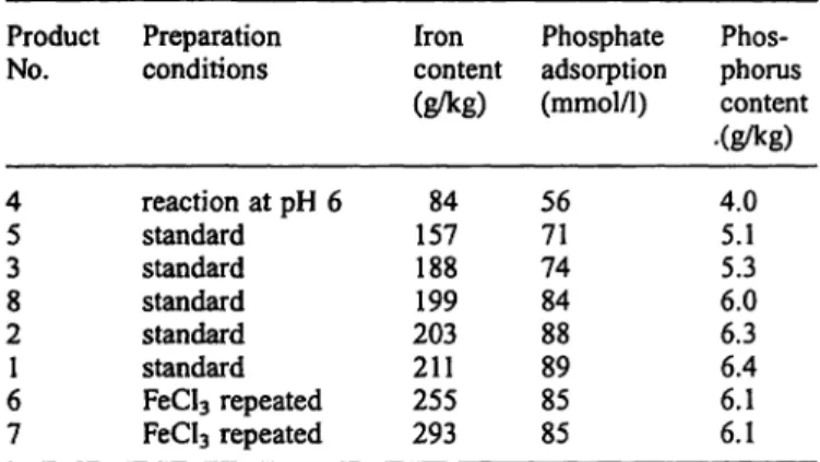
Tab. l Iron content and phosphate adsorption capacity of iron- dextran complexes.
10 20 30 40 50 60 70 80 90 100 120 140 160
Phosphate solution [ml]
Fig. 2 Removal of phosphate from a Standard solution. Columns packed with 3 inl of ironrdextran adsorbent (iron: 188 g/kg) were perfused at l ml · min"1 flow rate and the phosphate content of the eluate was determined. Tlie horizontal line represents the initial concentration (Pj, 100 mg/1). A: Dried adsorbent, not sterilized. ·:
Dried material sterilized at 121 °C.
Product No.
45 38 21 67
Preparation conditions
reaction at pH 6 Standard Standard Standard Standard Standard FeCl3 repeated FeCl3 repeated
Iron content (g/kg)
15784 188199 203211 255293
Phosphate adsorption (mmol/1)
5671 7484 8889 8585
Phos- phorus content .(g/kg)
4.05.1 6.05.3 6.36.4 6.16.1
phate binding capacity (tab. 1). Higher amounts of iron, produced by repeated treatment of the adsorbent in FeCl
3solution, did not raise the phosphate binding ca- pacity. In equilibrium binding experiments, sterile adsor- bents coated with 210 g/kg iron typically bound 250 mmol (7.7 g) Pj per kg or 90 mmol (2.6 g) P
{per litre adsorbent. These values were obtained by equilibrating 3 ml of sterilized adsorbent in a column for 18h with 500 ml circulating phosphate Standard solution, fol- lowed by removal of unbound inorganic phosphate by washing the column with 50 ml distilled water. Absorp- tion of phosphate from an aqueous solution onto the iron-dextrari complex could also be monitored by
31P- NMR (not shown).
The phosphate adsorption capacity was generally re- duced about 30% after heat sterilization (fig. 2). On the other hand, heat sterilization ppears to lead to a stabi- lized iron(III) oxide-hydroxide modified dextran at acidic pH vaiues. Thus, the sterilized adsorbent does not liberale iron even around pH 2.
Eur. J. CKn. Chem. Clin. Bioohem. / Vol. 32,1994 / No. 10
centration, or of distinct enzyme activities were ob- served during the treatment. The profile of the elimina- tion curve shows that additional phosphate can be ad- sorbed when higher blood phosphate concentrations are present.
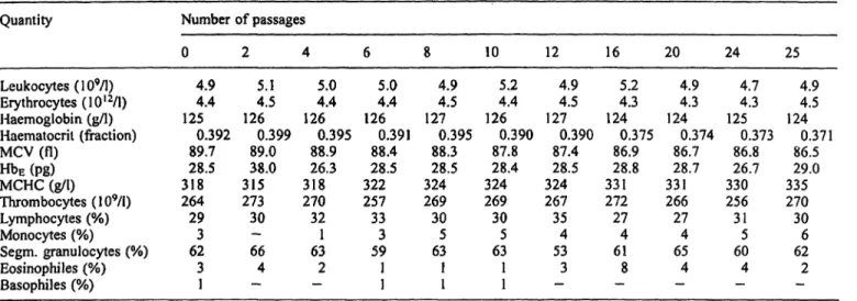
Investigation of the haemocompatibility of the adsorbent in a 250 ml-cartridge showed that the phosphate concen- tration of circulating whole blood decreased from 1.1 mmol/1 to 0.09 mmol/1 (3.4 mg/dl to 0.3 mg/dl PI). At a flow rate of 100 ml/min, a pressure of 80 mbar was built up. Even after 25 circulations the material did not induce haemolytic reactions, and there were no significant alter- ations in common blood quantities äs shown in table 2.
Elimination of calcium ions • \
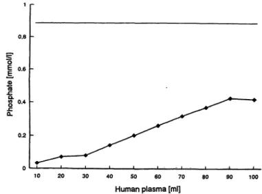
The behaviour towards calcium ions was determined by perfusing the adsorbent in a 3 ml column with 100 ml of human serum, heparinized plasma or standardized calcium solution (fig. 4). Calcium was only bound to the adsorbent in the presence of phosphate but not in phosphate-free solution. A molar ratio of calcium and phosphate elimination of approximately l : l was reached when both concentrations were varied.
In vivo experiments
The iron(III)oxide-hydroxide dextran adsorbent was tested in extracorporeal apheresis experiments with sheep. Laboratory data are reported in table 3. The aim of these tests was to investigate functional aspects such
0.2
40 50 60 70
Human plasma [ml]
90 100
Fig. 3 Elimination of phosphate from 100 ml human plasma (ini- tial P content, 2.8 mg). Conditions äs in Fig. 2.
cartridge phosphate concentratiqn from 1.93 mmol/1 (6,0 mg/dl) to 0.06 mmol/1 (0.2 mg/dl) P
fat the end of treat- ment (l h dialysis). There was neither an increase iii the iron nor a decrease in the calcium concentration öf the blood during treatnient with conventional dialysis solu- tions containing calcium. By appropriate dosage of hep- arin, the flow rate and pressure could be kept at normal levels without inducing haemolysis or significant alter- ations of the blood cells. Only thrombocytes were fe- duced during treatment. This was possibly due to some fibrin clotting which was not observed in in vitro experi- ments.
Discussion
Patients with kidney diseases, especially those with chronic renal failure, who have been on haemodialysis for a lofig time, often have Symptoms ässociated with highly elevated concentrations of inorganic phosphate in the blood. Lafge amounts of phosphate äbove normal levels (0.6—1.3 mmol/1) result in a decrease of calcium ion concentration which in turn induces the parathyroid to secrete an excess amount of parathyroid hormone.
The hormone increase and the inability of the diseased kidney to hydroxylate 25-hydroxycholecalciferol to the active forms of vitamin D are the principal biochemical factors underlying most of the related Symptoms (22, 23). In the case of chronic renal failure the filtration rate of glomeruli is less than 8-10 ml/min. Pathological phosphate concentrations ranging between 2.25 and 3 mmol/1 normally decrease to 1.3 — 1.6 mmol/1 after haemodialysis (24). In this context, it has recently been shown that extracorporeal elimination of phosphate is more effective usihg Standard dialysers than high-flux- dialysers (7! Eisenhauer, C. Ronco, unpublished). This effect is due to the limited phosphate concentration in the blood. Thus, the intravascular phosphate concentra- tion rather than the clearaiice seems to be rate-limiting.
The phosphate clearance, for exatnple of the Diacap cap- illary dialyser (B. Braun, Melsungen, Germany) lies be- tween 93 and 150 ml/min when pumping blood at 200 ml/min and dialysis solution at 500 ml/min.
The phosphate intake of a patient is 32 mmol per day or about 224 mmol per week. During one dialysis Session about 32 mmol phosphate are normally eliminated (4).
These rates of removal äfe insüfficient to prevent hyper- parathyroidism. The 8 5 »of about 130 minol phos- phate per week has to be addftionally elimiiiatedv Hence,
Eur. J. Clin! Chem. CHn. Biochem. / Vol. 32, 1994 /No. 10
Spengler et al.: New phosphate binding agent 737
1
Tab. 2 Blood count and erytlirocyte characteristics during perfusion of iron-dextran phosphate adsorbent with human blood.
Quantity
Leukocytes (109/1) Erythrocytes (10I2/1) Haemoglobin (g/l) Haematocril (fraction) MCV (fl)
HbE (pg) MCHC (g/l) Thrombocytes (109/1) Lymphocytes (%) Monocytes (%) Segm. granulocytes (%) Eosinophiles (%) Basophiles (%)
Number of passages 0
4.94.4 1250.392
89.7 28.5 318264
293 623 1
2 5.14.5 126
0.399 89.0 31538.0 27330 66— 4
— 4
5.04.4 1260.395
88.9 31826.3 27032 631
—2 6
5.04.4 126
0.391 88.4 32228.5 25733 593 11
8 4.94.5 1270.395
88.3 32428.5 26930 635 11
10 5.24.4 1260.390
87.8 32428.4 26930 635 11
12 4.94.5 1270.390
87.4 32428.5 26735 534
—3
16 5.24.3 1240.375
86.9 33128.8 27227 614
—8
20 4.94.3 1240.374
86.7 33128.7 26627 654
—4
24 4.74.3 1250.373
86.8 33026.7 25631
605
—4
25 4.94.5 124
0.371 86.5 33529.0 27030 626 2
— A cartridge containing 250 ml phosphate adsorbent was perfused
at 100 ml · mfcr1 under recirculating conditions with 500 ml hu- man blood containing 7 mmol/1 EDTA.
MCV = Mean corpuscular volume
H DE = Haemoglobin content per erythrocyte
MCHC = Mean corpuscular haemoglobin concentration
3
10 40 50 60 70
Human plasma [ml]
so 90 100
Fig. 4 Elimination of calcium from 100 ml human plasma (initial Ca content, 9.35 mg) concomitant with phosphate removal (cf.
fig-3).
Tab. 3 Electrolyte concentrations in sheep blood during an extra- corporeal treatment with iron-dextran complex.
Electrolyte
P (mmol/1) Fe ( / ) Ca (mmol/1)
Time (min) 0
1.94 2.65 2.52
15 0.03 2.63 2.87
30 0.03 2.75 2.54
45 0.13 2.79 2.64
60 0.06 2.40 3.29
75 0.06 2.72 2.85 Blood was pässed through a cartridge containing 250 ml of phos- phate adsorbent; flow rate, 70^90 ml/min.
most patients with en&stage renal failure and rnany with moderate tp marked renal insufficiency require phos- phate binders. Their applicätion is necessary to avoid
skeletal pain, changes in bone mineralization, spontane- ous ftactures, and calcium phosphate-like deposits in soft tissues and blood vessels (25, 26). In recent years it has been reported that the most seriously intoxicated patients had been treated with dialysing fluid containing high concentrations of aluminum and phosphate binders containing aluminum (27-29). The dialysis dementia syndrome was related to the increased concentration of aluminum in the plasma and in the brain tissue (30, 31).
For that reason many authors propagated the oral appli- cätion of aluminum-free compounds such äs calcium carbonate or calcium acetate (32, 33). However, medica- tion with the latter is associated with gastro-intestinal Problems and a high risk of hypercalcaemia (10).
From these considerations it follows that a more effec- tive and safer treatment of hyperphosphataemia can be achieved by extending the total dialysis time and by the optional applicätion of aluminum-free phosphate bind- ers. Prolonging dialysis time, however, is not applicable äs it additionally Stresses the patient. Instead, the desired systemic reduction of phosphate should be achieved by using, in addition to and simultaneously with conven- tional haemodialysis, an adsorbent which specifically re- moves the excess phosphate. To be clinically usefül, such an adsorbent has to be integrated into the extracor- poreal perfusion System and must be capable of elimi- nating phosphate directly from whole blood. Extracor- poreal removal of excess inorganic phosphate is espe- cially of interest for patients who refuse phosphate- lowering medication because of personal discomfort.
For a potentially chronic applicätion such a procedure must be free of even minor side effects. In addition, it should permit a blood flow of at least 100 ml/min.
Eur. J. Clin. Chem. Clin. Biocbem. / Vol. 32,1994 /No. 10
by Hjerten (34), only soluble complexes between iron hydroxide and mono- and polysaccharides have pre- viously been prepared (35, 36). Such complexes, e. g.
with polymaltose (Ferrum-Hausmann™), are used for the therapeutic treatment of iron deficiency anaemias in humans and animals (37, 38). Structural studies on solu- ble iron oxide-hydroxide complexes showed that they form chainlike, polynuclear condensation products (39) and that iron is present äs tetragonal ß-FeOOH (40).
Structure characterization of the newly synthesized, amorphous, insoluble iron-dextran complexes is less complete. Nevertheless, the ESCA and Mössbauer spectra confirm that only high-spin ferne irons with ox- ygen ligarids (sugar OH groups, water, hydroxide ions, - bridges) are present. The most typical iron mass Proportion of about 200 g/kg found in the dry material suggests that each iroii(III) centre is statistically coordi- nated to one glucose moiety (glucose-Fe(OH)
3, Fe = 195 g/kg). In fact, the magnetic susceptibility and its temperature dependence, measured in the ränge from 4 to 300 K, are very similar to a soluble, stoichiometric Fe-glucose complex (35).
A few reports describe the interaction of phosphate with iron-containing compounds (34, 37, 41, 42). Addition of phosphate, even in small amounts, may lead to strong effects, for example increased viscosity in iron-polymal- tose Solutions (37). FeOOH-modified non-porous agar- ose has been saturated with phosphate and used in this form äs ion exchanger for the chromatography of pro- teins (34).
nation of phosphate during haemodialysis. The patho- logical excess of about 130 mmol of phosphate per week in patients with end^stage renal failure, for example, would be elimiriated düring the common three dialysis sessions by an additional, integrated phosphate adsorp- tion apheresis using a cartridge packed with 450 ml of the new adsorbent.
On-line apheresis has the extra advantage that it can be precisely controlled by adjusting the eartridge size (i. e.
total phosphate binding capacity) to the blood völume.
Material costs can be significantly reduced by repeated regeneration and re^use of the adsorbent. Iii this context it was shown that regeneration by a simple treatment with NaOH did not cäuse a decrease of the phosphate binding capacity. The described in vivo experiments with sheep revealed no side efFects. Concomitant calcium depletion can be compensated by dialysis. Ide- ally, it may be expected that oral phosphate binders can be avoided and no other measures have to be taken.
In conclusion, the newly developed iron(III) oxide-hy- droxide modified dextran is suitäble for the selective elimination of inorganic phosphate from whole blood.
The excellent phosphate elimination capacity and blood compatibility make it attractive for the extracorporeal treatment of hyperphosphataemia in acüte and chronic renal failure.
Acknowledgement
This work was made possible by a grant from the B. Braun Mel- sungen AG. The authqrs thank Professor R von der Haar, Professor T. Eisenhauer and Dr. C. Ronco for helpful discussions and the B.
Braun laboratories for making facilities available.
References
1. Bricker, N. S., Slatopolsky, E., Preiss, E. & Aviolo, L. V.
(1969) Calcium phosphorus and bone renal disease and trans- plantation. Arch. Intern. Med. 123, 543-553.
2. Rubini, M. E., Coburn, J. W., Massry, S. G. & Shinaber, J. H.
(1969) Renal osteodystrophy - Some therapeutic considera- tions relative to long-term dialysis and transplantation. Arch.
Intern. Med. 124, 663-669.
3. Coburn, J. E. & Salusky, I. B. (1989) Control of serum phos- phorus in uremia. New England J. Med. 320, 1140-1142.
4. Günther K., Sperschneider, H., Stein, G. & Gaida, P. (1990) Phosphatrestriktion bei optimaler Eiweißzufuhr. Dial. J. 31, 29-34.
5. Ramirez, J. A., Emmett, W., White, M. G., Fathi, N., Ana Cas, Morawaski, S. G. & Fordtran, J. S. (1986) The absorption of o dietary phosphorus and calcium in haemodialysis-patients.
Kidney Int. 30, 753-759.
6. Yokel, R. A. (1989) Benefit vs. risk of oral aluminum forms:
Antacid and phosphate binding vs. absorption. Drug Chem.
Toxicol. 12, 277-286.
7. Bauman, J. L. (1987) Aluminum-induced bone disease in renal failure. Hospital Ther. 87, 48-55.
8. De Broe, M. E., D'Haese, P. C., Van De Vyver, F. L. & Lam- berts, L. V. (1991) Aluminiuminduzierte Osteopathie bei Patl·
enten mit chronischer Niereninsuffizienz. Nieren-Hochdruck- krankh. 20, 311-316.
9. Ritz, E. & Bommer, J. (1980) Störungen des Calcium- und Phosphatstoffwechsels bei Niereninsuffizienz. In: Chronische Niereninsuffizienz, pjp. 398—407, Verlag Chemie, Weinheim.
10. Sheikh, M. S., Maguire, J. A., Emmett, M., Santa Ana, C. A., Nicar, M. J., Schiller, L. R. & Fordtranj J. S. (1989) Reduction of dietary phosphorus absorption by phosphorus binders. J.
Clin. Invest. 83, 66-73. · *
Eur. J. Clin. Chem. Clin. Biochem. / Vol. 32, 1994 / No. 10
Spengler et al.: New phosphate binding agent 739 11. Barsotti, G., Morelli, E. & Guiducci, A. (1982) Reversal of
hyperparathyroidism in severe uremics following very low- protein and low-phosphate diet. Nephron 30, 310-313.
12. Herez, G. & Cobum, J. W. (1987) Prevention of phosphate retention and hyperphosphatemia in uremia. Kidney Int.
(Suppl.) 22, 215-220.
13. Boos, K.-S., Seidel, D., Rauh, A., Spengler, K. & Henke, G.
(1992) Verfahren zur selektiven Elimination von anorgani- schem Phosphat aus Flüssigkeiten mittels polynuclearen Metalloxidhydroxid-modifizierten Adso tionsmaterialien.
German Pat. Appl. P4139442.2.
14. Food and Drug Administration (FDA) (1976) Federal Register 41, No. 106.
15. Rick, W. (1990) Klinische Chemie und Mikroskopie. 6. Auf- lage, pp. 287-294. Springer Verlag Berlin, Heidelberg, New York.
16. Thomas, I, (1992) Labor und Diagnose. 4. Auflage, pp. 50, 121, 136, 342, 358, 390, Medizinische Verlagsgesellschaft, Marburg.
17. Empfehlungen der Deutschen Gesellschaft für klin. Chemie (1972) Z. Klin. Chem. Klin. Biochem. 70, 182.
18. Richterlich, R. (1971) Klinische Chemie - Theorie und Pra- xis. 3. erw. Auflage, p. 228. Karger Verlag, Basel.
19. Marr, I. L., Cresser, M. S. & Ottendorfer, L. J. (1983) Analyti- sche Chemie för die Praxis - Umweltanalytik (Hulpke, H., Hartkamp, H. & Tölg, G., eds.) Thieme-Verlag, Stuttgart, pp.
243-251.
20. Welz, B. (1981) Atomspektroskopische Spurenanalytik. Verlag Chemie, Weinheim.
21. Carver, J. C, Schweitz, G. K., Carlson, T. A. (1972) Use of X-ray photoelectron spectroscopy to study bonding in Cr, Mn, Fe and Co compounds. J. Chem. Phys. 57, 973.
22. Heckmann, C., Rudorff, K.-H. & Saueressig, U. (1991) Klini- sche Problematik des sekundären Hyperparathyreoidismus.
Nephrologisches Jahresgespräch 1991, Kassel.
23. Ritz, E., Matthias, S. & Reichel, H. (1991) Therapeutische Strategien beim sekundären (renalen) Hyperparathyreoidis- mus. Nephrologisches Jahresgespräch 1991, Kassel.
24. Henning, H. V. (1988) Therapie mit Phosphatbindern bei chro- nischen Dialysepatienten. Dial. J. 22, 10-16.
25. Henning, H. V. & Fuchs, C. (1984) Renale Osteopathie. Nie- ren-Hochdruckkrankh. 13, 235-253.
26. Croucher, P. L, Wright, C. D. R, Garrhan, N. J., Kudlac, H., Williams, A. J. & Compston, J. E. (1992) Characteristics of trabecular bone resorption cavities in patients with chronic re- nal failure. Bone Min. 169 139-147.
27. Mahurkar, S. D., Smith, E. C., Mamdani, B. H. & Dunea, G.
(1978) Dialysis dementia. The Chicago experience. J. Dial. 2, 447-458.
28. Rosas, V. V., Port, F. K. & Rutt, W. M. (1978) Progressive dialysis encephalopathy frorn dialysate aluminum. Arch. In- tern. Med. 138, 1375-1377.
29. Walker, G. S., Aaron, J. E., Peacock, M., Robinson, P. J. A. &
Davison, A. M. (1982) Dialysate aluminum concentration and renal bone disease. Kidney Int. 27, 411—415.
30. Alfrey, A. C., Le Gendre, G. R. & Kachny, W. D. (1976) The dialysis encephalopathy syndrome: Possible aluminum intoxi- cation. New England J. Med. 294, 184.
31. Sideman, S. & Manor, D. (1982) The dialysis dementia syn- drome and aluminium intoxication. Nephron 7, 1 — 10.
32. Schaefer, K. (1993) Alternative phosphate binders: An update.
Nephrol. Dial. Transplant. 7, 35-39.
33. Mai, M. L., Emmett, M., Sheikh, M. S., SAnta Ana, C. A., Schiller, L. & Torattran, J. S. (1989) Calcium acetate, an effec- tive phosphorus binder in patients with renal failure. Kidney Int. 36, 690-695.
34. Hjerten, S., Zelikmann, L, Lindenberg, J., Liao, J.-I., Eriksson, K.-O. & Mohamrnad, J. (1984) High-performance adsorption chromatography of proteins on deformed non-porous agarose beads coated with insoluble metal compounds. J. Chromat.
481, 175-186.
35. Pulla Rao, C., Geetha, K. & Raghavan, M. S. S. (1994) Fe(III) complexes of D-glucose and D-fructose. BioMetals 7, 25-29.
36. Rieh, H. W., Hegetschweiler, K., Streit, H. M., Erni, I. &
Schneider, W. (1991) Mononuclear, oligonuclear, and polynu- clear iron(III)complexes with polyalcohols formed in alkaline aqueous media. Inorg. Chim. Acta 187, 9-15.
37. Müller, A. (1967) Makromolekulare Eisen(III)-Hydroxid- Komplexe. Arzneim. Forsch. 77, 921-931.
38. Schwengers, D. (1990) Water soluble iron dextran and a pro- cess for its manufacture. United States Patent, No. 4. 927. 756.
39. Yang, C.-Y., Bryan, A. M., Theil, E. C., Sayers, D. E. & Bro- wen, L. H. (1986) Structural variations in soluble iron com- plexes of models for ferritin. J. Inorg. Biochem. 28, 393-405.
40. Marshall, P. R. & Rutherford, D. (1971) Physical investigations on colloidal iron-dextran complexes. J. Colloid Interface Sei.
37, 390-402.
41. Lijkiema, L. (1980) Interactions of orthophosphate with iron(III) and aluminum hydroxides. Environm. Sei. Technol.
14, 537-541.
42. Thole, S. (1992) Einfluß der Wassermatrix auf die Adsorption von Phosphat an Eisenoxidhydratschlämmen. Vom Wasser 79, 313-321.
Prof. Dr. Hartmut Follmann Fachbereich Biologie-Chemie der Universität
Heinrich-Plett-Straße 40 D-34109 Kassel Germany
Eur. J. Clin. Chem. Clin. Biochem. / Vol. 32,1994 / No.