Systematic Structural and Functional Characterization of the Family of Geranylgeranylglyceryl Phosphate
Synthase-like Enzymes
Dissertation
Zur Erlangung des Doktorgrades der
Naturwissenschaften (Dr. rer. nat.) der Fakultät für
Biologie und Vorklinische Medizin der Universität Regensburg
vorgelegt von David Peterhoff
aus Erding
im Jahr 2014
Das Promotionsgesuch wurde eingereicht am:
14.01.2014
Die Arbeit wurde angeleitet von:
Prof. Dr. Reinhard Sterner
Unterschrift:
Index of contents ... 1
Abstract ... 4
Kurzfassung der Arbeit ... 6
1 Synopsis ... 9
1.1 Homology guided functional assignment of proteins ... 9
1.2 The family of GGGPS-like enzymes ... 10
1.2.1 The GGGPS is a key enzyme in the early evolution of Archaea ... 10
Membrane lipids define the domains of life ... 10
In the focus of evolution: key enzymes for the synthesis of membrane lipids ... 11
1.2.2 On the reaction catalyzed by GGGPS-like enzymes ... 13
A stereo-specific O-alkylation reaction ... 13
Two alternative reaction mechanisms are considered for GGGPS-like enzymes ... 14
1.2.3 Phylogenetic background of the family of GGGPS-like enzymes ... 16
Ambiguities in denomination ... 16
Phylogenetic relationships in the family of GGGPS-like enzymes ... 17
Sequence similarity networks reveal phylogenetic subgroups with different traits ... 20
1.2.4 The differing substrate specificities among GGGPS-like enzymes ... 23
Members of the novel characterized bacterial group IIb exhibit archaeal substrate specificity ... 23
1.2.5 Structural features of GGGPS-like enzymes ... 26
An ancient fold for an ancient enzyme ... 26
Structural adaptations ... 27
1.2.6 Structure-function relationships in the family of GGGPS-like enzymes ... 30
Two disparate substrate binding sites ... 30
Length limitation mechanism for the hydrophobic substrate ... 31
Structural prerequisites for stereo-selective catalysis ... 32
The quaternary structure influences substrate specificity ... 33
An aromatic “anchor” amino acid mediates hexamerization ... 35
1.2.7 Functional and physiological context of GGGPS-like enzymes ... 39
On the substrates and products of the reaction of the GGGPS-like enzymes ... 39
2 Abbreviations ... 42
3 Literature ... 46
4 List of publications ... 54
5 Personal contribution ... 55
6 Publications... 56
Publication A ... 56
Supporting Information for Publication A: ... 64
Publication B ... 71
Supporting Information for Publication B: ... 87
7 Appendix: Generation of codon scanned gene libraries ... 103
7.1 Introduction: Methods for protein engineering ... 103
7.2 Optimizing a protocol for scanning mutagenesis ... 104
7.3 Protocol ... 113
7.3.1 Requirements and materials: ... 113
7.3.2 Procedure: ... 114
1st working day: Transposition reaction. ... 114
2nd working day: Library cleaning. ... 115
3rd working day: Transposon removal – part 1. ... 115
4th working day: Transposon removal – part 2. ... 116
5th working day: Reading frame selection. ... 117
6th working day: Subcloning (optional). ... 119
7.3.3 Sequences ... 120
pIG-plasmid ... 120
pDF-plasmid ... 121
Frame selectable transposon ... 122
pTNA-B/S ... 122
pInSal-B/S ... 123
Acknowledgement ... 124
Abstract
A characteristic difference between the three phylogenetic domains of life is the chemical composition of the lipids forming their cell membranes. In Bacteria and Eukarya phospholipids are based on a sn-glycerol-3-phosphate (G3P) which is esterified with two fatty acids, while in phospholipids from Archaea the enantiomer sn-glycerol-1-phosphate (G1P) forms two ether-bonds with isoprenoid chains. As a result of these differences, the chemical, physical and biological properties of the archaeal membranes are drastically altered compared to bacterial and eukaryotic membranes. These changed conditions have been considered to be crucial for separation of archaea from a bacteria-like last universal common ancestor (LUCA).
Consequently, the two enzymes that provide stereo-specificity in the synthesis of archaeal phospholipids have been regarded as key enzymes in the early evolution of Archaea and have been assumed to be strictly archaea-specific. These are the G1P-providing enzyme, glycerol- 1-phosphate dehydrogenase (G1PDH), and the enzyme transferring an activated polyprenyl substrate (mostly geranylgeranyl diphosphate, GGPP) stereo-specifically to G1P, geranylgeranylglyceryl phosphate synthase (GGGPS). In contradiction to the presumed restriction of GGGPS occurrence to the domain of Archaea, genome sequencing data recently revealed a significant distribution of these enzymes among Bacteria. A phylogenetic analysis of GGGPS-like enzymes showed their partition into two distinct groups (group I and II), both comprising archaeal and bacterial sequences. Group I contains bacterial sequences from Gram-positive Firmicutes and group II from Gram-negative Bacteroidetes. While several bacterial and archaeal group I enzymes have been subjects of recent extensive kinetic and structural analyses, only a few archaeal, but no bacterial group II enzymes have been characterized up to now.
The aim of this work was a systematic comparative characterization of representative members from all important subgroups of the family of GGGPS-like enzymes, with a strong focus on the functional assignment of the Bacteroidetes subgroup and the structure-function- relationship in group II enzymes. Therefore, the variability within the family was analyzed in detail by calculating a sequence similarity network, and 17 representative GGGPS-like enzymes were characterized biochemically regarding their catalytic activities and substrate specificities. Three complementary assays demonstrated that all tested archaeal enzymes preferred short chained polyprenyl substrates, certainly being GGPP in most species. The bacterial enzymes from group II showed a preference for short chained polyprenyl substrates comparable to the archaeal enzymes, but the physiological role of the ether lipid product in those species remains an enigma. In contrast, bacterial enzymes from group I preferred long
chained polyprenyl substrates, and a previous study has showed that the Bacillus subtilis enzyme produces heptaprenylglyceryl phosphate, which becomes dephosphorylated and acetylated in vivo.
Moreover, the first crystal structures of group II archaeal and bacterial enzymes have been solved within the framework of this thesis. These new structures expanded the knowledge on the mechanism of substrate specificity. In all GGGPS-like enzymes, the length of the polyprenylic substrate is measured by a “hydrocarbon ruler” that uses different residues to act as limiters in the binding pocket to provide the different substrate specificities. Interestingly, these limiter residues also differ between the GGPP-specific group I and group II enzymes.
Ongoing ambiguities on the native oligomerization interface of the group I enzyme from B. subtilis, PcrB, were resolved by means of a combined approach including computational, biophysical and biochemical methods. Mutagenesis-mediated monomerization identified the native interface among two computationally predicted alternatives. The result was verified by site-specific incorporation of an unnatural amino acid, which allowed for cross-linking when located in the native interface. Interestingly, monomerization did not affect thermal stability of PcrB, but limited the length of accepted polyprenyl diphosphates to three isoprene units in an in vitro assay, whereas the native substrate contains seven isoprene entities. A plausible hypothesis how dimerization determines substrate specificity of PcrB by affecting its secondary structure could be provided.
While these experiments confirmed that all group I enzymes and a number of group II enzymes share the same dimeric architecture, it was surprising that a significant portion of archaeal and bacterial group II enzymes revealed to be hexamers, as shown by crystallization and static light scattering experiments. In a rational mutagenesis study, which was aided by the novel structures, an aromatic “anchor” residue was found to be an inevitable prerequisite for hexamerization. By substituting this aromatic residue, the hexamers could be disrupted to dimers in the prototypical conformation. Because these dimeric mutants showed catalytic activities and thermal stabilities comparable to the hexameric wild types, the functional reason for hexamerization remains unclear.
In summary, the presented systematic comparative characterization leads to a deepened understanding of the mechanisms of substrate specificity and stereo-selectivity and a detailed description of the enzyme architecture in the two subgroups of the family of GGGPS-like enzymes.
Kurzfassung der Arbeit
Ein charakteristisches Unterscheidungsmerkmal zwischen den drei Domänen des Lebens ist die Zusammensetzung ihrer Zellmembranen. Die Phospholipide von Bakterien und Eukaryoten basieren auf sn-Glycerin-3-Phosphat (G3P), welches mit zwei Fettsäuren verestert ist, während in den Phospholipiden von Archaeen das Enantiomer sn-Glycerin-1-Phosphat (G1P) zwei Etherbindungen mit Isoprenolen ausbildet. Folglich unterscheiden sich die chemischen, physikalischen und biologischen Eigenschaften archaeeller Membranen drastisch zu bakteriellen und eukaryotischen Membranen. Man nimmt an, dass diese veränderten Bedingungen entscheidend für die Abspaltung der Archaeen von einem bakterienartigen, letzten gemeinsamen Vorfahren (last universal common ancestor, LUCA) waren. Folglich werden die beiden Enzyme, die Stereospezifität in der Synthese der archaeellen Phospholipide vermitteln, als Schlüssel-Enzyme in der frühen Evolution der Archaeen betrachtet und wurden bisher als strikt Archaeen-spezifisch angesehen. Diese sind das G1P-bereitstellende Enzym, Glycerin-1-Phosphat-Dehydrogenase (G1PDH), und die Geranylgeranylglycerylphosphat Synthase (GGGPS), welche ein aktiviertes Polyprenyl-Substrat (meistens Geranylgeranylpyrophosphat, GGPP) stereospezifisch auf G1P überträgt. Entgegen der allgemein angenommenen Beschränkung des Vorkommens der GGGPS auf die Domäne der Archaeen haben Genomsequenzierungsdaten in der letzten Zeit eine signifikante Verbreitung dieser Enzyme unter Bakterien gezeigt. Eine phylogenetische Analyse von GGGPS-artigen Enzymen zeigte ihre Spaltung in zwei distinkte Gruppen (Gruppe I und II), die beide archaeelle und bakterielle Sequenzen umfassen. Gruppe I enthält bakterielle Sequenzen von Gram-positiven Firmicutes und Gruppe II enthält Sequenzen von Gram-negativen Bacteroidetes. Während in jüngster Zeit verschiedene bakterielle und archaeelle Enzyme aus der Gruppe I Gegenstand eingehender kinetischer und struktureller Analysen waren, wurden aus der Gruppe II bisher nur wenige archaeelle und keine bakteriellen Enzyme charakterisiert.
Ziel dieser Arbeit war eine systematische, vergleichende Charakterisierung von repräsentativen Mitgliedern aller wichtigen Untergruppen der Familie der GGGPS-artigen Enzyme, mit einem starken Fokus auf die Funktionsaufklärung der Bacteroidetes- Untergruppe sowie auf die Struktur-Funktions-Beziehungen in Gruppe II-Enzymen. Hierzu wurde die Variabilität in der Familie durch Berechnung eines Sequenzähnlichkeits- Netzwerkes detailliert analysiert, und 17 repräsentative GGGPS-artige Enzyme wurden biochemisch hinsichtlich ihrer katalytischen Aktivität und Substratspezifität charakterisiert.
Drei komplementäre Tests zeigten, dass alle archaeellen Enzyme kurzkettige Polyprenyl- Pyrophosphate als Substrat bevorzugten, welches in den meisten Spezies sehr wahrscheinlich
GGPP ist. Die bakteriellen Enzyme aus Gruppe II zeigten ebenso eine Präferenz gegenüber kurzkettigen Polyprenylsubstraten, vergleichbar zu den archaeellen Enzymen. Die physiologische Rolle der Etherlipidprodukte in diesen Spezies bleibt unbekannt. Bakterielle Enzyme der Gruppe I bevorzugten dagegen langkettige Polyprenylsubstrate, und eine vorangehende Studie konnte zeigen, dass das Enzym aus Bacillus subtilis Heptaprenylglycerylphosphat produziert, welches in vivo dephosphoryliert und acetyliert wird.
Weiterhin wurden im Rahmen dieser Arbeit die ersten Kristallstrukturen von Gruppe II- Enzymen gelöst. Diese neuen Strukturen erweiterten das Wissen über den Mechanismus der Substratspezifität. In allen GGGPS-artigen Enzymen wird die Länge des Polyprenylsubstrates durch einen „hydrocarbon ruler“ („Kohlenwasserstoff-Lineal“) gemessen, welches verschiedene Residuen in der Bindetasche als „limiter“ (Begrenzer) nutzt, um die unterschiedlichen Substratspezifitäten zu ermöglichen. Interessanterweise unterscheiden sich diese Residuen auch zwischen GGPP-spezifischen Enzymen der Gruppe I und II.
Mithilfe eines kombinierten Ansatzes aus bioinformatischen, biophysikalischen und biochemischen Methoden konnten andauernde Unklarheiten bezüglich der nativen Oligomerisierungs-Kontaktfläche des Gruppe I Enzyms aus B. subtilis, PcrB, geklärt werden.
Mittels mutagenesevermittelter Monomerisierung konnte die native Kontaktfläche unter zwei bioinformatisch vorhergesagten Alternativen identifiziert werden. Das Ergebnis wurde durch ortsspezifischen Einbau einer nichtkanonischen Aminosäure verifiziert, welche bei Lokalisierung in der nativen Kontaktfläche ein Quervernetzen der Untereinheiten erlaubte.
Interessanterweise beeinflusste die Monomerisierung nicht die thermische Stabilität von PcrB, aber limitierte in einem in vitro Test die Länge der akzeptierten Polyprenylpyrophosphate auf drei Isopreneinheiten, während das native Substrat sieben Isopreneinheiten enthält. Eine plausible Hypothese konnte aufgestellt werden, wie die Dimerisierung die Substratspezifität von PcrB durch Beeinflussung von dessen Sekundärstruktur bedingt.
Während diese Experimente bestätigten, dass alle Enzyme der Gruppe I und einige der Gruppe II eine gemeinsame Dimer-Architektur aufweisen, war es überraschend, dass eine erhebliche Anzahl an archaeellen und bakteriellen Gruppe II Enzymen Hexamere bilden, wie durch Kristallisations- und Lichtstreuungsexperimente gezeigt werden konnte. In einer durch die neuen Strukturen unterstützten Mutagenesestudie konnte eine aromatische „Anker“- Residue als unabdingbare Voraussetzung für die Hexamerisierung identifiziert werden. Durch Austausch dieser aromatischen Residue konnten die Hexamere in Dimere mit prototypischer Konfiguration getrennt werden. Da diese Dimer-Mutanten zum Wildtyp vergleichbare
katalytische Aktivitäten und thermische Stabilitäten zeigten, bleibt der funktionelle Grund der Hexamerisierung vorerst unklar.
Zusammenfassend führt die vorgelegte systematische Charakterisierung zu einem vertieften Verständnis der Mechanismen der Substratspezifität und der Stereoselektivität sowie zu einer detaillierten Beschreibung der Enzym-Architektur in den zwei Untergruppen der Familie der GGGPS-artigen Enzyme.
1 Synopsis
1.1 Homology guided functional assignment of proteins
The challenging legacy of the genomics era is a hardly manageable mass of sequence data. As a result of large-scale genome sequencing projects, a huge number of putative genes is deposited in databases, while only few of their products have been characterized yet. In fact, the UniProtKB/TrEMBL protein database statistics (Release 2013_09 of 18-Sep-2013, 42.821.879 entries) show, that only for 0.05 % of its entries the existence of the protein has been proven experimentally. For further 1.91 % of the entries at least expression data (e.g.
cDNA(s), RT-PCR or Northern blots) indicates the existence of the protein, and 23 % of the deposited proteins are inferred by homology, meaning that their existence is likely because orthologs exist in related species. The remaining 75 % of the entries are pure predictions, i.e.
no evidence at protein, transcript, or homology level exists. The systematic experimental validation of these predictions is one of the biggest challenges of the post genomic era.
To annotate the function of uncharacterized proteins, predictions based on sequence similarity can be helpful. Although such predictions often turned out to be incorrect due to divergent evolution (Gerlt et al. 2000; Friedberg et al. 2006; Schnoes et al. 2009), they are on the other hand reasonable and advantageous when only taken as a rough estimation of the function.
Besides this, the quality of the predictions has strongly improved due to novel prediction strategies involving interconnected knowledge (Loewenstein et al. 2009, Radivojac et al.
2013) – a principle which gene ontology initiatives are currently streamlining (Ashburner et al. 2000; Berardini et al. 2010; Gaudet et al. 2011). Hence, a careful homology guided functional assignment approach is a feasible strategy to unravel novel protein functions.
Against this background and due to our special interest in proteins encountering the (βα)8- fold, we started to investigate the group of sn-glycerol-1-phosphate geranylgeranyltransferase-like (GGGPS-like) enzymes. In an integrative approach combining biochemical and computational methods we opted for a systematic and comparative characterization of this interesting group of enzymes, including the assignment of function to so far uncharacterized members.
1.2 The family of GGGPS-like enzymes
1.2.1 The GGGPS is a key enzyme in the early evolution of Archaea Membrane lipids define the domains of life
A characteristic difference between the three superkingdoms of life (Bacteria, Archaea and Eukarya) is the chemical composition of the lipids forming their membranes (Kates 1993;
Koga et al. 1993; Wächtershäuser 2003; Matsumi et al. 2011). Among the large variety of membrane lipids, the most important lipid chemistry in all domains of life is the class of phospholipids (Cronan 2003; van Meer et al. 2008; Albers et al. 2011). Although exhibiting the same architecture of a polar head group and a long hydrophobic tail, major differences appear when comparing the phospholipids of Bacteria and Eukarya to archaeal phospholipids.
In Bacteria and Eukarya, phospholipids are based on a sn-glycerol-3-phosphate (G3P) which is esterified with two fatty acids, while in phospholipids from Archaea the enantiomer sn- glycerol-1-phosphate (G1P) forms two ether-bonds with isoprenoid chains (Koga et al. 2007;
Ulrih et al. 2009; figure 1).
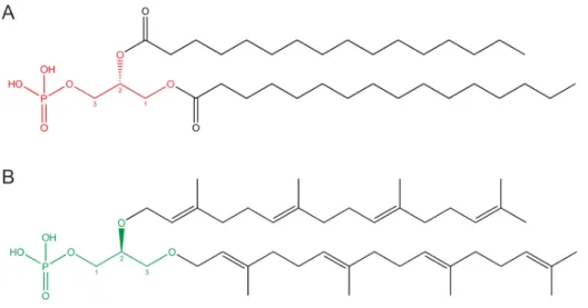
Figure 1: Bacterial and archaeal core phospholipids.
In Bacteria and Eukarya, G3P (red) is bound to fatty acids by ester linkages (A), while in Archaea, G1P (green) is bound to polyprenyl derivatives by ether linkages (B).
A large number of diverse modifications of these core phospholipids exist, and their specific combination in the membrane is characteristic for every organism (De Rosa et al. 1988;
Albers et al. 2011). This circumstance guided attempts to phylogenetically classify Archaea based on their membrane lipids (Koga et al. 2008).
In the focus of evolution: key enzymes for the synthesis of membrane lipids
Huge efforts have been made, to elucidate the characteristics and evolution of a primitive ancestor organism (last universal common ancestor, LUCA) at the root of the domains of life, and the mechanisms underlying the separation of the three superkingdoms (Woese 1998;
Glansdorff 2000; Glansdorff et al. 2008). A recent model on the evolutionary process of the separation of the Archaea from a bacterial-like LUCA proposes, that changes of the lipid composition of the cell membranes had a strong impact on this speciation event (Payandeh et al. 2007; Lombard et al. 2012; Lombard et al. 2012). On one hand the strongly altered chemical properties of the archaeal membranes are considered to act as a genetic barrier, while on the other hand they might have allowed for the colonization of extreme niches due to their high thermal and chemical stability as well as their impermeability (van de Vossenberg et al. 1998; Ulrih et al. 2009), thereby separating early Archaea from the bacterial genetic pool (Payandeh et al. 2007).
In this context, two enzymes involved in the synthesis of archaeal phospholipids have been widely discussed to act as key enzyme in the early evolution of Archaea (Payandeh et al.
2007; Lombard et al. 2012; Lombard et al. 2012). These are a G1P-providing enzyme and an enzyme transferring an activated polyprenyl substrate stereo-specifically to G1P. In accordance with this hypothesis, all Archaea have a glycerol-1-phosphate dehydrogenase (G1PDH, Interpro family IPR023002), which catalyzes the NADH-dependent reduction of dihydroxyacetone phosphate (DHAP) to G1P, and a sn-glycerol-1-phosphate geranylgeranyltransferase, commonly called geranylgeranylglyceryl phosphate synthase (GGGPS), which transfers the polyprenyl moiety of geranylgeranyl diphosphate (GGPP, consisting of 20 C-atoms) stereo-specifically to the C3 hydroxyl group of G1P. Other than GGGPS, the (S)-2,3-di-O-geranylgeranylglyceryl phosphate synthase (DGGGPS) transfers the second polyprenyl moiety to (S)-3-O-geranylgeranylglyceryl phosphate (GGGP) without stereo-selectivity. Hence, the chirality of the archaeal membrane is determined by the stereo- specific GGGPS reaction, which consequently has been considered to be the committed step in the synthesis of archaeal membrane lipids (Payandeh et al. 2007). Figure 2 gives an overview of the synthesis pathways of bacterial and archaeal core phospholipids.
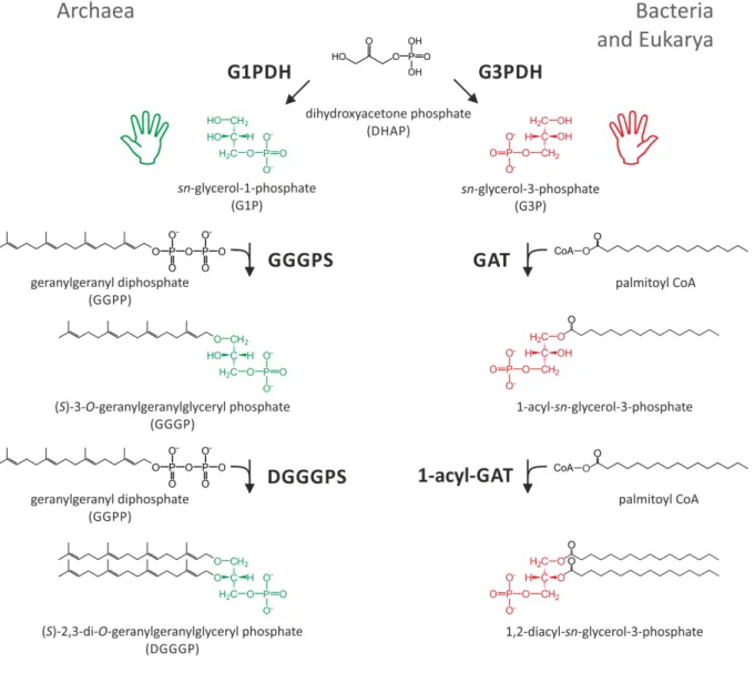
Figure 2: Synthesis pathway of bacterial and archaeal core phospholipids.
G1P (green) in Archaea is provided by a G1PDH, G3P (red) in Bacteria and Eukarya is provided by a glycerol-3-phosphate dehydrogenase (G3PDH). Both enzymes exhibit no structural homology. In Archaea, GGGPS and DGGGPS subsequently transfer the polyprenyl moiety of GGPP to G1P. In Bacteria and Eukarya, a G3P acyltransferase (GAT) and a 1-acyl-G3P acyltransferase (1-acyl-GAT) subsequently transfer the acyl moiety of acyl Coenzyme A (here palmitoyl CoA) to G3P.
Accordingly it has been proposed, that the key enzymes of the synthesis pathway of archaeal membrane compounds, G1PDH and GGGPS, are restricted to the domain of Archaea (Koga et al. 1998; Boucher et al. 2004; Hemmi et al. 2004; Pereto et al. 2004; Payandeh et al. 2007).
In contrast to this hypothesis, phylogenetic analyses revealed that the genomes of some Firmicutes and Bacteroidetes encode GGGPS-like proteins (Boucher et al. 2004; Doud et al.
2011; Lombard et al. 2012, for details see 1.2.3). It has recently been shown for Firmicutes that they produce heptaprenylglyceryl phosphate (HepGP) instead of GGGP, which subsequently becomes dephosphorylated and acetylated (Guldan et al. 2011). These GGGPS- like variants from Firmicutes bear for historical reasons the name PcrB (the well characterized
PcrA protein is a DNA helicase, encoded in the same operon, Petit et al. 1998). Whether the occurrence of these enzymes in Bacteria is a consequence of one or more lateral gene transfer events (LGT, Boucher et al. 2003) or a vestigial feature of the early evolution of Archaea from the LUCA, remains to be clarified. However, the existence of ether lipids in such species (Guldan et al. 2011), as well as the presence of a specific product processing phosphorylase and acetyltransferase (Linde 2013), indicate their physiological relevance. Furthermore, it was shown that a bacterial G1PDH (termed AraM) exists in Bacillus subtilis (Guldan et al. 2008).
1.2.2 On the reaction catalyzed by GGGPS-like enzymes A stereo-specific O-alkylation reaction
GGGPS-like enzymes catalyze the formation of an ether bond between sn-glycerol-1- phosphate and a polyprenylic compound with a varying number of isoprene units (Figure 3) (Chen et al. 1993; Zhang et al. 1993; Soderberg et al. 2001; Nemoto et al. 2003; Payandeh et al. 2006; Guldan et al. 2011). Stereo-specificity of the reaction is enabled by the stereo- specific binding of G1P by the enzyme.
Figure 3: The reaction catalyzed by the family of GGGPS-like enzymes.
GGGPS-like enzymes catalyze the transfer of a polyprenyl diphosphate (2) with a varying number (n+2) of isoprene units to the C3-oxygen of G1P (1). The reaction leads to the formation of an ether bond (red arrow) in the polyprenylglyceryl phosphate product (3), and is energetically driven by the release of diphosphate (4). A magnesium ion is an essential cofactor of the reaction.
In Archaea, where the polyprenyl substrate consists of 4 isoprene units (n=2, GGPP) in most cases, the ether product serves as a precursor of various archaeal cell membrane compounds (Matsumi et al. 2011). In this case the enzyme is called GGGPS (EC 2.5.1.41), being an eponym of the whole family of enzymes. Bacterial GGGPS-like variants from Firmicutes prefer a longer substrate (Guldan et al. 2011) which consists of seven isoprene units (n=5, heptaprenyl diphosphate, HepPP). Consequently these enzymes, which have initially been denominated as “PcrB”, are termed heptaprenylglyceryl phosphate synthases (HepGPS, EC 2.5.1.-).
An essential cofactor of the reaction is Mg2+, which binds in complex with the diphosphate moiety of the prenyl substrate (Chen et al. 1993; Soderberg et al. 2001; Nemoto et al. 2003;
Payandeh et al. 2006; Ren et al. 2013). In fact, many prenyltransferases depend on divalent cations, including farnesyl diphosphate synthase, undecaprenyl diphosphate synthase and squalene synthase (Christianson 2006).
Two alternative reaction mechanisms are considered for GGGPS-like enzymes
Two alternative catalytic mechanisms have been discussed: electrophilic alkylation and nucleophilic substitution (Chen et al. 1993; Soderberg et al. 2001; Nemoto et al. 2003;
Payandeh et al. 2006; Ren et al. 2012; Ren et al. 2013). While during an electrophilic alkylation a highly electrophilic allylic carbocation is formed (geranylgeranyl cation intermediate) which subsequently alkylates G1P (figure 4 A), in a nucleophilic substitution the activated (deprotonated) C3-hydroxyl group oxygen of G1P attacks the C1-atom of the polyprenyl diphosphate and the diphosphate acts as leaving group (figure 4 B). For the GGGPS-like family electrophilic alkylation has been suggested first by Poulter and co- workers (Zhang et al. 1993). To address this problem experimentally, they used three analogous substrates (figure 4 C), of which only two could perform an electrophilic alkylation.
Figure 4: Two possible reaction mechanisms of GGGPS-like enzymes, and substrate analoga to discriminate between them.
A) Electrophilic alkylation mechanism. The diphosphate moiety (orange) leaves the substrate, forming the highly electrophilic allylic carbocation, which subsequently reacts with the C3-oxygen of G1P (green). A catalytic glutamate carboxyl group from the GGGPS (blue, compare 1.2.6) accepts the released proton. B) Nucleophilic substitution mechanism. The catalytic glutamate deprotonates the C3 hydroxyl group. The latter attacks the C1 carbon atom of the polyprenyl substrate which subsequently releases the diphosphate leaving group. C) The three different substrates to discriminate between the proposed alternative mechanisms: GGPP (1), phytyl diphosphate (2) and phytanyl diphosphate (3).
The position, where a double bond provides electron density to stabilize the carbocation intermediate of an electrophilic alkylation is given in red.
The native substrate GGPP (figure 4 C (1)), and phytyl diphosphate (figure 4 C (2)) bear an allylic double bond (between the C2 and C3 carbon atom) thereby being able to stabilize the positively charged intermediate during an electrophilic alkylation, while the completely reduced phytanyl diphosphate (figure 4 C (3)) ought to form a highly unstable carbocation during an electrophilic alkylation. In contrast, all three substrates are equally likely to undergo a nucleophilic substitution. The GGGPS (which was provided in a cell free preparation of Methanothermobacter thermautotrophicus) only showed activity with the two substrates which are capable of electrophilic alkylation. While radiolabeled phytyl diphosphate was incorporated at 18 % of the maximal rate (GGPP rate), phytanyl diphosphate gave no signal for turnover.
The presumption of an electrophilic alkylation is consistent with the proposals for other prenyltransferases (Poulter et al. 1978; Liang et al. 2002), but nucleophilic substitution reactions have also been described for protein prenyl transferases (Long et al. 2002).
1.2.3 Phylogenetic background of the family of GGGPS-like enzymes Ambiguities in denomination
The group of enzymes transferring a polyprenyl moiety to G1P is not clearly defined and precisely termed and therefore not represented consistently in the current databases of protein families. The manually curated Pfam database (Punta et al. 2012) subsumes 644 sequences (July 2013, release 27) from Bacteria and Archaea to the “PcrB family” (PF01884). The Interpro database (Hunter et al. 2012) matches 1205 sequences (July 2013, release 43.1) form Bacteria and Archaea to a “Geranylgeranylglyceryl phosphate synthase/Heptaprenylglyceryl phosphate synthase” family (IPR008205) which divides into multiple inconsistent subfamilies (IPR010946, IPR026417, IPR026438). One reason for these ambiguities is the emergence of hundreds of bacterial sequences over the last decade, which undermined the conjecture of consistent substrate specificities among the family members. Due to these ambiguities we use the term “GGGPS-like” enzymes to address all members of the group. An alternative more general and unbiased denomination, legitimate for all members of the family, would be
“glyceryl-1-phosphate polyprenyl transferase”.
Phylogenetic relationships in the family of GGGPS-like enzymes
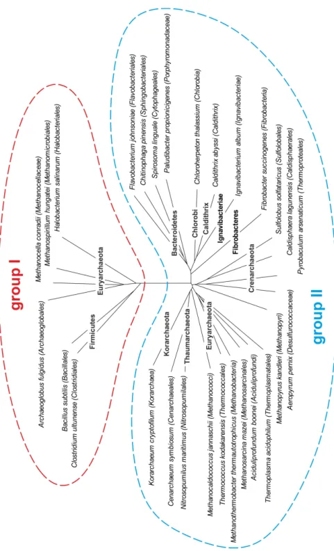
A phylogenetic analysis of the amino acid sequences of GGGPS-like enzymes reveals a partition into two distinct groups (figure 5).
Figure 5: Phylogenetic tree of the family of GGGPS-like enzymes.
Taxa possessing GGGPS-like enzymes were identified using BLAST (Altschul et al. 1990).
Representative sequences were selected for every taxonomic order, a multiple sequence alignment was calculated using Clustal Omega (Sievers et al. 2011), and a phylogenetic tree was generated by means of SplitsTree (Huson et al. 2006). Note, that the tree does not reflect the frequency of occurrence among the members of a shown taxon.
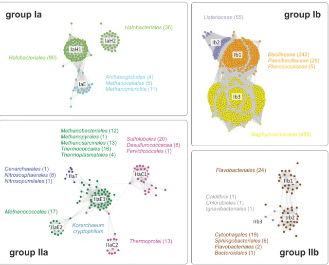
Group I includes sequences of species from the phylum Firmicutes (mostly Gram-positive Bacillales, group Ib) and some Euryarchaeota (group Ia), while group II assembles sequences from Gram-negative Bacteroidetes, Chlorobi, Caldithrix, Ignavibacteriae, Fibrobacteres (group IIb) and from all archaeal phyla except Nanoachaeota (group IIa), which is consistent with the fact that Nanoarchaeum equitans obtains its membrane lipids from its host Ignicoccus hospitalis (Jahn et al. 2004). The sequence identities between group I and group II enzymes are less than 20 %, while members belonging to the same group share sequence identities of more than 40 %. While for Firmicutes and Bacteroidetes a broad distribution of sequences among their numerous members can be found, Chlorobi and Ignavibacteriae are only represented by single sequences, although these phyla contain multiple sequenced species. For the phyla Caldithrix and Fibrobacteres only a single genome sequence is available. This suggests that the GGGPS variants of these representatives were obtained recently via LGT (probably from the Bacteroidetes), while Bacteroidetes and Firmicutes received their variants in an earlier stage of evolution from the Archaea (or vice versa). A sequence similarity network, which was calculated within the framework of this thesis, illustrates these relationships and reflects the number of sequenced genomes per order (figure 6).
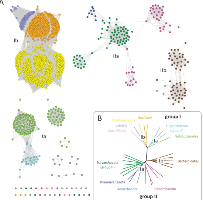
Figure 6: Sequence similarity networks of members of the GGGPS-family.
A) A sequence similarity map, containing 1205 sequences (IPR008205 from InterPro, release 43.1, 7/2013) comprising all currently known GGGPS-like protein sequences, was calculated using the methods developed by Atkinson et al. 2009. A network with an E-value cut-off of 10-60 was visualized using the organic layout in Cytoscape 2.8.3 (Smoot et al. 2011). The coloring of the nodes corresponds to B) where the associated phyla and orders are shown (miscellaneous rare occurrences in grey, not shown in B)). B) Phylogenetic tree of representative sequences from the main phyla possessing GGGPS-like enzymes.
In principle, in a sequence similarity network, nodes represent sequences, and edges represent the BLAST E-values from pairwise local alignments. Edges are drawn when a specified BLAST e-value cutoff is outvalued. This gives a two dimensional representation of clustering similarity patterns (Barabasi et al. 2004; Atkinson et al. 2009). Sequence similarity networks have been shown to correlate well with phylogenetic trees (Kalyanaraman et al. 2008; Lukk et al. 2012).
The network of the GGGPS-like enzymes, which contains the 1205 sequences of the IPR008205 family from the Interpro database, reflects the common archaeal taxonomy as well as the phylogeny as deduced from ribosomal protein sequences (Brochier-Armanet et al.
2011; Olsen et al. 1993), and the already mentioned splitting of the family into four subgroups (groups Ia, Ib, IIa, IIb; compare publication B, figure 1). Due to imbalances in the number of sequenced genomes, particularly the group Ib sequences are strongly overrepresented.
Due to a bug in the scripts used for calculating the network (Biocluster, University of Illinois at Urbana-Champaign, http://biocluster.igb.illinois.edu/, as of Sept. 2013), the BLAST hits per sequence, and therefore the number of edges per node, was limited to 250. Tests in Dec.
2013 with a revised script showed that this only affected cluster Ib, which now was much more compact (data not shown). Because all other clusters were completely unaffected, but the limit to 250 hits significantly improved the resolution of subdivisions within cluster Ib, the original version of the network was used within this work.
Sequence similarity networks reveal phylogenetic subgroups with different traits
Interestingly, the clustering in the network visualizes differences in the family, which are not well resolved in a phylogenetic tree. Some specific sequence features lead to a further splitting into subgroups in some orders, which is illustrated in figure 7. First, the Halobacteriales divide in two completely separated groups (IaH1, IaH2), one of the groups being connected to the rest of the sequences of group I Euryarchaeota (IaE). Second, the sequences of the Euryarchaeota from group II split into two subgroups (IIaE1, IIaE2) of which one is connected to the sequences of Crenarchaeota (IIaC1, IIaC2) and Thaumarchaeota (IIaT). The sequence from Korarchaeum cryptofilum is located between the two group II Euryarchaeota subgroups. Third the sequences of Crenarchaeota split into two subgroups (IIaC1, IIaC2), which are partially connected by the sequences from group II Euryarchaeota. Fourth, the sequences from Bacteroidetes split into two subgroups (IIb1, IIb2), one of them being connected to the three bacterial sequences from other taxa in group II (IIb3).
Figure 7: Taxonomic details of the main clusters in the sequence similarity network
All taxonomic orders occurring in a cluster (color of the taxon labels corresponds to the cluster color) are given together with the number of their members (in brackets). Because in case of cluster Ib all members are from the order Bacillales, the family names of the clustering members are given instead.
Checking the taxonomic details we found that many Halobacteria possess two different GGGPS enzymes, consistent with former findings (Boucher et al. 2004). It seems that there exist two paralogues of a GGGPS-like enzyme in these species. Interestingly, the two distinct subgroups (IaH1 and IaH2) are formed by these paralogues. The occurrence of a IaH2 type enzyme is in almost all cases connected to the occurrence of an enzyme of type IaH1. Only two Halobacteria strains (Halovivax asiaticus, Halovivax ruber) contain exclusively a type IaH2 enzyme. Boucher et al. (2004) suggest that the existence of two paralogues in some Halobacteria could be the reason for the occurrence of farnesylgeranylglyceryl phosphate (FGGP) and geranylgeranylglyceryl phosphate (GGGP) since the two enzymes might be the corresponding G1P-prenyl transferases with specificities for C25 and C20 substrates, respectively (Boucher et al. 2004). We resume this discourse in publication B, by discussing how an insertion of 25 amino acids (on average, figure 8), which led to the splitting into the
two halobacterial subgroups, is embedded into the structure of the GGGPS and that it might influence substrate specificity of the enzyme.
Figure 8: Consensus sequence and conservation logo illustration of the two variants of GGGPS- like enzymes in Halobacteria.
A multiple sequence alignment of the sequences of the two main clusters from Halobacteria in the sequence similarity network (see figure 6 and 7), which represent the two paralogues, was calculated using Clustal Omega (Sievers et al. 2011). The subgroup of sequences possessing the insertion (IaH2) contained 37 sequences while the group without insertion (IaH1) included 86 sequences. The conservation logo (colored, above the line) and the consensus sequence (black, below the line) for the
two subgroups was generated using Jalview (Waterhouse et al. 2009). The residues are colored according to their physicochemical properties (Zappo color code) as follows: Aliphatic/hydrophobic in pink (ILVAM); aromatic in orange (FWY); positive in blue (KRH), negative in red (DE), hydrophilic in green (STNQ), conformationally special in magenta (PG), and cysteine in yellow (C). The position of a proposed substrate length limiting residue, as discussed in 1.2.6 and, publication B is marked by a red arrow and box.
In chapter 1.2.6 evidence is provided, that the splitting of the Bacteroidetes and Crenarchaeal sequences is connected to their different oligomerization states. Another reason for the splitting of the Crenarchaeota into two subgroups could be the occurrence of a highly positively charged N-terminal stretch, which might be important for the interaction with the membrane as it has been proposed in case of the Archaeoglobus fulgidus enzyme (Payandeh et al. 2006). The structural basis for the separation of the two clusters of group II Euryarchaeota is currently unclear.
1.2.4 The differing substrate specificities among GGGPS-like enzymes
It has been mentioned before, that different members of the group of GGGPS-like enzymes have two different substrate specificities (see 1.2.2). While three archaeal enzymes (one from group Ia and two from group IIa) have been shown to prefer GGPP (20 C-atoms, Chen et al.
1993; Zhang et al. 1993; Soderberg et al. 2001; Nemoto et al. 2003; Payandeh et al. 2007), bacterial group Ib enzymes prefer HepPP (35 C-atoms, Guldan et al. 2011).
Members of the novel characterized bacterial group IIb exhibit archaeal substrate specificity
In publication B, the substrate specificities of a large subset of members of the GGGPS family were compared, including representatives from the so far uncharacterized group IIb. Three different assays (I, II, III) were used, all based on radioactively labeled substrates.
I) In a series of in vivo assays 17 GGGPS-like genes were overexpressed in a B. subtilis ΔpcrB strain (a B. subtilis strain lacking the GGGPS-like gene (pcrB), Kobayashi et al. 2003) in presence of radiolabeled G1P (Guldan et al. 2011). The B. subtilis ΔpcrB strain synthesizes various isoprenyl diphosphates, including farnesyl diphosphate (FPP, 15 C- atoms), GGPP (20 C-atoms), HepPP (35 C-atoms), and undecaprenyl diphosphate (UndPP, 55 C-atoms) by specific short-, medium- and long-chain prenyltransferases (Takahashi et al.
1980; Takahashi et al. 1981; Takahashi et al. 1982). Together with the radiolabeled G1P, the different isoprenyl diphosphates are substrates for the heterologously expressed GGGPS-like enzymes, which produce ether lipids according to their substrate specificity. After the labeling
experiment, the lipids were extracted and analyzed by thin layer chromatography and autoradiography (publication B, figure 2).
II) Purified GGGPS-like enzymes (publication B, figure S2) were subjected to an in vitro assay, where we used the (commercially available) short chained substrates geranyl diphosphate (GPP, 10 C-atoms), FPP (15 C-atoms) and GGPP (20 C-atoms) together with radiolabeled G1P (publication B, figure S3).
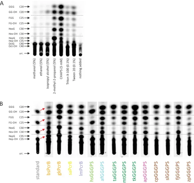
III) Because B. subtilis does not provide substrates of chain length C25, C30 and longer than C40, a coupled two-step in vitro assay was established and optimized to monitor the activity of the different variants with such substrates. A mixture of substrates (C20-C40) was synthesized and simultaneously radiolabeled by an octaprenyl diphosphate synthase from E.
coli (ecOPPS), followed by the conversion of these substrates by the different GGGPS variants in a second step together with G1P (Ren et al. 2013). Effects of detergents on the product chain-length spectrum of polyprenyl diphosphate synthases have been discussed recently (Pan et al. 2013). We therefore tested the effect of some alcohols and detergents on ecOPPS and found that 2-methyl-2-propanol worked best to achieve a broad product spectrum while no interference with the GGGPS reaction is present (figure 9 A). Figure 9 B shows all purified GGGPS-like variants which were characterized with the two-step in vitro assay.
Figure 9: A two-step coupled in vitro assay to analyze specificities for C20-C40 polyprenyl diphosphates.
The unlabeled substrates for ecOPPS, GGPP (Sigma-Aldrich) and FPP (Santa Cruz Biotechnology, Inc.) were obtained in methanol/NH4OH 7:3 (v/v). FPP (25 nmol) and GGPP (5 nmol) were combined, the solvent was removed by a vacuum concentrator, and the substrates were redissolved in 50 µl of reaction mixture, containing 50 mM HEPES (pH 7.5), 50 mM KCl, 0.5 mM MgCl2, 5 % 2-methyl-2- propanol, 1 µM ecOPPS (with N-terminal thioredoxin-fusion) and 100 μM 14C-isopentenyl diphosphate (American Radiolabeled Chemicals, Inc.). The mixture was incubated at 37 °C for 30 min upon slow shaking. In the next step, 15 µM of a GGGPS variant, 4 mM MgCl2 and 20 mM G1P were added, and the slowly shaking mixture was incubated for 6 h at 37 °C. To remove the phosphate of the polyprenyl glycerol phosphates for subsequent TLC analysis, potato acid phosphatase (Sigma-Aldrich) and 100 μL of 40 mM sodium acetate (pH 4.7), 20 % 1‐propanol, 0.1 % Triton X‐100 was added.
After overnight incubation at 37 °C with slow shaking, the lipids were extracted as described before (Bligh et al. 1959; Kates 1986; Guldan et al. 2011). The concentrated extracts were spotted onto a C18 reverse phase HPTLC plate (Merck Millipore) which was subsequently developed in acetone/methanol 2:8 (v/v). The products were visualized by autoradiography. The substrates and products are labeled as follows: GGG, geranylgeranylglycerol; GG-OH, geranylgeraniol; FGG, farnesylgeranylglycerol; FG-OH, farnesylgeraniol; HexG, hexaprenylglycerol; Hex-OH, hexaprenol;
HepG, heptaprenylglycerol; Hep-OH, heptaprenol; OctG, octaprenylglycerol; Oct-OH, octaprenol.
Red arrows show the band shift due to the conversion of polyprenyl diphosphates to polyprenyl glycerols for bsPcrB. A) Influence of several alcohols and detergents on the assay, using bsPcrB as
GGGPS variant. In the last sample (“nothing added”) no detergent or alcohol was added. An equal amount of polyprenyl diphosphate substrates of 20 to 40 carbon atoms is obtained when adding 2- methyl-2-propanol or CHAPS to the reaction mixture. Optimal turnover of the different substrates and G1P by bsPcrB is obtained in the presence of 2-methyl-2-propanol. B) Different GGGPS-like enzymes were incubated with G1P and the radiolabeled mixture of C20-C40 polyprenyl diphosphates produced by ecOPPS (due to insufficient resolution in the HPTLC, only the position C20-C35 is marked by arrows).
In the in vivo assay, where the native substrates C20 and C35 and in addition C15 and C55 were available as substrates, group Ib enzymes preferred HepPP, while group II enzymes only used GGPP significantly as substrate. The group Ia variants provided ambiguous signals:
while the GGGPS from Halobacterium salinarum was inactive the enzyme from A. fulgidus seems to be more promiscuous to longer chained substrates compared for group II enzymes (publication B, figure 2). The in vitro assay with commercially available substrates (C10, C15, C20) showed all enzymes to accept short chained polyprenyl diphosphates (publication B, figure S3). In addition the two step in vitro coupled assay provides C25, C30 and C40 as substrates. In this assay a slight promiscuity towards farnesylgeranyl diphosphate (FGPP, C25) substrate appeared in all group Ia and group II enzymes, while group Ib enzymes accepted all substrates including octaprenyl diphosphate (C40; figure 9 B).
Interestingly, several archaeal organisms possess additional ether lipids based on the alternative polyprenyl building block FGPP (De Rosa et al. 1986; De Rosa et al. 1988; Koga et al. 2005), but sn-glycerol-1-phosphate farnesylgeranyl transferases have not been identified yet. The Crenarchaeon Aeropyrum pernix displays an exceptional core lipid composition. The membranes of A. pernix consist solely of 2,3-di-O-sesterterpanyl-sn-glycerol (both polyprenyl chains connected to G1P consist of 5 isoprene units; Morii et al. 1999). In chapter 1.2.6 it is discussed how a differing mechanism in substrate length determination could lead to a slight substrate promiscuity of the GGGPS towards longer substrates. In addition, A. pernix features a polyprenyl diphosphate synthase of altered specificity (Tachibana et al. 2000), which produces C25 polyprenyl diphosphate.
1.2.5 Structural features of GGGPS-like enzymes An ancient fold for an ancient enzyme
The (βα)8-barrel fold is the most frequent fold among single domain proteins (Sterner et al.
2005). It constitutes 10 % of all proteins with known three-dimensional structure. In the SCOP database, 33 superfamilies are connected to the fold while (βα)8-barrels catalyze more than 60 different reactions. Hence it is not surprising that (βα)8-barrels are also present in the
large and heterogeneous family of prenyltransferases (Oldfield et al. 2012). Two types of prenyltransferases have been shown to be (βα)8-barrels: the GGGPS-like prenyltransferases and MoeO5, an enzyme involved in the biosynthesis of the antibiotic moenomycin, which catalyzes the trans-to-cis isomerization of its substrate farnesyl diphosphate when it is transferred to the second substrate 3-phosphoglycerate (Doud et al. 2011). Considering the spacial extent of the substrates of GGGPS-like enzymes, it is fascinating how the comparatively small (βα)8-barrel scaffold can display such a perfect adaptivity. In fact, HepGP (the product of PcrB) in a stretched conformation measures nearly 42 Å, while the average diameter of a (βα)8-barrel is with approximately 50 Å only slightly larger. Thus, large sections of the protein are in contact with the substrates.
Structural adaptations
Several crystal structures of three group I enzymes (from A. fulgidus, B. subtilis and Staphylococcus aureus) have been solved in complex with G1P as well as analogues of the hydrophobic prenyl substrate, revealing important catalytic residues and leading to a detailed hypothesis of the mechanism of substrate specificity (Badger et al. 2005; Payandeh et al.
2006; Guldan et al. 2011; Ren et al. 2013). Furthermore, a crystal structure of the closely related prenyltransferase MoeO5 has been solved in complex with its substrates (Ren et al.
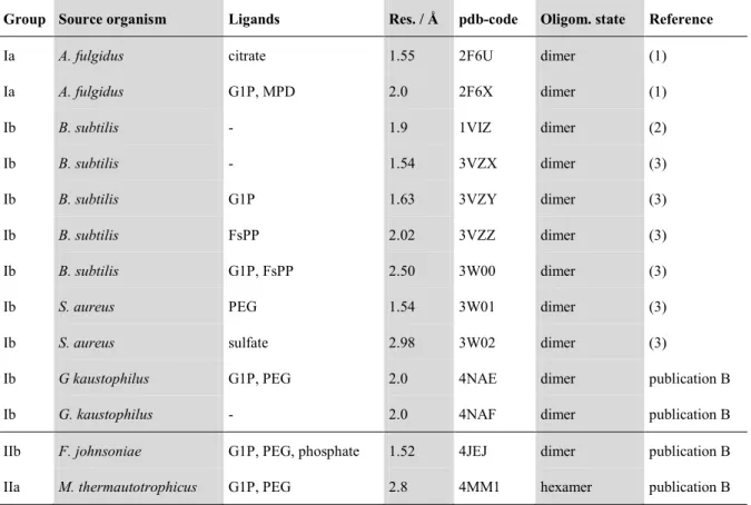
2012). In publication B, four additional structures are provided. Two structures are from group II species: one from the group IIa organism M. thermautotrophicus and one from the group IIb organism Flavobacterium johnsoniae, both solved in complex with G1P. These structures are the first solved structures for group II enzymes. The two other structures are from the group I enzyme from Geobacillus kaustophilus (one apo structure and one with its ligand G1P) which is closely related to the PcrB enzyme from B. subtilis (58 % sequence identity). Both group II and the group I holo structure (with G1P) bound a polyethylene glycol molecule in their binding pocket for the hydrophobic substrate (originating from the crystallization buffer). The basic data for all published structures of the GGGPS-like enzyme family, including our novel structures (for details see publication B, table S3), is summarized in table 1.
Table 1. Structures solved from the family of GGGPS-like enzymes.
Group Source organism Ligands Res. / Å pdb-code Oligom. state Reference
Ia A. fulgidus citrate 1.55 2F6U dimer (1)
Ia A. fulgidus G1P, MPD 2.0 2F6X dimer (1)
Ib B. subtilis - 1.9 1VIZ dimer (2)
Ib B. subtilis - 1.54 3VZX dimer (3)
Ib B. subtilis G1P 1.63 3VZY dimer (3)
Ib B. subtilis FsPP 2.02 3VZZ dimer (3)
Ib B. subtilis G1P, FsPP 2.50 3W00 dimer (3)
Ib S. aureus PEG 1.54 3W01 dimer (3)
Ib S. aureus sulfate 2.98 3W02 dimer (3)
Ib G kaustophilus G1P, PEG 2.0 4NAE dimer publication B
Ib G. kaustophilus - 2.0 4NAF dimer publication B
IIb F. johnsoniae G1P, PEG, phosphate 1.52 4JEJ dimer publication B
IIa M. thermautotrophicus G1P, PEG 2.8 4MM1 hexamer publication B
Grp. = group; Res. = Resolution; Oligom. = Oligomerization; FsPP = S-thiolo-farnesyl diphosphate; MPD = (4S)-2-methyl-2,4-pentanediol;
PEG = polyethylene glycol; (1) = Payandeh et al. 2006; (2) = Badger et al. 2005; (3) = Ren et al. 2013.
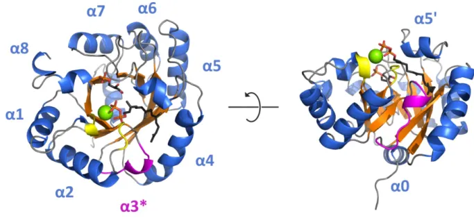
Figure 10 exemplarily shows the GGGPS (βα)8-barrel from F. johnsoniae with its distinct structural features.
Figure 10: Structural features of the group II GGGPS-like enzyme from F. johnsoniae with bound ligands.
Secondary structure elements and G1P (stick representation) of the crystal structure are shown (pdb- code: 4JEJ). GGPP (stick representation) and Mg2+ (green sphere) were modeled using YASARA Structure Version 13.4.21 (employing the YAMBER3 force field (Krieger et al. 2004)) based on the position of HepPP in PcrB from B. subtilis (Guldan et al. 2011) and the position of Mg2+ in MoeO5 (pdb-code: 3VKB). Ligand carbon atoms are given in black, oxygen atoms in red, phosphate atoms in orange. The surface exposed α-helices are given in blue, the central β-sheets in orange, the loops of the Mg2+ binding site in yellow, and the flexible “swinging door” helix α3* in magenta. For clarity one protomer of the homodimer is shown. Taken from figure 3 A, publication B.
The overall structure reflects the (βα)8-fold with some characteristic variations. As in other closely related (βα)8-barrels such as the α-subunit of the tryptophan synthase (TrpA, Hyde et al. 1988), the secondary structure of the GGGPS-like proteins exhibits an additional N- terminal helix α0. This helix has been proposed to mediate the interaction of GGGPS with the membrane, since many members of the family feature a number of positively charged and hydrophobic residues, as a multiple sequence alignment (Payandeh et al. 2006) and our sequence similarity networks show (compare 1.2.3). The most striking difference to the canonical (βα)8-barrel fold is the substitution of helix α3 by a long loop (in the following addressed with α3*). This loop has been termed “swinging door” due to its high flexibility (as suggested by high B-factors). It is located at the distal end of the binding pocket and might serve as a gateway for the prenyl substrate (Payandeh et al. 2006). Helix α4 and α5 are somewhat contorted to the outward, which allows them to form the interface with the second protomer of the dimer (Payandeh et al. 2006; Ren et al. 2013; publication B).
Our novel group II structures differ to some extent from the well known group I structures.
The most interesting differences appear in the G1P binding site and in the quaternary structure. The hydrophobic part of the G1P binding site is more pronounced in group II
enzymes due to the exchange of a lysine by a leucine (compare figure 11 in chapter 1.2.6 and publication B, figure 4). In fact, the lysine is conserved among group I enzymes, while the leucine is conserved among group II enzymes (Payandeh et al. 2006). The structure of the GGGPS from M. thermautotrophicus revealed a hexameric architecture (compare chapter 1.2.6 and publication B, figure 3). In publication B we describe three additional oligomerization interfaces for hexamerization (interface 2, 3a, and 3b), in addition to the standard interface (interface 1) for dimerization (publication B, figure S5). Interestingly the location of the additional interfaces in the hexameric protein is partially identical with the location of the two alternative interfaces of the dimeric protein PcrB from B. subtilis which we analyzed publication A. It is tempting to speculate about an evolutionary background of this finding (see chapter 1.2.6).
1.2.6 Structure-function relationships in the family of GGGPS-like enzymes Two disparate substrate binding sites
The different chemical traits of the two substrates of GGGPS-like enzymes are reflected in their binding sites in the enzyme. While the small hydrophilic G1P binds at the top inner rim of the barrel with its phosphate group attached to the standard phosphate binding motif of the (βα)8-fold (Nagano et al. 2002; Payandeh et al. 2006), the long prenyl substrate is bound into a hydrophobic cleft which extends to a tunnel at its end near the above described “swinging door”. This “greasy slide” (Payandeh et al. 2006) is basically formed by helix α3*, β-strand β4 and partially β5 as well as helix α4 and α5’.
The diphosphate moiety of the hydrophobic substrate has been discussed to bind in complex with magnesium (Payandeh et al. 2006; Ren et al. 2013). This is supported by the observation that GGGPS-like enzymes are inactivated by the addition of EDTA to the reaction (Chen et al. 1993; Zhang et al. 1993; Nemoto et al. 2003). Furthermore, the same is common in other prenyltransferases like MoeO5 (Ren et al. 2012) or the more distantly related polyprenyl diphosphate synthases (Christianson 2006; Peisajovich et al. 2007; Oldfield et al. 2012). In fact, the active site of the well studied group of polyprenyl synthases contains Mg2+ ions.
They hold the diphosphates of the substrates in place and are coordinated by aspartates from DXXDD motifs (Oldfield et al. 2012). In comparison, in the GGGPS-like enzymes a conserved aspartate in a negatively charged patch, formed by residues at the rim of the barrel (loops βα1 and βα2), has been shown to be essential for the reaction (Payandeh et al. 2007;
Ren et al. 2013). A bound magnesium in the structure of MoeO5 locates in this region when
superimposing it with the available structures of GGGPS-like enzymes. Consequently, this region can be assumed to be the binding site for the diphosphate moiety of the hydrophobic substrate in group I GGGPS-like enzymes.
In publication B we show, based on our novel structures, that this feature is likewise present in group II enzymes (publication B, figure S4). In fact, the negative patch on the protein surface is even more pronounced due to additional negatively charged residues in this region.
Length limitation mechanism for the hydrophobic substrate
Payandeh and co-workers first proposed a mechanism for the length determination of the hydrophobic prenyl substrate similar to the mechanism adopted by the polyprenyl diphosphate synthases (Tarshis et al. 1996; Payandeh et al. 2006). In this model, a bulky amino acid side chain (mostly aromatic) limits the binding pocket at its end by steric hindrance. For the GGGPS-like enzymes this means, that such a “limiter residue” might exist at the end of the “greasy slide” near the “swinging door”. For the archaeal group I GGGPS from A. fulgidus, Payandeh et al. predicted a tryptophan in this region to undertake the limiter-function which could be confirmed by Guldan et al. (Payandeh et al. 2006; Guldan et al. 2011). Guldan et al. could furthermore show, that the restriction to GGPP as a substrate is revoked in case of the closely related bacterial group I enzyme PcrB from B. subtilis, where the tryptophan is substituted by an alanine. Consequently, the enzyme accepts longer substrates. Ren et al. recently suggested a more remotely located tyrosine to carry over the limiter function for the HepPP substrate in B. subtilis PcrB (Ren et al. 2013).
Along this line we could identify limiter residues in the two GGGPS variants from group IIa and IIb, whose structures we solved (publication B, figure 5). Since the tryptophan limiter does not exist in group II enzymes, we searched for alternative residues, which could limit the substrate spectrum of those enzymes to a maximal length of 20 C-atoms. We tested several residues by means of alanine exchanges in the group IIb enzyme of F. johnsoniae. Subjecting them to the in vivo (publication B, figure 5) and the in vitro coupled assay (figure 11), we found that one variant (I90A) located in loop α3* exhibited a substrate spectrum shifted towards longer substrates. We obtained the same result, when we tested the analogous variant (V86G) of the enzyme from M. thermautotrophicus.
Figure 11: The limiter mutants in the two step coupled in vitro assay.
Wild-type GGGPS-like variants from F. johnsoniae and M. thermautotrophicus and their limiter mutants were incubated with G1P and the radiolabeled mixture of C20-C40 polyprenyl diphosphates produced by ecOPPS. For a detailed description of the protocol compare figure 9.
Interestingly, the identified position is not homologous to the position of the tryptophan limiter from group Ia enzymes, which is located in helix α4. These findings support the hypothesis of two independent LGT events in the evolution of the family of GGGPS-like enzymes (Boucher et al. 2004).
Structural prerequisites for stereo-selective catalysis
Several requirements for the catalysis of the reaction of the GGGPS family are essential and consequently reflected by conserved residues in the proteins. Considering the G1P binding site, two prerequisites have to be taken into account – stereo-selectivity and activation. Stereo- selectivity is obtained by the anchoring of the G1P phosphate group to the (βα)8-barrel standard phosphate binding motif (Nagano et al. 2002; Vega et al. 2003; Payandeh et al.
2006), and by the architecture of the binding pocket, especially surrounding the asymmetric C2 carbon atom (figure 12).
Figure 12: Elements in stereo-selective binding of G1P by GGGPS-like enzymes.
(I) The G1P molecule is anchored with its phosphate group to the standard phosphate binding motif in the (βα)8-barrel (blue). (II) A conserved carboxyl side chain (glutamate or aspartate) binds the hydroxy group of the chiral C2 carbon atom. (III) The C3 hydroxy group is bound by the conserved glutamate and a conserved tyrosine. (IV) Binding of the enantiomer G3P is disfavored, partially due to sterical hindrance by the hydrophobic part (violet) of the binding site (illustrated by the side chain of a leucine, as it occurs in group II enzymes).
Aided by our new structures, we propose in publication B, that the hydrophobic part of a lysine (group I enzymes) or the completely hydrophobic side chain of a conserved leucine (group II enzymes) in the active site is an important feature of the mechanism of stereo- selectivity. Positioning and activation of the C3 oxygen for the electrophilic attack by the C1 carbon atom of the polyprenyl diphosphate is accomplished by a conserved glutamate and tyrosine (figure 4, publication B). The rupture of the carbon-oxygen bond in the isoprenoid diphosphate substrate preceding the electrophilic alkylation is most probably assisted by the negatively charged patch as discussed above (compare figure S4, publication B).
The quaternary structure influences substrate specificity
Two different quaternary structures have been described for the family of GGGPS-like enzymes. While all previously characterized group I enzymes apparently exhibit dimeric oligomerization states (Badger et al. 2005; Payandeh et al. 2006; Ren et al. 2013), one group II GGGPS from M. thermautotrophicus has been predicted to be a pentamer (Chen et
al. 1993; Soderberg et al. 2001) and another group II GGGPS from Thermoplasma acidophilum to be a dimer (Nemoto et al. 2003). Although Pai and co-workers speculated on a mutual interference of the two active sites in the homodimeric GGGPS from A. fulgidus (Payandeh et al. 2006), little was published on this matter when we started our investigations on the group of GGGPS-like enzymes.
In publication A we addressed the question of the native dimer configuration of the group Ib enzyme PcrB from B. subtilis. This first published structure of a GGGPS-like protein, solved within a structural genomics project (Badger et al. 2005), was deposited (pdb-code: 1VIZ) in an implausible spatial orientation. The predicted interface was, relative to the protein size, small and differed from the dimerization interface of the homologous group Ia enzyme from A. fulgidus, which was published one year later (Payandeh et al. 2006). Using the program PresCont (Zellner et al. 2012), which was recently developed in our group, we predicted additional putative interfaces (publication A, figure S1) leading to three possible dimer configurations (publication A, figure 1). We evaluated these configurations using the Rosetta force field (Leaver-Fay et al. 2011). To this end, we basically used the force field to calculate the energetic effects of amino acid exchanges in the interfaces of the different complexes on their stability (publication A, figure S2). The results favored the A. fulgidus configuration.
Next, we investigated the three predicted alternative configurations by amino acid substitutions which hindered the assembling of the dimer (publication A, figure 1). The dimer was disrupted, as tested by size exclusion chromatography, only in case of mutations in the interface that was homologous to the interface in the GGGPS from A. fulgidus (publication A, figure 2). In an alternative experiment, we incorporated an unnatural amino acid into the predicted interfaces (publication A, figure 3 A), which allowed for cross-linking of the two protomers. These experiments confirmed the findings from the monomerization experiments (publication A, figure 3 B and figure S4), thereby proving that the quaternary structure of PcrB from B. subtilis corresponds to the one from the A. fulgidus enzyme.
Next, we were interested in the significance of oligomer formation. To test a potential impact on stability, we examined the thermal stability of the monomeric proteins. As a result, we found them to be only slightly less stable than wild-type PcrB (publication A, figure S5). In contrast, the spectrum of accepted hydrophobic substrates was dramatically altered for the monomeric variants (publication A, figure 4). They accepted only the short chained substrates GPP (consisting of two isoprene units) or FPP (consisting of three isoprene units) although the native substrate consists of seven isoprene units (Guldan et al. 2011). Comparing structures with modeled substrate and the apo structure, we found significant movements of
secondary structure elements towards the adjoining protomer and postulate that they are necessary for substrate binding (publication A, figure 5). These findings, together with the altered substrate spectrum of the monomeric PcrB variants, provide evidence for an essential impact of oligomerization on activity and substrate specificity in PcrB.
An aromatic “anchor” amino acid mediates hexamerization
While we investigated the effects of dimerization in publication A, we addressed the ongoing question (Soderberg et al. 2001; Payandeh et al. 2006) of the occurrence of higher oligomerization states in the group of GGGPS-like enzymes in publication B. Therefore we analyzed the oligomerization states of 14 variants from all subgroups of the family by light scattering measurements (publication B, table S4). It turned out that most group I enzymes are dimers, except the characterized variant from Halobacteria that gave a signal for a monomer.
This might be an experimental artifact due to the special properties of proteins from Halobacteria (Virnekas et al. 1994; Ono et al. 1995; Kayushin et al. 1996).). Group II enzymes are either dimers or hexamers (publication B, figure 1 and figure S1).
Aided by our structure of the hexameric GGGPS variant from M. thermautotrophicus, we could pinpoint residues which are important for hexamerization (publication B, figure S5). A tryptophan is essential in hexamer formation in the GGGPS from M. thermautotrophicus (“aromatic anchor”). We tested this by mutating the tryptophan to alanine, which lead to a dimeric protein (publication B, table S2 and S4). We furthermore mutated the aromatic amino acids in the homologous position in two other hexameric variants, one from the group IIa organism Thermococcus kodakaraensis and one from the group IIb organism Chitinophaga pinensis (publication B, table S2 and S4). As before, an alanine in the position led to dimeric proteins. Examining the position in a multiple sequence alignment containing all sequences from the GGGPS-like enzymes from the similarity network (1205 sequences, compare figure 5), we found two groups of amino acids with different traits to be predominant. One group contains a hydrophobic aromatic amino acid (tryptophan, tyrosine or phenylalanine) while the other group contains a positively charged amino acid (arginine, lysine or histidine).
Interestingly, in most clusters of the sequence similarity network, the occurrence of either one of these groups is predominant (except for sequences from Halobacteria or group Ib1 sequences, where a broad spectrum of other residues occurs). We could furthermore show, that in case of an aromatic amino acid in the position of the anchor, a positively charged residue in the interacting interface (interface 3b, compare figure 15) is correlated (publication B, figure 5). The distribution of amino acids at the anchor position and the correlated position
for all clusters in the sequence similarity network is given in figure 13 (and partially in publication B, figure 5).
Figure 13: Correlation of similarity network clustering and oligomerization states of group II GGGPS-like proteins.
The network shown here is identical to figure 6. Variants that have been characterized in this study are shown in large symbols, and their quaternary structure is illustrated by shaping: hexagons symbolize