Dwenger et al.: Analysis of bronchoalveolar lavage fluid, blood and leukocytes in traumatized patients 73 J. Clin. Chem. Clin. Biochem.
Vol. 24, 1986, pp. 73-88
© 1986 Walter de Gruyter & Co.
Berlin · New York
Bronchoalveolar Lavage Fluid and Plasma Proteins, Chemiluminescence Response and Protein Contents of Polymorphonuclear Leukocytes from Blood and Lavage Fluid in Traumatized Patients 1 )
By A. Dwenger, G. Schweitzer
Abteilung für Klinische Biochemie der Medizinischen Hochschule Hannover and G. Regel
Klinik für Unfallchirurgie der Medizinischen Hochschule Hannover
(Received August 26/November 18, 1985)
Summary: The technique of bronchoalveolar lavage was used to obtain serial samples of lavage every two days from non-contused hing areas of seven traumatized patients and four normals; blood was drawn simultaneously. Urea, total protein, albumin, aj-proteinase inhibitor, a
2-macroglobulin, lactate dehydroge- nase, ß-N-acetyl-glucosaminidase, myeloperoxidase, and elastase enzyme activity, äs well äs complexed and total elastase concentrations were determined in bronchoalveolar lavage fluids and plasma samples. Lavage fhiid cell pattern was counted. Polymorphonuclear leukocytes were isolated from lavage fluids and blood samples. Granulocyte Contents of elastase enzyme activity, complexed and total elastase concentrations, and myeloperoxidase and lactate dehydrogenase activity were determined. Polymorphonuclear leukocyte stimulatory functions were measured by luminol-enhanced Chemiluminescence.
The following results were obtained for the patient group: Patterns of lavage fluid cells were shifted in favour of polymorphonuclear leukocytes and lymphocytes. The protein determinations of bronchoalveolar lavage fluids and plasma samples gave Information about the extent of alterations of permeability of the capillary- mterstitia^alveolar space (albumin/urea and a
rproteinase inhibitor/urea ratios) äs well äs about the amounts of cytoplasüiic and lysosomal enzymes released by phagocytes (lactate dehydrogenase/urea, ß-N-acetyl- glucosaminidase/urea, elastase/urea ratios). Polymorphonuclear leukocytes isolated from bronchoalveolar lavage fluids contained a decreased content of myeloperoxidase and elastase enzyme activities and total elastase concentration; the content of complexed elastase was found to be increased more than 100 fold.
From Chemiluminescence measurements there was evidence for decreased zymosan-induced stimulatory function, while the photon emission rate öf polymorphonuclear leukocytes after passage into the alveolar space was increased.
Bronchoalveolar Lavage- und Plasma-Proteine, Chemilumineszenz und Proteinkonzentrationen polymorphkerni- ger Leukocyten aus Blut und Lavage bei Traumapatienten
Zusammenfassung: Mit Hilfe der bronchoalveolären Lavage-Technik wurden in zweitägigen Abständen lösliche und zelluläre Komponenten aus kontusionsfreien Lungenarealen traumatisierter Patienten und Gesunder gewonnen sowie venöse Blutproben entnommen. In Lavage-Flüssigkeiten und Plasmaproben wurden Harnstoff, Gesamtprotein, Albumin, -Proteinasen-Inhibitor, ei2-Makroglobulin, Lactatdehydroge- nase, ß-N-Acetylglueosaminidase, Myeloperoxidase, Elastase-Aktivität sowie komplexierte und totale Elasta-
*) This work was supported by the 'Deutsche Forschungsgemeinschaft', project II B 6
74
Dwcnger et al.: Analysis of bronchoalveolar lavage fluid, blood and leukocytes in traumatized patientsse-Konzentrationen gemessen, die Zellmuster in Lavage-Flüssigkeiten ermittelt, Granulocyten aus Blut und Lavage-Flüssigkeiten isoliert, ihre Gehalte an Elastase-Aktivität, komplexierter und totaler Elastase- Konzentration, Myeloperoxidase- und Lactatdehydrogenase-Enzymaktivitäten ermittelt sowie ihre Stimulier- barkeit mit Hilfe der Luminol-verstärkten Chemilumineszenz gemessen.
Folgende Ergebnisse wurden für die Patientengruppe gefunden:
Die Zellmuster in bronchoalveolärer Lavage-Flüssigkeit sind zugunsten polymorphkerniger Üeükocyten und Lymphocyten verschoben. Die Proteinmessungen in Lavage-Flüssigkeiten und Plasma geben über das Ausmaß der Permeabilitätsveränderungen der kapillär-interstitiell-alveolären Strecke Auskunft (Albumin/Harnstoff- und a
rProteinasen-Inhibitor/Harnstoff-Relationen) sowie zur Menge der aus Phagocyten freigesetzten cyto- plasmatischen und lysösomalen Enzyme (Lactatdehydrogenase/Harnstoff-, ß-N-Acetyl-glucosaminidase/
Harnstoff-, Elastase/Harnstoff-Relationen). Aus Lavage-Flüssigkeiten isolierte polymorphkernige Leukocyten weisen erniedrigte Gehalte an Myeloperoxidase- und Elastase-Aktivitäten und an totaler Elastase- Konzentration auf, der Gehalt an komplexierter Elastase ist mehr als 100-fach erhöht. Chemilumineszeriz- Messungen zeigen, daß nach Passage der Blut/Luft-Barriere das Ausmaß der Zymosan-vermittelten Stimulier- barkeit polymorphkerniger Leukocyten geringer ist, die Photonenemission dagegen schneller erfolgt.
Introduction
Bronchoalveolar lavage methods have been used to characterize inflämmatory and immune processes of the lower respiratory tract in different chronic and acute forms of pulmonary diseases. Evaluation of lavage fluid serves to assess composition, concentra- tions and functions of soluble and cellular bronchoal- veolar lavage fluid components for diagnostic, pro- gnostic and therapeutic purposes (1). Furthermore, from the data already acquired by this technique, it is apparent that bronchoalveolar lavage has and will continue to yield major inSights into the pathogenesis, staging, and therapy decisions involved in pulmonary disorders (2).
Inftammatory processes are the product of cellular Stimulation and humoral effects with the common pathological result of pläsma exudation and leüko- cyte Sequestration and accumulation. A variety of severe physißal, chemical or infectiöüs insults are frequently followed by the Adult Respiratory Distress Syndrome. The precise pathophysiological events that contribute to this syndrome formation are lar- gely unknown. However, substantial evideiice strongly suggests that the pulmohary lesions known to be associated with the Adult Respiratory Distress Syndrome may result from polymorphonuclear leu- kocyte Sequestration in the pulmonary microcircula- tion and their migration across the vascular-alveolar barrier. An increased release of toxic and damaging products like lysosomal proteinases äs well äs distur- bances of the proteinase-proteinase inhibitor imbal- ance have been discussed äs possible mediators of this syndrome (3 — 8).
In a prospective study we addressed our attention to the serial evaluation of soluble and cellular biochemi- cal factors and events that take place at the site of
inflammation in developing hing injury in trauma- tized patients. The present investigation was per- formed to evaluate a procedure, firstly fbr getting Information about some pathomechanisms of lung inflammation and injury, especiälly about markers for permeability and granulocyte function alterations and secondly, hereby to establish ä System for the prediction of improvement or deterioration of the patients' lung Situation from the serial course.
Specifically, the protein patterns of bronchoalveolar lavage fluid and pläsma were examined during post- traumatic inflammation in the lung. The enzyme Con- tent of polymorphonuclear leukocytes isolated from lavage fluid and from blood wais also investigated.
The granulocyte stimulatory capacity was evaluated by lumiiiol-enhanced chemiluminescence responses of lavage arid blood derived granulocytes to establish possible changes of polymorphonuclear leukocyte functions caused by biochemical events occurring during passage from the blood into the alveolar space.
Materials and Methods Study population
A group of 7 traumatized patients predisposed to the develop- ment of the Adult Respiratory Distress Syndrome was studied.
Associated injuries included blunt thoraqic trauma with rib fractures, blunt abdominal trauma and multiple extremity frac- tures. All had suffered to some degree from an early phase of hypovolaemic shock after trauma. The clinical characteristics of the patients and their injury pattern are listed in table 1. A control group (n = 4) was also studied. After informed consent patients undergoing operative removal of osteosynthetic foreign bodies were lävaged shortly after intubation arid before the beginning of the operative procedure.
Bronchoscopy was performed routinely every 48 hours for therapeutic reasons, and additionally the non-contused part of tbe lung was lävaged under the described-eonditions.
J. Clin. Chem. Clin. Biochem. / Vol. 24,1986 / No. l
Dwenger et al.: Analysis of bronchoalveolar lavage fluid, blood and leukocytes in traumatized patients 75 Tab. 1. Clinical charactcristics ofstudy population selected
Group Clinical characteristics Time of lavage proccdure (d)
Normals (n = 4)
Traumatized patients (n = 7)
No. l
No. 2
No. 3 No. 4 No. 5 No. 6 No. 7
Patients undergoing operative removal of osteosynthetic foreign bodies
Subtotal amputation of the arm with right humerus fracture;
right 2—8 rib fractures; right lung contusion; right haematothorax; right tibial fracture
Left clavicular fracture; left 4—6 rib fractures; left lung contusion 4- haemato-pneumothorax; right proximal femur fracture; distal right tibial fracture; left proximal tibial fracture
Blunt thoracic trauma; right femur fracture; right tibial fracture
Left lung contusion; left l -2 rib fracturcs
Skull fracture; cerebral contusion; right 2—9 rib fractures;
right lung contusion and haemato-pneumothorax
Left lung contusion with endobroncheal bleeding; left scapula fracture; left proximal humerus fracture
Left lung contusion with endobroncheal bleeding; left 4—6 rib fractures, haemato-pneumothorax
After intubation; before surgery
1,2,4,8, 10
2,4,8
l, 4, 6, 8 1,4 1,4 1,3,5 1,4,6
Bronchoalveolar lavage technique
Bronchoscopy and bronchoalveolar lavage procedure were done on the normal control group immcdiately before surgery and on the traumatized patients at the time of their admission to the intensive care unit and thereafter at the times indicated in table 1. Lavage fluid was recovered with a modification of the method of McGuire et al. (9). Briefly, evaluation of the airways was carried out with a flexible fiberoptic bronchoscope (Olympus BF type l T 10, Olympus Corporation of America, New Hyde Park, N. Y.) and the tip of the bronchoscope was wedged in bronchus 4 or 5 of the right middle lobe (or lingula) of the non-contused lung. This ensured a complete blockage of a 2nd order bronchus and allowed for an isolated segmental lavage without an appreciable amount of contamination with tracheo-bronchial blood. Each lavage was performed by the injection of 40 ml of sterile saline (9 g/l) followed by its suction removal through the bronchoscope and collection in a sterile graduated plastic Container: Quantities of 25 —30 ml were re- covered. After collection of lavage fluid, sodium citrate solution (31.3 g/l) was added in a l : 10 proportion to avoid coagulation.
The fluid was filtered through a two-fold layer of surgical gauze and the volume was measured. After removal of a l or 2 ml aliquot for differential cell counting, all of the remaining lavage fluid was divided and layered on a modified (10) two-step discontinuous Percoll gradient (3 ml each of l .077 and l .095 g/cm3 densities) in four to six 13 ml polystyrene tubes and centrifuged at 400g at 22 °C for 20 min. The upper layers of.the tubes were combined, centrifuged (12000g at room temperature for 2 min) to remove remaining cells, and several l ml aliquots were frozen at —70 °C until determination of the Contents. One aliquot was diluted with an equal volume of normal plasma for the immunological determination of the total elastase content. Granulocytes were harvested from all tubes, combined, washed twice with phosphate buffered saline by centrifugation (600 g for 10 min at 22 °C), resuspended with 200—500 μΐ of Minimal Essential Medium buflfer solution and counted using a Neubauer haemocytometer after cell staining with Twr/c's solution.
The resultant polymorphonuclear leukocyte Suspension was used for chemiluminescence measurements and protein determi- nations.
Prevention of bronchoalveolar lavage fluid contami- nation
Only lung Segments without bronchoscopic evidence of contu- sion were studied with bronchoalveolar lavage in order to avoid the contamination of lavage fluid with blood.
False positive lavage protein concentrations are quite often detected if more than 20 μΐ of blood are detected in lavage fluid samples of about 30 ml. Assuming a contamination of 20 μΐ of blood in 30 ml of lavage fluid and taking into account the normal concentrations of different proteins in plasma and bronchoalveolar lavage fluid, one can calculate an increase up to 1.5 fold for the concentrations of several proteins in lavage fluid. Therefore, a check of blood contamination was per- formed s follows: after harvesting granulocytes by Percoll gradient centrifugation (see above) red blood cell pellcts from all the centrifugation tubes were combined, lysed by l ml of distilled water and haemoglobin measured by the cyanomethae- moglobin method with Drabkin's reagent. If more than 3 g/l
•haemoglobin in the lysate were found, it was assumed that the original lavage sample had been contaminated with more than 20 μΐ of blood, and it was discardcd.
Differential counts of bronchoalveolar lavage fluid cells
Lavage cells of a l or 2 ml aliquot of filtered lavage fluid (see above) were concentrated by centrifugation (600 g al 22 °C for 10 min) and resuspended with 0.1 ml of Minimal Essential Medium buffer solution. Smears were prepared on a microscopc glass slide, and stained with May-Gr nwald and Gicmsa solution according to Pappenheim. Differential counts were then made using a Universal microscope (Zeiss, Oberkochen, FRG) and immersion oil, magnification χ 1000. The fractions of lym- phocytes, alveolar macrophages and polymorphonuclear leuko- cytes were determined.
Polymorphonuclear leukocyte isolation from blood Within one hour bclbrc or after the lavage procedure, vcnous blood was drawn and anticoagulated by ihe addition of 31.3
76
Dwengcr et al.: Analysis of bronchoalvcolar lavagc fluid, blood and leukocytes in traumatized patients g/l sodium citratc solution (9 voJ of blood 4· l vol of citratcsolution). Four ral wcre laycred on a two-stcp discontinuous Pcrcoll gradicnt (dcnsitics of 1.077 and 1.095 g/cm3) and pro- ccssed äs dcscribed in thc scclion 'bronchoalveolar lavagc tcch- nique'. The rcsulting granulocyte Suspension was used for thc measurements of chemilumincscencc and protein conccntra- tions.
Plasma samples
An aliquot of the venous blood samples (see above section) was removed and centrifuged al 800 g for 15 min at 22 °C. The resulting piasma was kept frozen at -70 °C until use.
ine dihydrochloride äs Substrates, l unit of myeloperoxidase was defmed äs the activity that catalysed the reaction of l umol of H2O2 per minute under the test conditions employed.
ß-N'Acetyl-glucosaminidase
The enzyme activity was measured fluorimetrically according to Yatziv et al. (14) with 4-methylumbelliferyl-N-acetyl-ß-/>- glucosaminide äs a Substrate and 4-methylumbelliferone äs a Standard, l unit of ß-N-acetyl-glucosaminidase was defined äs the activity that catalysed the reaction of l of Substrate per minute under the test conditions employed. Fluorescence was measured with a Model RF-510 spectrofluorophotometer (Shimadzu, Japan).
Polymorphonuclear leukocyte lysis
In order to measure the protein contents of granulocyles (except for total elastase) l volume of the granulocyte Suspension (isolated from lavage fluid and citrate blood, respectively) was diluled with l volume of lysing mixture (1.5 g of digitonin and 100 ml of distilled water containing l g of NP 40 were stirred for 3 hours, the undissolved digitonin was allowed to settle and the clear supernatant was used), sonicated for 10s in ice (Position 60 on the Braun-Sonic 125 sonifier, Quigley-Roches- ter, Inc. N. Y., U. S, A.), aliquoted and frozen at -70 °C until determination. For total elastase content l volume of granulo^
cyte Suspension was diluted with l volume of normal piasma (to complex free elastase and to avoid adsorption of the basic protein elastase on plastic material) and 2 volumes of the above lysing mixture. After sonication for 10s, aliquots were frozen at — 70 °C until determination. After thawing, these lysates were sonicated for 10s in ice, and the samples were then measured without previous centrifugation.
Test procedures Protein
The total protein concentrations of piasma samples and bron- choalveolar lavage fluid were determined with the biuret method according to Kingsley (11). Bovine serum albumin (70 g/l) in saline (9 g/l) served äs a protein Standard.
Laclate dehydrogenase
The lactate dehydrogenase activities of lavage fluid, piasma and lysate samples were determined with the test combraation
*LDH opt.' (BoebringerrMannheim, FRG).
Elastase activity
Granulocyte elastase in bronchoalveolar lavage fluid, piasma and lysate samples was determined üsing the protocol and the reagents of Kabi Diagnostica. The Substrate pyroGlu-Pro^Val- /?NA (S 2484, Kabi Diagnostica, Sweden) was used in the initial rate method, recording the absorbance change at 405 nm and 37 °C. l unit of elastase activity was defmed äs the Substrate turnover of l /inin under the test conditions employed.
Elastase concentration
The immunological determination of elastase concentrations in lavage fluid, piasma and lysate samples was performed with the enzyme immunoassay test combination *PMN Elastase' (E.
Merck-Darmstadt, FRG). Lavage fluid and piasma samples were measured with and without the addition of normal piasma, to investigate the ratio of complexed-to ßree elastase. The values were corrected for dilution and the elastase content of added normal piasma.
Albumin
Albumin concentrations of piasma samples and lavage fluids were measured by the bromocresol green method according to Schirardin & Ney (12). A 70 g/l bovine serum albumin in saline (9 g/l) Standard solution was used.
Haemoglobin
The haemoglobin concentration of erythrocyte lysates was measured by the cyanomethaemoglobin method with Drabkirfs reagent.
Urea
Urea concentrations of lavage fluid and piasma samples were determined enzymatically with the test combination 'Harnstoff S' (Boehringer-Mannheim, FRG).
cti-Proteinase inhibitor (v,{-antitrypsin) and <JLTmacroglobulin Both proteins were determined in lavage fluid and piasma samples nephelometrically using reagent sets, protocols and Instrumentation of Immuno Diagnostika GmbH, Heidelberg, FRG.
Myeloperoxidase
The activity of myeloperoxidase was determined photometri- cally according to Henson et al. (13) with H2O2 and o-dianisid-
Chemiluminescence
Luminol-enhanced ehemiluminescence of isolated polymor- phonuclear leukocytes was determined by employment of the six channel Biolumat LB 9505 in combination with a hardware configuration of an Apple II plus Computer, a floppy disk II, a video display and an Epson MX 82 F/T printer (Laborato- rium Prof. Dr. Berthold, Wildbad, FRG) in 3 ml polystyrene ehemiluminescence vials. For the ehemiluminescence measure- ments the following reagents were prepared: luminol in Minimal Essential Medium Dulbecco, 22.6 mmol/1 containing triethyl- amine, 40 mmol/1, daily fresh preparation; zymosan A, washed twice with phosphate buffered saline, oiice with Mhiimal Essen- tial Medium (centrifugation at 800g for 10 min ät 22 °C) and resuspended in Minimal Essential Medium, 100 g/l, frozen in aliquots at — 70 °C. ehemiluminescence measurements were performed at 37 °C with prewarmed reagents. Chemilumines^
cence reaction mixtures were composed äs foHows (volumes in
J. Clin. Chem. Clin. Biochem. / Vol. 24,1986 / No. l
Dwenger et al.: Analysis of bronchoalveolar lavage fluid, blood and leukocytes in traumatized patienls
77
Minimal Essential Medium Dulbecco buffer solution Lumin öl solution
Zymosan A Suspension AB plasma
Granulocyte Suspension (about 25 000 cells)
Channel 1/2 'Stimulation 1*
(CL1) 50010
2020 20
Channel 3/4 'Stimulation 2*
(CL2) 52010
•20— 20
Channel 5 blank 1 (Kl) 52010
20— 20
Channel 6 blank 2 (K 2) 54010
— 20—
After pipetting the granulocyte Suspension the chemilumines- cence tubes were agitated gently, chemiluminescence measure- ments were started simultaneously in 6 channels, and the photon emission was recorded continuously for at least 60 min.
Two chemiluminescence parameters were calculated from the measurements:
(i) peak maximum counts/min values of Stimulation reactions corrected for the corresponding blanks by subtraction and normalised to 25 000 cells, and
(ii) peak time values (time in min after starting the biolumat to reach the maximum of light emission; peak time values were not corrected).
Chemicals and reagents
E. Merck-Darmstadt, FRG: Turk's solution (acetic acid gentian violet solution) for leukocyte counting; sodium hydroxide; so- dium potassium tartrate tetrahydrate; cupric sulphate penta- hydrate; sodium iodide; bromocresol green (3', 3", 5', 5"-tetra- bromo-Aw-cresolsulphonephthalein); hydrogen peroxide 300 g/l solution; sodium azide; digitonin; May-Grünwald stain; Giemsa stain; N-2-hydroxyethylpiperazine-N-2-ethanesulfonic acid; so- dium Chloride; triethylamine for synthesis; Test combination TMN Elastase'.
Pharmacia Fine Chemicals, Sweden: Percoll for density gradient centrifugation.
Boehringer-Mannheim GmbH, FRG: luminol (powder); Mini- mal Essential Medium Duibecco for chemiluminescence with N-2-hydroxyethylpiperazine-N-2-ethanesulfonic acid without phenol red, without glutamine; phosphate buffered saline Dul- becco; test combination 'LDH opt.'; test combination 'Harnstoff S'.
Laboratorium Prof. Dr. Bertholci, Wildbad, FRG: chemilumi- nescence vials.
Sigma Chemical Co. St. Louis, MO, U.S. A.: zymosan A from S. cerevisiae yeast; o-dianisidine (3,3'-dimethoxy benzidine) di- hydrochloride purifled crystalHne; 4-methylumbelliferyl-N-ace- tyl-ß-jD-glucosamiriide; 4-methylumbelliferone; albumin, bov- ine fraction V, 96^99%; Prabkin's reagent.
Fresenius-Bad Homburg, FRG: sodium citrate solution (31.3
g/i).
Kabi Diagnostica, Sweden: S 2484, pyroGlu-Pro-Val-/>NA.
Fluka, Switzerland: Nonidet P 40 (ethylphenylpölyethylenegly- col, NP 40).
Immuno Diagnostika-Heidelberg, FRG: anti-human-a,-anti- trypsin from goat; anti-human-a2-macroglobulin from goat;
immunoneph Reference Standard Human Proteins; immuno- neph Norm Control Human Proteins; Immuno-Video-Nephelo- meter; polyethyleneglycol buffer concentrate 400 g/l.
Blood bank, Medizinische Hochschule Hannover: normal pooled plasma from blood of healthy donors anticoagulated with 31.3 g/l sodium citrate solution (9 vol of blood 4- l vol of citrate solution); AB plasma, normal plasma from an AB/Rh pos. blood donor.
Results
Differential cell counts
Table 2 summarizes the distribution of bronchoalveo- lar lavage cells between lymphocytes, macrophages and polymorphonuclear leukocytes. The total num- ber of granulocytes isolated from lavage fluids ranged between 0.06 and 10.0 · l O6 cells. As compared with the composition of lavage cells in normals (macro- phages 0.93, lymphocytes 0.07, granulocytes < 0.01 (15)), the distribution pattern was highly shifted in favour of polymorphonuclear leukocytes for all patients. Furtfiermore, the lymphocyte counts were initially increased in patients l and 2 and tended to decrease with time.
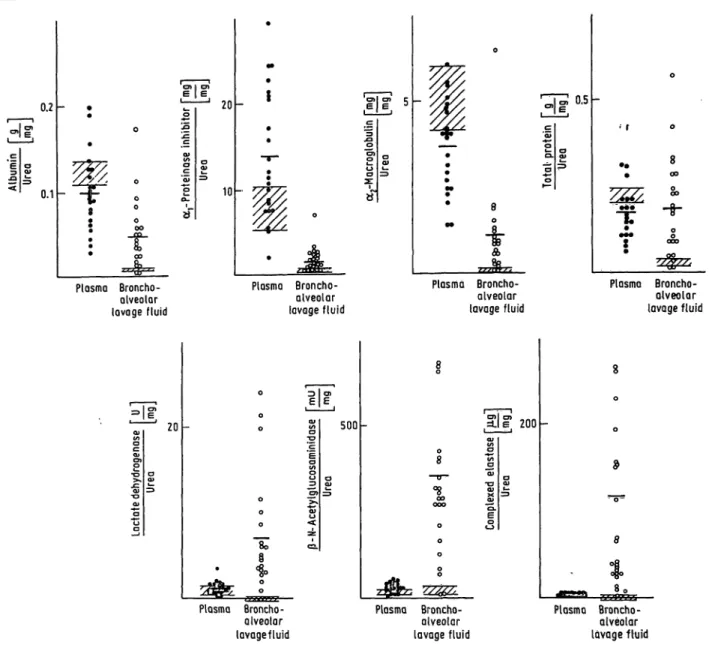
Bronchoalveolar lavage concentrations of urea and proteins
Two different types of soluble substances were found in lavage fluid:
(I) those of vascular origin such äs urea, albumin, ar
proteinase inhibitor, and a2-macroglobulin (a2-ma- croglobulin may also be secreted by alveolar macro- phages) (16) that can appear in lavage fluid via diffu- sion from blood, and
(II) those of predominantly cellular origin from in- vading mobile cells (lymphocytes, macrophages, poly- morphonuclear leukocytes) found after secretion or release.
Examples include lactate dehydrogenase, ß-N-acetyl- glucosaminidase, elastase and the elastase-arprotein- ase inhibitor complex. If the urea concentrations in plasma and lavage fluid serve äs an indicator of the extent of plasma exudation the difference between calculated and measured amounts of different pro- teins in bronchoalveolar lavage fluid should depend on their permeabilities for type (I) proteins and addi- tionally on the extent of secretion or release by mi- grating and phagocytosing or dying cells for type (II) proteins. Figure l demonstrates this behaviour for the lavage protein concentrations in normals. The protein pattern clearly showed that the calculated amounts of total protein, albumin, arproteinase in-
78
Dwenger et al.: Analysis of bronchoalveolar lavage fluid, blood and leukocytes in traumatized patients Tab. 2. Distribution of cells derived from bronchoalveolar lavage fluidsPatient No.
1
2
3
4 5 6
7
Day of lavage procedure
21 48 10 24 8 41 68 14 41 31 5 41 6
Fractions of Lymphocytes 0.24
0.090.07 0.05— 0.100.22 0 0.08 0.08— 0.10— 0.07—
——
— 0.040.05 0.08
Macrophages 0.150.64
0.71·*- 0.63 0.290.42 0.52 0.32— 0.22— 0.40
— 0.13— -
—— · 0.380.48 0.60
Polymorphonuclear leukocytes
0.61 - 1 0.270.22 0.32— 0.360.61 0.48 0.60— 0.70— 0.50— 0.80^ -*
<-
— 0.58 0.470.32
— = not determined
l» 50 '50
rfi
120
«10
s·§ 10
.§ 2
i
~5i
S 0.50 -
' 0.25
Fig. 1. Means of bronchoalveolar lavage fluid protein afhounts of normals (x ± s; n = 4) of totally fecovered bron- choäiveöiar lavage fluid
eafculated ^ measured
Plasma and bronchoalveolar lavage fluid urea concen- trations served äs an exudation marker System
hibitor and a
2-macroglobulin in lavage fluid were higher than those measured, indicating a lower per- meability of any of these proteins in comparison to urea. In contrast, the calculated amounts for ß-N- acetyl-glucosamihidase, complexed elastase, and total elastase were found to be lower than those measured, presumably due to their origin from migrating cells.
These migrating cells are probably predominantly polymorphonuclear leukocytes, äs indicated by the gfanulocyte-specific elastase that would be released during constant low grade basic phagocytosis by granulocytes of airway-invadingmicroorgänisms. En- zyöiatically active elastase was not observed in either lavage fluid or plasmä, probably due to the presence of inhibitöf (a
rproteinase inhibitor and a2-macröglo- bulin) in sufficient qüantity to completely bind free elastase.
Figure 2 stimmarizes the concentrations of ten dif- ferent fefqndhöalveolar lavage constituents. Lower and uppftr limits of concentrations determined in lavage fluids of four normals (black columns) äs well äs limits of all lavage fluids for each individual patient are indicated. The concentrations of neärly all lavage fluid components were generally elevated for the patient group in comparison with normals, except for enzymatically determined elastase wjiich was detected only in one lavage sample each from patients 2 and
J. Clin. Chem. Clin. Biochem^ / Vol. 24,1986 / No. l
Dwenger et ah: Analysis of bronchoalveolar lavage fluid, blood and leukocytes in traumatized patients 79
JE
2 200
o
n
S, 10je
l
10
s l
40= J!
o> .°- lo2 5
sl
-
__
-
-o-P=]
• 2000
l 10
'S» r-
=c
II n =1
r~
s
D ; zooo I]
Patient N 1 2 3 4 5 6 7
n 4 6 3 7 2 2 3 5 N 1 2 3 4 5 6 7
4 6 3 7 2 2 3 5
Fig. 2. Concentrations of urea and proteins in bronchoalveolar lavage fluids of normals (N; black columns) and patients. Upper and lower limits of concentrations are indicated for all bronchoalveolar lavage fluids (n) of each patient.
6, indicating incoinplete Inhibition by <Xi-proteinase inhibitor and/or a
2-macroglbbulin. Since an entirely suitäble denominator on which to base relative changes in various protein concentrations in lavage fluids does not exist, lavage protein values can be expressed in relation to a reference substance like wea (personal communication by C. G. Cochrane, Scripps Clinic and Research Foundation, La Jolla, California, U. S. A.) that freely diffuses across bio- logical membranes of the respiratory tract and ac- companies water shifting. Its presence in bronchoal- veolar lavage fluid reflects transüdation from the intravascular cpmpartment.
Bronchoalveolar lavage fluid and plasma concientrations of urea and proteins
i;Hie ratio of the concentrations of a specific substance to urea concentration in lavage fluid should decrease, compared with plasma, with the decreasing permea-
bility of this substance, and vice versa. Figure 3 shows the protein/urea ratios in plasma and lavage fluid for all patients äs well äs for normals.
Albumin
In plasma the mean ratio for patients (0.099) was somewhat lower than in normals (0.123), presumably äs a result of albumin loss; the lavage ratio (0.044) was found to be about 5.6 times higher than in normals (0.0079) indicating an increased permeability of the blood-air barrier.
, -Proteinase inhibitor
The mean plasma ratio for the patient group (14.0)
was found to be higher than in normals (7.77) äs a
result of an acute phase 'reaction. The mean lavage
ratio (1.56) was found to be 7.1 times higher than
that of normals (0.22), which is further evidence of
increased permeability.
80 Dvvcnger cl al.: Analysis of bronchoalveolar lavage fluid, blood and leukocytcs in traumatized patients
0.2
0.1
20
10
.£
A
"
8
-i-
<&>
Plasma Broncho- alveolar lavage fluid
Plasma Broncho- alveolar lavage fluid
Plasma Broncho- alveolar lavage fluid
Plasma Broneho- alveolar lavage fluid
20 500 Sie 200
8 <
Plasma Broncho- alveolar lavagefluid
Plasma Broncho- alveolar lavage fluid
Plasma Broncho- alveolar l a vage fluid
Fig. 3. Patient protein/urea ratios of bronchoalveolar lavage fluid and plasma components
= means
1ZZZ2. = normal ränge (x ± s; n = 4) O = plasma protein/urea ratio
O = bronchoalveolar lavage fluid protein/urea ratio
0.2-Macroglobulin
Because of an increased consumption of a2-macroglo- bulin, the mean plasma ratio of the patient group (3.7) was below the normal ränge (5.1), whereas the mean lavage ratio of patients (1.14) was elevated 16.3 fold above normals (0.07). This high lavage ratio is probably partially caused by additional a2-macroglo- bulin secreted from alveolar macrophages, leading to an overestimation of permeability disturbances.
Total prolein
Loss of total protein results in a decreased mean plasma ratio of patients (0.173) äs compared to nor- mals (0.22), whereas the lavage ratio of patients
(0.185) was 7 fold higher than the normal ränge (0.027). The mean ratios for plasma and lavage fluid were found to be nearly equal äs a consequence of the contribution of type (II) lavage proteins released by inflammatory cells, thus distorting the true condi- tions for exact transudatipn calcülation.
Lactate dehydrogenase
Whereas the plasma ratios of patients (1.22) and normals (1.03) were nearly eqtial, the mean lavage ratio increased from 0 for normals to 6.7 for the patient group, clearly indicating that this elevation could only be caused by cellulär release rather than by transudation. , j
J. Glin. Chem. Clin. Brachem, / Vol. 2A91986 / No. l
Dwenger et al.: Analysis of bronchoalveolar lavage fluid, blood and leukocytes in traumatized patients 81 ß-N-A cetyl-glucosaminidase
The same was true for the corresponding ß-N-acetyl- glucosaminidase/urea ratio data. In this case the mean lavage ratio of patients (349.6) was 15.8 fold higher than that of normals (22.2) and 11 fold higher than the mean plasma ratio (31.9) in patients, indicat- ing its cellular origin in the alveolar space.
Elastase
The above observations were further supported and confirmed by the elastase-a
rproteinase inhibitor complex. In plasma the mean ratio for patients (1.43) was 4.6 fold above normals (0.31). In lavage fluid this ratio was 77.7 fold higher and the patients' lavage ratio was 116.6 in comparison with 1.43 for plasma, i. e. a 81.5 fold increase.
Course of bronchoalveolar lavage protein/
urea ratios
In figure 4 the course of lavage protein/urea ratios is demonstrated for single patients. Large differences between individuals in the sequential course of ratios were observed.
Patient 1. There was an elevation of all ratios to a maximum on the second day with a subsequent de- crease to normal values after 8 days.
Patient 2. High and increasing ratios of type (II) proteins, ß-N-acetyl-glucosaminidase and complexed elastase, which indicate an increased probability of damage, were accompanied by an increase of permea- bility markers, i. e. the albumin/urea, a
rproteinase inhibitor/urea and a
2-macroglobulin/urea ratios. The survival of this patient indicated that reversible im- provement at this stage of lung injury can occur.
Patient 3. During the posttraumatic course all pro- tein/urea ratios declined toward normal ratios. As indicated by low complexed elastase/urea ratios the elastolytic bürden of lung structure was negligible.
Patient 4. After an initial rise of all ratios on day l a rapid subsequent decrease was observed in all Parameters, indicating an improvement of the perme- ability Situation and a diminishing influence of re- leased enzymes.
Patient 5. The behaviour of the ratio course of this patient was quite similar qualitatively to that of patient 4, but less pronounced quantitatively.
Patient 6. This patient showed high type (I) protein ratios on day l and high type (II) protein ratios on day 2; both decreased rapidly in the posttraumatic period, indicating an improvement of the lung Situa- tion.
Patient 7. The ratio course of this patient equals that of patient 2. Type (I) protein ratios demonstrated a progressive increase of permeability; type (II) protein ratios showed increasing enzyme release from phago- cytes until day 6; nevertheless, the lung Situation improved and this patient survived.
Enzyme content of polymorphonudear leukocytes
Figure 5 demonstrates the content of lactate dehydro- genase, myeloperoxidase, and elastase enzyme activ- ity, äs well äs complexed and total elastase concentra- tions in granulocytes isolated from blood and lavage fhiid of patients and normals. Generally, in nearly all measurements the inter-individual scattering was highest in cells isolated from lavage fluids.
Lactate dehydrogenase
The cytoplasmic lactate dehydrogenase activity was lower in blood-derived cells of the patient group (105.4 U/10
9cells), the mean Contents of lavage- derived cells and normal cells (150.2 and 142.4 U/10
9granulocytes, respectively) being of comparable size.
Myeloperoxidase
Myeloperoxidase activity in lavage- and blood- derived patient cells were lower (49.2 and 40.7 U/10
9cells, respectively) than in normals (112.8 U/10
9cells).
Elastase activity
As with myeloperoxidase, the elastase enzyme activity was found to be lower in patient lavage- and blood- derived cells (46.4 and 66.9 U/10
9granulocytes, re- spectively) in comparison with normals (105.6 U/10
9cells). Together with the myeloperoxidase results, this could be explained by enzyme loss through regurgita- tion during feeding or by frustrated phagocytosis.
Complexed elastase concentration
In comparison with normals (1.34 g/10
9cells) the
complexed elastase concentrations were markedly el-
evated in patient blood cells (3.28 g/10
9granulocy-
tes) and drastically increased in lavage-derived
patient granulocytes (360.9 g/10
9cells).
82
Dwenger et al.: Analysis of bronchoalveolar lavage fluid, blood and leukocytes in traumatized patientsP-N-Acetyl- Loctote arProteinase Complexed elostase |>9l glucosaminidase [" U 1 dehydrogenase f U ~| Total protein [g ~| ocrMacroglobulin[mg~| inhibitor fmg"| Albumin |~g "|
PC 0.5
— 1
I
a>σ
10
— 1σ»
1
I 1 °
aO)t_
=5
σa>
=5
|? 50
σa>
^1000
-'
σo>
a01
n_
itieni
r-T~l 1
,Jh,
1
p^
H
— |
1 '.Π
H~H_
— ι
2
n
_j
-
Ί
Jtl.
Γ-ι_Γ~Ι
[ί
rT
p.
3
\\\n„
rltn
4
[L
ΓΗ
(l .
1-1
T
Ί
n-, _
5
ru
[L. ._
ru ^
rfl _
n
n.
n.
6
fln ."
Π
-n _
Jl-
n
7
HT1
rTfl -
ΗΊ-. _
ffll .
rrfl
IT-
ΓΚ1
2 4 8 N 1 46 B N 1 4 N 1 4 N 1 3 5 N 1 4 6 N
Fig. 4. Posttraumatic course of bronchoalveolar lavage fluid protein/urea ratios of patients 1-7 abscissa = time of bronchoalveolar lavage procedure (d)
N (black columns) = ratios of normals (χ; η = 4)
J. Glin. Chem. Clin. Biochem. / Vol. 24^ 1986 / No. l
Dwenger et al.: Analysis of bronchoalveolar lavage fluid, blood and leukocytes in traumatized patients 83
S 200
ΟΙ
g s
cn f.
1 100
'So»
"o σ
0
θο
1 200
σ»(J
o §
."* Ϊ
5 s - 1
- ·»· §· 100
φ β "β»« o *
0
200 J2
o
A 2 A o»
CLJ
8 __ "o"
0
i 1000
2
^
A O* σ
8 4- 'T 'S 500
1.* f : i
θ_SL
* 8 i
l
- o S 20000O
o J o ^
0 cu σ
~ 1000°
o o
§
-5- i A^~
0 0
o
Jt 0 A 0 8
• i
β
Fig. 5. Enzyme Contents of polymorphonuclear leukocytes isolated from blood (O) and bronchoalveolar iavage fluid (O) of seven patients and from blood of eight normals (A)
= means
Total elastase concentration
The total elastase content of patient blood and lavage granulocytes was lower (6154 and 7684 μ§/10
9cells, respectively) than in normals (9962 μ§/10
9cells). This observation may confirm the above spec lation about enzyme loss through phagocytic activity.
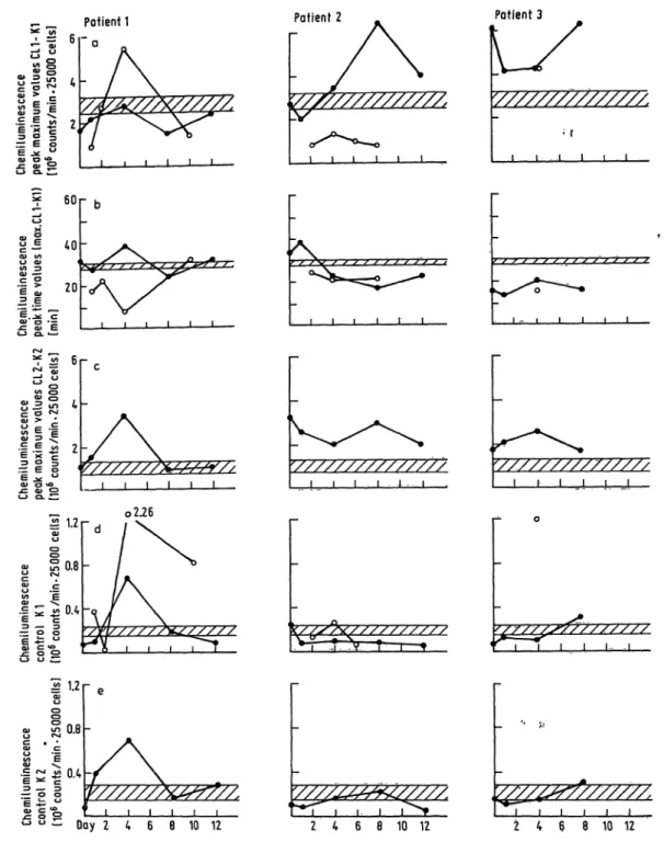
Chemiluminescence results
Absence of chemiluminescence data points in the figures indicates that there was an insufficient granul- ocyte count to investigate.
From chemiluminescence measurements we tried to obtain s much information about the functional Status of blood- and lavage-derived granulocytes s possible. The stimulatory capacity of the cells was therefore evaluated s follows:
1. peak maximum c Unts/min values (CL l —K 1):
magnitude of maximum photon emission from 25 000 granulocytes (CL 1) stimulated by zymosan and AB plasma corrected for the eounts/min val e produced in absence of zymosan b t in presence of AB plasma (Kl)
2: peak time in min:
time required to reach (CL 1); by using a constant opsonin concentration (constant vplume of AB plasma) this time can only vary s a result of changes in granulocyte receptor affinity and/or concentration 3. peak maximum counts/min values (CL 2—K 2):
magnitude of maximum photon emission from 25 000 granulocytes (CL 2) stimulated by zymosan without
AB plasma addition corrected for the counts/min value produced in the absence of zymosan and AB plasma (K 2)
4. control counts/min (K 1):
magnitude of maximum photon emission from 25 000 granulocytes without zymosan in presence of AB plasma
5. control counts/min (K 2):
magnitude of maximum photon emission from 25 000 granulocytes without zymosan or AB plasma
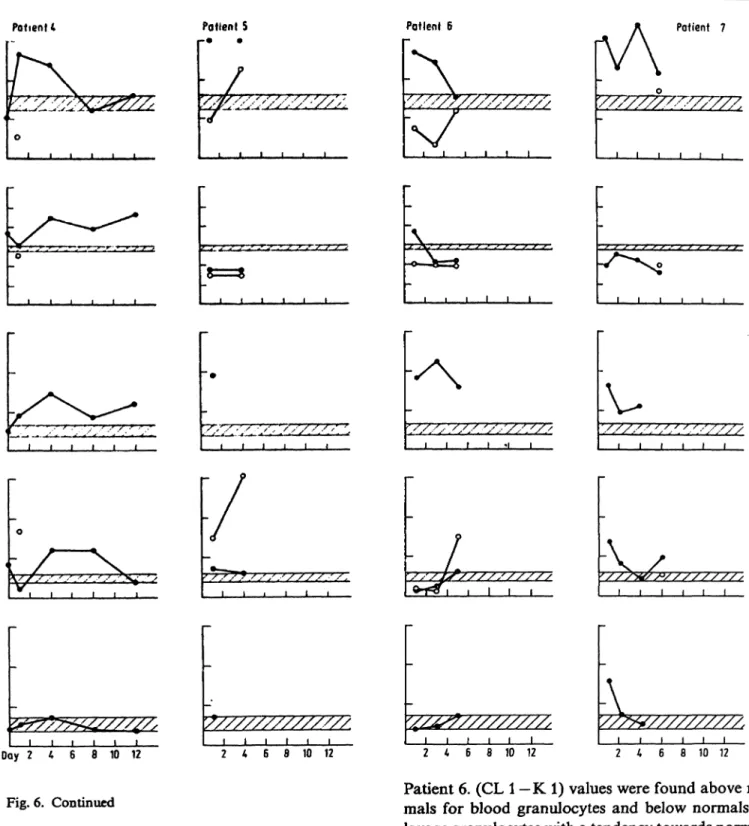
Figure 6 summarizes the chemiluminescence results.
Normal ranges (x ± s; n = 8, shaded areas) were determined for blood-derived granulocytes of normal healthy donors.
Patient l. In the posttraumatic course lavage-derived granulocyte chemiluminescence showed a distinct maximum of zymosan H- AB plasma-stimulated va- lues above normals, indicating a higher degree of excitability than blood-derived cells.
The time to reach maximum values was prolonged
for blood-derived cells on day 4, and extremely
shortened for lavage-derived cells. This effect is
supposed to have been caused by granulocyte passage
across the blood-air barrier, during which the sensitiv-
ity of the cells seemed to increase, thus being stimu-
lated faster and to a higher degree. An unusually
high degree of Stimulation by zymosan without op-
sonin (AB plasma) was observed on day 4; the con-
trols also had elevated values. An explanation for
this phenoinenon cannot be given at present.
84
Dwcnger et al.: Analysis of bronchoalveolar lavagc fluid, blood and leukocytes in traumatized patients^ _ Patient 1 Patient 2 Patient 3
V//////////////,
\ J I I » » _J l - - l l l l
60r
οχ
J 40
l«0 o eu (Λ ο α»«Λ 3
|1 Ι ι
Ι-ϊ-ϊ
6 S.S
<χι _
S3 g 4
c σ f*J ο ι > '
< - > < = . £
<Λ ε ·£
α> => ^
•ιι 1 ζ
:?! Ι ·Ρ ^ «-»
1 1
o 2.26
-2 1.2 r
'e :
i,
Ξ-s g
§ i; "
5 § o
<_> O ^
0.4
ι ι ι ι ι l l l l
Day 2 4 6 8 10 12 2 4 6 8 10 12 2 4 6 8 10 12
Fig. 6. Course of chemiluminescence data of bronchoalveolar lavage fluid (O) and blood (·) derived polymorphonuclear leukocytes
abscissa = time of bronchoalveolar lavage fluid and blood sampling for polynlOφhonucleaΓ leukocyte Isolation TKZK = normal r nge (x ± s; n = 8) of blood-derived polymorphonuclear leukocytes
CL = chemiluminescence response after Stimulation
K = chemiluminescence response without Stimulation (blank)
Patient 2. In this patient quite different behaviour was observed. The stimulatory capacity (CL l —K 1) was higher for blood- than for lavage-derived granul- ocytes, especially on day 8. Beginning on day 2 the reaction times were shortened for both lavage- and blood-derived cells, possibly indicating a receptor al- teration. (CL2—K 2) values were observed to be higher than in normals.
Patient 3. With the exception of day 4, only blood granulocytes could be studied. During the complete posttraumatic co rse they Were found to be in a state of faster and higher stimulatory capacity compared with normals. The blood-derived cell Stimulation by zymosan without AB plasma was again found to be above that of normals.
J. Clin. Chem. Clin. Biochem. / Vol. 24,1986 / No. l
Dwcnger et aK: Analysis of bronchoalvcolar lavagc fluid, blood and leukocytcs in traumatizcd paticnts
85
PottenU Potlent S Pollen» S Potient 7
l t
Day 2 4 6 8 10 12 2 4 6 8 10 12
Fig. 6. Continued
Patient 4. In this patient äs well, only blood-derived cells could be investigated with the exception of day 1. The blood granulocytes were excitable to a higher degree of photon emission than normals on days l and 4. This was accompanied by s slower reaction than in normals. Zymosan Stimulation without AB plasma addition was again elevated.
Patient 5. Lavage-derived granulocyte Stimulation by zymosan and AB plasma showed an increase from subnormal to supranormal values between days l and 4, accompanied by short reaction times. Blood- derived cell values were several fold higher than those of normals, and increasingly so with time.
l
6 8 10 12 2 4 6 8 10 12
Patient 6. (CL l —K 1) values were found above nor- mals for blood granulocytes and below normals for lavage granulocytes with a tendency towards normali- zation throughout the course. Photon emission times were low for lavage cells during the whole course, whereas blood-derived cell times were higher on day l and lower on days 3 and 5 äs compared with normals. (CL 2—K 2) values were several fold higher than those of normals.
Patient 7. In comparison with the only lavage
granulocyte value on day 6 the blood granulocyte
stimulatory potentials was found to be far above the
normal ränge and the Stimulation times were below
the normal ränge. As for the majority of patients, the
zymosan-stimulated chemiluminescence without AB
plasma was above normal values.
86
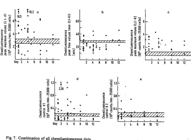
Dwenger et al.: Analysis of bronchoalveolar lavage fluid, blood and leukocytes in traumatized patientsFigure 7 comprises all Chemiluminescence patient data by projecting corresponding values of the same Parameter on to a single figure. From this, semiquan- titative conclusions were drawn: The Stimulation by zymosan and AB plasma of blood-derived granulocy- tes produced a Chemiluminescence response usually above that of normals, while the Chemiluminescence response of lavage-derived granulocytes was below that for normals. Stimulatory times seemed to be below the normal r nge for lavage-derived cells and both above and below the value for normals for blood-derived cells. The r nge of Stimulatory re- sponse of patients' blood granulocytes to zymosan and without AB plasma was clearly above the normal r nge. Control values (K 2) without zymosan and AB plasma were generally within the normal r nge, whereas controls (K 1) without zymosan and with AB plasma were not consistently distributed either above or below the normal r nge.
Discussion
In addition to its acceptance s a technique for the diagnosis, staging, and follow-up of interstitial lung diseases, bronchoalveolar lavage has gained impor- tance s a research tool for evaluating the dynamic inflammatory and immune mechanistns operating in the local environment of the lung. A serial analysis of soluble and cellular components in lavage fluid s well s in blood is therefore useful. In a group of traumatized patients predisposed to the development of the Adult Respiratory Distress Syndrome (Injury Severity Score > 30) the course of lavage and blood component concentrations s well s lavage cell pat- tern shifting and alterations of enzyme contents and Chemiluminescence response of lavage- and blood- derived polymorphonuclear leukocytes were moni- tored. Although no patient s bsequently developed the severe Adult Respiratory Distress Syndrome pro-
3|·
v> once val
h > e εin. .ρ-
<=.=
II
ε 4/>5SL2
f ?16.2 10.5 ·
5 60 l
SS83 II 20"ε Λ: Έα> σ · —
5α> εex — 0 8
α> 3 c E ε 5ε i 2
Ooy 2 4 6 θ 10 12
'/////7///S///SS/.
2 4 6 8 10 "12" 2 4 6 8 10 12
121.4,
= ο g4
ε i= °
(U C «β
5 SS
2.26 §ο
m^0.8
1
|||°
ΛssS i
Oay 2 4 6 8 10 12 2 4 6 8 10 12
Fig. 7. Combination of all Chemiluminescence data
O = blood-derived polymorphonuclear leukocyte Chemiluminescence
O = bronchoalveolar lavage fluid-derived polymorphonuclear leukocyte chemilummescence TZZZZ. = normal r nge (x ± s, n = 8)
abscissa = time pf bronchoalveolar lavage fluid and blood sampling CL = Chemiluminescence response after Stimulation «
K = Chemiluminescence response without Stimulation (blank)
J. Clin. Chem. Clin. Biochem. / Vol. 24,1986 / No. l
Dwengcr et al.: Analysis of bronchoalveolar lavage fiuid, blood and leukocytes in traumatized patients 87
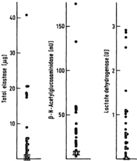
gressing to death, bronchoalveolar lavage fluids of
the non-contused part of the hing clearly indicated distinct signs of inflammation and differentiation during the posttraumatic course. In comparison with normals (1) the bronchoalveolar lavage cell distribu- tion was altered towards an increased number of lymphocytes and a markedly increased number of polymorphonuclear leukocytes äs evidence of inflam- mation. The lactate dehydrogenase activity found in the patients
9lavage fluids greatly exceeded the transudated portion, indicating enzyme release by cell damage in the interstitial-alveolar space. To judge whether only granulocyte lysis contributed to the lavage fluid's enzyme concentration, the hypothetical number of totally damaged granulocytes was calcu- lated from elastase-a
rproteinase inhibitor complex, ß-N-acetyl-glucosaminidase and lactate dehydroge-
20
10
150
i 100
l
50
S.2
fr
»0»
Fig. 8. Elastase (äs total protein) and ß-N-acetyl-glucosamini- dase and lactate dehydrogenase activities in patient bronchoalveolar lavage fluid
= normal ranges (x ± s; n = 4)
Hypothetical number of originally present and totally destroyed polymqtphonuclear leukocytes calculated from total elastase, ß-N-acetyl-glucosaminidase and lac- tate dehydrogenase contents in bronchoalveolar lavage fluids of seven patients
Number of polymorphonuclear leukocytes (l O6) According to
Xs n
total elastase 0.790.95
27
ß-N-acetyl- glucosaminidase 24.126.8
27
lactate dehydrogenase 5.756.28
28
Normal contents (x from n = 8) served äs a reference:
| perlO6
9.96 g elastase-qti-proteinase inhibitor poly- 1.45 mU ß-N-acetyl-glucosammidase morpho- 0.14 U lactate dehydrogenase nuclear
J leukocytes
nase contents in patient lavage fluids by using the normal values äs also determined in this study. The results are presented in figure 8. From the granulo- cyte-specific elastase concentrations of all patient lav- age fluids a mean of 0.79 · l O
6totally lysed polymor- phonuclear leukocytes was calculated. Based on the ß-N-acetyl-glucosaminidase (and lactate dehydroge- nase) content a mean of 24.1 · 10
6(5.75 · 10
6) granulo- cytes was calculated. This leads to the conclusion that cells other than polymorphonuclear leukocytes, presumably alveolar macrophages, took part in the liberation of the enzymes. In order to overcome the variability of lavage fluid sampling and the difficulty of comparing lavage protein concentrations, all con- centration values were related to urea äs an internal reference substance instead of to albumin (17) or an external reference such äs methylene blue (l 8). By this method the albumin/urea and a
rproteinase inhibitor/
urea ratios in lavage fluid and plasma clearly indi- cated an increased permeability of the blood-air bar- rier. a
2-Macroglobulin/urea and total protein/urea ratios did not *demonstrate such a change. On the other hand, lactate dehydrogenase, ß-N-acetyl-gluco- saminidase, and elastase were predominantly released by phagocytes rather than transudated from plasma.
Thus, ß-N-acetyl-glucosaminidase and elastase could be used äs markers of the degree of predisposition to lung injury development (e. g. Adult Respiratory Distress Syndrome), that may be initiated by neutral proteases like elastase (3, 7).
Careful consideration of type (I) and type (II) lavage protein/urea ratios could allow the prediction of im- provement or deterioration of the patients' lung Situa- tion. In comparison to normals, decreased myeloper- oxidase and elastase activities, äs well äs elastase concentrations of patient lavage- and blood-derived polymorphonuclear leukocytes, demonstrated en- zyme loss, presumably through phagocytosis. Com- plexed elastase was found to be more than 100 fold elevated in lavage-derived granulocytes. Further in- vestigations are needed to elucidate whether polymor- phonuclear leukocytes are able, like macrophages to incorporate free or complexed elastase by phago- cytosis (19-21).
From chemiluminescence data it was evident that
lavage-derived granulocytes had partially lost stimu-
latory capacity, in comparison with above normal
activity in blood-derived cells. Furthermore, lavage
granulocytes seemed to acquire an increased receptor
efficiency during blood-air passage, documented by
shortened photon emission times. The migration of
polymorphonuclear leukocytes from blood to al-
veolus seemed to be linked with a transformation
process that resulted in a less aggressive type of cells
88
Dwenger et al.: Analysis of bronchoalveolar lavage fluid, blood and leukocytes in trauraatized patients with respect to stimulatory mechanisms which releasetoxic and damaging mediators of lung injury. Such altered granulocytes are believed to be exhausted, their content partially released into the lavage fluid and their chemiluminescence response diminished.
This is supported by the simultaneous comparison of lavage type (II) protein/urea ratios and the stimulat- ory capacities of lavage- and blood-derived polymor- phonuclear leukocytes. In patient 2 on day 8 when
ß-N-acetyl-glucosaminidase and elastase concentra- tions were at their highest, the simultaneous chemilu- minescence response exhibited a maximal difference between the stimulatory functions of blood (highest) and lavage (lowest) granulocytes. Exactly the same correlation was observed in patient 4 t(day 1) äs well äs in patient 5 (days l and 4), and ha patient 6 (days l and 3). This striking relationship needs to be confirmed by further studies on patients developing severe Adult Respiratory Distress Syndrome.
References
1. Hunninghake, G. W., Gadek, J. E., Kawanami, O., Fer- rans, V. J. & Crystal, R. G. (1979) Am. J. Pathol. 97, 149-206.
2. Daniele, R. R, Elias, J. A., Epstein, P. E. & Rossman, M. D. (1985) Ann. Intern. Med. 102, 93-108.
3. Lee, C. T., Fein, A. M., Lippmann, M., Holtzman, H., Kirabel, P. & Weinbaum, G. (1981) N. Engl. J. Med. 304, 192-196.
4. Schraufstatter, :, Revak, S. D. & Cochrane, C. G. (1984) Fed. Proc. 43, 2807-2810.
5. Merritt, T. A., Cochrane, C. G., Holcomb, K., Bohl, B., Hallman, M., Strayer, D., Edwards III, D. K. & Gluck, L.
(1983) J. Clin. Invest. 72, 656-666.
6. Ogden, B. E., Murphy, S. A., Saunders, G. C., Pathak, D. & Johnson, J. D. (1984) Am. Rev. Respir. Dis. 130, 817-821.
7. Janoff, A. (1985) Annu. Rev. Med. 36, 207-216.
8. Bernhard, G. R. & Brigham, K. L. (1985) Annu. Rev. Med.
36, 195-205.
9. McGuire, W. W., Spragg, R. G., Cohen, A. B. & Cochrane, C. G. (1982) J. Clin. Invest. 69, 543-553.
10. Hjorth, R., Jonsson, A. & Vretblad, P. (1981) J. Immunol.
Methods 43, 95-101.
11. Kingsley, G. R. (1939) J. Biol. Chem. 131, 197-200.
12. Schirardin, H. & Ney, J. (1972) Z. Klin. Chem. Klin.
Bipchem. 10, 338-344.
13. Henson, P. M., Zanolari, B., Schwartzman, N. A. & Hong, S. R. (1978) J. Immunol. 121, 851-855.
14. Yatziv, S., Kahane, L, Abeliuk, P., Cividalli, G. & Rachmi- lewitz, E. A. (1979) Clin. Chim. Acta 96, 67-72.
15. Crystal, R. G., Gadek, J. E., Ferrans, V. J., Fulmer, J. D., Line, B. R. & Hunnmghake, G. W. (1981) Am. J. Med. 70, 542-568.
16. White, R., Janoff, A. & Godfrey, H. P. (1980) Lung 158, 9-14.
17. Reynolds, H. Y., Fulmer, J. D., Kazmierowski, J. A., Ro- berts, W. C., Frank, M. M. & Crystal, R. G. (1977) J. Ciin.
Invest. 59, 165-175.
18. Baughman, R. R, Bosken, C. H., Loudon, R. G., Hurtu- bise, P. & Wesseler, T. (1983) Am. Rev. Respir. Dis. 128, 266-270.
19. Debanne, M. T., Bell, R. & Dolovich, J. (1975) Biochim.
Biophys. Acta 411, 295^304.
20. Dolovich, J., Debanne, M. T. & Bell, R. (1975) Am. Rev.
Respir. Dis. 112, 521-525.
21. Campbell, E.J., White, R. R., Senior, R. M., Rodriguez, F. J. & Kühn, C. (1979) J. Clin. Invest. 64, 824-833.
Dr. Alexander Dwenger · Abteilung für Klinische Biochemie Medizinische Hochschule Hannover Konstanty-Gutschow-Straße 8 D-3000 Hannover 61
J. Clin. Chem. Clin. Biochem. / Vol. 24,1986 / No. l