• Keine Ergebnisse gefunden

liver injury and cancer

N/A
N/A
Protected

Academic year: 2021

Aktie "liver injury and cancer "

Copied!
127
0
0

Wird geladen.... (Jetzt Volltext ansehen)

Volltext

(1)

liver injury and cancer

Inaugural-Dissertation zur Erlangung des Doktorgrades

der Mathematisch-Naturwissenschaftlichen Fakultät der Universität zu Köln

vorgelegt von Trieu My Van

aus Köln

Köln 2017

(2)

Köln, 2017

(3)

Berichterstatter: Prof. Dr. Manolis Pasparakis Prof. Dr. Kay Hofmann

Tag der mündlichen Prüfung: 23.05.2017

(4)

Zusammenfassung

Die Serin/Threonin Rezeptor-interagierende Protein Kinase (RIPK)1 ist ein zentrales Protein, welches den Ausgang von TNF Rezeptor 1 (TNFR1)- und Toll-ähnlicher Rezeptor ( TLR)- vermittelten Signalen durch Kinase-abhängigen und Kinase-unabhängigen Funktionen determiniert. Bis heute ist es unklar, in wie fern RIPK1 in der Regulierung von Leberfunktionen agiert. Deshalb wurde ein konditionales RIPK1-Allel erzeugt, welches die Cre-Recombinase vermittelte Deletion von RIPK1 in Leber-parenchymalen Zellen (LPZ) ermöglicht. Zusätzlich wurden RIPK1-Mutanten verwendet, welche, aufgrund eines Aminosäureaustausches an der Position 138 von Asparaginsäure zu Asparagin, eine mutierte Form von RIPK1 exprimieren (RIPK1D138N) und deshalb keine Kinaseaktivität besitzen. Mit Hilfe dieser Mutanten sollten die Kinase-abhängigen und Kinase-unabhängigen Funktionen von RIPK1 in der Regulierung bzw.

Induzierung von Leberschäden und in der Initiierung von Leberkrebs untersucht werden. Die LPZ-spezifische Deletion von RIPK1 beeinflusste weder die Leberentwicklung noch die Leber Homöostase unter normalen Konditionen, sensibilisierte RIPK1-defiziente Hepatozyten jedoch zu spontaner Apoptose in vitro. RIPK1

LPZ-KO

Mäuse zeigten eine erhöhte Sensitivität gegenüber einer Lipopolysaccharid (LPS)-Injektion aufgrund von erhöhter Apoptose von Hepatozyten. Die LPZ-spezifische Deletion von TNFR1, Fas-assoziiert via Death Domain (FADD) oder TNFRSF1A-assoziiert via Death Domain (TRADD) schützte RIPK1-defiziente Hepatozyten vor Zelltod, wodurch die hohe Sensitivit ä t von RIPK1

LPZ-KO

Mäusen nach einer LPS-Injektion vermindert werden konnte. Die Inaktivierung der Kinase Aktivität von RIPK1 schützte Mäuse vor LPS/D-Galactosamin (D-GalN) induzierten Leberschäden. Die LPZ-spezifische Deletion von RIPK1, aber nicht die Inaktivierung der Kinase Aktivität in Mäusen reduzierte Diethylnitrosamin (DEN)-induzierten Leberkrebs, was auf eine Kinase-unabhängige Funktion von RIPK1 in der Initiierung von Leberkrebs hindeutet. Darüber hinaus reduzierte die LPZ-spezifische Deletion von RIPK1 zum Teil DEN-induzierte Tumorentstehung in einem Mausmodel mit induzierter Adipositas. Eine reduzierte Entstehung von DEN-induziertem Leberkrebs korrelierte mit einer erhöhten Anzahl von frühzeitiger Caspase-8-vermittelter Apoptose und reduzierten DNS Schäden in RIPK1

LPZ-KO

Mäusen. Die Inhibierung des frühzeitigen Zelltodes durch die zusätzliche LPZ-spezifische Deletion von TNFR1 stellte die volle Entstehung von DEN- induziertem Leberkrebs in RIPK1

LPZ-KO

Mäusen wieder her. Zusammenfassend kann geschlussfolgert werden, dass die Resultate dieser Arbeit eine neue, wichtige Kinase- unabhängige Funktion von RIPK1 für die Expression von Überlebenssignalen enthüllen wodurch das Überleben von Hepatozyten beeinflusst wird. Durch diese Funktion reduziert RIPK1 durch LPS Injektion induzierte akute Leberschäden und verhindert somit Leberversagen.

Wohingegen RIPK1 durch seine Überlebensfunktion erhöhten Zelltod nach DEN Injektion

blockiert und dadurch zu erhöhter Tumorentstehung nach einer DEN Injektion führt.

(5)

Summary

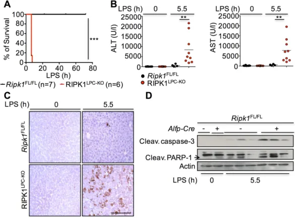
The serine/threonine kinase receptor-interacting protein kinase (RIPK) 1 with pro-death kinase-dependent and pro-survival kinase-independent functions has emerged as an important regulator of cell death, survival and inflammation downstream of death receptors (DR) and Toll-like receptors (TLR), including tumor necrosis factor receptor (TNFR) 1 and TLR4 respectively. The functions of RIPK1 in regulating liver homeostasis, inflammation and cell death are poorly understood and remain to be elucidated. In this study we aimed to investigate the biological significance of the kinase-dependent and -independent functions of RIPK1 specifically in maintaining liver homeostasis and in the regulation of acute liver injury and carcinogenesis using well-established murine models. Accordingly, mice lacking RIPK1 or its kinase activity specifically in liver-parenchymal cells (LPC) were generated using the Cre/LoxP system. LPC-specific RIPK1 deficiency did not affect normal liver development and homeostasis but sensitized RIPK1-deficient hepatocytes to spontaneous apoptotic death in vitro. In vivo, RIPK1

LPC-KO

mice were highly sensitive to lipopolysaccharide (LPS)- induced injury showing massive hepatocyte apoptosis that resulted in early death of mice.

LPC-specific TNFR1, Fas-Associated via Death Domain (FADD) or TNFRSF1A-Associated via Death Domain (TRADD) ablation prevented LPS-induced liver injury in RIPK1

LPC-KO

mice, identifying TNFR1-TRADD-FADD-induced hepatocyte apoptosis as the cause of liver damage. Inactivation of RIPK1 kinase activity (D138N) did not phenocopy the high sensitivity of RIPK1

LPC-KO

mice to LPS-induced liver injury suggesting a RIPK1 scaffolding function in preventing LPS-induced toxicity. However, inactivation of RIPK1 kinase activity partially prevented TNFR1-induced cell death caused by LPS/D-Galactosamine (D-GalN) suggesting a RIPK1 kinase-dependent function in mediating LPS/D-GalN-induced death.

LPC-specific RIPK1 knockout but not loss of its kinase activity reduced Diethylnitrosamine (DEN)-induced liver carcinogenesis in mice suggesting a kinase-independent function of RIPK1 in promoting liver tumorigenesis. Moreover, LPC-specific RIPK1 deficiency also reduced liver tumor formation in a model of dietary obesity-driven DEN-induced hepatocarcinogenesis. Reduced liver tumor development correlated with increased early DEN-induced Caspase-8-mediated hepatocyte apoptosis and reduced γ H2AX levels in the liver of RIPK1

LPC-KO

mice. LPC-specific TNFR1 deficiency prevented increased early hepatocyte apoptosis and restored liver tumor development in DEN-injected RIPK1

LPC-KO

TNFR1

LPC-KO

mice.

Taken together, the results presented in this work reveal a novel, important kinase-

independent scaffolding function of RIPK1 in the regulation of liver cell survival, acute liver

damage and liver cancer development.

(6)

Table of Content

Zusammenfassung ... I Summary ... II Table of Content ... III Abbreviations ... VI Table of Figures ... IX List of Tables ... VI

1. Introduction ... 1

1.1. Receptor-interacting protein kinases (RIPKs) ... 1

1.2. NF-κB signaling ... 3

1.3. Death receptor signaling ... 4

1.3.1. TNF signaling pathway ... 5

1.3.1.1. TNFR1-induced pro-inflammatory and pro-survival signaling...

....

6

1.3.1.2. TNF-induced complex-IIa/-IIb-mediated apoptosis ... 10

1.3.1.3. TNF-induced complex-IIc mediated necroptosis ... 14

1.4. TLR signaling ... 15

1.5. Anatomy and function of the liver ... 17

1.5.1. Murine models of liver inflammation and liver injury ... 18

1.5.2. Murine model of hepatocellular carcinoma ... 19

1.5.2.1. Diet-induced obesity and its role in DEN-induced HCC ... 21

1.6. The role of RIPK1 in tissue homeostasis, liver injury and HCC...22

1.7. Project description ... 23

2. Material and Methods ... 24

2.1. Chemicals and biological materials ... 24

2.2. Molecular biology ... 28

2.2.1. Isolation of genomic DNA ... 28

2.2.2. PCR for genotyping ... 28

2.2.3. Agarose gel electrophoresis ... 30

2.2.4. Isolation of RNA ... 30

2.2.5. cDNA synthesis ... 31

2.2.6. qRT-PCR ... 31

2.2.7. Multiplex assay ... 32

2.3. Cellular biology ... 33

2.3.1. Haematoxylin and Eosin staining of liver tissue sections ... 33

2.3.2. Immunhistochemistry ... 33

2.3.2.1. Quantification of positively stained cells ... 34

2.4. Biochemistry ... 34

2.4.1. Preparation of protein extracts ... 34

2.4.2. Subcellular fractionation ... 34

2.4.3. Western blot analysis ... 34

2.5. Mouse experiments ... 35

2.5.1. Animal care ... 35

2.5.2. Causes recombination (Cre)/LoxP conditional gene targeting ... 36

2.5.3. Generation of mice ... 37

2.5.4. Serum levels of alanine (ALT), aspartate amino transferase (AST) and Low-density- lipoprotein-cholesterol (LDL-C) ... 38

2.5.5. Intraperitoneal injection (i.p.) of LPS and D-GalN ... 38

2.5.6. Isolation of hepatocytes ... 38

2.5.7. Cell survival assay (Lactate Dehydrogenase assay) ... 38

2.5.8. Intraperitoneal injection of DEN ... 39

2.5.8.1. DEN-induced carcinogenesis ... 39

2.5.8.2. Histopathological evaluation of HCC development in DEN-injected 32 and 36-week-old mice .. 39

2.5.8.3. acute dose of DEN ... 39

(7)

2.5.8.4. Glucose tolerance test ... 39

2.6. Computer analysis ... 39

2.6.1. Software ... 39

2.6.2. Statistical analysis ... 40

3. Results ... 41

3.1. RIPK1 is not involved in normal liver homeostasis and development ... 41

3.1.1. Generation of mice with LPC-specific RIPK1 deficiency ... 41

3.1.2. LPC-specific deficiency of RIPK1 does not result in spontaneous liver pathology .... 41

3.2. RIPK1 prevents LPS-induced liver injury by inhibiting TNF-induced hepatocyte apoptosis ... 43

3.2.1. RIPK1

LPC-KO

mice are prone to LPS-induced liver injury ... 43

3.2.2. RIPK1 scaffolding function protects mice from LPS- but RIPK1 kinase activity drives LPS/D-GalN induced liver injury ... 44

3.2.3. RIPK1 prevents TNF-dependent and -independent apoptotic death of primary hepatocytes ... 45

3.2.4. RIPK1 deficiency impairs TNF-induced NF-κB activation in primary hepatocytes .... 46

3.2.5. LPC-specific deficiency of RIPK1 sensitizes hepatocytes to FADD-dependent apoptosis in response to LPS ... 47

3.2.6. LPC-specific deficiency of RIPK1 sensitizes hepatocytes to TNFR1-dependent apoptosis in response to LPS ... 49

3.2.7. LPC-specific deficiency of RIPK1 sensitizes hepatocytes to TNF-induced TRADD/FADD-dependent apoptosis in response to LPS ... 50

3.2.8. LPC-specific deficiency of TNFR1, FADD or TRADD protects RIPK1-deficient primary hepatocytes from spontaneous death in vitro ... 51

3.2.9. LPC-specific deletion of RIPK1 does not sensitize to RIPK3-dependent necroptosis 52 3.2.10. LPC-specific deficiency of RIPK1 does not affect the expression of pro-survival proteins ... 53

3.2.11. Constitutive LPC-specific expression of IKK2 protects RIPK1

LPC-KO

mice from LPS- induced death ... 54

3.3. LPC-specific deficiency of RIPK1 reduces DEN-induced carcinogenesis by sensitizing hepatocytes to early apoptosis ... 55

3.3.1. LPC-specific ablation of RIPK1 reduces DEN-induced tumor initiation and development ... 55

3.3.2. Loss of RIPK1 kinase activity does not affect DEN-induced carcinogenesis ... 59

3.3.3. LPC-specific deficiency of RIPK1 reduces DEN-induced γH2AX levels ... 61

3.3.4. RIPK1

LPC-KO

livers display highly elevated Caspase-3 cleavage after DEN injection 63 3.3.5. Increased hepatocyte apoptosis correlates with reduced levels of γH2AX+ cells ... 64

3.3.6. LPC-specific deficiency of RIPK1 results in extensive Caspase-8- and Caspase-9- mediated hepatocyte apoptosis ... 65

3.3.7. LPC-specific deficiency of RIPK1 does not affect hepatocyte proliferation in response to acute DEN ... 66

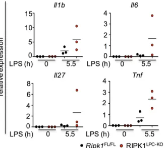
3.3.8. LPC-specific deficiency of RIPK1 does not promote cytokine induction...67

3.3.9. Kinase-inactive RIPK1 mice do not show elevated DEN-induced hepatocyte apoptosis ... 68

3.3.10. Loss of RIPK1 kinase activity does not affect DEN-induced DNA-damage ... 70

3.3.11. Injection with an acute dose of DEN results in the induction of Tnf and Trail gene expression ... 70

3.3.12. TNFR1 signaling promotes apoptotic death 3h after acute DEN injection ... 71

3.3.13. LPC-specific deficiency of TNFR1 restores γH2AX levels in RIPK1

LPC-KO

mice upon acute DEN. ... 74

3.3.14. TNFR1 signaling mediates the protective effect in RIPK1

LPC-KO

mice in response to DEN-induced carcinogenesis ... 75

3.3.15. LPC-specific deficiency of RIPK1 ameliorates obesity-induced liver carcinogenesis

... 77

(8)

4. Discussion ... 79

4.1. RIPK1, a crucial regulator of cell survival in vivo and in vitro ... 79

4.2. RIPK1, a regulator of NF-κB activation in hepatocytes ... 80

4.3. RIPK1 as a key regulator of cell death in response to LPS-induced toxicity ... 83

4.4. Apoptosis as the preferable mechanism of hepatocyte death ... 85

4.5. The pro-survival scaffolding function of RIPK1 promotes DEN-induced hepatocarcinogenesis ... 86

4.6. The role of RIPK1 in spontaneous HCC formation ... 89

4.7. RIPK1 in human cancer ... 90

4.8. Concluding Remarks ... 91

5. References ... 93

6. Acknowledgement ... 107

7. Erklärung zur Dissertation ... 108

Curriculum vitae...110

(9)

Abbreviations

ABC Avidin-Biotin-Complex ALF Acute liver failure Alfp α-fetoprotein

ALS Amyotrophic lateral sclerosis ALT Alanine aminotransferase ANK Ankyrin repeats

APAF-1 Apoptotic protease activating factor 1

APAP Acetaminophen

AST Aspartate aminotransferase ATM Ataxia telangiectasia mutated ATR Ataxia telangiectasia and

Rad3-related protein

BAFF B-cell activating factor of the TNF family

BCL-2 B-cell lymphoma 2

Bid BH3 interacting domain death agonist

BILTS Total bilirubin special BMDMs Bone marrow derived

macrophages

BSA Bovine Serum Albumin

BW Body weight

CC3/8/9 Cleaved Caspase 3/8/9 CD40L Cluster of differentiation 40L cDNA Complementary DNA cFLIP Cellular FLICE-like Inhibitory

Protein

Chk Checkpoint kinase

CHX Cycloheximide

cIAP1/2 Cellular Inhibitor of Apoptosis Protein 1/2

ConA Concanavalin A Cre Causes recombination CYLD Cylindromatosis (Protein) DAI/Zbp1 DNA-dependent activator of

IFN-regulatory factors DAB Diaminobenzidine

DD Death Domain

ddH

2

O Double-distilled water DED Death effector domain DEN Diethylnitrosamine D-GalN D-Galactosamine

DISC Death-Inducing Signaling Complex

DMEM Dulbecco’s Modified Eagle Medium

DMSO Dimethyl sulfoxide DNA Desoxyribonucleic acid dNTPs Desoxyribonucleotides

DR Death Receptor

DTT Dithiothreithol

ECL Enhanced

Chemiluminescence

EDTA Etylene Diamine Tetraacetate ERK Extracellular signal related

kinases

FADD Fas (TNFRSF6)-Associated via Death Domain

FAS TNF Receptor Superfamily Member 6

FasL Fas ligand

FCS Fetal Calf Serum

GTP Guanosintriphosphate GTT Glucose-tolerance test HBV Hepatitis B virus

HCC Hepatocellular carcinoma HCL Hydrochloride acid HCV Hepatitis C virus HE Hematoxylin and Eosin HEPES 4-2-hydroxyethyl-1-

piperazineethanesulfonic acid HFD High-fat diet

HOIL-1 Heme-Oxidized IRP2 Ubiquitin Ligase 1 Homolog

HOIP HOIL-1 Interacting Protein

HRP Horseradish peroxidase

(10)

HRP Horseradish peroxide

ID Intermediate domain

IFN-γ Interferon-γ

IHC Immunhistochemistry

IL Interleukin

(p-)IκBα (phospho-) Inhibitor of NF-κB alpha

IKK1 Inhibitor of κB Kinase 1 IKK2 Inhibitor of κB Kinase 2 IKK2ca Inhibitor of κB Kinase 2

constitutively active IRAK interleukin-1 receptor-

associated kinase i.p. Intraperitoneal ITT Insulin-tolerance test JNK c-Jun N-terminal kinase

KD Kinase dead

KO knock-out

LDH Lactatedehydrogenase LDL-C Low–density-lipoprotein-

cholesterol

LPC Liver parenchymal cell LPS Lipopolysaccharide LRR Leucine rich repeat LoxP Locus of X-over P1 LUBAC Linear Ubiquitin Chain

Assembly Complex MAPK Mitogen-Activated Protein

Kinases

MCL-1 Myeloid leukemia cell differentiation protein

MEF Murine Embryoinc Fibroblasts MLKL Mixed-lineage kinase domain

like

MyD88 Myeloid Differentiation Primary Response Gene 88

NCD Normal-chow diet

nec-1 Necrostatin-1

NEMO Nuclear Factor-kappa B essential modulator

NF-κB Nuclear Factor kappa light polypeptide gene enhancer in B-cells

NIK NF-κB inducing kinase NOXA phorbol-12-myristate-13-

acetate-induced protein 1 NP-40 Nonident P40

PAMP Pathogen Associated Molecular Pattern

PARP-1 poly(ADP-ribose)-polymerase 1

PBS(T) Phosphate Buffered Saline (Tween)

PCR Polymerase Chain Reaction PFA Paraformaldehyde

PUMA p53 upregulated modulator of apoptosis

RHIM Receptor (TNFRSF)-

Interacting Protein Homotypic Interaction Motif

RIPK1 Receptor (TNFRSF)-

Interacting Serine-Threonine Protein Kinase 1

RIPK3 Receptor (TNFRSF)-

Interacting Serine-Threonine Protein Kinase 3

RNA Ribonucleic acid

ROS Reactive Oxygen Species

RT Room temperature

qRT-PCR Quantitative Real-Time PCR SDS Sodium Dodecyl Sulfate SEM Standard error of the mean SHARPIN SHANK-Associated RH

Domain Interacting Protein SI SMAC Second mitochondria derived

activator of Caspases

TAB1 TAK1-Binding Protein 1

TAB2 TAK1-Binding Protein 2

TAK TGF-β-Activated Kinase 1

Tbp TATA-binding protein

(11)

TE Tris EDTA Buffer TEMED N,N,N’,N’ Tetra

methylenediamine

Tg Transgenic

TLR Toll-Like Receptor TNF Tumor Necrosis Factor TNFR1/2 TNF Receptor 1/2

TRADD TNFRSF1A-Associated via Death Domain

TRAF2 TNF Receptor-Associated Factor 2

TRAF5 TNF Receptor-Associated Factor 5

TRAF6 TNF Receptor-Associated Factor 6

TRAIL(R) TNF-related apoptosis- inducing ligand (receptor) TRIF TIR Domain-Containing

Adaptor Inducing IFN-β TWEAK TNF-like weak inducer of

apoptosis

UV Ultraviolett

WT Wildtype

XIAP X-linked Inhibitor of Apoptosis zVAD-fmk Carbobenzoxy-valyl-alanyl-

aspartyl-[O-methyl]- fluoromethylketone

Abbreviations of units

bp Base pair

cm centimetre

µm micrometer

cm centimeter nm nanometre gram g gram

mg milligram µg microgram ng nanogram kDa kilodalton

l litre

ml millimetre

µl microlitre

M mole

mM millimol

µM micromol

nM nanomol

mA milliampere

mol mole

rpm revolutions per minute

V Volts

W Watt

°C degree celsius

% percentage

h, min, s hours, minutes, seconds

x g G force

(12)

Table of Figures

Figure 1. Schematic representation of the structure of the RIPK family members ... 1

Figure 2. Schematic representation of the RIPK1 domain structure ... 2

Figure 3. Canonical and non-canonical pathways of NF-κB activation ... 4

Figure 4. TNFR1-signaling induces cell survival, inflammation, anti-microbial processes and cell death. ... 6

Figure 5. TNF-induced complex-I signaling ... 7

Figure 6 (De-)Ubiquitination events regulating proximal TNF signaling...9

Figure 7. Various stimuli activate MAPK by inducing a phosphorylation cascade. ... 10

Figure 8. TNF-induced complex-IIa and -IIb formation. ... 11

Figure 9. The extrinsic and intrinsic apoptotic pathways ... 12

Figure 10. TNF-induced necrosome formation. ... 15

Figure 11. LPS-mediated TLR4 signaling activation ... 17

Figure 12. DEN-induced DNA-damage response ... 20

Figure 13. Cre/LoxP-mediated conditional gene targeting ... 37

Figure 14. Conditional deletion of RIPK1 in LPCs ... 41

Figure 15. LPC-specific deficiency of RIPK1 did not affect normal liver homeostasis .. 42

Figure 16. LPC-specific ablation of RIPK1 sensitized mice to LPS-induced liver injury ... 43

Figure 17. LPS-injected RIPK1

LPC-KO

mice showed mildly elevated cytokine expression levels in the liver ... 44

Figure 18. LPS-treated Ripk1

FL/FL

and RIPK1

LPC-KO

mice showed similar serum TNF levels ... 44

Figure 19. Ripk1

D138N/D138N

mice were protected to LPS- and LPS/D-GalN mediated liver injury ... 45

Figure 20. Loss of RIPK1 sensitized hepatocytes to TNF-dependent and –independent apoptosis ... 46

Figure 21. zVAD-fmk stimulation did not affect TNF-induced NF-κB activation ... 46

Figure 22. Loss of RIPK1 in hepatocytes impaired TNF-induced NF-κB activation ... 47

Figure 23. LPC-specific ablation of FADD prevented LPS-induced death of RIPK1

LPC-KO

mice ... 48

Figure 24. LPC-specific ablation of TNFR1 prevented LPS-induced death of RIPK1

LPC- KO

mice ... 50

Figure 25. LPC-specific ablation of TRADD prevented LPS-induced death of RIPK1

LPC- KO

mice ... 51

Figure 26. LPC-specific ablation of TNFR1, FADD or TRADD reduced spontaneous hepatocyte death in vitro ... 52

Figure 27. Primary hepatocytes underwent RIPK3-independent death in vitro ... 53

Figure 28. LPC-specific deficiency of RIPK1 did not affect gene expression and the stability of pro-survival proteins ... 54

Figure 29. LPC-specific expression of constitutive active IKK2 protected RIPK1

LPC-KO

mice from LPS-induced death ... 55

Figure 30. LPC-specific deficiency of RIPK1 reduced tumor incidences and growth ... 57

Figure 31. RIPK1

LPC-KO

mice showed delayed tumor growth and reduced tumor numbers and incidences at 36 weeks of age ... 58

Figure 32. RIPK1

LPC-KO/D138N

mice showed similar tumor development compared to Ripk1

FL/D138N

mice ... 60

Figure 33. RIPK1

LPC-KO

mice showed reduced γH2AX levels but similar p53-signaling compared to Ripk1

FL/FL

mice upon an acute dose of DEN ... 62

Figure 34. LPC-specific deficiency of RIPK1 sensitized hepatocytes to DEN-induced apoptosis ... 64

Figure 35. DEN-induced apoptosis happened in a DNA-damage-independent manner ... 65

Figure 36. DEN-injection induced Caspase-8- and Caspase-9-dependent apoptosis in

mice...66

(13)

Figure 37. LPC-specific ablation of RIPK1 did not affect hepatocyte proliferation ... 67

Figure 38 LPC-specific ablation of RIPK1 did not increase Il1b gene...67

Figure 39. Deficiency of RIPK1 kinase activity prevented DEN-induced apoptosis ... 69

Figure 40. Loss of RIPK1 kinase activity did not affect γH2AX levels ... 70

Figure 41. LPC-specific loss of RIPK1 did not alter gene expression patterns of DRs and their ligands in response to DEN ... 71

Figure 42. Loss of TNFR1 reduced CC3 and CC8 levels 3h after DEN injection ... 74

Figure 43. RIPK1

LPC-KO

TNFR1

LPC-KO

mice showed similar γH2AX+ levels compared to Ripk1

FL/FL

Tnfr1

FL/FL

mice upon acute DEN injection ... 74

Figure 44. TNFR1 signaling promoted DEN-induced liver tumorigenesis ... 76

Figure 45. LPC-specific deficiency of RIPK1 mildly reduced obesity-induced liver carcinogenesis ... 78

Figure 46. RIPK1 prevents LPS-induced liver injury ... 84

Figure 47. RIPK1 promotes DEN-induced HCC development by preventing early TNF-

mediated apoptotic death ... 87

(14)

List of Tables

Table 1. Reagents/Chemicals used in this study ... 24

Table 2. Kits used in this study ... 25

Table 3. Buffers and solutions used in this study ... 26

Table 4. Primer-sequences for genotyping PCRs and PCR-amplified fragment sizes . 29 Table 5. PCR-programes for genotyping PCRs ... 29

Table 6. Taqman probes used for qRT-PCR ... 32

Table 7. Primary antibodies used for IHC in this study ... 33

Table 8. Primary antibodies and conditions used for immunoblot analysis ... 35

Table 9. Secondary antibodies and conditions used for immunoblot analysis ... 35

(15)

1. Introduction

1.1. Receptor-interacting protein kinases (RIPKs)

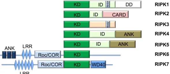
The RIPK family consists of seven known family members, which are classified as serine-threonine kinases due to their highly conserved kinase domain (KD) (Figure 1) (Zhang, Lin, and Han 2010; Festjens et al. 2007). A number of different domain structures such as the intermediate domain (ID) and the Caspase activation and recruitment domain (CARD) are found in different RIPK family members defining the specific function and the interaction partners of each protein (Figure 1). RIPKs participate in different biological processes implicated in the regulation of cell survival and cell death in response to a variety of stimuli (Zhang, Lin, and Han 2010;

Pasparakis and Vandenabeele 2015).

Figure 1. Schematic representation of the structure of the RIPK family members (adapted from Zhang, Lin and Han 2010)

The RIPK family consists of seven family members with all of them sharing a conserved KD. In addition to the KD each RIPK displays additional domain structures which defines their additional function or interaction partners. RIPK1, RIPK2, RIPK4 and RIPK5 contain in addition to the KD an ID. Only RIPK1 has a death domain (DD). Together with RIPK3 it contains a receptor homotypic interacting motif (RHIM). RIPK2 has an additional CARD domain. RIPK4 and RIPK5 contain a C-terminal ankyrin repeats (ANK) domain, which is also present in RIPK6.

Like RIPK7, RIPK6 has a leucine-rich repeat (LRR) motif and a Ros of complex proteins/C- terminal of Roc (Roc/COR) domain. RIPK7 further harbors a C-terminal WD40 motif.

1.1.1 RIPK1

RIPK1 was initially identified in 1995 as an interaction partner of the death receptor (DR) Fas (CD95) using a yeast-two hybrid screen and was described to be activated downstream of other DRs, such as tumor necrosis factor receptor 1 (TNFR1), and toll- like receptors (TLRs) including TLR4 (Stanger et al. 1995; Zhang, Lin, and Han 2010).

Human and murine RIPK1 proteins share 68% homology with the highest similarity in

(16)

the DD. Human RIPK1 is located on chromosome 6 whereas mouse Ripk1 has been mapped to chromosome 13 (Hsu, Huang et al. 1996; Stanger et al. 1995). RIPK1 consists of an N-terminal KD, an ID and a C-terminal DD (Figure 2) (Festjens et al.

2007; Stanger et al. 1995; Kelliher et al. 1998).

Until now, the kinase-dependent cellular functions of RIPK1 are still not fully understood. So far, only RIPK1 is known to be a target of its kinase activity. Degterev et al. identified various serine, threonine and tyrosine residues as phosphorylation sites (Degterev et al. 2008). Accumulating evidence suggests that the KD of RIPK1 enables its autophosphorylation on Ser14/15, Ser20, Ser161 and Ser166 and that site-specific autophosphorylation of RIPK1 defines its function (Degterev et al. 2008; de Almagro et al. 2017; Zhang et al. 2017)

In addition to the KD, RIPK1 consists of an ID, which harbors a Caspase-8 cleavage site, ubiquitination sites, and a receptor homotypic interacting motif (RHIM) domain (Zhang, Lin, and Han 2010). Like autophosphorylation, site-specific ubiquitination of RIPK1 was recently reported to promote specific functions of RIPK1 (de Almagro et al.

2017). Moreover, the RHIM domain of RIPK1 is required for the interaction with other RHIM domain containing proteins, such as RIPK3, TIR domain-containing adaptor inducing IFN-beta (TRIF) and DNA-dependent activator of IFN-regulatory factors (DAI/ZBP1) (Sun et al. 2002; Kaiser and Offermann 2005; Rebsamen et al. 2009; Lin et al. 2016; Newton et al. 2016).

Furthermore, the DD of RIPK1 mediates homotypic interactions with other DD- containing proteins, including TNFR1, TNFR superfamily 1A-associated via death domain (TRADD) and Fas (TNFRSF6)-associated via death domain (FADD) thus promoting tumor necrosis factor (TNF)-induced cell death or nuclear factor kappa light polypeptide gene enhancer in B-cells (NF-κB) activation (Zhang, Lin, and Han 2010) (Figure 2).

Figure 2. Schematic representation of the RIPK1 domain structure

RIPK1 is composed of a N-terminal KD, an ID, containing a RHIM domain, and a C-terminal

DD. RIPK1 interacts with RIPK3, TRIF and DAI/ZBP1 via its RHIM domain while its DD allows

its interaction with TNFR1, FADD and TRADD.

(17)

1.2. NF-κB signaling

NF-κB was identified as a transcription factor that binds to the intronic enhancer of the kappa light chain gene in B cells (Sen and Baltimore 1986a, 1986b). The NF-κB transcription factor family consists of p50, its precursor p105, p52, its precursor p100, RelA (p65), c-Rel and RelB with all of them sharing a Rel homology domain (Mitchell, Vargas, and Hoffmann 2016; Hayden and Ghosh 2014). The Rel domain allows NF-κB family members to homo- or heterodimerize and sequence-specific DNA binding. p50 and p52 rely on dimerization with RelA, c-Rel and RelB, which contain a transactivation domain, to positively induce transcription (Mitchell, Vargas, and Hoffmann 2016;

Hayden and Ghosh 2014; Rothwarf and Karin 1999; Chen and Greene 2004). In resting cells, NF-κB transcription factors remain in an inactive state in the cytoplasm through the binding to inhibitor of κB proteins (IκBs) (IκBα, IκBβ and IκBγ) and the precursor proteins p100 and p105 (Hayden and Ghosh 2014).

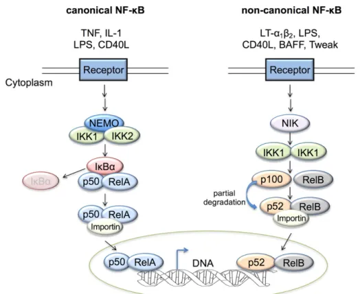
NF-κB signaling can be distinguished between canonical and non-canonical pathway (Figure 3). TNF, interleukin (IL)-1, lipopolysaccharide (LPS) and cluster of differentiation (CD)40L are potent inducers of canonical NF-κB involving p50/RelA heterodimerization (Figure 3) (Ghosh and Hayden 2008; Hayden and Ghosh 2008).

Binding of the ligands to their receptor induces the ubiquitination of RIPK1 which serves as a platform for the recruitment of the inhibitor of κB kinase (IKK) complex consisting of the regulatory subunit nuclear factor kappa B essential modulator (NEMO) and the kinases IKK1 and IKK2. The IKK complex triggers the phosphorylation of IκBα on Ser32 and 36 and thereby targets it for K48-ubiquitination and subsequently proteasomal degradation (Woronicz et al. 1997; Mercurio et al.

1997; Wang et al. 2001). Degradation of IκBα induces rapid translocation of the canonical NF-κB heterodimer p65(RelA):p50 to the nucleus initiating the transcription of target genes such as pro-inflammatory cytokines including Tnf and Il-6 and the anti- apoptotic/anti-necroptotic gene Cflar (Mitchell, Vargas, and Hoffmann 2016; Hayden and Ghosh 2014). In contrast, binding of B cell activating factor of the TNF family (BAFF), TNF-like weak inducer of apoptosis (Tweak) and CD40L to their respective receptors induces non-canonical NF-κB activation. In the non-canonical NF-κB pathway, NF-κB inducing kinase (NIK)-dependent activation of IKK1 mediates processing of p100 to p52 leading to the formation of a transcriptionally active p52- RelB complex. In summary, RIPK1, IKK1/2 and NEMO are key drivers of the canonical NF-κB pathway while the non-canonical NF-κB pathway acts independently of RIPK1, IKK2 and NEMO but exclusively relies on NIK and IKK1 (Figure 3) (Senftleben et al.

2001).

(18)

Figure 3. Canonical and non-canonical pathways of NF-κB activation (adapted from Horie and Umezawa 2012)

Under resting conditions, NF-κB dimers are bound to an IκB protein. Binding of a ligand to its receptor mediates the degradation of the inhibitor resulting in the release of the NF-κB dimer, which translocates to the nucleus mediating transcription of pro-inflammatory cytokines. There are two types of pathways that can induce NF-κB activation, the canonical and the non- canonical. The canonical NF-κB pathway can be induced by ligation of TNF, IL-1, LPS and CD40L with their respective receptor triggering the degradation of the IκB protein in a NEMO/IKK1/2-dependent manner resulting in the nuclear translocation of the NF-κB heterodimer mostly consisting of p65/RelA. In contrast, the non-canonical pathway is activated by the binding of CD40L, BAFF and lymphotoxin-β, LPS and TWEAK to their receptors, involving NF-κB inducing kinase (NIK)-dependent IKK1-mediated phosphorylation of p100, which associates with RelB. This induces a partial degradation of p100 to p52 leading to the formation of the transcriptionally active p52-RelB complex.

1.3. Death receptor signaling

The TNF receptor superfamily consists of various family members important for the regulation of cell survival and cell death in response to inflammatory cytokines and infectious particles (Locksley, Killeen, and Lenardo 2001). DRs, which are a subgroup of the TNF receptor superfamily, are characterized by their DD motif (Wajant 2003;

Wilson, Dixit, and Ashkenazi 2009). The DR family consists of 6 family members that

are activated by distinct ligands. Tumor necrosis factor superfamily member (Tnfrs) 1a

(TNFR1) is activated by ligation with TNF or lymphotoxin α (LTα), Tnfrsf6 (Fas or

CD95) is activated by FasL/CD95L, TNF-related apoptosis-inducing ligand receptor 1

and 2 (TRAILR1 and TRAILR2) are activated by TRAIL while DR3 (Tnfrsf25) and DR6

(19)

are activated by TLA1 and amyloid precursor protein (APP) respectively (Silke and Hartland 2013; Wilson, Dixit, and Ashkenazi 2009). In contrast to the other DRs, DR3 and DR6 are not efficient in inducing cell death (Wajant 2003). Activation of DRs by binding to their ligands induces the recruitment of DD-containing adapter proteins via homotypic interactions. This interaction results in the formation of a receptor associated signaling complex mediating downstream signaling including cell death or inflammatory gene expression (Wilson, Dixit and Ashkenazi 2009).

1.3.1. TNF signaling pathway

TNF is a cytokine produced by immune, epithelial and endothelial cells and binds to

two plasma membrane receptors, TNFR1 and TNFR2. Unlike TNFR1, which

recognizes soluble and membrane-bound TNF, TNFR2 binds with a much higher

affinity to the membrane-bound TNF precursor and is likely to be restricted to T cells

(Brockhaus et al. 1990; Dembic et al. 1990; Schall et al. 1990; Zheng et al. 1995; Kim

and Teh 2001; Depuydt et al. 2005). Until now, TNFR1 is the most well-studied

receptor with respect to cell death or cell survival pathways. TNF ligation to TNFR1

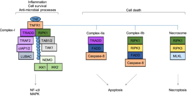
induces inflammatory, cell survival and anti-microbial processes via complex-I but in

addition also mediates apoptosis via complex-IIa/-IIb and necroptosis through the

necrosome (Figure 4) (Micheau and Tschopp et al 2003; Li et al. 2012; Varfolomeev

and Vucic 2016). TNF alone is not sufficient to induce cell death in most cell types

while in combination with inhibitors of transcription (e.g. D-Galactosamine) or

translation (e.g. cycloheximide) TNF potently triggers cell death suggesting that NF-κB-

dependent gene expression negatively regulates TNF-induced cell death (Morikawa et

al. 1996; Wang, Du and Wang 2008).

(20)

Figure 4. TNFR1 signaling induces cell survival, inflammation, anti-microbial processes and cell death.

TNF ligation to TNFR1 results in formation of a receptor-bound complex (complex-I) that activates NF-κB and mitogen-activated protein kinase (MAPK) signaling triggering inflammation, cell survival and anti-microbial processes. Under certain conditions, TNF causes complex-IIa or IIb-mediated TRADD or RIPK1-dependent apoptotic death. However, inhibition of complex-IIa and IIb TNF mediates necroptosis through the necrosome formation. (for details see 1.3.1.1-3)

1.3.1.1. TNFR1-induced pro-inflammatory and pro-survival signaling

Binding of TNF to TNFR1 induces pro-inflammatory and anti-apoptotic functions by inducing canonical activation of the transcription factor NF-κB and mitogen-activated protein kinase (MAPK) pathways (Varfolomeev and Vucic 2016; Baud and Karin 2001).

TNF induces the trimerization of TNFR1 resulting in a rapid formation of a receptor proximal complex-I consisting of RIPK1, TRADD, TNF receptor-associated factor 2 (TRAF2), cellular Inhibitor of Apoptosis Protein 1/2 (cIAP1/2) and linear ubiquitin chain assembly complex (LUBAC) (Figure 4 and 5) (Legler et al. 2003; Hayden and Ghosh 2014; Hsu, Huang, et al. 1996). RIPK1 and TRADD are recruited via their DD to the intracellular DD of TNFR1. TRADD in turn recruits the E3 ubiquitin ligase TRAF2 via interaction through their TRAF binding domain (Hsu, Huang, et al. 1996; Hsu, Shu, et al. 1996; Hsu, Xiong, and Goeddel 1995; Pobezinskaya et al . 2008). TRAF2 further binds the E3 ligases, cIAP1 and cIAP2, and recruits them to the TNFR1 complex (Shu, Takeuchi, and Goeddel 1996). The recruitment of the E3 ligases cIAP1/2 to complex-I promotes K11-, K63- and K48-linked ubiquitination of themselves and RIPK1 (Figure 5) (Park, Yoon and Lee 2004; Varfolomeev et al. 2008; Dynek et al. 2010; Mahoney et al.

2008). K63-specific modification of cIAP1/2 supports the engagement of LUBAC

consisting of haem-oxidized IRP2 ubiquitin ligase-1 (HOIL-1L), HOIL-1 interacting

(21)

protein (HOIP) and SHANK-associated RH domain-interacting protein (SHARPIN) (Ikeda et al. 2011; Tokunaga et al. 2011). K63-specific modification of RIPK1 supports the recruitment of transforming growth factor b activated kinase (TAK1) and TAK1 binding protein 1 and 2 (TAB1/TAB2) (Wang et al. 2001; Kanayama et al. 2004).

LUBAC in turn promotes linear ubiquitination (M1) of TNFR1, TRADD, RIPK1 and NEMO (Haas et al. 2009; Tokunaga et al. 2009). NEMO together with the IKKs are recruited to the TNFR1 complex through the interaction of NEMO with M1-, K11- and K63-linked ubiquitin chains, with the highest affinity for M1-ubiquitin chains, via its Ub binding in ABIN and NEMO (UBAN) domain (Wu et al. 2006; Rahighi et al. 2009;

Dynek et al. 2010). Trans-autophosphorylation of TAK1 induces the activation of IKK2 which mediates the activation of the NF-κB signaling pathway (for details see 1.2) (Wang et al. 2001).

Figure 5. TNF-induced complex-I signaling

Upon TNF ligation to TNFR1 a receptor- associated complex-I is being assembled, which mediates NF-κB activation resulting in pro-inflammatory and pro-survival signaling. For details see text.

Ub Ubiquitination (blue circles) P Phosphorylation

Stabilization of complex-I signaling is mediated by the modification of several complex-

I members with ubiquitin chains (O'Donnell et al. 2007; Mollah et al. 2007; Tokunaga et

al. 2009) (Figure 6). Genetic studies in mice revealed impaired canonical NF-κB

activation in response to TNF in absence of HOIL-1 and HOIP or after mutation of

Sharpin (Mollah et al. 2007; Tokunaga et al. 2009). RIPK1 displays multiple

(22)

ubiquitination sites, however, it remains controversial whether ubiquitination of RIPK1 is fundamental for NF-κB activation. Several studies indicated that ubiquitination of RIPK1 was indeed essential for NF-κB activation since mutation of the ubiquitination site at K377 abolished the recruitment of the TAK1- and IKK-complex to complex-I as well as IκBα phosphorylation after TNF stimulation (Ea et al. 2006; Hsu, Huang, et al.

1996; Li et al. 2006). On the contrary, an independent study reported that mutated ubiquitin, which could not engage K63 chains, did not result in reduced IκBa phosphorylation upon TNF stimulation suggesting a ubiquitin-dependent, K63- independent mechanism of TNF-induced NF-κB activation (Xu et al. 2009). In contrast to ubiquitination, autophosphorylation of RIPK1 is largely dispensable for TNF-induced NF-κB activation (Lee et al. 2004). However, unlike autophosphorylation, phosphorylation of RIPK1 by IKK1/IKK2 within complex-I was recently described to be important to restrain RIPK1 in complex-I in order to suppress its pro-death function and stabilize complex-I signaling (Dondelinger et al. 2015; Koppe et al. 2016).

Positive and negative regulation of NF-κB by TNF is partially achieved through the transcription of A20 (Mevissen et al. 2013; Ritorto et al. 2014; Wertz et al. 2004). A20 recruitment to complex-I requires the M1-chain forming function of LUBAC since absence of HOIP or M1-ubiquitin chains abolishes A20 recruitment to complex-I (Draber et al. 2015). Through its Zinc finger domains 4 and 7, A20 binds to K63- and M1 linkages respectively (Bosanac et al. 2010; Tokunaga et al. 2012; Verhelst et al.

2012). A20 has been so far implicated in the hydrolysis of K11-, K63- and K48- but not M1-ubiquitin chains to positively and negatively regulate NF-κB activation (Figure 6).

On the one hand, A20 stabilizes complex-I and favors signal transduction that leads to gene transcription by binding M1-ubiquitin chains. On the other hand, A20 negatively regulates TNF-induced NF-κB activation by hydrolyzing K63-ubiquitin chains via its OTU domain, leading to K48-ubiquitination and proteasomal degradation of RIPK1 (Wertz et al. 2004; Zilberman-Rudenko et al. 2016). In addition, accumulation of M1- ubiquitin-bound A20 can switch from positive to negative regulation of NF-κB activation by restricting NEMO/IKK complex binding to M1-ubiquitin chains (Zilberman-Rudenko et al. 2016). Together, A20 exhibits dual functions in NF-κB regulation by acting as an ubiquitin-stabilizing protein promoting NF-κB activation and as an ubiquitin hydrolyzing protein impairing NF-κB activation.

In addition to A20, the deubiquitinase cylindromatosis (CYLD) is recruited to TNFR1-

associated signaling complex-I by associating with HOIP-1 induced by SPATA2 at

which it regulates gene activator signaling and cell survival/death (Figure 6) (Takiuchi

et al. 2014; Draber et al. 2015). Unlike A20, CYLD recruitment most probably depends

(23)

on its interaction with HOIP but is independent of LUBAC’s M1-chain-forming capacity.

CYLD antagonizes and hydrolyzes K63-ubiquitin and M1 chains via its USP domain sensitizing cells to TNF-induced cell death (Figure 6) (Trompouki et al. 2003; Wright et al. 2007; Ritorto et al. 2014; Sato et al. 2015). In summary, controlled regulation of complex-I mediated signaling is achieved by A20 and CYLD. A20 positively regulates complex-I by binding to M1-ubiquitin chains but like CYLD it also negatively regulates complex-I signaling by hydrolyzing K63-ubiquitin chains.

Figure 6. (De-)Ubiquitination events regulating proximal TNF signaling.

TNF-induced NF-κB activation is tightly regulated by (de)ubiquitination events. Ubiquitination of several complex-I members including TNFR1, TRADD and RIPK1 is mediated by cIAP1/2 and LUBAC resulting in the stabilization of complex-I promoting NF-κB activation. A20 stabilizes complex-I by binding to M1-ubiquitin chains but together with CYLD it also negatively regulates complex-I signaling by hydrolyzing K63-ubiquitin chains.

P Phosphorylation

In addition to NF-κB signaling, TNF ligation to TNFR1 also activates MAPKs such as c- Jun n-terminal kinases (JNK), extracellular signal-related kinases (ERK) and p38 via TAK1-mediated activation. TAK1 acts as a MAP3K and consequently starts the phosphorylation cascade (Sabio and Davis 2014; Sakurai et al. 2000). MAPKs are evolutionarily conserved serine-threonine kinases regulating cellular processes such as cell proliferation, differentiation, survival, death and regulation of gene expression.

They are activated by phosphorylation cascades or by interaction with small

guanosintriphosphate (GTP)-proteins of the Ras/Rho family in response to oxidative

and osmotic stress, heat shock and pro-inflammatory cytokines including TNF (Figure

7) (Pearson et al. 2001; Treisman 1996).

(24)

Figure 7. Various stimuli activate MAPK by inducing a phosphorylation cascade.

Various stimuli such as oxidative or osmotic stress and cytokines activate MAP4K or GTPases. Activation of MAP4K and GTPases induces the activation of downstream kinases by a phosphorylation cascade.

MAPK activation results consequently in cell proliferation, differentiation, cell death and inflammatory response.

1.3.1.2. TNF-induced complex-IIa/-IIb-mediated apoptosis

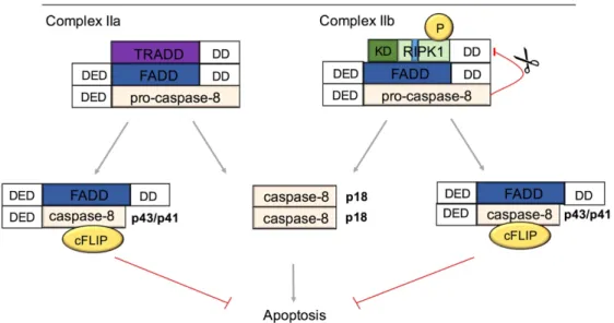
In addition to complex-I, binding of TNF to TNFR1 also results in apoptosis mediated by complex-IIa and IIb. Formation of complex-IIa through the association of TRADD and FADD via homotypic DD-DD interactions is induced e.g. after inhibition of pro- survival protein translation caused by cycloheximide (CHX) (Hsu et al. 1996; Hsu, Xiong and Goeddel 1995; Wang, Du and Wang 2008; Micheau and Tschopp 2003).

FADD then recruits the initiator Caspase, Caspase-8, via its death effector domain (DED) (Figure 8) (Micheau and Tschopp 2003; Wang, Du, and Wang 2008).

Alternatively, under conditions such as TNF stimulation in the presence of an IAP inhibitor, knockout of IAPs, TAK1 inhibition or knockdown, and NEMO knockout, a cytosolic complex-IIb can be formed consisting of RIPK1, FADD and Caspase-8 to execute RIPK1-kinase-dependent apoptosis (Figure 8) (Dondelinger et al. 2013;

Moulin et al. 2012; Legarda-Addison et al. 2009; Wang, Du, and Wang 2008; Wilson,

Dixit, and Ashkenazi 2009). This complex mediates apoptosis in a RIPK1 kinase-

activity dependent manner (Kondylis et al. 2015). Accumulating evidence suggests that

deubiquitination of RIPK1 is crucial for the execution of TNF-induced apoptosis (de

Almagro et al. 2017; O’Donnell et al. 2007). Moreover, cleavage of RIPK1 at the

Caspase cleavage site located in its ID by Caspase-8 is believed to prevent NF-κB

activation and necroptosis but promotes complex IIa-dependent apoptotic death in

response to DR signaling (Lin et al. 1999; Rajput et al. 2011).

(25)

Figure 8. TNF-induced complex-IIa and -IIb formation

Destabilization of complex-I results in the assembly of complex-IIa and -IIb in response to TNF.

Assembly of complex-IIa and -IIb promotes Caspase-dependent apoptotic death. TNF-induced complex-IIa signaling is TRADD dependent whereas complex-IIb signaling is RIPK1-kinase activity dependent.

In both complexes, homodimerization of pro-Caspase-8 results in a conformational change inducing the autocatalytic cleavage at Asp374 and the generation of multiple Caspase-8 fragments such as p43/p41 and p10 (Figure 8). After an additional cleavage at Asp216 the catalytic active p18 fragment and the inactive p26/24 prodomain are produced (Medema, Scaffidi, et al. 1997; Medema, Toes, et al. 1997).

The p18 fragment forms a homodimer which induces the cleavage and activation of the effector Caspases such as Caspase-3, -6 and -7 mediating extrinsic apoptosis.

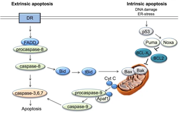
Moreover, activated Caspase-8 also triggers the activation of the pro-apoptotic BH3 interacting domain death agonist (Bid) resulting in the mitochondrial-mediated amplification of the apoptotic signal induced by extrinsic stimuli (Figure 9) (Li et al.

1998; Luo et al. 1998). Bid induces the release of mitochondrial proteins such as

cytochrome C and second mitochondrial derived activator of Caspases (SMAC)

leading to APAF-1-dependent apoptosome formation and pro-Caspase-9 activation

(Luo et al. 1998; Gross et al. 1999). Activated Caspase-9 activates effector Caspases

such as Caspase-3 and consequently amplifies apoptotic signaling also in cells with

low levels of Death-Inducing Signaling Complex (DISC) components (Wilson, Dixit, and

Ashkenazi 2009). Independent of Caspase-8, the mitochondria-mediated form of

apoptosis is induced among others by DNA-damage and ER-stress and is classified as

intrinsic apoptosis.

(26)

Figure 9. The extrinsic and intrinsic apoptotic pathways

Apoptosis is induced via mitochondrial (intrinsic) or via DR-mediated (extrinsic) pathways. The extrinsic apoptotic pathway is dependent on FADD-pro-Caspase-8 interaction resulting in the processing of pro-Caspase-8 to catalytically active Caspase-8, which consequently activates Caspase-3/6/7. In contrast, intrinsic apoptosis is activated in response to DNA-damage and ER- stress that induces p53 activation. p53 signaling activation results in the upregulation of p53 upregulated modulator of apoptosis (PUMA) and phorbol-12-myristate-13-acetate-induced- protein 1 (NOXA) that neutralizes the anti-apoptotic BCL-2 proteins, B-cell lymphoma (BCL)-2 and BCL-XL resulting in the activation of Bid (tBid). tBid in turn activates Bax and Bak resulting in the disruption of mitochondrial integrity causing the release of cytochrome C and SMAC and consequently resulting in apoptosome-dependent activation of Caspase-9. This leads to the activation of Caspase-3/6/7 and ultimately to apoptosis. However, extrinsic apoptosis induces also intrinsic apoptosis via Caspase-8-dependent activation of Bid to tBid.

Effector Caspases activate cytoplasmic endonucleases and proteases to induce the degradation of nuclear material and cytoskeletal proteins, respectively. These processes influence the morphological characteristics of apoptosis characterized by cell shrinkage and pyknosis, a result of chromatin condensation (Kerr, Wyllie, and Currie 1972). Moreover, extensive plasma membrane blebbing followed by karyorrhexis, separation of cell fragments into apoptotic bodies and nuclear fragmentation are characteristics of apoptosis (Kerr, Wyllie, and Currie 1972).

Apoptotic bodies are subsequently phagocytosed by macrophages, parenchymal cells or neoplastic cells and degraded within phagolysosomes (Poon et al. 2014).

Due to the immediate clearance of apoptotic bodies, apoptosis is considered to be

non-immunogenic (Lamkanfi and Dixit 2010; Green et al. 2009). Conversely, current

(27)

studies also identified a pro-inflammatory role of apoptosis under cancerous conditions (Poon et al. 2014).

Apoptotic death has to be tightly regulated since inappropriate apoptosis is involved in neurodegenerative diseases, autoimmune disorders and many types of cancer including hepatocellular carcinoma (HCC) (Poon et al. 2014; Kondylis et al. 2015).

Accordingly, a study using a genetic model of HCC formation revealed that the liver- parenchyma cell (LPC)-specific deficiency of NEMO triggered apoptotic death resulting in an inflammatory response inducing spontaneous HCC development in mice (Kondylis et al. 2015). Furthermore, IKK1/IKK2

LPC-KO

mice developed increased RIPK1- dependent cholangiocyte death resulting in strong hepatocellular damage, severe biliary destructions and cholestasis, which are fatal at 7-9 months of age (Luedde et al.

2008; Koppe et al. 2016). Conversely, inhibition of extrinsic apoptosis also results in a developmentally fatal outcome since Caspase-8 deficiency is embryonically lethal accompanied by embryonic vascular, cardiac and haematopoietic defects (Varfolomeev et al. 1998; Kaiser et al. 2011). Similarly, inhibition of intrinsic apoptosis by Caspase-9 deficiency resulted in embryonic/neonatal lethality showing enlarged and malformed cerebellum (Kuida et al. 1998; Hakem et al. 1998). Taken together, both extensive but also diminished apoptosis strongly affect tissue homeostasis and the viability of mice. Therefore, tight regulation of Caspase-dependent apoptosis is essential for development and tissue homeostasis.

NF-κB-dependent expression of cellular FLICE-like inhibitory protein (cFLIP) is

considered to negatively regulate extrinsic apoptotic signaling through modulation of

Caspase-8 activity (Dillon et al. 2012; Feoktistova et al. 2011). cFLIP is a catalytically

inactive homolog of Caspase-8 with two isoforms, cFLIP

L

and cFLIP

S

in humans and

cFLIP

L

and cFLIP

R

in mice. Caspase-8 cFLIP heterodimerization results in primary

Caspase-8 cleavage and the generation of a p10-fragment but lacks secondary

cleavage inhibiting p18-fragment generation (Pop et al. 2011). Interaction between the

p43/p41 Caspase-8 fragment and cFLIP

L

negatively regulates apoptosis and is thought

to inhibit necroptosis. In detail, a Caspase-8-cFLIP heterodimer has been suggested to

cleave CYLD, RIPK1 and RIPK3 degrading key drivers of TNF-induced necroptosis

(see 1.3.1.3.). There is evidence suggesting that low levels of cFLIP

L

stabilize complex

IIa by binding to Caspase-8 allowing its oligomerization to execute apoptosis and block

necroptosis while high levels of cFLIP

L

prevent Caspase-8 oligomerization and thereby

block apoptosis (Hughes et al. 2016; Fu et al. 2016). This hypothesis is based on data

obtained by Fas/FasL signaling and yet needs to be confirmed in a TNFR1-dependent

setting (Pop et al. 2011; Wachter et al. 2004; Hughes et al. 2016; Fu et al. 2016).

(28)

1.3.1.3. TNF-induced complex-IIc mediated necroptosis

When the formation of complex-IIa and-IIb are blocked by FADD, Caspase-8 deficiency or inhibition of Caspase-8 activity by zVAD-fmk, TNF induces the assembly of a third death-inducing complex, called complex-IIc/ necrosome, consisting of RIPK1, RIPK3 and mixed lineage kinase domain-like (MLKL) (Figure 10) (Newton et al. 2014;

Pasparakis and Vandenabeele 2015). Accordingly, Fadd

-/-

or Caspase-8

-/-

mice showed embryonic lethality that was prevented by additional deletion of the necroptosis mediator RIPK3 or MLKL (Kaiser et al. 2011; Dillon et al. 2012; Alvarez- Diaz et al. 2016) To date, necroptotic death is considered to occur independently of Caspases in cells with elevated levels of RIPK3 and MLKL. TNF-induced necroptotic death is dependent on RIPK1 kinase activity as several studies reported that mutation of the kinase activity or using the allosteric inhibitor of the kinase activity of RIPK1, necrostatin-1 (nec-1), prevented necroptotic death (Ni et al. 2016; Polykratis et al.

2014; Degterev et al. 2008; Degterev et al. 2005; Kondylis et al. 2015; Vlantis et al.

2016; Newton et al. 2014). Indeed, autophosphorylation of RIPK1 on Ser161 or Ser166 was recently reported to be important for necroptotic death (de Almagro et al. 2017;

Zhang et al. 2017). Autophosphorylation at the residue Ser161 was shown to be triggered in response to TNF-induced ROS (Zhang et al. 2017). However, it remains to be elucidated whether only autophosphorylation or phosphorylation by other kinases at one or multiple residues are crucial for RIPK1 functions particularly in the necrosome.

It was recently identified that not only phosphorylation, but coordinated ubiquitination and phosphorylation of RIPK1 within complex-IIc are crucial for the induction of necroptosis (Figure 10) (de Almagro et al. 2017 and 2015). Mutation of the ubiquitination site of RIPK1 at K115 prevented K63- and linear ubiquitination of RIPK1 and thereby reduced RIPK1 phosphorylation, necrosome formation and consequently necroptosis (de Almagro et al. 2017). However, the precise molecular mechanisms controlling RIPK1 activation in the necrosome are still unclear.

Activation of RIPK1 results in conformational changes allowing an interaction between RIPK1 and RIPK3 through their RHIM domains (Chan, Luz, and Moriwaki 2015; Cho et al. 2009). The heterodimeric interaction between RIPK1 and RIPK3 mediates the recruitment of another RIPK3 molecule to form a RIPK3-RIPK3 amyloid complex allowing RIPK3 autophosphorylation (Cho et al. 2009; He et al. 2009; Li et al. 2012;

Zhang et al. 2009; Sun et al. 2002). RIPK3 phosphorylation at Ser232 is crucial for the recruitment and activation of its downstream target MLKL (Zhao et al. 2012; Sun et al.

2012; Murphy et al. 2013). The activation of the pseudokinase MLKL is mediated by its

phosphorylation by RIPK3 at different phosphorylation sites including Ser345, Ser347,

(29)

Ser 352 and Thr 349 (Chen et al. 2013; Li et al. 2012; McQuade, Cho, and Chan 2013;

Cai et al. 2014). This phosphorylation of MLKL results in a conformational change that exposes the 4-helical bundle domain allowing its oligomerization (Cai et al. 2014; Chen et al. 2013; Dondelinger et al. 2014; Sun et al. 2012; Wang et al. 2014; Xie et al.

2013). It is still highly debated how MLKL executes necroptotic death. The current model suggests that MLKL binds to phospholipids of the cell membrane through its 4- helical bundle domain resulting in the influx of Ca

2+

and Na

+

that likely contribute to cell death (Cai et al. 2014; Chen et al. 2013; Dondelinger et al. 2014; Su et al. 2014; Wang et al. 2014).

Morphologically, necrotic cell death is characterized by organelle swelling and loss of plasma membrane integrity (Festjens et al. 2007; Krysko et al. 2008; Vanden Berghe et al. 2010). In contrast to apoptotic death, necroptotic death is currently believed to be highly immunogenic by releasing factors collectively described as damage-associated molecular patterns (DAMPs) (Kono and Rock 2008; Krysko et al. 2008; Lamkanfi and Dixit 2010). Consistently, FADD deficiency in both intestinal epithelial cells (IECs) and keratinocytes induces necroptotic death and tissue inflammation that was both prevented by RIPK3 deficiency (Welz et al. 2011; Bonnet et al. 2011).

Figure 10. TNF-induced necrosome formation.

TNF induces the assembly of a necrosome mediating Caspase- independent necroptotic death in a RIPK1-RIPK3 kinase activity dependent manner. Coordinated ubiquitination and phosphorylation of RIPK1 was reported to induce the complex formation between RIPK1 and RIPK3, which interact via their RHIM domain, resulting in the phosphorylation and activation of MLKL.

1.4. TLR signaling

TLRs are receptors involved in the activation of innate immune cells recognizing

specific patterns of microbial components (PAMPs) including bacteria, mycobacteria

and viruses or altered host-derived DAMPs that under specific conditions may trigger

(30)

an immunological response (Akira, Takeda and Kaisho 2001; Akira, Uematsu and Takeuchi 2006; Janeway and Medzhitov 2002). All TLRs share a conserved cytoplasmic TIR domain with which they can interact with TIR domain-containing adaptors such as myeloid differentiation primary response gene 88 (MyD88) and TRIF.

In addition to their TIR domain, TLRs contain a leucine-rich repeat (LRR) ectodomain which enables recognition of PAMPs and DAMPs (Kawai and Akira 2010). Until now, 12 TLRs have been identified in humans and 10 in mice. Unlike MyD88, which is implicated in the signaling induced by all TLRs except for TLR3, TRIF is implicated only in TLR3 and TLR4 signaling (Kawai and Akira 2010).

TLR4 induces an inflammatory response against gram-negative bacteria after recognizing lipopolysaccharide (LPS) on their outer-surface (Figure 11) (Hoshino et al.

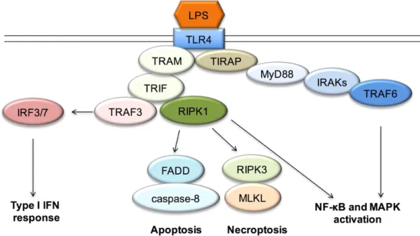
1999; Poltorak et al. 1998; Qureshi et al. 1999). TLR4 is the only TLR that mediates signaling via both MyD88 and TRIF adaptors. After LPS stimulation, TLR4 recruits the intracellular adapter MyD88 and TRIF via TIRAP/TRAM. MyD88 induces inflammatory cytokine production through NF-κB activation mediated by interleukin-1 receptor- associated kinase (IRAK) and TRAF6 interaction. TRIF associates with either TANK- binding kinase (TBK)1 or activates IFN regulatory factor 3 (IRF3) mediating type I IFN expression. In addition, TRIF interacts with RIPK1 via their RHIM domains to mediate MAPK and NF-κB activation (Figure 11). Similar to its role in TNF signaling, recruitment of RIPK1 to TLR4 via TRIF is followed by its polyubiquitination, which is possibly mediated by TRAF6. Polyubiquitination of RIPK1 mediates the recruitment of TAB1/2 and TAK1 resulting in MAPK activation and in the recruitment of the IKK1/2/NEMO complex inducing NF-κB activation (Cusson-Hermance et al. 2005).

TLR4-mediated TRIF-dependent cell death occurs in order to eliminate infected host-

cells. Therefore, a death-inducing complex containing TRIF, RIPK1, RIPK3, FADD,

Caspase-8 and cFLIP induces RIPK1 kinase-activity-dependent apoptosis and

necroptosis (He et al. 2011; Kaiser et al. 2005; Polykratis et al. 2014).

(31)

Figure 11. LPS-mediated TLR4 signaling activation

Binding of LPS to TLR4 induces the engagement of TIR domain-containing adaptor proteins TIRAP/MyD88 or TRAM/TRIF. This mediates the interaction between MyD88 with IRAKs and the adapter molecule TRAF6. Together they mediate NF-κB and MAPK activation. Via TRAM and TRIF TLR4 interacts with RIPK1 and TRAF3. Through TRAF3 TLR4 triggers IRF3/7- mediated Type-I-Interferon response while via RIPK1 TLR4 induces either NF-κB/ MAPK activation or cell death (apoptosis or necroptosis).

1.5. Anatomy and function of the liver

The liver is an essential organ in vertebrates consisting of two main lobes in humans and four main lobes in mice (Abdel-Misih and Bloomston 2010). The liver is a heterogeneous organ that is predominantly composed of the liver parenchyma cell compartment consisting of 60-65% hepatocytes and 3.5% biliary epithelial cells and the non-parenchymal cell compartment including 15-20% endothelial cells, as well as 8-12% liver-resident macrophages (Kupffer cells) and 3-8% lymphocytes and hepatic stellate cells (Racanelli and Rehermann 2006; Kmiec 2001). The liver mediates various functions that are implicated in fat and glucose metabolism, detoxification, protein synthesis, hormone production and glycogen storage (van den Berghe 1991; Dietschy, Turley, and Spady 1993). Deregulation of liver homeostasis can result in acute liver failure (ALF), in chronic liver diseases or in HCC (Dietschy, Turley, and Spady 1993;

Kmiec 2001; van den Berghe 1991; Wullaert et al. 2007).

The maintenance of liver homeostasis is dependent on the tight regulation of

inflammation and cell death. Although under physiological conditions the turnover rate

of differentiated hepatocytes is relatively low (200-400 days in mice/rats) compared to

other epithelial or epidermal cells, liver regeneration capacity is highly increased upon

extensive tissue loss during surgical resection or injury caused by massive alcohol

(32)

consumption, metabolic toxicity and viral or other microbial infections (Martins, Theruvath and Neuhaus 2008; Fausto 2004). Massive hepatocyte death exceeding the regenerative capacity of the liver mediates acute liver injury which can ultimately result in ALF causing high mortality rates of patients (Bantel and Schulze-Osthoff 2012).

Moreover, massive hepatocyte death or chronic inflammation coupled with compensatory liver regeneration induced by hepatocyte proliferation is thought to greatly increase the risk of HCC (Luedde, Kaplowitz, and Schwabe 2014; Guicciardi et al. 2013). HCC is one of the most frequent and difficult to treat types of cancer constituting the second most common cause of cancer-related deaths worldwide (Shariff et al. 2009; El-Serag 2011). Understanding the mechanisms regulating chronic inflammation and cell death in the liver is essential to find new therapeutic strategies against liver diseases such as ALF and HCC.

1.5.1. Murine models of liver inflammation and liver injury

Several murine models of liver injury provide distinct approaches to elucidate the inflammatory response and cell death signaling in the liver upon damage. A well- characterized murine model to study inflammatory signaling is the LPS-induced systemic inflammation model. A single intraperitoneal injection (i.p.) of sub-lethal doses of the gram-negative bacteria derived endotoxin LPS induces a TLR4-dependent acute inflammatory response, which resembles to a large extent those occurring during the early stages of septic shock (Martich, Boujoukos, and Suffredini 1993). LPS toxicity is associated with activation of KCs in the liver and the release of inflammatory cytokines such as TNF resulting in a strong inflammatory response that can ultimately lead to death by septic shock without inducing liver injury (Bautista et al. 1994; Luster et al.

1994; Bradham et al. 1998). Combined injection of LPS with a hepatic transcription inhibitor D-galactosamine (D-GalN), which is metabolized into an active metabolite exclusively in hepatocytes and blocks transcription by depleting uridine nucleotides, sensitizes hepatocytes to death resulting in hepatic damage caused by TNF resembling viral hepatitis accompanied by focal cell death and periportal inflammation (Keppler, Pausch, and Decker 1974; Freudenberg, Keppler, and Galanos 1986;

Morikawa et al. 1996; Xiong et al. 1999; Van Dien et al. 2001; Liu et al. 2008). Besides

the LPS/TNF-induced murine model, ConA-induced hepatic inflammation reflects the

overall physiopathological conditions observed in human viral-induced or autoimmune

hepatitis. ConA is an effective activator of T-cells resulting in the activation of cytotoxic

effectors such as TNF that trigger hepatocyte death consequently resulting in hepatitis

development (Mizuhara et al. 1994; Tiegs et al. 1992). Moreover, α-galactosylceramide

(α-GalCer), a potent NKT cell stimulator, causes liver injury through a pronounced

Abbildung

Figure 1. Schematic representation of the structure of the RIPK family members   (adapted from Zhang, Lin and Han 2010)
Figure 3. Canonical and non-canonical pathways of NF-κB activation   (adapted from Horie and Umezawa 2012)
Figure 4. TNFR1 signaling induces cell survival, inflammation, anti-microbial processes  and cell death
Figure 8. TNF-induced complex-IIa and -IIb formation
+7

Referenzen

ÄHNLICHE DOKUMENTE

Improved understanding of the immune cell-mediated mechanisms involved in hepatocyte cell death could be beneficial for the development of common therapeutic strategies

In conclusion, from this current study, by using PrPC knockout mice, we showed a critical role of PrPC in the liver of aging mice by regulating the glucose/lipid metabolism, which

In a sensitivity analysis including only patients with liver imag- ing > 30 days before onset of COVID-19, imaging evidence of hepatic steatosis remained associated with

A New Flavonoid C-Glycoside from Solanum elaeagnifolium with Hepatoprotective and Curative Activities against Paracetamol- Induced Liver Injury in Mice..

There was a signifi cant (P < 0.05) decrease in the protein content in the livers of the paracetamol-treated group compared to the control group, while the groups treated

Taken together this study shows that the liver endothelial layer, mainly LSE Cs, represent a direct target of the cytotoxic effect of paracetamol and that activation of

Synergistic induction of cell death by TRAIL and chemotherapeutics has been described in different tumors cell Iines.6-8,14 Similarly, we have previously reported

Consequently, there is an urgent need for liver models that allow to screen drug candidates for their potential to induce idiosyncratic DILI in the early non- clinical