Synthesis of Glycopeptides for Exploration of Mucin‐Type‐Threonine and HexNAc‐Tyrosine
Modifications
DISSERTATION
Zur Erlangung des akademischen Grades eines Doktors der Naturwissenschaften
(Dr. rer. nat.)
der Fakultät Chemie und Chemische Biologie der Technischen Universität Dortmund
Vorgelegt von M. Sc. Chem. Biol.
Manuel Schorlemer
aus Anröchte.
Dortmund 2018
Die vorliegende Arbeit entstand im Zeitraum von April 2011 bis März 2018 unter Anleitung von Prof.
Dr. Ulrika Westerlind an der Fakultät Chemie und Chemische Biologie der Technischen Universität Dortmund und dem Leibniz Institut für analytische Wissenschaften ISAS e.V. in Dortmund.
Erstgutachter: Prof. Dr. Herbert Waldmann
Zweitgutachterin: Prof. Dr. Ulrika Westerlind
Parts of this work have already been published in:
A Unified Strategy for the Synthesis of Mucin Cores 1‐4 and the Assembled Multivalent Glycopeptides
C. Pett, M. Schorlemer, U. Westerlind, Chem. Eur. J. 2013, 19, 17001‐17010
Assignment of Saccharide identities through Analysis of Oxonium Ion Fragmentation Profiles in LC‐
MS/MS of Glycopeptides
A. Halim, U. Westerlind, C. Pett, M. Schorlemer, U. Rüetschi, G. Brinkmann, C. Sihlbom, J. lengquist, G.
Larson, J. Nilsson, J. Proteome Res. 2014, 13, 6024‐6032
Distinctive MS/MS Fragmentation Pathway of Glycopeptide Generated Oxonium Ions Provide Evidence of the Glycan Structure
J. Yu, M. Schorlemer, A. Gomez Toledo, C. Pett, C. Sihlbom, G. Larson, U. Westerelind, J. Nilsson, Chemistry 2015, 22, 1114‐1124
Microarray Analysis of Antibodies Induced with Synthetic Antitumor Vaccines: Specificity against Diverse Mucin Core Structures
C. Pett, H. Cai, J. Liu, B. Palitzsch, M. Schorlemer, S Hartmann, N. Stergiou, M. Lu, H. Kunz, E. Schmitt, Chem. Eur. J. 2017, 23, 3875‐3884
Submitted papers:
Effective assignment of 2,3/2,6 sialic acid isomers in LC-MS/MS based glycoproteomics
C. Pett, W. Nasir, C. Sihlbom, B.‐M. Olsson, M. Schorlemer, R. Zahedi, G. Larson, J. Nilsson, U.
Westerlind, under review.
Table of Contents
1 Abstract ... 6
2 Zusammenfassung ... 8
3 Introduction ... 10
3.1 O‐glycosylation in eukaryotes ... 10
3.2 Mucin glycopeptides ... 13
3.2.1 Mucin glycoproteins in airway diseases ... 15
3.2.2 Mucins in cancer ... 17
3.3 HexNAc O‐glycosylation on tyrosine ... 22
4 Motivation ... 25
4.1 Project 1: Mucin glycosylation in cancer and airway diseases ... 25
4.2 Project 2: Chemical tools to explore HexNAc‐Tyr O‐glycosylation ... 28
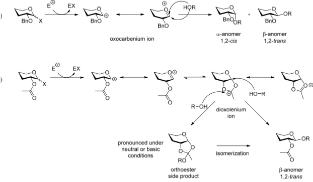
5 Synthetic strategies ... 32
5.1 Carbohydrate synthesis ... 32
5.2 The anomeric effect ... 34
5.3 Protecting groups in carbohydrate chemistry ... 35
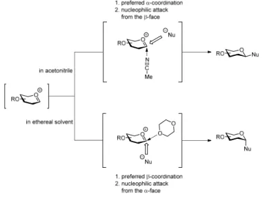
5.4 Solvent effects in carbohydrate synthesis ... 37
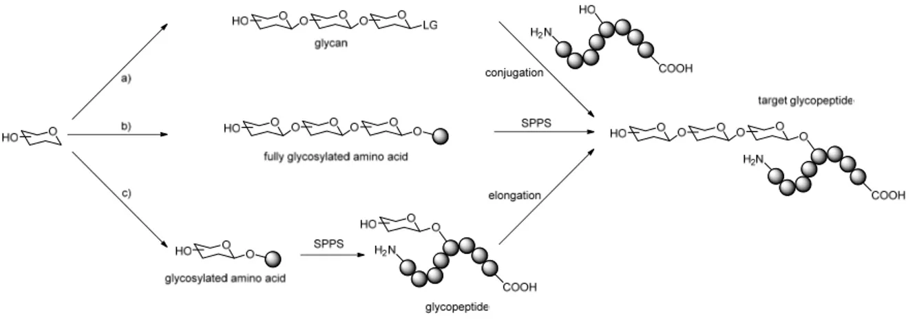
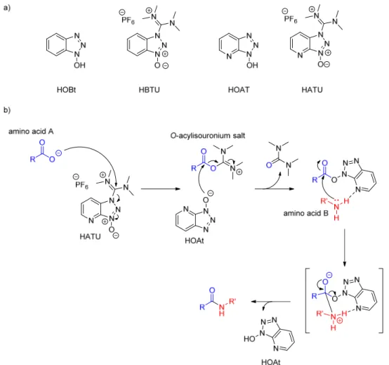
5.5 Glycopeptide synthesis ... 38
5.6 Enzymatic carbohydrate and glycopeptide synthesis ... 44
6 Bioanalytical Methods ... 47
6.1 Glycan and glycopeptide microarrays for studies of binding interactions ... 47
6.1.1 Immobilization methods for glycopeptide microarrays ... 48
6.1.2 General principle of glycopeptide microarray analysis ... 50
6.2 Mass spectrometry‐based analysis of the glycoproteome ... 51
6.2.1 Common glycoproteomic methodologies... 51
7 Results & Discussion ... 54
7.1 Project 1: Mucin glycosylation in cancer and airway diseases ... 54
7.1.1 Synthesis of mucin‐type glycosylated amino acid building blocks ... 54
7.1.2 Synthesis of the T
N‐antigen acceptor ... 57
7.1.3 The LacNAc disaccharide ... 60
7.1.4 Synthesis of the LacNAc type‐1 disaccharide donor ... 61
7.1.5 Synthesis of the LacNAc type‐2 disaccharide donor ... 66
2
TABLE OF CONTENTS
7.1.6 Synthesis of the T‐antigen glycosyl acceptor ... 67
7.1.7 Synthesis of the core 2 type‐1 glycosylated amino acid ... 68
7.1.8 Synthesis of the core 2 type‐2 glycosylated amino acid ... 70
7.1.9 Synthesis of the core 3 type‐1 acceptor ... 71
7.1.10 Synthesis of the core 3 type‐2 acceptor ... 72
7.1.11 Synthesis of the core 4 type‐1 glycosylated amino acid ... 74
7.1.12 Synthesis of the core 4 type‐2 glycosylated amino acid ... 76
7.1.13 Synthesis of Fmoc‐GalNAc‐D
3‐threonine ... 77
7.1.14 Generation of a MUC1 glycopeptide library ... 78
7.1.15 Ligand specificity of adenovirus lectins ... 81
7.1.16 Binding specificities of human galectin‐3 ... 82
7.1.17 Evaluation of antitumor vaccines towards antibody specificity ... 83
7.1.18 Oxonium ion‐based HexNAc identification ... 85
7.2 Project 2: Chemical tools to explore HexNAc‐Tyr O‐glycosylation ... 89
7.2.1 Overview of HexNAc‐Tyr building block synthesis ... 89
7.2.2 Synthesis of the α‐GalNAc‐Tyr building block ... 90
7.2.3 Synthesis of the α‐GlcNAc‐Tyr building block ... 91
7.2.4 Synthesis of the β‐GlcNAc‐Tyr building block ... 92
7.2.5 Generation of the HexNAc glycopeptide library ... 93
7.2.6 Evaluation of lectins for HexNAc detection and enrichment ... 102
7.2.7 Oxonium profiles of HexNAc‐Tyr O‐glycans ... 112
7.2.8 β‐GlcNAc‐Tyr enzyme profiling ... 119
8 Conclusion ... 133
8.1 Project 1: Mucin glycosylation in cancer and airway diseases ... 134
8.2 Project 2: O‐glycosylation on tyrosine ... 142
9 Experimentals ... 150
9.1 Building block synthesis ... 150
9.1.1 General ... 150
9.1.2 Synthesis of mucin‐type glycosylated amino acid building blocks ... 151
9.1.3 Synthesis of the HexNAc‐Tyr amino acid building blocks ... 209
9.2 Solid phase peptide synthesis ... 218
9.2.1 General protocol for solid phase (glyco‐)peptide synthesis ... 218
9.2.2 Synthesis of mucin‐type glycosylated peptides ... 220
9.2.3 Synthesis of HexNAc glycopeptides ... 227
9.3 Microarray experiments ... 246
9.3.1 General procedure ... 246
3
TABLE OF CONTENTS
9.3.2 Lectins and antibodies ... 246
9.3.3 General spotting conditions ... 247
9.3.4 Microarray slides MA1, MA2 and MA3 ... 247
9.3.5 Protocol for microarray binding assays ... 255
9.4 HCD‐based oxonium ion profiling ... 256
9.4.1 Profiling with single collision energy on MUC1 glycopeptides ... 256
9.4.2 Profiling with stepped collision energy on MUC1 glycopeptides ... 256
9.4.3 Profiling with stepped collision energy on tryptic glycopeptides ... 256
9.5 Enzyme incubation experiments ... 258
9.5.1 OGA‐incubations ... 258
9.5.2 hOGA incubations ... 259
9.5.3 hOGT incubations ... 260
10 References ... 261
11 Appendix ... 277
11.1 NMR spectroscopic data ... 277
11.2 HPLC chromatograms ... 303
Acknowledgements ... 323
Abbreviations
gg gauche‐gauche
Glc D‐glucose
GlcNAc N‐acetyl‐D‐glucosamine GSL II griffonia simplicifolia lectin II gt gauche‐trans
GTP Guanosine‐5’‐triphosphate
h hours
HATU O‐(1H‐7‐azabenzotriazol‐1‐yl)‐
1,1,3,3‐tetramethyluronium hexafluorophosphate
HBTU O‐(1H‐benzotriazol‐1‐yl)‐1,1,3,3‐
tetramethyluronium hexafluorophosphate HMBC heteronuclear multiple bond
correlation
HOAt 7‐aza‐1‐hydroxybenzotriazole HOBt 1‐hydroxybenzotriazole hOGA human O‐GlcNAc hydrolase hOGT human O‐GlcNAc transferase HPLC high performance liquid
chromatography HR high resolution
HSQC heteronuclear single quantum coherence
Hz hertz
Ig immunoglobulin iPr isopropyl J coupling constant
KLH Keyhole Limpet Hemocyanin LacNAc N‐acetyllactosamine LC liquid chromatography Le
x/Le
aLewis x/Lewis a LG leaving group LWAC lectin weak affinity
chromatography
m multiplet
M molarity
MALDI matrix assisted laser desorption ionization
mbar millibar
MD Molecular dynamics
Me methyl
MeCN acetonitrile
MGL macrophage c‐type lectin MHC major histocompatibility complex
min minutes
MS mass spectrometry NCE normalized collision energy
AA amino acid
Aβ amyloid‐β
Ac acetyl
All allyl
arom aromatic
AspAT aspartate amino transferase Atp5a ATP‐synthase subunit α Atp5b ATP‐synthase subunit β Boc tert‐butyloxycarbonyl BSA bovine serum albumin
Bu butyl
Bn benzyl
c concentration
calc. calculated cat. catalytic
CAZy carbohydrate active enzymes CID collision induced dissociation CFG Consortium for Functional
Glycomics
COPD chronic obstructive pulmonary disease
COSY correlated spectroscopy Cox4i1 cytochrome‐c oxidase subunit 4
isoform 1
d duplet, day
DBU 1,8‐diazabicyclo[5.4.0]undec‐7‐
ene
DCC N,N’‐dicyclohexylcarbodiimide DCM dichloromethane
dd duplet of duplet
DHB 2,5‐dihydroxy benzoic acid DIC N,N’‐diisopropylcarbodiimide DIPEA diisopropylethylamine DMAP 4‐(dimethylamino(pyridine) DMF dimethylformamide DMSO dimethyl sulfoxide
ELISA enzyme‐linked immunosorbent assay
eq. equivalents
ESI electron spray ionization ETD electron transfer dissociation FA formic acid
Fmoc N‐(9H‐fluoren‐9‐yl)‐
methoxycarbonyl Fuc L‐fucose
FliD flagella hook associated protein 2 Gal D‐galactose
GalNAc N‐acetyl‐D‐galactosamine
5
ABBREVIATIONS
VNTR variable number of tandem repeats
VVA vicia villosa agglutinin WGA wheat germ agglutinin α specific optical rotation δ chemical shift
λ wavelength
Amino acid codes
Ala A Alanine Arg R Arginine Asn N Asparagine Asp D Aspartate Cys C Cysteine Gln Q Glutamine Glu E Glutamate Gly G Glycine His H Histidine Ile I Isoleucine Leu L Leucine Lys K Lysine Met M Methionine Phe F Phenylalanine Pro P Proline Ser S Serine Thr T Threonine Trp W Tryptophan Tyr Y Tyrosine Val V Valine Neu5Ac N‐acetylneuraminic acid
NHS N‐hydroxysuccinimide NIS N‐iodosuccinimide
NMR nuclear magnetic resonance NucB2 nucleobindin‐2
OGA O‐GlcNAc hydrolase OGT O‐GlcNAc transferase
p para
PBS phosphate buffered saline PBST PBS + Tween‐20
PcrV type III secretion system protein PEG polyethylene glycol
PG protecting group
Ph phenyl
pH potentia hydrogenii PMP para‐methoxy phenyl pos positive
ppGalNAcT polypeptide N‐
acetylgalactosamine transferase ppm parts per million
p‐TsOH para‐toluenesulfonic acid
Pyr pyridine
q quartet
Rbl2 retinoblastoma‐like protein 2 R
fretention factor
s singulet
SLe
x/SLe
asialyl‐Lewis x/sialyl Lewis a SPPS solid phase peptide synthesis ST sialyl‐Thomsen‐Friedenreich STD saturation transfer difference ST
Nsialyl‐T‐antigen nouvelle
Su succinimide
t triplet
T Thomsen‐Friedenreich antigen TBAF tert‐butylammonium fluoride TBS tert‐butylammonium fluoride tBu tert‐butyl
TEG triethylene glycol tert tertiary
TFA trifluoroacetic acid
TfOH trifluoromethanesulfonic acid tg trans‐gauche
THF tetrahydrofuran TIPS triisopropylsilance TLC thin layer chromatography TMS tetramethylsilyl
T
NT‐antigen nouvelle
TOCSY Total correlation spectroscopy Troc trichloroethoxycarbonyl Trt trityl
UDP‐GlcNAc Uridine diphosphate N‐
acetylglucosamine
UV ultraviolet
1 Abstract
Protein O‐glycosylation is a structurally and biologically versatile modification. The mucin‐type O‐
glycosylation on extracellular mucin proteins is for instance involved in cell‐cell and cell‐matrix interactions, and play key roles in airway inflammations of COPD, asthma and cystic fibrosis patients by serving as binding receptors for virulence factors of airway disease‐related pathogens (bacteria and viruses). Insights into molecular details in the binding specificities of virulence factors like adhesion proteins (lectins) or sialidases would support the development of glycomimetic antiadhesive drugs or enzyme inhibitors to suppress infections. Moreover, adenocarcinoma cells (from for instance breast, colon, lung and ovary cancer) have been found to exhibit altered mucin glycosylation and expression levels, which has been related to tumor progression and metastasis. MUC1‐based vaccine constructs have been identified as promising for induction of a tumor‐directed immune response, which in consequence might eradicate the tumor burden. However, without a structurally defined MUC1 glycopeptide library, a quality control and a precise epitope mapping of the induced antibodies is not possible. This work focuses on the synthesis of a well‐defined peptide library (including extended core 2 and core 4 glycosylation) for application in microarray experiments to map lectin/glycopeptide and antibody/glycopeptide binding specificities. The gained information will aid to tackle the above‐
described challenges in airway disease and cancer progression. Further, this work contributed in the development of a new oxonium ion‐based mass‐spectrometric methodology for improved structural characterization of glycopeptides.
O‐glycosylation is commonly found on serine (Ser) or threonine (Thr), but recent identifications
suggest that similar modifications also occur on tyrosine (Tyr). These modifications are initiated by
addition of a HexNAc (GalNAc or GlcNAc) monosaccharide residue to the respective amino acid. A
major challenge for identification of the recently discovered HexNAc‐Tyr modifications was to
distinguish the structural similar isomers αGalNAc‐, αGlcNAc‐ and βGlcNAc‐Tyr. Assignments of the
glycan structures were only possible by general knowledge of biosynthetic pathways or being based
on lectin glycan‐binding preferences when applied in lectin‐affinity enrichment of protein‐digested
peptide fragments. However, lectin binding‐preferences of HexNAc‐Tyr have not been explored and
lectins are not always capable to discriminate between different glycan isomers. In previous
glycoproteomic studies complex Tyr glycosylation was assumed as mucin type αGalNAc‐Tyr
modification, based on knowledge of lectin specificity to Ser/Thr glycopeptides and the fact that
mucin‐type glycopeptides are generally extended with additional glycans. The αGlcNAc‐Tyr was found
7
ABSTRACT
on host GTPases, a modification caused by intracellular infections and was assigned by enzymatic in vitro studies and NMR structural characterization. Further, in an extensive glycoproteomic study several HexNAc‐Tyr modifications were found on mitochondrial proteins and some of the identified sites are also known phosphorylation sites. Therefore, the identified HexNAc‐Tyr modifications were likely to be βGlcNAc‐Tyr (although to date this modification is still not verified). In order to enable identification of HexNAc‐Tyr as a possible modification by glycoproteomic analysis, lectins commonly used for glycopeptide affinity enrichment were evaluated towards binding recognition of HexNAc‐Tyr glycopeptides compared to Ser/Thr analogs. To this extent, a versatile library of HexNAc model glycopeptides was synthesized, providing the basis for the lectin binding studies. The contribution of structural elements towards recognition by lectins commonly used for glycopeptide enrichment (Vicia villosa agglutinin ‐ VVA, Wheat germ agglutinin ‐ WGA and Griffonia simplicifolia lectin II ‐ GSL II) was tested by using glycopeptide microarrays. The binding recognition influence of glycan (αGalNAc/αGlcNAc/βGlcNAc), acceptor amino acid (Tyr/Ser/Thr) and mono vs divalent glycan presentation was evaluated. The WGA and GSL II lectins showed a clear preference for Tyr glycopeptides over Ser/Thr. In contrast no clear Tyr preference was found for VVA. The WGA lectin showed strong binding to both GlcNAc‐ and GalNAc‐Tyr glycopeptides and could not be considered for structural assignment of HexNAc‐Tyr glycopeptides. In contrast VVA was highly specific for
αGalNAc‐containing glycopeptides and GSL II recognized both αGlcNAc‐ and βGlcNAc modified
peptides. In analysis of WGA the impact of divalent glycan presentation was evaluated and found to
be dependent on the peptide spacing between the glycans. A distance of 2‐5 amino acids resulted in
a generally enhanced recognition. Taken together, these results will enable a more reliable application
of lectins for HexNAc‐Tyr glycopeptide enrichment. Further, mass spectrometric oxonium ion
fragmentation was elucidated for applicability towards HexNAc‐Tyr glycopeptide identification. The
methodology was found to be fully applicable for discrimination of GalNAc vs GlcNAc epimers, but not
for assignment of the anomeric configuration. The knowledge about lectin affinities in combination
with the oxonium‐based glycan identification will enable large‐scale glycoproteomic studies taking
account of Tyr O‐glycosylation, which eventually might help to clarify, whether βGlcNAc‐Tyr is actually
an existing modification. However, the ultimate proof of existence of the modification would be the
observation of Tyr‐specificity by the βGlcNAc biosynthetic enzymes O‐GlcNAc transferase (OGT) and
O‐GlcNAcase (OGA). Accordingly, selected peptides and glycopeptides were used as substrate
candidates for OGT and OGA. Thereby, OGA showed a clear preference for βGlcNAc‐Tyr substrates
over Ser/Thr analogs. The OGT substrate studies were inconclusive and needs further evaluation.
2 Zusammenfassung
Die Protein O‐Glykosylierung ist eine biologisch sowie strukturell vielseitige Modifikation. Die O‐
Glykosylierung an extrazellulären Mucinproteinen ist z.B. involviert in Zell‐Zell‐ sowie Zell‐Matrix‐
Interaktionen und spielt eine Schlüsselrolle bei Atemwegsinfektionen von COPD‐, Mukoviszidose‐ und Asthmapatienten. Dabei dienen die O‐Glykane als Rezeptoren für Virulenzfaktoren von Bakterien und Viren. Detailliertere Einblicke in die Bindungsspezifitäten von Virulenzfaktoren wie Lektine oder Sialidasen würde die Entwicklung von glykomimetischen Antiadhäsionsmedikamenten und Enzyminhibitoren zur Infektionsbekämpfung unterstützen. Zudem wurde gezeigt, dass Adenokarzinomzellen veränderte Mucinglykosylierung und ‐expression aufweisen, was wiederum mit Tumorwachstum und Metastasierung in Verbindung gebracht wird. MUC1‐basierte Vakzinkonstrukte gelten als vielversprechend, um eine tumorgerichtete Immunantwort zu induzieren, jedoch ist eine Qualitätskontrolle sowie präzise Aufklärung der induzierten Antikörper ohne eine Bibliothek bestehend aus MUC1‐Gklykopeptiden definierter Struktur unmöglich. Diese Arbeit konzentriert sich auf die Synthese einer wohldefinierten Peptidbibliothek (mit verlängerter Core 2‐ und Core 4‐
Glykosylierung) zur Anwendung in Microarrayexperimenten um schließlich Lektin/Glykopeptid‐ und Antikörper/Glykopeptidspezifitäten aufzuklären und somit bei der Lösung der o.g. Herausforderungen bzgl. Atemwegserkrankungs‐ und Krebsverlauf zu helfen. Zudem trug diese Arbeit zur Entwicklung einer neuen massenspektrometrischen Methode zur verbesserten Charakterisierung von Glykopeptidstrukturen bei.
Jüngste Identifikationen zeigten, dass O‐Glykosylierungen auch auf Tyr stattfinden können. Eine Herausforderung bei den kürzlich entdeckten HexNAc‐Tyrosin‐Modifikationen war die Unterscheidung zwischen den Isomeren αGalNAc‐, αGlcNAc‐ und βGlcNAc‐Tyr. Eine Zuordnung der Glykanstrukturen war nur möglich durch das generelle Wissen um Biosynthesewege oder beruhend auf bekannten Lektin/Glykan‐Bindungspräferenzen zur Lektinaffinitätsanreicherung. Jedoch sind die Lektinbindungspräferenzen ggü. HexNAc‐Tyr bislang unerforscht. Zudem können Lektine nicht immer zwischen verschiedenen Glykanisomeren unterscheiden. In bisherigen Studien wurde komplexe Tyr‐
Glykosylierung als mucinartige αGalNAc‐Tyr‐Modifikation betrachtet, basierend auf bekannten Lektinspezifitäten ggü. Ser/Thr‐Glykopeptiden und der Tatsache, dass mucinartige Glykopeptide generell um weitere Glykane verlängert vorliegen. αGlcNAc‐Tyr wurde an Host‐GTPasen gefunden.
Eine Glykanzuordnung fand mittels enzymatischen In‐Vitro‐Studien sowie NMR‐basierten
Strukturbestimmungen statt. Weitere HexNAc‐Tyr‐Modifikationen wurden im Rahmen einer
9
ZUSAMMENFASSUNG
umfangreichen Glykoproteom‐Studie an mitochondriellen Proteinen gefunden, wobei einige der identifizierten Glykosylierungsstellen gleichzeitig bekannte Phosphorylierungsstellen darstellen.
Daher handelt es sich hier wahrscheinlich um βGlcNAc‐Tyr (obwohl diese Modifikation bis dato nicht verifiziert werden konnte). Um eine Identifikation von HexNAc‐Tyr als mögliche Modifikation durch Glykoproteomanalysen zu ermöglichen, wurden Lektine, welche üblicherweise zur Glykopeptidaffinitätsanreicherung verwendet werden, hinsichtlich ihrer Bindungserkennung von HexNAc‐Tyr und im Vergleich zu dessen Ser/Thr‐Analoga, untersucht. Zu diesem Zweck wurde eine vielseitige Bibliothek bestehend aus HexNAc‐Modellglykopeptiden synthetisiert, welche die Grundlage für Lektinbindungsstudien bildet. Der Beitrag von Glykopeptidstrukturelementen zur Erkennung durch üblicherweise zur Glykopeptidanreicherung verwendete Lektine (Vicia Villosa Agglutinin ‐ VVA, Wheat Germ Agglutinin – WGA und Griffonia Simplicifolia Lectin II – GSL II) wurde anhand von Glykopeptidmicroarrays getestet. Dabei wurde der Einfluss von Glykan (αGalNAc/αGlcNAc/βGlcNAc), Akzeptoraminosäure (Tyr/Ser/Thr) sowie die Art der Glykanpräsentation (mono‐ vs divalent) untersucht. WGA und GSL II, nicht aber VVA, zeigten eine deutliche Präferenz für Tyr‐Glykopeptide ggü. Ser/Thr. WGA zeigte eine starke Bindung sowohl an GlcNAc‐Tyr‐ als auch an GalNAc‐Tyr‐Glykopeptide und kommt somit für eine Strukturzuordnung von HexNAc‐Tyr nicht in Betracht. Im Gegensatz dazu wurde für VVA eine hohe Spezifität für ausschließlich αGalNAc‐Glykopeptide gefunden. GSL II erkannte sowohl αGlcNAc‐ als auch βGlcNAc‐modifizierte Peptide. Für WGA wurde ein längenabhängiger Einfluss von divalenter Glykanpräsentation beobachtet, wobei eine Distanz von 2‐5 Aminosäuren in erhöhter Glykopeptiderkennung resultierte.
Zusammengenommen ermöglichen diese Ergebnisse eine verlässlichere Verwendung von Lektinen zum Zwecke der HexNAc‐Tyr‐Glykopeptidanreicherung. Des Weiteren wurde die o.g.
massenspektrometrische Oxoniumfragmentierung hinsichtlich der Anwendbarkeit zur HexNAc‐Tyr‐
Glykopeptididentifikation untersucht. Es ergab sich volle Anwendbarkeit zur Unterscheidung von GalNAc vs GlcNAc, jedoch nicht zur Zuordnung der jeweiligen anomerischen Konfiguration. Das hier generierte Wissen über Lektinaffinitäten in Kombination mit der oxoniumionenbasierten Glykanidentifikation wird großangelegte Glykoproteomstudien ermöglichen, in welchen die Tyr‐O‐
Glykosylierung mit in Betracht gezogen wird. Dies könnte schließlich zur Klärung beitragen, ob βGlcNAc‐Tyr eine tatsächlich existierende Modifikation ist. Der ultimative Beweis für dessen Existenz wäre jedoch die Beobachtung einer Tyr‐Spezifität ausgehend von den βGlcNAc‐Biosyntheseenzymen O‐GlcNAc‐Transferase (OGT) und O‐GlcNAcase (OGA). Entsprechende, hier durchgeführte Inkubationen von Substratkandidaten mit OGA ergaben eine deutliche OGA‐Präferenz ggü. βGlcNAc‐
Tyr‐Substraten im Vergleich zu Ser/Thr‐Analoga. Entsprechende Inkubationen mit OGT verliefen
bislang ergebnislos und benötigen weitere Untersuchungen.
3 Introduction
3.1 O‐glycosylation in eukaryotes
Among post‐translational modifications (PTMs), protein glycosylation is one of the most abundant and versatile of its kind. Depending on the linkage connection protein glycosylation is divided into two main classes, the N‐glycans, which are attached to asparagine (Asn) residues through an amide bond and the O‐glycans that are linked to serine (Ser), threonine (Thr) or tyrosine (Tyr) amino acids forming a O‐glycosidic bond (an acetal). N‐glycans are divided into three major classes, the high‐mannose, hybrid and complex type, which all contain a common pentasaccharide (Man
3GlcNAc
2) core structure.
In N‐glycan biosynthesis a presynthesized triantennary tetradecasaccharide (Glc
3Man
9GlcNAc
2) is transferred from a dolichol pyrophosphate glycolipid to a newly synthesized protein that often contain the Asn‐Xaa‐Ser/Thr motif
1. Enzymatic processing in ER and Golgi are then followed to form more mature N‐glycans.
The O‐glycans are a more diverse group of modifications and among them the mucin‐type O‐
glycosylation, attachment of α‐N‐acetylgalactosamine (α‐GalNAc) to Ser/Thr/Tyr, and O‐
GlcNAcylation, attachment of the structural epimer β‐N‐acetylglucosamine (β‐GlcNAc) to Ser/Thr, are most common.
Mucin‐type O‐glycosylation is the dominant glycan species found on mucin glycoproteins although the appearance is not limited to this group of proteins
2,3. The biosynthesis is started by α‐N‐
acetylgalactosamine (GalNAc) transfer to either Ser or Thr (the pathway for Tyr glycosylation is not explored), resulting in α‐GalNAc‐O‐Ser/Thr, also known as T‐antigen nouvelle or T
N‐antigen
4. The initial GalNAc transfer takes place under consumption of a uridine‐diphosphate‐N‐acetylgalactosamine (UDP‐GalNAc) molecule and is mediated by one of over twenty different tissue specific polypeptide N‐acetylgalactosaminyl transferases (ppGalNAcTs), which are located in the luminal compartment of the rough ER or Golgi
45‐11. Some of the ppGalNAcT isoenzymes contain ricin‐like lectin domains
12that recognize O‐GalNAc residues, which determines the site selectivity and glycan density on mucin polypeptide tandem repeats
13.
After GalNAc transfer, the glycoproteins are translocated via Golgi stacks to various destinations.
During this transport, the glycans are further elongated inside the Golgi stacks to linear or branched
structures by a set of thirty different glycosyl transferases
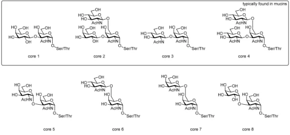
14‐16. These structures are divided into eight
classes according to their core motifs
17of which only core 1‐4 are frequently found in nature while
11
INTRODUCTION
core 5‐8 are rather uncommon (Figure 1). Elongation of the T
N‐antigen by the core 1 β‐1,3‐galactosyl transferase (core1 β3GalT) forms the core 1 motif, also called Thomsen‐Friedenreich‐ or T‐antigen.
Transfer of a β‐GlcNAc to the 6‐OH of GalNAc at the T‐antigen acceptor by the core 2 β‐1,6‐N‐
acetylglucosaminyl transferase I‐III (core 2 β6GlcNAcT I‐III) or the I‐branching enzyme (IGnT) generates the core 2 structure. Alternatively, the T
N‐antigen can be extended at C‐3 by the core 3 β‐1,3‐N‐
acetylglucosaminyl transferase (core 3 β3GlcNAcT) forming the core 3 structure, which in turn can be elongated at GalNAc C‐6 by the core 4 β‐1,6‐N‐acetylglucosaminyl transferase (core 4 GlcNAcT) or core 2 β6GlcNAcT II or IGnT to form the core 4 structure.
18‐19Figure 1: Overview of the O‐glycan core structures.
The obtained mucin‐type O‐glycan core structures can be elongated by alternating Gal or GlcNAc residues in a linear or branched manner, e.g. elongation by one or several Galβ1,3‐GlcNAcβ (LacNAc type‐1) or Galβ1,4‐GlcNAcβ (LacNAc type‐2) repeats. This occurs more often at the GlcNAcβ1‐6 residue of core 2 or core 4 structures and is catalyzed by a family of isoenzymes (β3GalT
20‐21, β4GalT
22‐25
, iGnT
26and IGnT
27‐28). Finally, the glycan chains can be sialylated, fucosylated, sulfated, methylated or acetylated
29, forming a considerable variety of glycan structures including histoblood group antigens such as the A, B or H or Lewis antigens (Le
a/b/SLe
a/bor Le
x/y/SLe
x/y, Figure 2)
30. Among the terminal modifications, sialylation and sulfation provide a negative charge to glycoproteins and fucosylation increase the hydrophobicity. On mucins these terminal modifications contribute dramatically to physical and biological properties
19. For instance, the mucosal glycobiome varies within different tissues depending on the different functional requirements of the mucosal surfaces and is altered in airway diseases and cancer as described in more detail in chapter 3.2
31, 18, 32.
12
INTRODUCTION
Figure 2: Selection of terminal antigenic determinants of mucin‐type O‐glycans. Shown are the histo blood group antigens H, A and B as well as the Lewis type antigens. Lea and Leb are mainly found in surface epithelium while Lex and Ley are commonly expressed in mucous cells.18, 30
The above described mucin‐type O‐glycosylation is usually found on proteins at the extracellular side of the cell plasma membrane being involved in important cell‐cell and cell‐matrix interactions. In contrast, another common group of O‐glycosylation, O‐GlcNAcylation, the transfer of a β‐GlcNAc monosaccharide to Ser or Thr residues of a protein, is an intracellular modification that usually takes place in the nucleus
33‐36, cytoplasm
37or mitochondria
38‐39. This is mediated by one single human enzyme, the O‐linked N‐acetyl‐glucosaminyl transferase (OGT)
37under consumption of a UDP‐GlcNAc molecule
40. In turn, β‐GlcNAc hydrolysis is mediated by the OGT counterpart O‐GlcNAcase (OGA)
41. Both enzymes are ubiquitously expressed and distributed in most mammalian tissues. As a consequence, O‐GlcNAc is one of the most abundant PTMs within the nucleocytoplasmic compartments of mammalian cells. However, to date only in one case mammalian O‐β‐GlcNAcylation has been observed at the extracellular side of the cell plasma membrane
42. However, on protozoa extracellular α‐GlcNAcylation seems to be a common modification
43. β‐O‐GlcNAcylation is a nutrient‐
responsive modification and is involved in the regulation of various processes
44like signal transduction
45, cell cycle
46‐47, gene expression
48‐49and proteasomal degradation
50‐51.
Unlike α‐GalNAc, the β‐GlcNAc modification is usually not elongated or further modified and thus exists as monomeric glycan. A specific characteristic of β‐O‐GlcNAcylation is to show some
β1,3
H
α1,2α1,2 α1,3β1,3
A
α1,2 α1,3β1,3
B
β1,3 α1,4
Le
aLe
bβ1,3 α1,4
α2,3
SLe
aα1,2 β1,3 α1,4
β1,4 α1,3
β1,4 α1,3 α1,2
α2,3 β1,4
α1,3
Le
xLe
ySLe
xtype‐1 type‐2
GlcNAc GalNAc Gal Fuc Sia
13
INTRODUCTION
resemblance to protein O‐phosphorylation. Both, β‐O‐GlcNAcylation and O‐phosphorylation take place on Ser and Thr (and probably Tyr) and often show rapid cycling, commencing at a higher rate than protein turnover
52‐53. In addition, the enzymes responsible for β‐GlcNAc cycling are regulated by O‐phosphorylation and β‐O‐GlcNAcylation
37like kinases and phosphatases
54. Further, cross‐talk of O‐
phosphorylation and β‐O‐GlcNAcylation is evident, as mammalian cells with attenuated O‐
GlcNAcylation show altered O‐phosphorylation levels
55, in some cases O‐GlcNAcylation could even be shown to be directly reciprocal to phosphorylation
56‐57. However, while phosphorylation could be linked to certain primary sequences, no such consensus motif has been identified for O‐GlcNAc, although many O‐GlcNAc sites contain nearby proline or valine residues
58‐59. Even though O‐
GlcNAcylation is distributed throughout the whole nucleocytoplasmic part of the cell, particularly high O‐GlcNAc levels are found for example on nuclear pore proteins
33, 60and the nuclear transcriptional regulatory machinery
36. These regulatory proteins, are known to contribute to oncogenic phenotypes, which in turn suggests an involvement of O‐GlcNAcylation in cancer. A prominent example is the c‐
myc oncogene, which is known to be O‐GlcNAc modified in its transactivation domain and represents the mutational hotspot in many Burkitt’s lymphomas patients
57, 61. O‐GlcNAcylation gains even more clinical relevance as it is hypothized
62, that a decreased glucose metabolism in aging neurons leads to reduced O‐GlcNAcylation of central cytosolic or nuclear proteins. This might then result in abnormal phosphorylation of many proteins, which eventually results in neurodegenerative diseases. Finally, high cellular glucosamine levels (hyperglycemia) seem to promote insulin resistance (diabetes type 2)
63‐64. This might be explained with the fact, that glucosamine is a precursor in the UDP‐GlcNAc formation, which might lead to hyper‐O‐GlcNAcylation of transcription factors or signaling proteins, which are involved in the insulin response
65. In a few recent studies it was further shown that many of the metabolic enzymes involved in glycolysis and oxidative phosphorylation are highly O‐GlcNAc modified, which probably is connected to functional regulation of these enzymes
66‐67.
3.2 Mucin glycopeptides
In mammals, the epithelial surfaces of for instance the respiratory, reproductive and gastrointestinal tracts are coated by a mucus layer, an aqueous gel comprising ions, proteins, glycoproteins and lipids.
This gel serves for lubrication and in addition, is a part of the innate immune system, that protects the
host against pathogens and toxins
68. The major constituent of the mucus layer are the secreted mucin
glycoproteins for instance MUC2, MUC5AC, MUC5B and MUC6
69that are covering the epithelial
tissues. The secreted mucins constitute a mass amount of 10‐50% (wt/wt) of the complete aqueous
networks, which are held together by numerous disulfide bonds
70. Below the mucus layer, the
14
INTRODUCTION
membrane mucins are covering the epithelial cells, for instance MUC1, MUC4 and MUC16. Due to their extended (rod‐like) conformation, these mucins are extended further from the membrane surface than most cell surface receptors, which enables a disruption of cell and pathogen adhesion
71‐72
. Further they play important roles in the organization of cells and their cell surface‐proteins (cell polarization)
73and are involved in a number of signaling events related to for instance cellular motility, growth
74‐75, adhesion
76and proliferation
77as well as T cell activation
78.
79Mucins exhibit a high and variable number of proline‐, serine‐ and threonine‐rich tandem repeats (VNTRs), which serve as scaffolds for dense O‐glycosylation leading to ‘bottle‐brush’ structures with a carbohydrate mass fraction of 50‐90 % with respect to the whole mucin glycoproteins. With the different degrees of glycosylation and the variable number (resulting in variable glycoprotein length), the mucin VNTRs influence the biophysical properties of the mucus
80‐82. The mucins are classified according to the MUC protein backbones which are encoded by over twenty different genes and every mucin gene product shows a characteristic and unique tandem repeat sequence. The mucin genes are abbreviated with MUC, followed by a number, which reflects the order that the proteins were cloned for the first time. According to these Human Genome Mapping conventions, the MUC1 gene, which codes for a membrane tethered mucin glycoprotein, was the first mucin gene to be cloned. The corresponding glycoprotein exhibits a large extracellular N‐terminal domain, comprising a signal sequence, which is responsible for mucin peptide insertion to the ER and translocation to the cytoplasm membrane. Further, the N‐terminal domain contains 25 to 125 tandem repeats
83with the sequence PAHGVTSAPDTRPAPGSTAP and a C‐terminal region consisting of a hydrophobic membrane‐
spanning domain as well as a short phosphorylated cytoplasmic tail that is involved in signaling events.
The membrane bound MUC1 glycoprotein is commonly expressed at the apical cell surface of normal epithelial tissues
84. Besides the function of mucins in protection and lubrication, antibody staining experiments with the complete mucus gel have suggested that MUC1 is involved in the tethering of the mucus gel to the mucosal surface
85‐86. Furthermore, MUC1 play many important roles in cell adhesion lectin interactions of pathogens
72, 87, immune cells and tumor cell metastasis
19, 32, 84. Additionally, the O‐glycan structure, site specificity and density and the number of MUC1‐VNTRs varies within different tissues and disease stages, which further influence the properties and functions of MUC1
88‐90.
Besides MUC1 further mucins have received ongoing attention by researchers due to their
involvement in chronic diseases: For instance increased expression of MUC4 has been linked to
pancreatic cancer
91. MUC5AC has been identified as marker for colorectal
92and pancreatic cancer
93,
15
INTRODUCTION
but altered MUC5AC levels have also been related to gastric cancer
94‐95. Decreased MUC5AC and MUC5B levels have been found in cystic fibrosis airways
96, while hypersecretion of these mucins have been found in the sputum of COPD patients
97. Finally, MUC5AC and MUC6 serve as receptors for Heliobacter pylori, which enables colonization of the human gastric epithelium and thus promotes gastric inflammation
98.
3.2.1 Mucin glycoproteins in airway diseases
Mucins play an important role in airway diseases like asthma, chronic obstructive pulmonary disease
(COPD), or cystic fibrosis (CF). If challenged with environmental toxins, allergens or infectious
pathogens, a healthy respiratory tract reacts with the activation of lung immune response mediators
of which some induce mucin hypersecretion by activation of a secretory cascade
19. These
hypersecreted mucins could entrap particles
99or bind receptors of inflammatory cells via MUC
domains or glycans
100. Typically, these hypersecretions decrease to baseline levels after a couple of
days, which might be connected to anti‐inflammatory mediators
101. However, patients with chronic
airway diseases show a permanent overproduction of the airway mucins MUC5AC and MUC5B
97caused by goblet cell hyperplasia and upon external insults, which mainly are triggered by bacteria
and virus infections (in 75% of the cases), acute attacks (exacerbations) take place which result in
shortage of breath and pulmonary obstruction. Every exacerbation leads to an advancement in
disease stage and an increase in frequency of the attacks, which eventually lead to death. Even if the
general symptoms in chronic pulmonary diseases are similar, the origin of these diseases vary. The
fundamental cause of asthma is not known, but a combination of genetic predisposition and
environmental exposure to inhaled substances that trigger allergy are considered major factors. One
theory is that an imbalance in the T helper type 2 (Th2) cellular cytokine response is involved in the
development of an asthma phenotype
102‐103. Cystic fibrosis is caused by mutations in the CFTR gene
104,
which leads to a malfunction or failure of the chloride channel protein, resulting in a chloride ion
imbalance between extra‐ and intracellular fluid. In consequence, the osmotic pressure leads to a
water drain from the extracellular fluid, which increases its viscosity and eventually clogs the lung
capillaries. Development of COPD is caused by repetitive insults of the airways triggered by extensive
exposure to tobacco smoke
105or in low income countries by pollution by biomass fuels from cooking
and heating
106. In addition to hypersecretion, characteristic changes in O‐glycosylation can be
attributed to pulmonary infections and inflammations. For example, increased fucosylation, sialylation
and sulfation has been found in secreted mucins from CF patients
107‐108, while the membrane bound
mucins of these patients have been found to exhibit decreased sialylation
109‐111. Increased expression
16
INTRODUCTION
of SLe
xepitopes, GlcNAc‐6‐sulphate and Gal‐3‐sulfate has been found in sputum of CF and COPD patients
112, which is in accordance with an observed shift from α2,6‐sialylation to α2,3‐sialylation on MUC5B from CF patients
113. Glycans on mucins often serve as ligands for invading bacteria or viruses and the altered glycosylation pattern might promote infections.
Pseudomonas aeruginosa, the most prominent pathogen responsible for morbidity and mortality in CF, for instance often recognizes SLe
xand 6‐sulfo‐SLe
x114
. To date, a few adhesins are known which serve as virulence factors for P. aeruginosa by mediating the bacterial adhesion to glycan structures.
For example the type‐4 pilus was found to recognize a GalNAcβ(1‐4)Gal disaccharide motif as found internally in fucosylasialo‐GM1 and asialo‐GM1 as well as terminally in asialo‐GM2
115‐116. LecB, which is secreted by P. aeruginosa, binds to fucosylated oligosaccharides and in particular to Le
bepitopes
117. The flagellar cap protein, FliD, has been found to mediate P. aeruginosa adhesion, SLe
xand Le
xepitopes seem to be involved in these interactions
118. The protein PcrV is a structural component of the type III secretion system of P. aeruginosa and serves as a virulence factor. This was demonstrated by neutralization experiments
119which led to significantly reduced airway inflammation. Glycan structures are believed to be involved in PcrV adhesion, but to date the specific ligands are still unknown.
Adherence of Haemophilus influenzae, the most common pathogen involved in COPD, proceeds via fimbriae. Here, its subunit HifA could be identified to contain the adhesive domain for mucin binding but also in this case the underlying glycan ligand motifs are not identified yet.
For Streptococcus pneumoniae the adhesin PsrP (pneumococcal serine‐rich repeat protein) was found to bind keratin10 in lung cells
120‐121. This particular binding was shown to be independent of a lectin‐
carbohydrate interaction, however S. pneumoniae has been found to interact with mucin glycoproteins and it is reasonable to expect that binding to glycans take place by other adhesin proteins.
Given the crucial role of adhesins in in the process of bacterial infections, a deeper understanding of the corresponding lectin specificities could pave the way for the development of antiadhesive drugs as part of an efficient pathogen‐specific antivirulence therapy.
The above described bacterial adhesion via cell surface glycoproteins and glycolipids is further
supported by sialidases (also known as neuraminidases), which are expressed by the major airway
disease‐related pathogens P. aeruginosa, H. influenzae and S. pneumoniae
122‐126. These enzymes
catalyze the hydrolysis of terminal sialic acid residues, which can be metabolized and serve as nutrition
for the invading pathogen or alternatively, be implemented to the bacterial glycocalyx to mimic the
17
INTRODUCTION
host sialylation pattern and therefore deceive the immune system. Further, the host sialic acid removal reveals underlying glycan structures which sometimes can be recognized by corresponding bacterial cell surface receptors (adhesins)
127. The bacterial sialidases are also involved in biofilm formation
122, 125. In this case the desialylation results in physical changes of charge and hydrophobicity of the environment. Being involved in numerous virulence mechanisms, sialidases represent important and promising therapeutic targets. Knowledge about the substrate specificities of different sialidases would be of high value for the development of inhibitors, however past studies on sialidases were based on bacterial genetic analyses and specific sialidase substrate specificities remain to be
elucidated.
1273.2.2 Mucins in cancer
In a plethora of epithelial cancers aberrant mucin O‐glycosylation and altered mucin expression levels have been observed, for instance in breast‐, colon‐, gastric and lung cancer
70. By influence on cell‐cell‐
and cell‐matrix‐interactions as well as cell‐recognition, trafficking and signaling, changes in O‐
glycosylation have an impact on tumor progression and metastasis
128. A common characteristic of O‐
glycosylation in epithelial cancer tissue is that the glycans are truncated and short saccharides are formed with increased terminal sialylation compared to glycans in corresponding non‐cancerous tissues
129‐130. On the mucin MUC1 which is highly overexpressed on epithelial cancer cells, the aberrant glycosylation additionally lead to an exposure of the immunodominant peptide backbone and specific glycopeptide epitopes are formed. This in turn enables induction and antibody recognition of unique glycopeptide structures, as exploited in the detection of aberrant presentation of MUC1 in breast carcinomas by SM‐3 and DF‐3P
84, 131‐132. Common glycans on cancer mucins are T
N‐, Sialyl‐T
N‐, T‐, Sialyl‐
T‐ and Le
x‐antigens
133, whose dominance has several reasons. For example mutations in Cosmc, a
chaperone responsible for the correct folding of the core‐1‐galactosyltransferase (C1GalT)
134, leads to
the expression of the T
N‐antigen in tissues with high core 1 and core 2 expression
135. Alternatively, if
α‐N‐acetylgalactosaminide α‐2,6‐sialyltransferase 1 (ST6GalNAc‐I) is expressed, mutation of Cosmc
results in formation of Sialyl‐T
Ninstead. Another reason for high T
N‐, ST
N‐, T‐ and ST‐antigen expression
is the stimulation of the proto‐oncogene Src, which regulates trafficking between Golgi and
Endoplasmatic Reticulum. While mucin‐type O‐glycosylation is mainly initiated in the Golgi, activation
of Src results in the relocation of ppGalNAc‐Ts to the ER
136. The resulting increased T
N‐antigen density
on the affected mucins blocks further glycosylation by C1GalT and C2GlcNT, which would facilitate the
generation of core 1 and core 2 otherwise. However, the most common reason for aberrant mucin O‐
18
INTRODUCTION
glycosylation lies within altered expression levels of the different glycosylatransferases. These changes
are versatile and dependent on the different cancerous tissues.
137On the glycopeptide level an overexpression of Mucins has been found in tumors, especially in adenocarcinomas. This correlates with a high risk of metastasis and as a consequence to lowered chances of survival. The mucin overexpression has been found to influence tumor cells in numerous approaches, for instance by cell growth, transformation, invasion, differentiation and adhesion. Here, MUC1 is a very prominent example, being massively overexpressed in cancer and not restricted to protein localization at apical surfaces
137. Instead it is additionally found in other sub‐cellular compartments and on the whole surface of tumor cells
84.
3.2.2.1 The role of galectin‐3 in cancer
MUC1 overexpression plays a key role in the galectin mediated tumor survival and metastasis.
Galectins are a family of lectins, which share affinity for β‐galactosides
138. Galectins are not membrane‐bound and are localized in the nucleus, the cytosol as well as secreted in the extracellular space. They are involved in a number of cellular functions, for instance in cell adhesion to the extracellular matrix, signaling, cell cycle progression or apoptosis
139. One of the most‐studied galectins is galectin‐3, which shows altered expression states in various carcinomas compared to normal cells
140. The high amount of galectin‐3 secreted from tumor cells has shown to be immunosuppressive towards T‐ and B‐lymphocytes by induction of apoptosis of the affected immune cells upon galectin‐3 exposure (Figure 3, mechanism A)
141‐145. Here, a high significance of lymphocyte O‐glycan ligands have been found for the galectin‐3 recognition
146. In contrast tumor cells are protected from galectin‐3 induced apoptosis by altered glycosylation. After sialidase treatment of colon carcinoma cells and subsequent contact with galectin‐3, apoptosis was induced on those cells
147. In these cells, the galectin‐3 receptors were found to be heavily α2,6‐sialylated. Consequently, it was proposed, that overexpression of galectin‐3 is advantageous for the secreting tumor cells in several ways. It enables immune evasion from B‐ and T‐cells via induction of apoptosis on those cells, the tumor itself is protected from this mechanism (Figure 3, mechanism B). In addition, the interplay of extracellular galectin‐3 with overexpressed, altered and membrane‐bound MUC1 supports the metastasis of primary tumors.
Cancer associated MUC1 has been found to be a natural target for galectin‐3
148via the tumor‐
associated T‐antigen
149. It is proposed, that overexpressed MUC1 on circulating tumor cells is captured by galectin‐3, leading to clustering of MUC1. This might reveal shorter cell adhesion molecules on the cell surface, which are then able to interact with corresponding binding partners on endothelial cells.
This facilitates settling of the circulating tumor cell to the endothelium and in consequence mediates
19
INTRODUCTION
metastasis (Figure 3, mechanism C)
149. Alternatively, circulating cells can interact with each other, forming circulating tumor cell clusters. This cell adhesion protects the aggregated tumor cells from anoikis, an induced apoptosis by the loss of cell‐cell or cell‐matrix interactions
150‐151. The tumor cell clusters can – like the single tumor cells – settle to the surface of endothelial cells (Figure 3,
mechanism D)
152.
153The mechanisms of metastasis promotion are also shared by other galectins
154. Furthermore, the galectin‐3 expression is enhanced by the cytosolic part of MUC1 via stabilization of the galectin‐3 mRNA transcript. Accordingly, the tumor characteristic MUC1 overexpression directly correlates with the galectin‐3 expression. In turn, galectin‐3 binds to MUC1 N‐glycosylation, bridging MUC1 to ERB1, which integrates MUC1 to EGFR signaling
155. Summarized, galectins play an important role in the tumor progression of many carcinomas and are therefore an interesting subject for detailed studies of lectin/carbohydrate interactions.
Figure 3: Proposed effects of galectin‐3 on tumor immune escape and metastasis.
Primary tumor
Overexpression of galectin‐3, MUC1 and tumor‐associated antigens (TN‐, T‐, STN‐, ST‐antigen)
Secondary tumor
Strong adherence via integrins
Circulatinig galectin‐3
Apoptotic T‐cell
B
Blocking of galectin‐3 mediated tumor cell apoptosis by extensive 2,6‐sialylation of surface receptors
Metastasis
A
Galectin‐3 mediated apoptosis
Tumor Immune escape
C
Exposure of cell adhesion molecules by MUC1 cell surface polarization
D
Galectin‐3 induced cell aggregation prevents suspension‐induced apoptosis (anoikis)
Endothelial cells
Galectin‐3 (monomer) Galectin‐3 (pentamer) MUC1
Cell adhesion molecules Integrin
2,6‐sialylated β1‐integrin