Nesprin-1 is required to maintain genomic stability and prevent
tumorigenesis
Inaugural-Dissertation
zur
Erlangung des Doktorgrades
der Mathematisch-Naturwissenschaftlichen Fakultät der Universität zu Köln
vorgelegt von
Ilknur Sur
aus Istanbul, Türkei Köln 2013
Berichterstatter: Prof. Dr. Angelika A. Noegel
Berichterstatter: Prof. Dr. Siegfried Roth
Prüfungsvorsitzender: Prof. Dr. Peter Nürnberg
Tag der mündlichen Prüfung: 22.01.2014
The present research work was carried out from November 2010 to November 2013 at the Centre for Biochemistry, Institute of Biochemistry I, Medical Faculty, University of Cologne, Cologne, Germany, under the supervision of Prof. Dr.
Angelika A. Noegel.
Die vorliegende Arbeit wurde in der Zeit von Nov 2010 bis Nov 2013 unter der Anleitung von Prof. Dr. Angelika A.Noegel am Biochemischen Institut I der Medizinischen Fakultät der Universität zu Köln angefertigt.
I am dedicating this thesis to my beloved family for giving me the strength to keep working to make my dreams come true, and especially to my grandmother, Ayşe Sur, who always prays for me. I wish she were here to see her prayer and our dream come true.
.
The letters of alphabet won`t be enough to express my deep sense of gratitude (bold g) to Prof. Dr. Angelika Noegel for giving me the valuable opportunity to study in her department. I am fortunate to know her as a great scientist and I could not achieve so much today without her invaluable guidance, encouragement and insights throughout my PhD. I owe her thousands of thanks for being busy with my thesis not only within working hours but also after work.
I would like to thank the members of my thesis committee Prof. Dr. Siegfried Roth and Prof. Dr. Peter Nürnberg. My deepest thanks belong to Dr. Muhammad Sajid Hussain (Sila-Saira) for his continuous encouragement and support from the beginning of my first day to this time. He is a great instructor, but more importantly a great friend who introduced me to plenty of new things. I wish to thank Dr. Sascha Neumann for giving me the experimental introduction to the very interesting world of nuclear envelope. A special word of thanks goes to Dr. Vivek Peche who waited for me in the institute on my first day. It was very meaningful for me. Thank you! Dr.
Raphael Rastetter is also acknowledged for teaching us the cell migration experiments and inviting all of us to his summer house.
I am very thankful to all Lab14 members, Atul (bench mate), Kosmas (CAP2-/-), Napo (Rack1), Karthic (what are you doing?), Salil(i), Ping (SUN lady), Pranav (Nesprin-2), Tanja (Taran-Yuna), Rashmi, and Eva (Jouana) for working hand by hand with me. I would also like to acknowledge Bärbel, Sonja, Brigitte for providing laboratory reagents. Special thanks to Rosi (my cherry) and Maria (sunshine) for giving the absolute access to the confocal microscopy. I am indebted to Martina and Rolf for their technical support. I give special thanks to Berthold (hardworking guy) for never shouting at me, or breaking my heart during these three years. I also want to thank Budi and Gudrun for their computing assistance and providing me a nice
environment during my thesis writing. I wish to thank Eike and Oliver for repairing the equipment (also my earrings). I am indebted to Dörte Püsche for her kind support concerning the official things and making the life easy in Cologne where it is difficult to find a residence. I would like to express my sincere thanks to Liu, Xin (Xueli), Kalle, Bhagyashri (Arna), Juliane, Christoph, Jaqueline, Tim, Emrah, Ilyas, Jana, Anne, Susanne, Natalie, Marja, Qiuhong, Ramesh, Khalid, Linh, Sze Man, and Margit for having shared with me the many nice moments of these three years. I am deeply grateful to my all friends, Dr. Sandra Herzog (Axel), Ping, Kurchi (Unni), Sandra S (Andi), Anja, Clara, Carmen (Michael), and Aseel who helped me at any time and provided me with their pleasant company also outside the institute. Lunch time will never be the same anywhere else. I give special thanks to Sandra (Andi) for making our office like a flower garden. I would like to extend my thanks to Dr. Anja Ziemann (endnote), Dr. Jan Matthias (RT-PCR) for their heIp without hesitation.
Thanksgiving to my almighty God who is the source of my dreams, life, and strength.
I am forever grateful to Prof. Dr. Orhan Arslan for his support on making my dreams come true. Last but not least, I wish to thank my family, Safigülü Sur, Riza Sur, Cihan Sur, Ali Sur (x2), Metin Yilmaz, Mahmut Demir, and Zeki Sur. They taught me responsibility, honesty, and dedication and they gave me the most unconditional love that could exist, they always trusted me no matter what. Above all, I would like to thank my grandparents Aynur Yılmaz (died before I was born, heart attack), Ayşe Sur (stroke), Mehmet Sur (diabet), and Mustafa Nuri Yılmaz (prostate cancer) for their prayers. When I was 7 years old, my grandmother (Ayse Sur) had a stroke. Day by day she was getting worse; she wanted me to become a doctor and find a cure to treat diseases. I promised her to become a doctor one day. Therefore, this thesis is dedicated to her (especially) and all my family. I hope, people will not suffer owing to cancer, and age related-diseases.
Abbreviations...6
Summary ...8
1. Introduction...1
1.1 Nuclear envelope and LINC complex ... 1
1.1.1 INM components of LINC complexes, and their functions ... 2
1.1.2 Nesprins, ONM components of LINC complexes and their functions ... 4
1.1.3 Nesprin-1 ... 8
1.2 The LINC complex in human diseases and its cancer connections ... 10
1.3 DNA repair pathways and mechanisms ... 14
1.3.1 Non-homologous end-joining (NHEJ) pathway ... 15
1.3.2 Nucleotide excision repair ... 17
1.3.3 Mismatch repair network ... 19
1.4 Aim of the project ... 21
2. Results...22
2.1 Nesprin-1 interactions ... 22
2.1.1 Interactions of N-terminal Nesprin-1 spectrin repeats ... 22
2.1.2 The C-terminus of Nesprin-1 interacts with Nesprin-2 ... 24
2.1.3 Nesprin-3 is able to recruit vimentin to the nucleus ... 25
2.2 Nesprin-1 role in tumorigenesis ... 27
2.2.1 Nesprin-1 isoform expression in cancer cell lines ... 27
2.2.2 Hep3B and Huh7 have nuclear shape defects and alterations in components of the nuclear envelope ... 30
2.2.3 The centrosome-nucleus distance is increased in Hep3B and Huh7 cells 34
2.3 Loss of Nesprin-1 ... 36
2.3.1 Knock down of Nesprin-1 elicits changes that are observed in cancer cell lines ... 36
2.3.2 The centrosome-nucleus distance is increased in Nesprin-1 KD cells ... 42
2.3.3 Loss of Nesprin-1 leads to cytoskeletal alterations ... 44
2.3.4 Senescence is increased in Nesprin-1 knock down fibroblasts ... 46
2.4 Nesprin-1 and DNA damage response (DDR) network ... 47
2.4.1 N-terminal Spectrin repeats of Nesprin-1 interact with DNA mismatch repair proteins MSH2 and MSH6 ... 47
2.4.2 Loss of Nesprin-1 affects the DDR network ... 57
3. Discussion ...63
4. Materials and Methods ...70
4.1 Materials ... 70
4.2 Molecular biological methods ... 74
4.2.1 Primer design ... 74
4.2.2 Annealing of oligonucleotides ... 75
4.2.3 Digestion of pSHAG-1 vector ... 75
4.2.4 Ligation and cloning procedure ... 76
4.2.5 DNA Midi/Maxi preparation ... 77
4.2.6 RNA isolation and cDNA generation for quantitative RT-PCR analysis .... 77
4.3 Protein chemical and immunological methods ... 79
4.3.1 Protein extraction from E.coli and mammalian cells ... 79
4.3.2 Western blotting... 80
4.3.3 Recombinant protein purification and pull downs ... 81
4.3.4 Co-immunoprecipitation (Co-IP) ... 82
4.3.5 Immunofluorescence ... 83
4.4 Cell culture and transfections ... 84
4.5 Cell biological assays ... 85
4.5.1 Heat stress experiments ... 85
4.5.2 Senescence-associated β-galactosidase ... 85
4.5.3 Cell migration assay ... 85
4.5.4 DDR assays ... 86
5. References ...87
Abbreviations
Abbreviations
Aa Amino acid
ABD Actin-binding domains
cDNA Complementary DNA
CH Calponin homology
Co-IP Co-Immunoprecipitation
Da Dalton
DAPI 4’-6-diamidino-2-phenylindole DMEM Dulbecco`s modified Eagle medium
DMSO Dimethyl sulfoxide
DNA Deoxyribonucleic acid
dNTP Deoxynucleoside triphosphate DTT Dithiothreitol
EDTA Ethylene diamine tetraacetic acid ECL Enhanced chemiluminescence EDMD Emery Dreifuss Muscular Dystrophy F-actin Filamentous actin
FBS Fetal bovine serum
GAPDH Glycerinaldehydephosphate dehydrogenase GFP Green fluorescent protein
GST Glutathione S-transferase
HEPES 4-(2-hydroxyethyl)-1-piperazineethanesulfonic acid HGPS Hutchinson-Gilford progeria syndrome
IgG Immunglobulin G
IPTG Isopropyl-β-Dithiogalactopyranoside
IF Immunofluorescence
IP Immunoprecipitation
KASH Klarsicht, ANC1 and SYNE1 homology
KD Knock down
KO Knock out
mRNA messenger ribonucleic acid
MS Mass spectrometry
Nesprins Nuclear envelope spectrin repeat proteins NPC Nuclear pore complex
PBS Phosphate buffered saline PCR Polymerase chain reaction PFA Paraformaldehyde
pH negative decadic logarithm of protein concentration PIC Proteinase inhibitor cocktail
rpm Rotation per minute
qRT-PCR Quantitative real time PCR
SDS-PAGE Sodium dodecyl sulfate polyacrylamide gel electrophoresis
shRNA Short hairpin RNA
SR Spectrin repeats
SUN Sad1 and UNC-84
TBS Tris buffered saline UV Ultraviolet light
WB Western blot
WT Wild type
X-gal 5-bromo-4-chloro-3-indolyl-D-galactopyranosid
Summary Summary
Nuclear envelope (NE) proteins have fundamental roles in maintaining nuclear structure, cell signaling, chromatin organization and gene regulation, and mutations in genes encoding NE components were identified as primary cause of a number of age associated diseases and cancer. Nesprin-1 belongs to a family of multi-isomeric NE proteins that are characterized by spectrin repeats. Our results imply interactions between spectrin repeats and an interaction of Nesprin-1 with Nesprin-2.
Furthermore, we analysed NE components in various tumor cell lines and found that Nesprin-1 levels were strongly reduced associated with alterations in further NE components. By reducing the amounts of Nesprin-1 by RNAi mediated knock down we could reproduce those alterations in mouse and human cell lines pointing towards a key role for Nesprin-1 in the maintenance of nucleus morphology, centrosome positioning, nuclear membrane structure, cytoskeleton organization, and cellular senescence. In a search for novel Nesprin-1 binding proteins we identified MSH2, MSH6, and DDB1 proteins of the DNA damage response pathway as interactors. We found alterations in the mismatch repair pathway in cells with lower Nesprin-1 levels.
We also noticed an increased number of γH2AX foci in the absence of exogenous DNA damage as was seen in tumor cells. The levels of phosphorylated kinases Chk1 and 2 were altered in a manner resembling tumor cells and the levels of Ku70 were low and the protein was not recruited to the DNA after HU treatment. Our findings indicate a role for Nesprin-1 in the DNA damage response pathway and propose Nesprin-1 as novel regulator of tumorigenesis and genome instability. Loss of Nesprin-1 might play a significant role in cancer progression.
Zusammenfassung
Kernhüllenproteine haben eine wesentliche Rolle bei der Aufrechterhaltung der Kernstruktur, Chromatinorganisation, Genregulation und bei Signaltransduktionsprozessen, und Mutationen in Genen, die für Kernhüllenkomponenten kodieren, wurden als primäre Ursache für eine Reihe von Erkrankungen und Krebs identifiziert. Nesprin-1 gehört zu einer Familie von multi- isomeren Proteinen der Kernhülle, die von Spectrin Wiederholungen gekennzeichnet sind. Unsere Ergebnisse zeigen, dass Nesprin-1 mit Nesprin-2 über Wechselwirkungen ihrer Spectrin Repeats miteinander interagieren. Weiterhin haben wir die Kernhüllenkomponenten in verschiedenen Tumor-Zelllinien analysiert und dabei festgestellt, dass die Nesprin-1-Spiegel stark reduziert und mit Veränderungen in weiteren Kernhüllenproteinen und anderen Kern-assoziierten Prozessen verbunden waren. Durch die Reduzierung der Mengen an Nesprin-1 durch einen RNAi-vermittelten Knockdown konnten wir diese Veränderungen in murinen und humanen Zelllinien reproduzieren. Diese Ergebnisse verweisen auf eine Schlüsselrolle für Nesprin-1 bei der Aufrechterhaltung der Kernmorphologie, Zentrosomenpositionierung, Struktur der Kernmembran, Organisation des Cytoskeletts und der zellulären Seneszenz. Bei der Suche nach neuen Nesprin-1- bindenden Proteinen identifizierten wir MSH2, MSH6 und DDB1 als Interaktionspartner, die Proteine von DNA-Reparaturwegen sind. In Übereinstimmung damit haben wir Veränderungen im Mismatch-Reparaturweg in Zellen mit niedrigen Nesprin-1-Spiegeln beobachtet. Wir haben auch eine erhöhte Anzahl von γH2AX Foci in Abwesenheit von exogenen DNA-Schäden festgestellt, wie es in Tumorzellen gesehen wird. Die Mengen der phosphorylierten Kinasen Chk1 und 2, die an Kontrollpunkten im Zellzyklus auftreten, waren in einer ähnlichen Weise
Zusammenfassung
in Tumorzellen verändert und die Mengen an Ku70 waren niedrig und das Protein wurde nicht an die DNA nach HU Behandlung rekrutiert. Unsere Ergebnisse zeigen eine Rolle für Nesprin-1 im DNA-Reparaturweg und schlagen Nesprin-1 als neuartigen Regulator für Tumorentstehung und Genominstabilität vor. Der Verlust von Nesprin-1 könnte eine wichtige Rolle in der Tumorprogression spielen.
1. Introduction
1.1 Nuclear envelope and LINC complex
The genome is contained within the nucleus in eukaryotic cells and is separated from the cytoplasm by a selective barrier, the nuclear envelope (NE). The proteins of the NE regulate the nucleo-cytoplasmic traffic and connect the nucleoplasm to the cytoplasm (Maraldi et al., 2010; Shimi et al., 2012). The NE consists of an outer (ONM) and inner nuclear membrane (INM) which enclose the perinuclear space (PNS). The ONM is contiguous with the endoplasmic reticulum, the INM contacts the nuclear lamina and chromatin (Figure 1).
Figure 1: The structure of the nuclear envelope (NE). The NE is a barrier separating the nucleus from the cytoplasm and consists of ONM and INM, nuclear pore complexes (NPC), proteins of the INM, ONM, lamins and other proteins. ABD:
Actin-binding domain; PBD: Plectin-binding domain. This model was taken from the lab website of Dr. Didier Hodzic. The question mark points out unknown functions of these connections.
Nuclear pore complexes (NPC) are inserted into the NE and connect the nucleus with the cytoplasm. The protein composition of the NE is complex as more than 100
Introduction
proteins have been described and differs between the ONM and INM (Schirmer et al., 2003). The NE proteins are involved in a variety of cellular processes including genome organization, gene expression and stability (Therizols et al., 2006; Chow et al., 2012).
An important component of the NE is the LINC (linker of nucleoskeleton and cytoskeleton) complex which connects the nucleus with the cytoskeleton. The LINC complex is present in a wide variety of organisms including amoebae, yeast, worms, flies, vertebrates, and plants (Schneider et al., 2008; Schulz et al., 2009; Graumann and Evans, 2010; Starr and Fridolfsson, 2010). The central components of mammalian LINC complexes are SUN (Sad1p, UNC-84) domain proteins and KASH- domain containing proteins, the Nesprins (Nuclear envelope spectrin repeats), which connect to Emerin, Lamins and chromatin at the nucleoplasmic side and to F-actin, microtubules, intermediate filaments and plectin at the cytoplasmic side (Figure 1) (Padmakumar et al., 2005; Crisp et al., 2006).
1.1.1 INM components of LINC complexes, and their functions
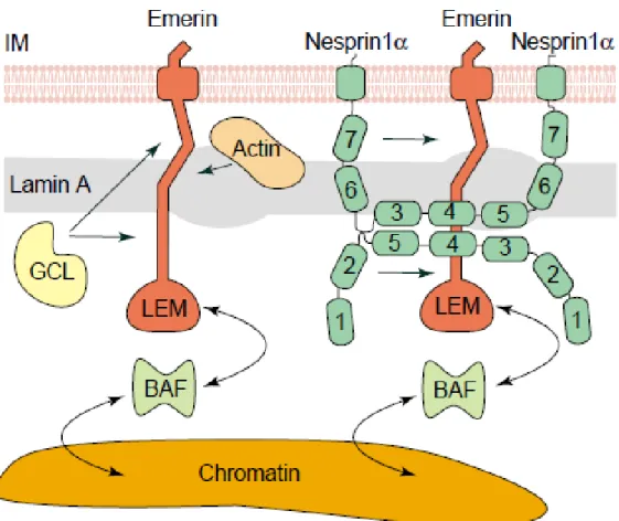
Among the first INM components recognized were members of the LEM domain family of proteins, which are named for the founding members, LAP2, Emerin and MAN1 (Figure 2) (Hetzer et al., 2005). The LEM domain is composed of a motif of approximately 40 amino acids that mediates binding to BAF (barrier-to- autointegration factor) which is an abundant chromatin-associated protein (Lin et al., 2000; Laguri et al., 2001; Shumaker et al., 2001). LAP2 (lamina associated polypeptides 2), one of the LEM domain proteins, has several isoforms in mammals (Berger et al., 1996). Specifically, LAP2β is the most ubiquitous LAP2 isoform and interacts with chromatin and Lamin B via a specific region at its C-terminus.
Emerin is a 254 amino acid protein and has an N-terminal LEM-domain. It was the first NE protein which was linked to a human disease, Emery-Dreifuss muscular dystrophy (Bione et al., 1994) and is involved in several protein–protein interactions (Cartegni et al., 1997; Squarzoni et al., 1998; Liu et al., 2003). Emerin plays key roles in signal transduction, chromatin organization and gene expression. Additionally, Emerin links centrosomes to the nuclear envelope via a microtubule association (Holaska et al., 2004; Markiewicz et al., 2006; Salpingidou et al., 2007).
Figure 2: Illustration of LEM interactions. Emerin is anchored at the INM and interacts with lamin A, GCL (germ-cell-less), BAF, actin and a Nesprin-1α dimer. The seven SR domains in Nesprin-1α are numbered 1–7, beginning at the N-terminus.
The image is not drawn to scale (Bengtsson and Wilson, 2004).
LEM domain proteins have also been linked to nuclear shape (Lammerding et al., 2005) and transcriptional regulation (Nili et al., 2001) as well as signaling cascades (Pan et al., 2005; Markiewicz et al., 2006).
Introduction
The nuclear lamina underneath the INM is a network of type V intermediate filaments, and subdivided to A- and B-type lamins (Gerace et al., 1978; McKeon et al., 1986).
All A-type Lamins, A, AΔ10, C, and C2 are encoded by the LMNA gene (Worman and Bonne, 2007). By contrast, B-type Lamins 1 and 2 are encoded by LMNB1 and LMNB2, respectively (Worman and Bonne, 2007). B-type LaminB3 is a splice variant of the LMNB2 gene (Furukawa and Hotta, 1993). Several studies suggested that Lamins participate in functions ranging from nuclear shape and stability to replication, transcription and splicing (Moir et al., 2000; Schirmer et al., 2001; Kumaran et al., 2002; Lammerding et al., 2004).
SUN proteins are prototypical type-II transmembrane proteins of the INM components of the LINC complex with their N-terminus facing the nucleoplasm, and a C-terminal conserved SUN domain localizing in the perinuclear space (PNS) between the INM and ONM. At the nuclear side they interact with Lamins (Padmakumar et al., 2005; Crisp et al., 2006; Haque et al., 2006). To date, five SUN proteins have been identified; SUN1 (UNC-84A), SUN2 (UNC-84B), SUN3, SUN4 (SPAG4), and SUN5 (SPAG4L). Remarkably, SUN proteins play important roles in genome stability and nucleus centrosome coupling (Zhang et al., 2009; Lei et al., 2012).
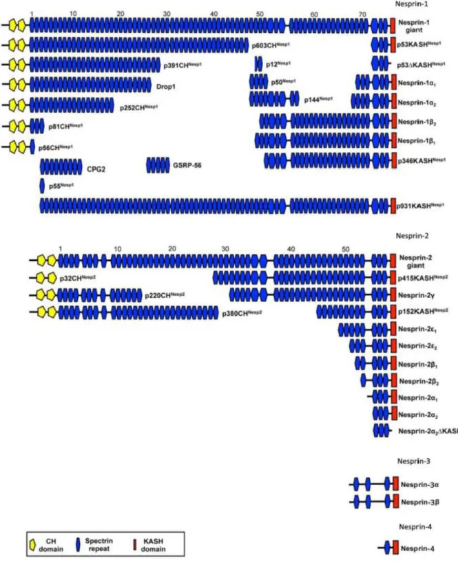
1.1.2 Nesprins, ONM components of LINC complexes and their functions Nesprins are type II transmembrane proteins, which connect the nucleus with the cytoskeleton. Nesprins localize to both nuclear membranes and have evolutionarily conserved orthologs in lower organisms including Schizosaccharomyces pombe (Kms1), Dictyostelium discoideum (interaptin), Caenorhabditis elegans (ANC-1, ZYG- 12 and UNC-83), and Drosophila melanogaster (Msp-300). To date, four Nesprins have been described (Nesprin-1, Nesprin-2, Nesprin-3, Nesprin-4). They are
encoded by separate genes (SYNE1, SYNE2, SYNE3, SYNE4) that give rise to multiple isoforms (Figure 3.1). Nesprins have a varying number of SR domains, each of which consists of approximately 106 residues that form a triple-helical bundle. SR domains facilitate protein-protein interactions, crosslink actin and microtubules, and function as molecular scaffolds or stabilizers (Rajgor et al., 2012).
The giant isoforms of Nesprin-1 (1 MDa) and Nesprin-2 (800 kDa) are greatly homologous to one another and share an N-terminal actin-binding domain (ABD) made from two calponin homology domains thereby linking the NE to the actin cytoskeleton (Zhen et al., 2002; Padmakumar et al., 2004). On the other hand, Nesprin-1 and Nesprin-2 can bind to microtubule motor proteins, kinesin and dynein, through specific spectrin repeats (Zhang et al., 2009; Schneider et al., 2011b; Yu et al., 2011). Nesprin-3 and Nesprin-4 are much smaller and lack N terminal ABDs.
Nesprin-3 interacts with plectin which provides a link to the intermediate filament system, Nesprin-4 can associate with kinesin-1 which establishes a link to microtubules (Wilhelmsen et al., 2005; Roux et al., 2009). In the LINC complex, Nesprins bind through their C-terminal luminal amino acids to the C-terminal SUN domain of SUN proteins. The structure of this complex has been recently solved and the presence of a trimer revealed (Sosa et al., 2012; Wang et al., 2012; Rothballer et al., 2013).
Introduction
Figure 3.1: Scheme of Nesprin isoforms. Nesprins share common structural features, a N-terminal calponin homology (CH) domain (yellow) and a C-terminal KASH domain (red) and a spectrin repeat containing rod (blue). Each Nesprin is encoded by a single gene that gives rise to many different isoforms. This Figure was taken from (Rajgor et al., 2012) and modified.
On the other hand, Nesprins can form self-interactions via their SRs and the ABD of Nesprin-1 and Nesprin-2 was shown to interact with N-terminal spectrin repeats of
Nesprin-3 (Mislow et al., 2002a; Lu et al., 2012; Taranum et al., 2012a). Based on these binding abilities, Nesprins enable a more orchestrated protein network along the nuclear envelope (Figure 3.2).
Figure 3.2: Schematic diagram of Nesprin interactions at the outer nuclear membrane surface. Nesprins form self-interactions via their SRs. The ABDs of Nesprin-1/-2 interact with F-actin, N-terminal Nesprin-1 spectrin repeats interact with Nesprin-3 (modified from (Taranum et al., 2012a)).
Due to the establishment of nuclear-cytoskeletal connections, Nesprins play key roles in biologically important functions and this is supported by several studies (Table 1).
Especially, Nesprin-1 and Nesprin-2 contribute to nuclear shape and position of the nucleus (Grady et al., 2005; Zhang et al., 2005; Kandert et al., 2007; Luke et al., 2008; Puckelwartz et al., 2009). A recent report also indicated that Nesprin-3 regulates cell morphology and cell migration (Morgan et al., 2011; Khatau et al., 2012). On the other hand, in Nesprin-4 knock out (KO) mice, nuclear position was changed in outer hair cells, thereby leading to hear loss (Horn et al., 2013). More recently, the effects of Nesprin-1, Nesprin-2, and Nesprin-3 on centrosome positioning have been reported (Zhang et al., 2009; Morgan et al., 2011).
Introduction
Table 1: Interactions of Nesprins with cellular components and functions of these connections. Nesprins have key roles in many aspects of cell functions due to their nuclear-cytoskeletal connections (modified from (Mellad et al., 2011)).
Connection Nesprin Function
Actin F-actin Nesprin-1 Nuclear positioning, Nesprin-2 Mechanotransduction Meckelin Nesprin-2 Ciliogenesis
IF Lamin A Nesprin1/2 Chromatin organization,
NE architecture Plectin Nesprin-3 NE-IF coupling
Microtubules Kinesin-1 Nesprin-2/4 Nuclear migration, polarity Kinesin-2 Nesprin-1 Vesicular transport
Dynein Nesprin-1/2 Nuclear migration, polarity Dynactin Nesprin-1/2 Nuclear migration, polarity
1.1.3 Nesprin-1
The human Nesprin-1 locus (SYNE1) at chromosome position 6q25 has 147 exons that encode up to 8,797 residues (Padmakumar et al., 2004). Nesprin-1 is a ~1 MDa protein with 74 predicted spectrin repeats (Figure 3.1). There are several isoforms for Nesprin-1 with many names including Syne-1 (Apel et al., 2000), Drop1 (Marme et al., 2008), GSRP56 (Kobayashi et al., 2006), MSP300 (Rosenberg-Hasson et al., 1996), Myne-1 (Mislow et al., 2002b), Enaptin (Padmakumar et al., 2004), CPG2 (Nedivi et al., 1996), ANC-1 (Starr and Han, 2002). One of the small isoforms, Nesprin-1α, localizes at the nuclear inner membrane and interacts with Emerin and Lamin A (Mislow et al., 2002a). The longest Nesprin-1 isoforms contain the ABD motif at their N-terminus, which colocalizes with F-actin in vivo (Starr and Han, 2002;
Padmakumar et al., 2004) and a highly conserved KASH domain at their C-terminus (Zhang et al., 2002). Nesprin-1-165 harbors the ABD and the first 11 spectrin repeats, CPG2 contains spectrin repeats 3 to 11.
In several cell lines, Nesprin-1 isoforms are localized to the nucleolus, to microtubules, stress-fibres, focal adhesions, and RNA processing bodies. It is important to note that the localization of individual Nesprin-1 isoforms can vary depending on which cell types express them, suggesting that any single Nesprin-1 isoform may have different functions in different cell lines. Beyond that, Nesprin-1 isoforms anchor to the Golgi apparatus and to mitochondria and overexpression of the Golgi-binding domain of Nesprin-1 causes the Golgi to collapse into a condensed structure near the centrosome (Gough et al., 2003). Moreover, Nesprin-1 has been reported to localize to the Golgi apparatus and over-expression of dominant-negative Nesprin-1 fragments composed of SRs within the central rod domain disrupt Golgi organization and function (Gough et al., 2003; Gough and Beck, 2004; Kobayashi et al., 2006). The candidate plasticity gene 2 (cpg2), brain-specific Nesprin-1 isoform, encompasses solely SRs and localizes to the neuronal postsynaptic endocytic zone surrounding dendritic spines where it regulates clathrin-mediated uptake and recycling of chemokine receptors (Nedivi et al., 1996; Cottrell et al., 2004).
Analysis of Nesprin-1 KO mice strongly indicate the importance of Nesprin-1 for nuclear morphology, NE organization, actin organization and cell motility (Grady et al., 2005; Zhang et al., 2007; Chancellor et al., 2010; Zhang et al., 2010), and in vitro studies demonstrated that knock down of Nesprin-1 led to nuclear defects and mislocalization of Emerin and SUN2 in U20S and fibroblast cells (Zhang et al., 2007).
Introduction
1.2 The LINC complex in human diseases and its cancer connections
Disruption of the nuclear-cytoskeletal connection has severe consequences: The stability, size and shape of the nucleus are altered, its position in the cell is disturbed, cell migration is affected, the mechanical properties of the cell and mechanotransduction from the extracellular space to the nucleus are impaired as well as signaling processes. The importance of the LINC complex is further underlined by the large group of diseases in which components of the LINC complex are mutated generating a variety of degenerative diseases affecting striated muscle and peripheral nerves, skeletal and fat development, and premature aging syndromes (Zaremba-Czogalla et al., 2011).
Disruption of the LINC complex via mutations in the genes encoding Nesprin-1 and Nesprin-2 or their binding partners such as Emerin and Lamin A/C gives rise to Emery-Dreifuss Muscular Dystrophy (EDMD) (Table 2). Other mutations in LMNA cause Hutchinson Gilford progeria syndrome (HGPS) or Charcot-Marie-Tooth (CMT) disorder and many other syndromes (Zaremba-Czogalla et al., 2011). Mutations in SYNE1 in addition to being responsible for some forms of Emery-Dreifuss muscular dystrophy cause cerebellar ataxia and arthrogryposis (Gros-Louis et al., 2007; Zhang et al., 2007; Attali et al., 2009). To date, more than 300 mutations in ten genes encoding proteins of the NE have been linked with laminopathies (Mejat and Misteli, 2010).
Table 2: Diseases caused by gene mutations in NE proteins. This table summarizes mutated genes and the resulting diseases. LMNA (A-type lamins), LMNB1 (lamin B1), LMNB2 (lamin B2), EDMD (emerin), SYNE1 (Nesprin-1), SYNE2 (Nesprin-2), TMEM43 (transmembrane protein 43), TMPO (thymopoietin), ZMPSTE24 (zinc metallopeptidase STE24), BANF1 (barrier to autointegration factor 1), LEMD3 (MAN1), LBR (lamin B receptor) (taken from (Schreiber and Kennedy, 2013)).
Introduction
During tumor formation several cellular activities are deregulated. This includes cell motility, adhesion, proliferation, metabolism and DNA damage response. Many of these features depend on the integrity and organization of NE and morphological changes of the NE and LINC complex are a hallmark of cancer. Although much of the emphasis has been on deciphering the aetiology of these specific and often devastating diseases, recent studies also shed new light on how cancer associated alterations of LINC complex protein expression levels may affect tumorigenesis and provide an informative parameter in tumor detection and characterization.
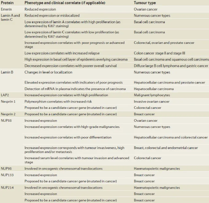
Several components of the LINC complex, Lamins (Broers et al., 1993; Moss et al., 1999), LAP2 (Somech et al., 2007), and Emerin (Capo-chichi et al., 2009) were discovered as biomarkers in a wide range of cancer types (Table 3). Interestingly, not only was mRNA encoding lamin B1 found in the blood circulation (Sun et al., 2010), and its detection in plasma indicate early stage hepatocellular carcinoma, but lamin B1 was also in a proteomic approach found to be upregulated in hepatocellular tumors (Lim et al., 2002). On the other hand, the nucleoporin NUP88 has been assessed as a cancer biomarker (Martinez et al., 1999) and was found in several studies to be overexpressed in malignant tissues (Gould et al., 2002; Agudo et al., 2004; Knoess et al., 2006; Brustmann and Hager, 2009; Schneider et al., 2010).
Down regulation of Drop1, an N-terminal isoform of Nesprin-1, has been observed in early tumor stages in a wide range of human carcinomas and may play a role in chromatin organization (Raffaele Di Barletta et al., 2000; Dou et al., 2005; Marme et al., 2008). Furthermore, mutations in SYNE1 were observed in ovarian and colorectal cancers (Sjoblom et al., 2006; Doherty et al., 2010). Additionally, the SYNE1 gene was frequently methylated in lung cancer cell lines, lung adenocarcinoma (Tessema et al., 2008) and colorectal cancer (Schuebel et al., 2007). By bioinformatic analysis of data from a collection of cancer genome samples Mascia and Karchin identified
SYNE1 as one of the genes that participated in glioblastoma progression (Masica and Karchin, 2011). They observed that mutations in SYNE1 were associated with a large number of differentially expressed genes.
Table 3: Nuclear envelope components and tumorigenesis. The table summarizes cancer-associated alterations in the nuclear envelope components (modified from (Chow et al., 2012)).
Introduction
1.3 DNA repair pathways and mechanisms
Biologically, nuclear DNA is the most important component in the cell since it has all the genetic information required for proper cell functions. It is well known that mutagenic compounds, ionizing radiation, oxidative stress, and also normal DNA metabolic activities like replication and recombination can cause alterations in the DNA. It is therefore not surprising that any damage that leads to a break in the DNA double helix triggers a quick cellular reaction.
To maintain genetic stability and relay genetic information from one cell to another, mechanisms are required to protect the DNA against the accumulation of DNA damage. These mechanisms have been defined as DNA repair pathways. The major DNA repair pathways are non-homologous end-joining (NHEJ), nucleotide excision repair (NER), mismatch repair (MMR), homologous recombination (HR), and base excision repair (BER) (Figure 4).
Figure 4: DNA repair pathways. Non-homologous end-joining (NHEJ), nucleotide excision repair (NER), mismatch repair (MMR), homologous recombination (HR), and base excision repair (BER) are the major DNA repair pathways (Melis et al., 2011).
More importantly, defects in DNA repair pathways lead to genome instability, tumorigenesis, and aging owing to accumulative DNA damage (de Boer and Hoeijmakers, 2000).
1.3.1 Non-homologous end-joining (NHEJ) pathway
One of the most dangerous forms of DNA damage is the DNA double-strand breaks (DSBs), which can result in genome instability if not repaired successfully (Hartlerode and Scully, 2009). DSBs initiate signalling cascades that trigger cell cycle checkpoints and change gene transcription to maintain genome stability (Valerie and Povirk, 2003; Cann and Hicks, 2007).
There are two main signalling networks in eukaryotic cells to repair DSBs:
homologous recombination (HR) and nonhomologous end-joining (NHEJ) (Figure 5).
A recent report indicated that HR uses a homologous chromosome or sister chromatid as template to repair the broken DNA and NHEJ re-ligates the two broken DNA ends together (Cann and Hicks, 2007). Based on this the NHEJ is an error- prone repair mechanism that might cause insertion and deletion of DNA sequences.
The DDR pathway is initiated by a phosphorylation cascade that triggers chromatin modifications which improve accessibility of the broken DNA to repair complexes and promotes the subsequent accumulation of DDR complexes into lesions at the site of damage (Riches et al., 2008). Ataxia telangiectasia mutated (ATM)-mediated phosphorylation of the histone variant H2AX is the initial step to create a platform to which other DDR complexes are able to bind (Marti et al., 2006). ATM triggers signaling cascades that activate cell cycle checkpoints leading to cell cycle arrest through phosphorylation of several proteins including CHK1, CHK2, p53, MDC1, and BRCA1 (Lavin and Kozlov, 2007).
Introduction
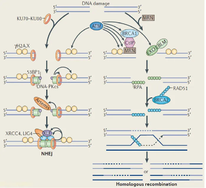
Figure 5: Overview of DNA DSBs repair pathways. DSBs in the DNA are induced by damaging agents. Phosphorylation of histone H2AX (γH2AX) is the initial step of these pathways. In the NHEJ pathway, the two broken ends are bound by the DNA- end-binding protein Ku, which recruits the DNA-dependent protein kinase catalytic subunit (DNA-PKcs) to the free DNA ends. Homologous recombination requires BRCA1, RAD51 (which forms filaments along the unwound DNA strand to facilitate strand invasions) and RAD52 (a DNA-end-binding protein) (modified from (Chowdhury et al., 2013)).
Ku is the DNA-binding component of the NHEJ repair machinery. Ku recruits DNA- PKcs to form the active protein kinase complex DNA-PK upon recognition and binding to the broken DNA end (Mahaney et al., 2009). Following this process, Ku appears to protect broken DNA from nuclease binding or activity (Downs and Jackson, 2004). In addition, Ku has also been shown to bind to telomeres and to
function in telomere maintenance, remarkably by anchoring telomeres to the nuclear periphery, thus contributing to telomeric silencing and preventing telomere shortening (Riha et al., 2006).
Sharpless et al. carried out a study which indicated that NHEJ-deficiency predisposes to cancer (Sharpless et al., 2001). Moreover, mice lacking p53, DNA-PKcs or Ku80 succumb in early postnatal life to progenitor B cell lymphomas (Guidos et al., 1996;
Nacht et al., 1996; Difilippantonio et al., 2000; Lim et al., 2000).
1.3.2 Nucleotide excision repair
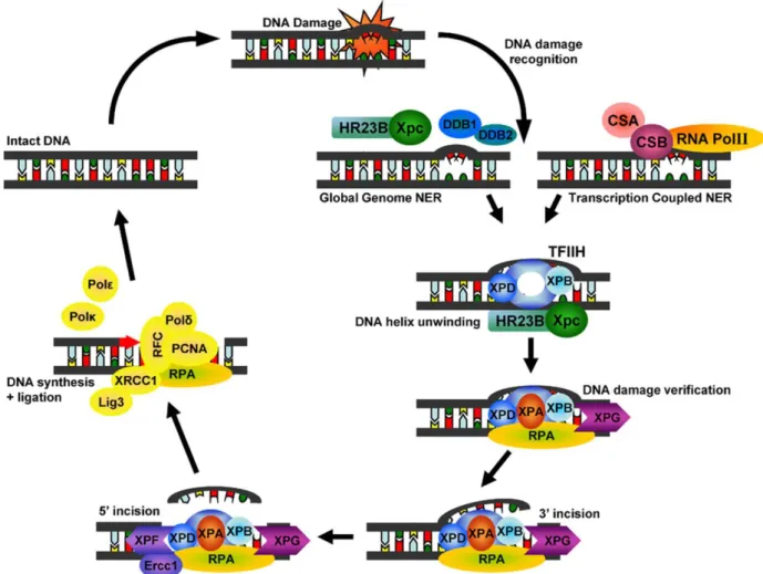
The nucleotide excision repair (NER) pathway is characterized by the removal of bulky DNA helix-distorting injuries of both exogenous and endogenous origin. These helix-distorting DNA lesions recruit the proteins of this pathway to the damaged DNA sites (Figure 6).
GG-NER (global genomic repair) and TC-NER (transcription-coupled repair) are two subpathways of NER (Bohr et al., 1985; Mellon et al., 1987). GG-NER is independent of transcription and is constantly screening the genome for the identification of distorting lesions in the DNA helix, whereas TC-NER is recruited to the transcribed DNA strand of active genes to repair transcription blocking lesions (Yasuda et al., 2007). About 30 proteins participate in the NER pathway, most function in GG-NER as well as in TC-NER (Christmann et al., 2003). However, they have different types of damage recognition and therefore contain different proteins that recognize the damage.
Introduction
Figure 6: NER repairs UV-induced pyrimidine dimers, protein-DNA and intra- strand crosslinks, and a wide range of bulky chemical adducts. Global GG-NER and TC-NER differ in their damage recognition. In GG-NER, damage recognition is accomplished by the XPC-HR23B protein complex whereas CSA and CSB proteins are responsible for the initial detection of damaged DNA in TC-NER (Melis et al., 2011).
GG-NER uses the XPC-HR23B protein complex as the primary recognition factor (Volker et al., 2001; Hanawalt, 2002). Recently, it has been reported that, for certain types of lesions, different proteins are responsible for the initial recognition.
Additionally, DDB2 (or XPE) bound to DDB1 can recognise CPD lesions. The XPE- DDB1 complex recruits the XPC-HR23B complex to the lesion, where it gets exchanged by the XPC-HR23B complex and repair takes place (Kulaksiz et al., 2005). In TC-NER, DNA damage is recognized by CSA and CSB proteins. After recognition, these proteins bind to the DNA lesion thereby distorting the DNA and
recruiting other factors of this repair system to reproduce the correct DNA sequence (Gillet and Scharer, 2006; Trego and Turchi, 2006).
Although somatic alterations in some of the NER factors lead to skin cancer, defects in the NER pathway associate with genetic disorders such as the Xeroderma pigmentosum (XP) syndrome (Yasuda et al., 2007).
1.3.3 Mismatch repair network
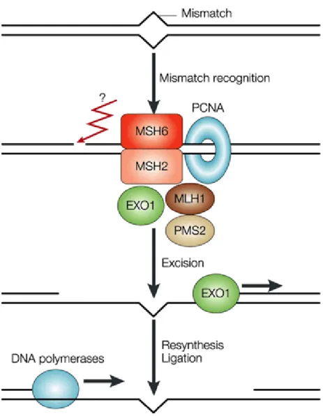
The mismatch repair (MMR) is a conserved biological pathway from bacteria to man that plays a critical role in maintaining genome stability. Its function is in correcting DNA mismatches occurred during DNA replication to prevent mutations in dividing cells. MMR is also required for damage-induced cell cycle checkpoint response.
Deficiency of MMR activity causes replication-associated errors leading to point and frameshift mutations and tumorigenesis (Jacob and Praz, 2002).
The initial step in MMR is identification of the mispaired region (Figure 8). Two separate heterodimers known as MutSα (Msh2/Msh6 in eukaryotes) (Iaccarino et al., 1996), and MutSβ (Msh2/Msh3) participate in this process (Habraken et al., 1996;
Palombo et al., 1996). MutS is a DNA‐binding protein, contains an ATPase domain and a protein‐protein interaction domain that allows two MutS molecules to interact in order to form dimers (Lamers et al., 2000; Obmolova et al., 2000). While MutSα binds primarily to single-base pair mismatches and small insertion/deletion loops, MutSβ binds to larger insertion/deletion loops (Acharya et al., 1996; Marsischky et al., 1996).
After mismatch recognition, MutL homologs are responsible to repair mismatches. At least 4 human MutL homologs, hMLH1, hMLH3, hPMS1, and hPMS2 have been identified (Bronner et al., 1994; Nicolaides et al., 1994; Papadopoulos et al., 1994;
Lipkin et al., 2000).
Introduction
Figure 7: Mismatch repair. MSH2–MSH6 and PMSH2-MLH1 heterodimers bind to single base pair mismatches in an ATP-dependent manner. The lesion is digested by EXO1, and then filled in by DNA polymerases. Question mark points out the unknown homologue of prokaryote MutH in eukaryotes (Martin and Scharff, 2002).
hMLH1 heterodimerizes with hPMS2, hPMS1, or hMLH3 to form hMutLα, hMutLβ, or hMutLγ, respectively (Kunkel and Erie, 2005). MutLα heterodimer (Mlh1/Pms1) initiates subsequent repair events (Prolla et al., 1994; Pang et al., 1997). hMutLα is required for MMR and hMutLγ plays a role in meiosis, but no specific biological role has been identified for hMutLβ (Kunkel and Erie, 2005). hMutLα possesses an ATPase activity and defects in this activity inactivate MMR in human cells. Defects in the MMR pathway have been associated with a number of different malignancies (Gazzoli et al., 2002).
1.4 Aim of the project
Goal of this study is to provide a picture of the interactions and roles of Nesprin-1.
This will allow defining its roles in health and disease more precisely and will provide mechanisms underlying these roles. We will specifically try
1. to understand the role of Nesprin-1 in tumorigenesis and genome stability 2. to identify possible interaction partners of Nesprin-1
3. to detect consequences of Nesprin-1 loss
4. to elucidate the role of Nesprin-1 in DDR and DNA repair pathways.
Results 2. Results
2.1 Nesprin-1 interactions
2.1.1 Interactions of N-terminal Nesprin-1 spectrin repeats
Earlier studies have demonstrated that the C-terminal isoform Nesprin-1α can self- associate through its third and fifth spectrin repeat to form an antiparallel dimer (Mislow et al., 2002a; Zhong et al., 2010). Whether other spectrin repeats of Nesprin- 1, in particular the N-terminal spectrin repeats, possess similar oligomerization properties is not known. When we compared Nesprin-1’s N-terminal spectrin repeats to the spectrin repeats of mouse α-actinin 2, SR1, 2, and 4 showed homology with SR2 from α-actinin, and SR10 and 11 resembled SR1 and SR4, respectively. The remaining SRs exhibited less homology with the ones of α-actinin. From earlier reports it is known that spectrin repeats of the α-actinin type can dimerize as in α- actinin or in spectrin determining the protein structure (Noegel et al., 1987; Djinovic- Carugo et al., 2002).
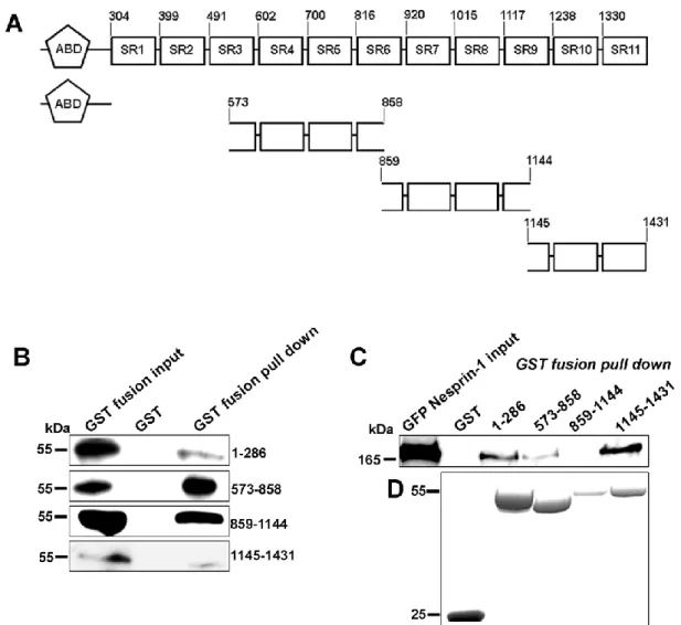
To understand whether N-terminal sequences of Nesprin-1 can interact with themselves, bacterially produced GST-fusion proteins encompassing several spectrin repeats of Nesprin-1-165 (aa 573-858, 859-1144 and 1145-1431; Figure 8A) was used to pull down the corresponding GFP-tagged proteins from COS7 cells.
While all fusion proteins pulled down their GFP-tagged counterpart, GST alone did not interact with anyone of the GFP fusion proteins (Figure 8B). Furthermore, Nesprin-1-165-GFP was transfected in COS7 cells and the GST-fusion proteins used in pull down experiments. Interestingly, Nesprin-1-165-GFP was precipitated with GST-573-858 and GST-1145-1431, but not with GST-859-1144. The polypeptide encompassing aa 859-1144 encodes part of SR6, SR7, 8 and part of 9 which in the comparison showed a lower resemblance to the ones of α-actinin. GST alone also
did not bind to Nesprin-1-165-GFP (Figure 8C, D). Our results imply that Nesprin-1 oligomerizes through N-terminal spectrin repeats.
Figure 8: N-terminal spectrin repeats of Nesprin-1 can self-associate through its amino terminal sequences. (A) Overview of Nesprin-1-165 and constructs used as GST and GFP fusion proteins. Numbers indicate the location of amino acids. (B, C) GFP-tagged ABD (aa 1–286), and spectrin repeats of Nesprin-1-165 (aa 573–858, 859–1144, and 1145–1431) and full-length Nesprin-1-165 (C) were expressed in COS-7 cells. (B) The cell lysates were incubated with either immobilized GST-fused aa 1–286, 573–858, 859–1144, and 1145–1431 or GST alone for control. The precipitated proteins resolved in 10% acrylamide gels by SDS-PAGE. The membranes were probed with GFP antibody mAb K3-184-2. (C) Interaction of N- terminal spectrin repeats with GFP Nesprin-1-165. (D) The GST-fusion proteins used for the pull down are shown by Coomassie Blue staining (bottom panel).
Results
2.1.2 The C-terminus of Nesprin-1 interacts with Nesprin-2
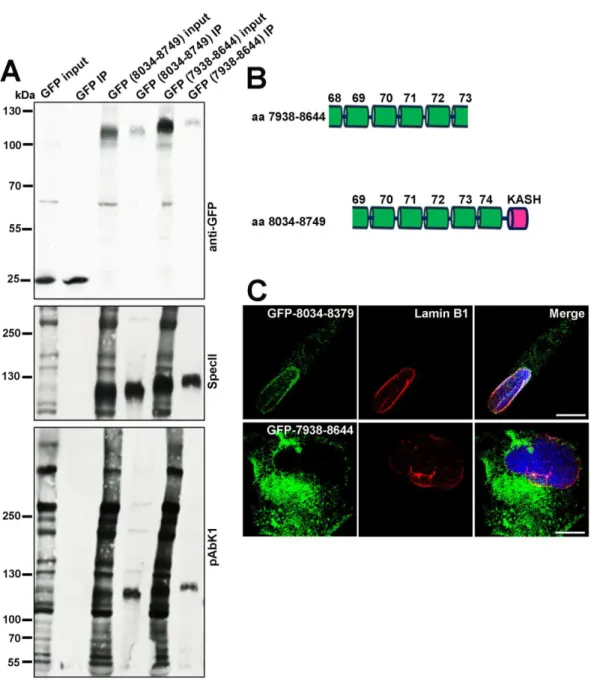
By immunoprecipitation experiments we next asked whether the possibility of an association between Nesprin-1 and Nesprin-2 exists. Human fibroblasts (HF) were transfected with plasmids encoding GFP-tagged Nesprin-1 polypeptides that were differently composed and possessed or lacked the KASH domain (aa 8034-8749;
7938-8644) (Figure 9A, B).
Figure 9: Nesprin-1 interacts with Nesprin-2. (A) GFP-tagged Nesprin-1 (aa 8034- 8749; 7938-8644) were expressed in HF cells. Numbers indicate the location of amino acids and used for immunoprecipitation experiments. Immunoprecipitation was carried out with GFP-TRAP beads. Detection of precipitated proteins was with mAb
K3-184-2. The blot was further probed with pAbK1 and SpecII antibodies specific for Nesprin-2 and Nesprin-1, respectively. (B) Schematic of the Nesprin-1 constructs. (C) HF cells were transfected with plasmids allowing GFP-Nesprin-1-8034-8379, and GFP-Nesprin-1-7938-8644 expression (green) and stained with Lamin B1 (red), and DAPI (blue). Scale bar, 10 μm.
GFP transfected cells served as negative control for Nesprin-1 and Nesprin-2 interaction. The proteins were immunoprecipitated using GFP-TRAP beads and the resulting blot probed with pAbK1 specific for Nesprin-2 and SpecII specific for Nesprin-1.
We found that Nesprin-2 proteins detected by pAbK1 which is directed against C- terminal Nesprin-2 sequences precipitated with GFP-Nesprin-1-8034-8749, but not with GFP-Nesprin-1-7938-8644. The interacting domain is therefore located within the C-terminal sequences of Nesprin-1. GFP alone did not bind to Nesprin-2 (Figure 9A). Furthermore, we studied the subcellular localization of GFP-Nesprin-1-8034- 8749 and GFP-Nesprin-1-7938-8644. GFP-Nesprin-1-8034-8749 localized mostly to the NE as revealed by Lamin B1 colocalization. GFP-Nesprin-1-7938-8644, which lacks the KASH domain containing the transmembrane region, was also present at the NE but was most prominent in the cytoplasm (Figure 9C).
2.1.3 Nesprin-3 is able to recruit vimentin to the nucleus
Our group had previously reported that N-terminal sequences of Nesprin-1 can associate with N-terminal spectrin repeats of Nesprin-3 (Taranum et al., 2012a).
Nesprin-3 binds to plectin, a huge protein which associates with intermediate filaments.
Earlier reports indicate that Nesprin-1 and -2 through their association with F-actin can assemble an F-actin cage around the nucleus (Khatau et al., 2009). In analogy we asked whether Nesprin-3 is able to recruit an intermediate filament network to the
Results
nucleus. Since Nesprin-3 is not normally expressed in COS7 cells (Wilhelmsen et al., 2005), HA-Nesprin-3 was expressed in these cells to study vimentin localization. In untransfected cells, vimentin staining was not particularly enriched around the nucleus. However, vimentin colocalized with Nesprin-3 in HA-Nesprin-3 expressing cells (Figure 10) extending recent report findings from zebrafish to mammalian cells (Postel et al., 2011). By contrast, GFP-tagged Nesprin-1 ABD was recruited to the nuclear envelope but it did not affect vimentin localization (Figure 10).
Figure 10: Nesprin-3 recruits intermediate filaments to the nuclear envelope in COS7 cells. COS7 cells stained for vimentin reveal the typical cytoskeletal staining.
In HA-Nesprin-3 transfected COS7 cells vimentin was recruited to the NE and co- localized with HA-Nesprin-3. GFP-fused ABD-Nesprin-1 was recruited to the nuclear envelope but it did not affect vimentin localization. Confocal images are shown in (Taranum et al., 2012a). Size bars, 10 μm.
2.2 Nesprin-1 role in tumorigenesis
2.2.1 Nesprin-1 isoform expression in cancer cell lines
As mutations in SYNE1 have been identified in different types of human cancers and Nesprin-1 transcripts were down regulated at early tumor stages in a wide range of human carcinomas ((Marme et al., 2008); www.oncomine.org), we probed several human and murine cancer cell lines with Nesprin-1 specific antibodies by immunoblotting and immunofluorescence analysis. Monoclonal antibody K43-322-2 generated against spectrin repeats 9, 10 and 11 (Figure 11A) recognized proteins of
~600, 400, 300, 250, 150, 55 and 50 kDa in CH310T1/2 cells. The proteins correspond in their molecular weights to Nesprin-1 isoforms described in a recent detailed analysis (Rajgor et al., 2012).
The ~600 and 400 kDa proteins were absent from all cancer cell lines and only the
~150 kDa protein was present with the exception of WIDR, where ~300, 250, 150 and 60 kDa proteins were detected. In the CT26 and Huh7 cell lysates the signal was rather faint, even after prolonged exposure (Figure 11B). Furthermore, a protein of high molecular weight which presumably corresponds to Nesprin-1 Giant (Taranum et al., 2012a) was detected in C2F3, HaCaT, and HeLa and Hep3B cell lysates.
Based on the low expression levels of the N terminal Nesprin-1 isoforms in Hep3B and Huh7 liver cancer cells compared to colon, cervic, and skin cancer cells, we focused our studies on these cell lines. Furthermore, recent data also suggested that Nesprin-1 expression levels are significantly reduced in liver cancer samples compared with matched normal tissue (www.oncomine.org).
Results
Figure 11: Nesprin-1 isoforms in various cell lines. (A) Location of the binding sites of Nesprin-1 antibodies. The largest isoform Nesprin-1 giant is depicted. ABD, actin binding domain. (B) Lysates were separated on a 3-15% SDS-PA gradient gel and probed with mAb K43-322-2 to detect N terminal isoforms. Arrow heads point to proteins discussed in the main text. Tubulin amounts were checked on a separate gel.
Nesprin-1 C-terminal isoforms were identified in Hep3B and Huh7 cell lysates with polyclonal SpecII antibodies directed against the C-terminus of Nesprin-1(Taranum et al., 2012b). In fibroblasts we detected a 400 kDa protein which was absent from Hep3B and Huh7. Instead, they harbored low levels of 100 kDa and in case of Hep3B of 250 kDa proteins (Figure 12A). When probing for Nesprin-2, we detected several isoforms of Nesprin-2 with polyclonal antibodies pAbK1 directed against the C-terminus of Nesprin-2. The amounts were significantly higher in Hep3B and Huh7 cells as compared to fibroblasts (Figure 12B).
Figure 12: Cancer cells have alterations in nuclear envelope components. (A) Nesprin-1 expression in human fibroblasts (HF), Hep3B and Huh7 cells using SpecII antibodies. The blots were probed with Emerin, Lamin A/C, Lamin B1, LAP2, SUN1 and SUN2 antibodies. Tubulin was used to assess equal loading. (B) Presence of Nesprin-2 as detected with pAbK1 directed against the C-terminus. NPC proteins were detected with mAb414. (C) Changes at the protein level in HF, Hep3B and Huh7 cell lines as determined by western blotting. Fold change of Emerin, Lamin A/C, Lamin B1, LAP2, SUN1, SUN2 in HF, Hep3B and Huh7 cells. Band intensities were normalized relative to the loading control (tubulin). Histogram representing fold changes in band intensity. The results are the average from 3 independent experiments (*p<0.05, **p<0.001).
Results
In further studies we compared Nesprin-1 expression in lysates from normal mammary tissue (N1, N2, N3) and tumor tissue (T1, T2, T3) of different patients. The SpecII antibodies recognized primarily a ~55 kDa protein which was strongly reduced in the tumor tissue (Figure 13A, B).
Figure 13: Nesprin-1 is reduced in human tumor tissues. (A) Nesprin-1 expression in normal (N1, N2, N3) and tumor (T1, T2, T3) mammary tissues using SpecII for detection. Upper panel, PonceauS staining of the nitrocellulose membrane.
(B) Histogram representing fold change in band intensity of Nesprin-1 for normal and tumor tissues (*p<0.05).
2.2.2 Hep3B and Huh7 have nuclear shape defects and alterations in components of the nuclear envelope
The nuclei of Hep3B and Huh7 cells were enlarged and often displayed a deformed morphology in contrast to the oval shape in HFs which we used for control. We further noted folds, lobulations, protrusions, blebs and micronuclei (Figure 14A, B).
In Hep3B, 37% of the cells had misshapen nuclei, in Huh7 26% and in control 7%.
Micronuclei were observed in 11% of the Hep3B cells, in 8% in case of Huh7 and 1%
of HF cells (Figure 14B). SpecII antibodies labeled the NE in fibroblasts and gave some cytoplasmic staining in the vicinity of the nucleus whereas the Nesprin-1 presence at the NE was strongly reduced in the cancer cells (Figure 14A).
Figure 14: Hep3B and Huh7 have nuclear shape defects and alterations in components of the LINC complex. (A) Staining was with SpecII (green) to detect Nesprin-1 and a mAb specific for Emerin (red). DAPI staining of DNA is in blue.
Arrow heads indicate nuclei with regular shape and staining for SpecII and Emerin.
Scale bar, 10 µm. (B) Huh7 cells have nuclear shape defects and alterations in components of the nuclear envelope. Staining was with polyclonal SpecII antibodies against Nesprin-1 (green), Lamin B1 (green), SUN1 (green) and mAb Emerin (red) antibodies. DAPI staining of DNA is in blue. Scale bar, 10 μm. Upper panel, statistical analysis of nuclear aberrations. 300 nuclei each for HF (passage 7), Hep3B and Huh7 were evaluated (**p<0.001).
Remarkably, Emerin was nearly absent from the NE in the cancer cells (Figure 14A, B). The absence of Emerin was also confirmed in western blots (Figure 12A, C).
Lamin A/C specific antibodies showed a rim like staining pattern in HFs. In Hep3B and Huh7 cells a discontinuous, patchy Lamin A/C distribution at the NE was observed. Lamin B1 staining of the NE was homogenous in HFs, in Hep3B and Huh7 cells the distribution was patchy (Figure 15A).
LAP2, a member of a group of NE proteins involved in tethering chromatin to the nuclear envelope and affecting gene expression, showed an unaltered localization at the NE in Hep3B and Huh7 cells. The expression level appeared to be significantly higher than in fibroblasts which expressed low amounts of LAP2 (Figure 15A).
NPC proteins regulate nuclear transport, are connected to chromatin and participate in the regulation of transcription. Increased expression of individual NPC components
Results
has been noticed in several tumor types. Hep3B and Huh7 cells exhibited NE staining with mAb 414, which recognizes several NPC proteins based on the presence of FXFG-repeats, however staining was reduced in nearly 45% of the cells (Figure 15B, C).
Figure 15: Nuclear envelope components are altered in Hep3B and Huh7 cells.
(A) Distribution of Lamin A/C, Lamin B1, and LAP2 in HF, Hep3B and Huh7 cells.
Arrow heads indicate the observed defects. Scale bar, 10 μm. (B) HF, Hep3B and Huh7 cells were stained with anti-Nesprin-1 SpecII (green), mAb NPC (red), DAPI (blue). Arrow heads point to normal shaped nuclei stained with SpecII and NPC.
Scale bar, 10 μm. (C) Statistical analysis of NPC staining. 200 cells per strain were analysed (*p<0.0001).
Analysing individual proteins by western blotting, we found that NUP153 levels were higher in Hep3B and Huh7 cells compared to the HF control and NUP116 levels were
decreased (Figure 12B). In colon cancer cell lines, localization of Nesprin-1 and other nuclear envelope components was unperturbed (Figure 16).
Figure 16: Nuclear envelope components in colon cancer cells. Distribution of Nesprin-1 detected by anti-ABD-Nesprin-1, Emerin, NPC, Lamin A/C, Lamin B1, Nesprin-2 as detected by pAbK1 in CT26, CMT93, WIDR, C2F3 cells. DAPI staining of DNA is in blue. Scale bar, 10 μm.
Immunofluorescence analysis for SUN proteins revealed a rim like staining for SUN1 in Hep3B and Huh7 cells. Some cells exhibited a brighter SUN1 staining which was associated with misshapen and enlarged nuclei (Figure 17, arrows). When we
Results
examined the amounts of SUN1 and SUN2 in western blots, we found that particularly the SUN1 levels were higher in Hep3B and Huh7 as compared to HFs (Figure 12A, C). Quantification of the mRNA levels by qRT-PCR showed that SUN1 and SUN2 mRNA were significantly increased in Hep3B and slightly increased in Huh7 cells (Figure 17B).
Figure 17: Cells with misshapen and enlarged nuclei exhibited a brighter SUN1 staining. (A) SUN1 (red) staining in Hep3B and Huh7 cells, DAPI, blue. Arrow heads point to cells with high SUN1 expression and misshapen and enlarged nuclei. Scale bar, 10 μm. (B) SUN1 and SUN2 transcript levels in control, Hep3B and Huh7 cells as determined by qRT-PCR. Significant up-regulation of SUN1 and SUN2 was detected in Hep3B, Huh7, and KD-HF cells compared to HF cells (*p<0.05,
**p<0.001). The SUN1 and SUN2 mRNA level in HF at passage 7 was taken for reference. For normalization, GAPDH was used.
2.2.3 The centrosome-nucleus distance is increased in Hep3B and Huh7 cells
Centrosomal aberrations are frequently observed in cancer cells. Normal cells in the G1 phase of the cell cycle have a single centrosome which is attached to the nucleus. In HFs, centrosomes were positioned near the nucleus at a mean distance of 0.33±0.25 μm. In Hep3B and Huh7 cells the distance between the centrosome and the nucleus was highly variable and cells with normal shaped as well as deformed nuclei displayed an increased centrosome-nucleus distance. In Hep3B and Huh7
cells we observed an increase to 6.29±4.24 μm and 3.56±3.0 μm, respectively (Figure 18A, B). The number of centrosomes also differed. 15% of Hep3B and 14.3%
of Huh7 cells had more than two centrosomes (Figure 18C). The centrosome number was not necessarily associated with nuclear shape changes.
Figure 18: Centrosome-nucleus-distance is altered in Hep3B and Huh7 cells.
(A) Centrosome-nucleus-distance is altered in Hep3B and Huh7 cells. γ-Tubulin (red) specific antibodies were used to label the centrosome. DAPI (blue), nuclear staining.
Scale bar, 10 μm. (B) Statistical evaluation of the centrosome-nucleus distance. (C) Statistical evaluation of cells with >2 centrosomes. Error bars indicate standard deviations (*p<0.001, **p<0.0001).
Results 2.3 Loss of Nesprin-1
2.3.1 Knock down of Nesprin-1 elicits changes that are observed in cancer cell lines
To test whether a loss of Nesprin-1 can cause the changes observed in the cancer cells, we reduced the amounts of Nesprin-1 by shRNA mediated knock down in C3H10T1/2 (KD-CH310T1/2) and in human fibroblasts (KD-HF) using knock down vectors targeting N-terminal and C-terminal sequences of human or mouse Nesprin-1 (see Materials and Methods) and analyzed the consequences.
For control we used untransfected cells (HF, CH310T1/2) and cells transfected with the empty pSHAG-1 vector used for cloning (C-HF, C-CH310T1/2). Western blot analysis with mAb K43-322-2 and SpecII labeling confirmed the knock down (KD) (Figure 19A, B). Labeling with mAb K43-322-2 showed that in Nesprin-1 KD cells particularly the 250 kDa and larger proteins of 400 and 600 kDa were significantly reduced in amounts (Figure 19A, arrow heads). The 130 kDa protein and smaller proteins were also less prominent. The expression level of the smallest ones were not altered (Figure 19A).
Reduction of Nesprin-1 was associated with a down regulation of Emerin, Lamin B1, NPC proteins and LAP2. By contrast, SUN1 and SUN2 protein amounts were increased (Figure 19A, C). This was also observed for SUN1 in CH310T1/2 in which the levels of endogenous SUN1 were quite low (Figure 19A). An increase was also seen at the transcript level as revealed by qRT PCR (Figure 17B).
Figure 19: Loss of Nesprin-1 elicits changes that are observed in cancer cell lines. (A) Immunoblot analysis of Nesprin-1 Giant knock down HF and CH310T1/2 cells. Detection was with mAb K43-322-2 and pAb SpecII. Tubulin served as control.
Emerin, Lamin A/C, Lamin B1, LAP2, SUN1 and SUN2 specific antibodies were used for analysis. Human and murine Emerin differ in their primary sequence explaining the observed difference in molecular weight. (B) The blot in (A) was reprobed with SpecII antibodies and mAb414 to detect NPC proteins. (C) Changes of NE
Results
components at the protein level after knock down of Nesprin-1. Knock down was carried out using plasmids targeting the N and C terminal regions of Nesprin-1.
Histogram representing fold changes in band intensities of Emerin, Lamin B1, LAP2, SUN1 for HF, C-HF, KD-HF, CH310T1/2, C-CH310T1/2, KD-CH310T1/2 cells (*p<0.05).
For Nesprin-1, alterations in transcript levels were assessed by qRT-PCR. KD-HF and Huh7 cells showed significant reductions (Figure 20).
Figure 20: The transcript levels of Nesprin-1 vary significantly in Hep3B, Huh7, HF, C-HF, and KD-HF cells as determined by qRT-PCR. Significant down- regulation of Nesprin-1 was detected in Hep3B, Huh7, and KD-HF cells compared to HF cells (*p<0.05, **p<0.0001). The primers used for amplification were located in the N terminus of Nesprin-1. For normalization GAPDH was used.
In immunofluorescence analysis the clear NE staining by SpecII and Emerin antibodies was lost and some residual punctate staining in the cytosol of KD-HF and KD-CH310T1/2 cells was seen (Figure 21A). The NPC antibodies labeled the nuclear envelope in control fibroblasts and in CH310T1/2, whereas in the knock down cells NPC staining is strongly reduced (Figure 21B).
Figure 21: Effect of Nesprin-1 knock down on NE components. (A, B) Cells were stained for Nesprin-1 with pAb SpecII (green), Emerin (red), mAb NPC (red) and DAPI (blue). Knock down was carried out with vectors targeting N and C terminal sequences of Nesprin-1. Arrow heads indicate the NE phenotypes described. Scale bars, 10 μm.
Nearly all cells that had an altered staining pattern exhibited nuclear shape defects.
SUN1 antibodies strongly stained the NE in KD-HF. Cells with particularly strong SUN1 staining exhibited a variety of nuclear shape defects including folds, lobulations, blebs and micronuclei (Figure 22).
Results
Figure 22: SUN1 staining revealed a variety of nuclear shape defects including folds, lobulations, blebs and micronuclei. SUN1 (red) staining in Hep3B, Huh7, C- HF, KD-HF, C-CH310T1/2, KD-CH310T1/2 cells. Nesprin-1 was detected with mAb K58-398-2 (green). Nuclei are stained by DAPI (blue). Scale bars, 10 μm.
Heat treatment is often used to probe the stability of the nuclear envelope (Vigouroux et al., 2001). To understand the link between Nesprin-1 loss and heat resistance, the cells were stained with LAP2 to evaluate nuclear shape changes after heat shock.
When we incubated cells for 30 min at 45°C, Nesprin-1 knock down fibroblasts, Hep3B and Huh7 cells exhibited increased nuclear deformations with folds and pleats after heat treatment (Figure 23A, B). Many nuclei also displayed notches, tears and herniations (Figure 23A, arrow heads). Knock down with plasmids targeting N- terminal or C-terminal sequences showed similar results as knock down experiments
where we used vectors targeting N-terminal and C-terminal sequences together (data not shown).
Figure 23: Loss of Nesprin-1 leads to hypersensitivity towards heat shock. (A) Cells were immunostained with SpecII (green) and LAP2 (red) antibodies to detect the nuclei deformations after heat shock at 45°C for 30 min. Arrow heads point to nuclear deformations. Scale bar, 10 µm. (B) Histograms representing the percentage of deformed nuclei of cells before (white bars) or after heat shock (black bars). Data are the mean ±SD from three samples per group of three independent experiments.
Statistically significant differences were determined between before and after heat shock groups (*p<0.05, **p<0.0001).
When we quantified the defects for each cell type, we found that Hep3B and Huh7 had more abnormal nuclei in general. This number increased only slightly upon heat
Results
treatment. Compared with unheated Hep3B (42%) and Huh7 (34%) cells, deformed nuclei after heat shock was 46% for Hep3B and 41% for Huh7 cells. Stronger increases in the number of deformed nuclei were observed for the KD-HF cells (35%±4.09 before and 41%±3.22 after heat shock). Moreover, no significant changes were observed among C-CH310T1/2 cells before (7% misshapen nuclei) or after (10%) heat shock. The extent of nuclear deformations caused in KD-CH310T1/2 (29.5%±4.37 before and 40.83%±2.86 after heat shock) was similar to heat shock experiments performed in KD-HF cells (Figure 23B). These results suggest that heat treatment leads to more deformed nuclei in Nesprin-1 KD, Hep3B and Huh7 cells.
2.3.2 The centrosome-nucleus distance is increased in Nesprin-1 KD cells
The LINC complex proteins play essential roles in centrosome biology (Salpingidou et al., 2007; Schneider et al., 2008; Zhang et al., 2009). Moreover, recent studies revealed that centrosome defects, including alterations in centrosome shape, size, number, position, composition lead to tumorigenesis (Lingle and Salisbury, 2001;
Fukasawa, 2005; Salisbury, 2005; Nigg, 2006; Hassold et al., 2007). To further address whether the Nesprin-1 is associated with centrosome position and number, the cells were stained with Ɣ-tubulin. We investigated the centrosome-nucleus distance and centrosome number upon loss of Nesprin-1 and found that centrosomes were positioned 0.35±0.29 and 3.20±2.34 μm away from the NE in C-HF and Nesprin-1 KD-HF cells, respectively. Similarly, in Nesprin-1 KD-CH310T1/2 cells the mean centrosome-nucleus distance increased from 0.44±0.27 μm in C-CH310T1/2 cells to 2.40±1.49 μm in Nesprin-1 KD-CH310T1/2 cells (Figure 24A, B).