connecting the nuclear interior with the cytoskeleton
INAUGURAL-DISSERTATION
zur
Erlangung des Doktorgrades
der Mathematisch-Naturwissenschaftlichen Fakultät der Universität zu Köln
vorgelegt von
Wenshu Lu
aus
Changchun, Jilin, P. R. China
Köln, 2007
Referees/Berichterstatter: Prof. Dr. Angelika A. Noegel Prof. Dr. Jens Brüning
Date of oral examination: 12.06.07 Tag der mündlichen Prüfung
The present research work was carried out under the supervision of Prof. Dr. Angelika A.
Noegel and Dr. Akis Karakesisoglou, in the Institute of Biochemistry I, Medical Faculty, University of Cologne, Cologne, Germany from April 2004 to December 2006.
Diese Arbeit wurde von April 2004 bis Dezember 2006 am Institut für Biochemie I der Medizinischen Fakultät der Universität zu Köln unter der Leitung von Prof. Dr. Angelika A.
Noegel und Dr. Akis Karakesisoglou durchgeführt.
1. Introduction
1.1 Nuclear envelope 1
1.1.1 Inner and outer nuclear membranes 1
1.1.2 The nuclear lamina 2
1.1.3 Nuclear envelope proteins 4
1.2 Nuclear envelope related human diseases 6 1.3 The nuclear envelope and nuclear positioning 7
1.4 KASH domain proteins 8
1.5 SUN domain proteins 11
1.6 Aim of the work 12
2. Results
2.1 Characterization, distribution and functional analysis of Sun1 13 2.1.1 Sun1 is the closest orthologue of C. elegans UNC-84 in mouse 13 2.1.2 Phylogenetic tree of SUN domain containing proteins 15
2.1.3 Sun1 is broadly expressed in mice 16
2.1.4 Subcellular localization of endogenous human Sun1 17
2.1.5 Domain analysis of Sun1 18
2.1.6 Sun1 has three transmembrane domains 20
2.1.7 Sun1 dynamics at the nuclear envelope 21
2.1.7.1 iFRAPs of GFP-Sun1 full-length 23
2.1.7.2 iFRAPs of GFP-Sun1-2TM-N and GFP-Sun1-TM-C 24 2.1.7.3 iFRAPs of GFP-Sun1-TM-SD1, 2 and GFP-Sun1-TM-∆CC-SUN 26
2.1.8 Sun1 interacts with chromatin 28
2.1.9 Sun1 oligomerizes via its coiled-coil domains 29 2.1.10 Sun1 forms SDS-resistant dimers under non-reducing conditions 35 2.1.11 The C-terminal domain of Sun1 forms SDS-resistant dimers under 36
non-reducing conditions through disulfide bond formation
2.1.12 Sun1 interacts with Sun2 39
2.1.13 Sun1 is required for proper Nesprin-2 NE localization 41 2.1.14 Sun1 affects the nuclear envelope localization of emerin and LAP2β 43
5µm
2.1.17 Characterization of HaCaT cells stably transfected with SPGFP-Sun1-C 46 2.1.18 Nesprin-2 isoforms are mislocalized in SPGFP-Sun1-C stably transfected 48
HaCaT cells
2.1.19 Nuclear shape changes in SPGFP-Sun1-C cells 50 2.1.20 Proliferative ability of SPGFP-Sun1-C stably transfected cells 53 2.1.21 Senescence analysis of SPGFP-Sun1-C stably transfected cells 56 2.1.22 The expression level of E-cadherin is increased in SPGFP-Sun1-C 57
stably transfected cells
2.2 Generation of a Nesprin-1 knock-in mouse model 59
2.2.1 Knock-in strategy and ES clone screening 59
2.2.2 Generation of Nesprin-1 knock-in chimeric mice 61
3. Discussion
3.1 Sun1 and Sun2 proteins display similar topologies at the nuclear envelope 63
3.2 Sun1 is a widely expressed INM protein 64
3.3 Sun1 displays a low lateral mobility at the NE 65 3.4 Sun1 oligomerizes via the coiled-coil domains 66
3.5 Sun1 binds to chromatin 68
3.6 Sun1 interacts with Nesprins to form a bridge across the NE 69 3.7 Overexpression of Sun1-C disrupts the SUN-Nesprin linkage across the NE 71 3.8 Proposed roles of Sun1 in higher eukaryotes 73
4. Summary/ Zusammenfassung 76
5. Materials and methods
5.1 Cloning strategies 79
5.2 Primer sequences for cloning 80
5.3 RNA isolation and RT-PCR analysis 82
5.4 Tissue culture and transfection 83
5.5 Establishment of stably transfected HaCaT cell lines 83 5.6 Inverse fluorescence recovery after photobleaching experiments (iFRAP) 83
5.7 Antibodies and immunofluorescence microscopy 84
5.10 Purification of GST fusion proteins and in vitro binding assays 87 5.11 Native polyacrylamide gel-electrophoresis (Native-PAGE) 87
5.12 Chemical cross-linking experiments 87
5.13 Yeast two-hybrid assay 88
5.14 Site-directed Mutagenesis 88
5.15 His-tag pull-down assays 88
5.16 BrdU incorporation 89
5.17 Generation of Growth Curves 89
5.18 PI/FACS (cell cycle) analysis 89
5.19 Senescence-Associated β-Galactosidase Assays 90
5.20 Nuclear shape analysis 90
5.21 Statistical analysis 91
6. References 92
7. Appendix
7.1 Abbreviations 104
7.2 Amino acid code 105
Acknowledgement 106
Erklärung 107
Curriculum Vitae/ Lebenslauf 108
1. Introduction
1.1 Nuclear envelope
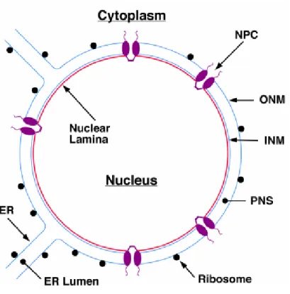
The nucleus is the hallmark of eukaryotic cells, and is separated from the cytoplasm by a selective structural barrier - the nuclear envelope (NE). The nuclear envelope (Fig 1.1) is composed of a pair of membranes, the inner and outer nuclear membranes (INM and ONM, respectively) joined together by the nuclear pore complexes (NPCs) (Gerace & Burke, 1988).
NPCs span the NE and are large macromolecular assemblies that form aqueous gated channels across the NE and regulate the transport of macromolecules between the nucleus and cytoplasm (Gorlich & Kutay, 1999; Macara, 2001). In addition to this function, the NE is essential in defining higher order nuclear structure by providing anchoring sites for chromatin at the nuclear periphery. A recent convergence of clinical and basic research has highlighted a link between NE functions and various human diseases, including cardiac and skeletal myopathies (Burke & Stewart, 2002), which underline the vital importance of NE functions.
1.1.1 Outer and inner nuclear membranes
The ONM is continuous with the peripheral endoplasmic reticulum (ER) and is functionally related to the ER (Franke et al., 1981). In fact, these two structures share a similar set of proteins as well as ribosomes (Gerace & Burke, 1988). The ONM also provides attachment sites for structural elements of the cytoplasm (Burke & Ellenberg, 2002). The INM, in contrast, is composed by a distinct set of membrane proteins, which perform close associations with the underlying nuclear lamina and chromatin (Stuurman et al., 1998). The INM is not known to participate in protein synthesis, and has no association with ribosomes.
The lumen between the two membranes that makes up the nuclear envelope is continuous
with the ER lumen, and is usually about 20 - 100 nm in width (Broers et al., 2006).
1.1.2 The nuclear lamina
The inner nuclear membrane (INM) is stabilized by an underlining filamentous meshwork called nuclear lamina. In mammalian cells, the nuclear lamina appears as a thin (20–50 nm) protein meshwork that lines the nuclear face of the INM (Dwyer & Blobel, 1976).
The nuclear lamina associates with both INM integral proteins and chromatin, performing thus fundamental roles in the functional organization of the nucleus. Principal constituents of the lamina are the intermediate filament lamin proteins (Franke, 1987). Nuclear lamins are type V intermediate filament (IF) proteins containing a central α-helical rod, which is flanked by N- and C-terminal non-helical domains. Most lamins, except for lamin C, are farnesylated at their carboxy termini via a CaaX motif (Hutchison et al., 2001) (Fig. 1.2). Based on their primary sequences and biochemical features, lamins are subdivided into A-type and B-type (Gerace et al., 1978).
Figure 1.1. Overview of nuclear envelope organization. The outer nuclear membrane (ONM) is coated
with ribosomes and is contiguous with the rough endoplasmic reticulum (ER). The inner nuclear membrane
(INM) is connected to the ONM at the periphery of each nuclear pore complex (NPC). The nuclear lamina
lines the nuclear face of the INM and is closely associated with peripheral heterochromatin (Burke et al.,
2001).
Both major (A and C) and minor (A∆10 and C2) A-type lamin species are encoded by a single developmentally regulated gene (LMNA), which arise through alternative splicing (Fisher et al., 1986). By contrast, the main B-type lamins (B1 and B2) are encoded by two separate genes (LMNB1 and LMNB2, respectively) (Hoeger et al., 1988; Hoeger et al., 1990).
A single minor B-type lamin (B3) is a splice variant of lamin B2 (Furukawa & Hotta, 1993).
A-type lamins are expressed in more differentiated cells (Rober et al., 1990) and are nonessential for cell viability (Sullivan et al., 1999). B-type lamins are expressed in all cell types and are essential for cell viability and normal development (Harborth et al., 2001; Lenz- Bohme et al., 1997; Vergnes et al., 2004). LMNB1 mutant mice are lethal after birth, but it is possible to culture and even immortalize LMNB1 mutant embryonic fibroblasts, suggesting a partial functional substitution by lamin B1 and lamin B2 proteins (Vergnes et al., 2004).
Figure 1.2. Schematic representations of somatic cell lamins. Colored rectangles represent α-helical coiled-coil domains; B-type lamins are shaded in green, and A-type lamins are shaded in red. The blue shaded area within coil 1b shows the position of the heptad repeat extension, which is unique to the lamins.
NLS=nuclear localization signal sequence; CaaX is the site for carboxy methylation, prenylation and
proteolytic cleavage. Colored regions in the C-terminal tail domains show the major sites of amino acid
residue variation among the lamin subtypes. The stylized cytoplasmic IF illustrates differences in
organization for comparison. The central rod domain is shorter (owing to the absence of the heptad repeat
extension in coil 1b). The head domain is generally longer and varies between IF types. The tail domain
lacks an NLS and CaaX (Hutchison et al., 2001).
Individual lamin polypeptides — both A- and B-type — readily self-associate to form parallel coiled–coil homodimers, which can, in turn, assemble into higher-order filamentous structures. Growing lines of evidence point to the involvement of the nuclear lamina in various fundamental nuclear activities. These include maintaining and integrating cell shape (Lenz-Bohme et al., 1997), anchoring chromatin (Gant et al., 1999), association with nuclear bodies (Jagatheesan et al., 1999), DNA replication (Jenkins et al., 1995) and transcription (Spann et al., 2002; Hutchison, 2002).
1.1.3 Nuclear envelope proteins
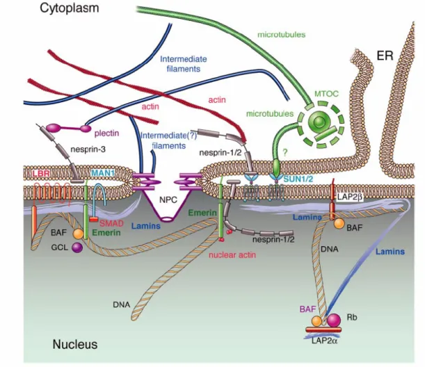
The first nuclear envelope proteins to be characterized were the nuclear lamins. In addition to lamins, several integral membrane proteins specifically localized to the INM were identified. However, the complete protein composition of the NE in vertebrates is not yet available. A recent proteomics analysis suggests that as many as eighty transmembrane proteins are localized to the INM in interphase cells (Schirmer et al., 2003). As shown in Figure 1.3, most of these INM proteins have large nucleoplasmic domains performing interactions with nuclear lamins and/or chromatin.
LBR (lamin B receptor) is one of the best-characterized INM integral proteins. The nucleoplasmic N-terminal domain of LBR interacts in vitro not only with B-type lamins, but also with chromatin via HP1 (Ye & Worman, 1996). LBR is involved in the reassembly of the nuclear envelope after cell division (Drummond et al., 1999; Okada et al., 2005).
Lamin-associated proteins (LAPs), LAP1A, LAP1B, LAP1C, and LAP2, were initially
identified in rat liver NE fractions (Foisner & Gerace, 1993). LAP1A, LAP1B and LAP1C,
are closely related and are probably alternatively spliced products of the same gene. LAP2
proteins are also expressed as a variety of isoforms (α, β, γ, δ and ε), all being alternatively
spliced products of a single gene. LAP2α does not contain a transmembrane segment and is
present diffusely throughout the nucleus, while other isoforms contain transmembrane
domains and are thus INM integral proteins. LAP2β associates specifically with lamin B1 and
chromatin. The affinity of LAP2β for lamin B1 and chromatin is reduced in the presence of
CDC2 activity (Foisner & Gerace, 1993). Microinjection of peptides corresponding to the
lamin binding domain of LAP2β into mitotic and early G1 phase cells was able to inhibit
lamina assembly, NE growth and entry into S phase (Yang et al., 1997). Similarly, GST
fusion proteins containing the N-terminal region of LAP2β also inhibited some aspects of NE
assembly and in particular, lamina assembly in cell-free extracts of Xenopus eggs (Gant et al.,
1999). Thus, these lines of evidence suggest that LAP2β mediate membrane-chromatin attachment, lamina assembly, and probably also promotion of replication by influencing chromatin structure.
In 1994, positional cloning resulted in the identification of a gene responsible for X- linked Emery-Dreifuss muscular dystrophy. Sequencing demonstrated that it encoded for a protein named emerin, that contained a stretch of about 40 amino acids with sequence similarity to a portion of LAP2 (Bione et al., 1994). Emerin was soon identified as an INM protein (Manilal et al., 1996), which interacts with A-type and possibly B-type lamins (Fairley et al., 1999; Clements et al., 2000).
Figure 1.3. Model of nuclear envelope proteins and their interactions with nearby proteins. Most
nuclear membrane proteins (LBR, LAP2, emerin, MAN1, nesprin-1 and -2) bind directly to lamins. LEM
domain proteins (LAP2, emerin, MAN1) interact with the chromatin associated protein BAF. Nesprins
interact with SUN proteins therefore connecting the nucleus with the cytoplasm. Question marks indicate
suggested but not proven interactions (taken from Broers et al., 2006).
Emerin, together with LAP2 (LAP2β, LAP2γ, LAP2δ and LAP2ε) and MAN1, belong to a growing family of proteins that is defined by the presence of a so-called “LEM domain”.
The LEM domain is composed of a motif of about 43 amino acids that is exposed to the nucleoplasm and interacts with BAF (barrier to autointegration factor), an abundant chromatin-associated protein (Laguri et al., 2001; Shumaker et al., 2001). Therefore, BAF might form a link between chromatin and the INM. Although the function of BAF is still poorly understood, it is an essential protein, as reduction of BAF levels is lethal in C. elegans embryos (Zheng et al., 2000).
Except for these NE proteins mentioned, there are two novel families of NE proteins, characterized by distinct domains (SUN or KASH). Proteins containing the KASH (Klarsicht, ANC-1 and Syne1 homology) domain interact with SUN domain (Sad1 and UNC-84 homology domain) containing proteins in the perinuclear space (Padmakumar et al., 2005;
Haque et al., 2006) (Fig 1.3), and are involved in nuclear positioning and possibly in other unknown cellular processes (Starr & Han, 2002; Patterson et al., 2004; Zhang et al., 2007).
1.2 Nuclear envelope related human diseases
For many years, the nuclear envelope was thought to function mainly as an architectural stabilizer of the nucleus, participating in assembly and disassembly processes during mitosis.
However, recent findings demonstrate that nuclear envelope proteins are involved in fundamental nuclear functions, such as gene transcription and DNA replication. To date, mutations in the genes encoding the nuclear envelope proteins were found to cause a wide range of human diseases, known collectively as nuclear envelopathies (Somech et al., 2005).
The first of these disorders to be recognized was Emery-Dreifuss Muscular Dystrophy (EDMD) (Emery & Dreifuss, 1966). This disorder is characterized by childhood onset with progressive muscle wasting and weakening. Late-onset defects of EDMD include abnormal cardiac rhythms, conduction block and cardiomyopathy that can lead to sudden cardiac arrest.
Mutations that cause X-linked EDMD were originally mapped to the EMERIN gene. A
clinically identical autosomal-dominant form of EDMD has been mapped to the LMNA gene
coding for lamin A/C (Bonne et al., 1999). Mutations in LMNA gene also associate with limb
girdle muscular dystrophy (LGMD) with atrioventricular conduction disturbances (Muchir et
al., 2000), dilated cardiomyopathy (DCM) with conduction system disease (Fatkin et al.,
1999), autosomal recessive axonal neuropathy (Charcot-Marie-Tooth disorder type 2) (De
Sandre-Giovannoli et al., 2002), Dunnigan-type familial partial lipodystrophy (Speckman et
al., 2000) and Hutchinson-Gilford progeria syndrome (Eriksson et al., 2003). Until now, more than 211 different mutations have been identified in the LMNA gene, and these mutations cause more than 15 different types of diseases (Broers et al., 2006). However, the mechanisms by which LMNA gene mutations cause these diverse tissue-specific phenotypes have not been elucidated.
Lmna
–/–mice exhibit growth retardation from 2–3 weeks of age with clinical and histological features of muscular dystrophy and uniform death by 8 weeks of age (Cutler et al., 2002). Lamin A/C deficiency primarily causes destabilization of nuclear lamina structure and enhances nuclear deformability, with intranuclear and extranuclear sequelae that promote the development of a severe form of DCM and limit compensatory hypertrophy (Nikolova et al., 2004). Lmna-/- cells have increased nuclear deformations, defective mechanotransduction, and impaired viability under mechanical strain (Lammerding et al., 2004). Lamin A/C gene deficiency in mice is also associated with mislocalization of emerin and other NE proteins. Thus, one plausible pathomechanism for EDMD, limb-girdle muscular dystrophy type 1B, hypertrophic cardiomyopathy and familial partial lipodystrophy is the presence of specific abnormalities of the nuclear envelope. The recognition of these various disorders now raises the novel possibility that the nuclear envelope may have functions that go beyond housekeeping and which impact upon cell-type specific nuclear processes.
1.3 The nuclear envelope and nuclear positioning
Nuclear positioning is essential for many cellular processes, including cell division, migration, differentiation, fertilization and polarization (Starr & Fischer, 2005; Wilhelmsen et al., 2006). In eukaryotes, the positioning of the nucleus within each cell is regulated. This positioning requires the activity of actin, microtubules and microtubule-dependent motors (Starr et al., 2001; Starr & Han, 2002). Remarkably, various studies reveal that nuclear positioning also depends on the nuclear lamina, involving KASH and SUN domain proteins.
The SUN domain containing protein, UNC-84 is an INM integral protein of C. elegans,
shown to be involved in nuclear migration and nuclear anchoring during C. elegans
development (Lee et al., 2002). UNC-84 (null) mutants result in both uncoordinated and
egg/laying defective animals and affect the nuclear envelope localization of ANC-1 (for
anchorage defective), a huge protein of 8546 residues (Starr & Han, 2002). ANC-1 also failed
to localize to the NE in alleles that have missense mutations in or near the conserved SUN
domain of UNC-84. Similar phenotypes exhibited by both ANC-1 and UNC-84 mutants in C.
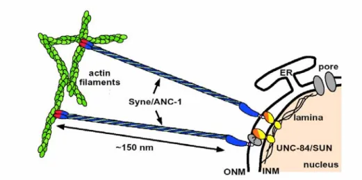
elegans suggested a genetic interaction between these proteins. The model in Figure 1.4 indicates that as an integral component of the inner nuclear membrane, the C-terminus of UNC-84 extends into the nuclear lumen where it interacts with the KASH domain of ANC-1.
The calponin domains of ANC-1 (red) attach to actin microfilaments (green) to effectively anchor nuclei in the cytoplasm. UNC-84 binds directly to another KASH domain protein of the ONM, UNC-83 in vitro (Starr et al., 2001). Mutations in either UNC-84 or UNC-83 affect the polarization of gut primordial cells and disrupt nuclear migration in P-cells (Malone et al., 1999). With the C-terminus facing the nuclear lumen, UNC-84 could retain other proteins at the outer nuclear membrane as well. These data suggest that ANC-1, UNC-83, UNC-84, the nuclear lamina, and other unknown proteins create a structural bridge across both nuclear membranes in C. elegans.
1.4 KASH domain proteins
The importance of KASH domains was first speculated through studies on several proteins including Klarsicht, ANC-1 and Syne1. Klarsicht is important for the development of the compound eye in Drosophila (Fischer-Vize & Mosley, 1994) and also involved in the transport of lipid droplets in Drosophila embryos (Welte et al., 1998). In C. elegans, ANC-1 tethers nuclei to the actin cytoskeleton (Starr & Han, 2002). The mammalian Syne1 protein
Figure 1.4. A model for nuclear anchorage in C. elegans. The KASH domain (light blue) is targeted to the outer nuclear membrane (ONM) through an interaction with the C-terminus of UNC-84 (yellow), which may occur through an unknown intermediate protein or complex (gray circle with question mark). The UNC-84/SUN N-terminus associates with lamins which are required for its proper NE positioning (Starr &
Han, 2003).
was originally identified in yeast two hybrid screens to identify binding partners of the MuSK, a tyrosine kinase of the postsynaptic membrane in muscle (Apel et al., 2000). Syne1 was also named Nesprin-1 for nuclear spectrin repeat containing protein 1. These proteins contained a highly conserved short C-terminal region (Mosley-Bishop et al., 1999; Apel et al., 2000; Starr & Han, 2002), which has been termed the KASH domain standing for Klarsicht, ANC-1 and Syne1 Homology. The KASH domain is a highly conserved C-terminal short segment, which consists of a predicted transmembrane domain followed by about 35 amino acids situated in the nuclear lumen. KASH domain containing proteins have been discovered in a variety of species. Till now, the identified members of this family include Kms1 in S. pombe, ANC-1, UNC-83, ZYG-12 in C. elegans, Klarsicht and MSP-300 in D.
melanogaster, Nesprin-1, -2 and -3 in mammals.
Many KASH domain proteins are giant molecules with a molecular weight of more than
800 kDa, such as C. elegans ANC-1 (Starr & Han, 2002), D. melanogaster MSP-300
(Rosenberg-Hasson, 1996; Zhang et al., 2002) and the mammalian proteins Nesprin-1 (also
termed as Enaptin, Syne1) and Nesprin-2 (also termed as Nuance, Syne2) (Padmakumar et al.,
2004; Zhen et al., 2002). They share the same overall domain architecture and have been
regarded as the membrane-anchored “dystrophins” of the nucleus due to their homology to
dystrophin (Zhang et al, 2001). They are composed of an N-terminal α-actinin type actin
binding domain (ABD), a massive spectrin repeat containing rod and a highly conserved C-
terminal KASH domain (Mosley-Bishop et al., 1999). The C-terminal KASH domain is
sufficient for nuclear envelope targeting of these proteins. ANC-1 functions to physically
tether the actin cytoskeleton to the ONM. Mutations in ANC-1 disrupt the positioning of
nuclei and mitochondria in C. elegans (Starr & Han, 2002). MSP-300 of Drosophila is
required for embryonic muscle morphogenesis and in the formation of embryonic muscle
attachments (Rosenberg-Hasson et al., 1996), whereas, maternal MSP-300 appears to play an
important role in actin-dependent nuclear anchorage during cytoplasmic transport (Yu et al.,
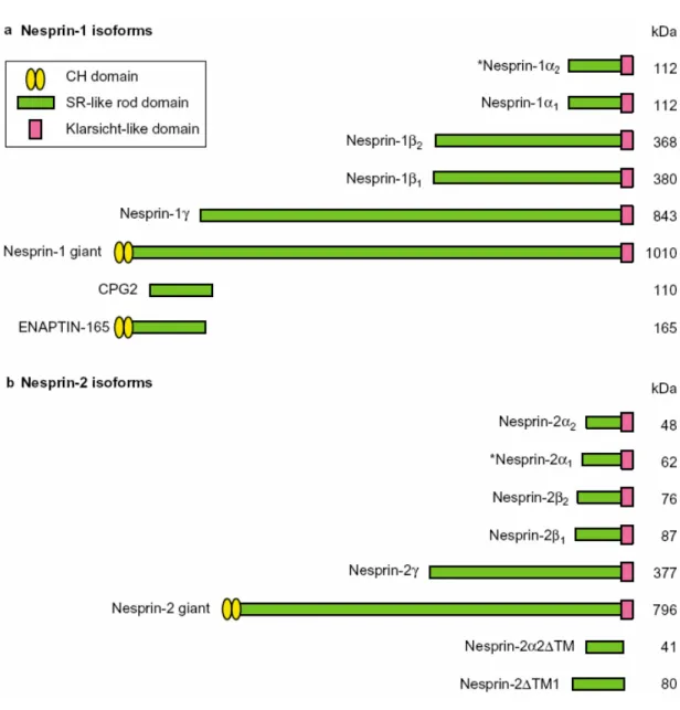
2006). The mammalian Nesprin-1 and Nesprin-2 genes display enormous complexity,
generating a wide variety of transcripts that differ in length, domain composition, expression
pattern and probably in their functional properties. To date, five smaller N-terminally
truncated isoforms (Nesprin-1α
1, Nesprin-1α
2,Nesprin-1β
1, Nesprin-1β
2and Nesprin-1γ) and
two C-terminally truncated isoforms (CPG2 and ENAPTIN-165) of Nesprin-1 were identified
(Apel et al., 2000; Mislow et al., 2002; Zhang et al., 2001; Nedivi et al., 1996). Similarly,
Nesprin-2 α-γ are small C-terminal truncated isoforms of Nesprin-2 Giant (Zhang et al., 2002)
(Fig 1.5). Nesprin-1 and Nesprin-2 encoded isoforms are broadly expressed. In contrast to Nesprin-2, which localizes predominantly to the nuclear envelope, Nesprin-1 has a rather heterogeneous subcellular distribution. Antibodies against the ABD of Nesprin-1 localize the protein at the F-actin rich structures throughout the cell as well as at the NE (Padmakumar et al., 2004). Consistent with their subcellular localizations, genetic studies involving their orthologues in lower eukaryotes suggest roles for these proteins in the attachment of intracellular membrane compartments to the actin cytoskeleton (Starr & Fischer, 2005;
Warren et al., 2005).
Figure 1.5. Domain structures of major isoforms generated from the Nesprin-1 and Nesprin-2 genes.
Isoforms are generated by alternative initiation and termination of transcription. The asterisks denote muscle
specific isoforms Nesprin-1α
2and Nesprin-2α
1(Warren et al., 2005).
1.5 SUN domain proteins
In 1999, Malone and his colleagues characterized UNC-84 as a nuclear envelope protein that is required for nuclear migration and anchoring during C. elegans development. UNC-84 consists of 1111 amino acids. It has a predicted transmembrane domain located approximately in the middle of the protein and a C-terminal region that has significant homology to a region in the S. pombe spindle pore body protein Sad1. This homologous region they called SUN domain (Sad1 and UNC-84 homology domain) (Malone et al., 1999). SUN domain proteins are evolutionarily conserved. To date, UNC-84 and matefin have been found in C. elegans, Sad1 in S. pombe, Dd-Sun1 in D. discoideum, predicted proteins Q9V996 and Q9VKG2 in Drosophila, and Sun1, Sun2 Sun3 and SPAG4 proteins in human. Besides the SUN domain, members of this family share other structural features to various extents. Most SUN domain proteins have at least one predicted transmembrane domain. Several of them span the membrane multiple times. This is a potentially useful property for proteins that are proposed to anchor mechanical-load-bearing structures and to transmit mechanical force (Tzur et al., 2006). Lastly most SUN domain proteins have at least one coiled-coil domain, which in human and mouse Sun1 is proposed to mediate homo-dimerization (Crisp et al., 2006;
Padmakumar et al., 2005).
In S. pombe, Sad1 localizes to the spindle pole body (the yeast microtubule organizing centre) and when ectopically overexpressed, Sad1 also localizes to the NE (Chikashige et al., 2006). Sad1 is required for setting up the mitotic spindle (Hagan & Yanmagida, 1995). In C.
elegans, UNC-84 is required for the localization of ANC-1 at the NE (Starr & Han 2002), whereas, matefin is involved in the attachment of centrosomes to the nucleus and also in nuclear migratory events (Malone et al., 2003). Matefin binds Ce-lamin (B-type lamin) directly in vitro but its NE localization does not depend on Ce-lamin in vivo (Fridkin et al., 2004). Matefin links the NE to the centrosome or microtubule organizing centre through ZYG-12, a NE localized KASH domain protein (Malone et al., 2003). In D. discoideum, consistent with the domain architecture of SUN proteins in C. elegens and human, Dd-Sun1 comprises an N-terminal nucleoplasmic domain, a single transmembrane domain, followed by two coiled-coil domains and the conserved SUN domain. Human Sun1, like UNC-84 in C.
elegans, is also an INM protein. Epitope-tagged Sun1 localizes to the NE in transfected cells
(Dreger et al., 2001; Padmakumar et al, 2005). The N-terminal domain of mouse and human
Sun1 is sufficient to target the protein to the NE and directly binds to A-type lamins (Haque et
al., 2006). However, unlike most INM proteins, Sun1 localization seems to be lamin A/C
independent in vivo (Padmakumar et al, 2005; Haque et al., 2006; Hasan et al., 2006). Hodzic et al. (2004) characterized Sun2 and reported that Sun2 localizes to the INM with the SUN domain located in the perinuclear space between the ONM and INM in human HeLa cells.
The N-terminus of Sun2 interacts with A-type lamins as well (Crisp et al., 2006). These findings implicate that Sun1 and Sun2 have similar topologies, with their N-terminal domains in the nucleoplasm and their SUN domains in the lumen of the nuclear envelope.
Current evidence indicates that INM positioned SUN domain proteins interact with ONM positioned KASH domain proteins, and in this manner creates protein “bridges” that span both nuclear membranes (Lee et al., 2002; Starr & Fischer, 2005; Padmakumar et al, 2005; Crisp et al., 2006).
1.6 Aim of the work
Knowledge about SUN domain containing proteins is restricted to lower eukaryotes
including fission yeast and C. elegans indicating functions in nuclear migration and
anchorage whereas very little is known about SUN domain proteins in higher eukaryotes. Do
mammalian SUN domain containing proteins share similar functions as their lower eukaryotic
orthologues? Are they also involved in nuclear migration? Do they form similar ONM-INM
complexes in higher eukaryotes? These were the main questions that we were trying to
address in the current PhD thesis. To answer these questions we applied a combination of
biochemical, cell biological and genetic approaches. Firstly, we examined the tissue and
subcellular distribution of Sun1 in mouse and human. Secondly, we investigated the mobility
and stability of Sun1 full length and various Sun1 domains by monitoring their NE dynamics
using the iFRAP methodology. Furthermore, we addressed also the NE targeting mechanism
of Sun1 by performing localization studies on various Sun1 domains, which were fused to
GFP. Thirdly, we applied a combination of yeast two-hybrid, RNAi and biochemical methods
to investigate the possible self-interaction of Sun1, and interactions between Sun1, Sun2 and
KASH domain proteins. Lastly, in order to reveal the Sun1 functions at the NE, we generated
and characterized stably transfected HaCaT cells expressing a dominant negative Sun1
polypeptide.
2. Results
2.1 Characterization, distribution and functional analysis of Sun1
2.1.1 Sun1 is the closest orthologue of C. elegans UNC-84 in mouse
UNC-84 is a nuclear envelope protein that is required for nuclear migration and nuclear anchoring during C. elegans development (Malone et al., 1999). The UNC-84 protein contains 1111 amino acids, and is composed of an N-terminal nucleoplasmic domain, one predicted transmembrane domain and a conserved C-terminal SUN domain. Current studies in C. elegans show that UNC-84 links the nucleus to the cytoskeleton by mediating interactions
Ce UNC-84 Mm Sun1 Mm Sun2 Mm Sun3 Mm SPAG4 Mm SPAG4L
998
Ce UNC-84 Mm Sun1 Mm Sun2 Mm Sun3 Mm SPAG4 Mm SPAG4L
Ce UNC-84 Mm Sun1 Mm Sun2 Mm Sun3 Mm SPAG4 Mm SPAG4L
801 146587 317 228
1058
1111 860 646 205 286 375
345 435 260699 913
Figure 2.1. Sun1 is the closest orthologue of Ce UNC-84 in mice. A blast search using the SUN domain of Ce UNC-84 (CAA94142) identified five SUN domain containing protein sequences in the mouse genome, namely Sun1 (BC048156), Sun2 (AY682987), Sun3 (AK132922), SPAG4 (CAM46026) and SPAG4L (AY307077). The alignment was done with the program BioEdit. Identical amino acids are shown in black and similar residues in gray. Mm Sun1, Mm Sun2, Mm Sun3, Mm SPAG4 and Mm SPAGL share 47.8%, 39.8%, 32% 28% and 27% identity to Ce UNC-84 SUN domain, respectively. Ce:
C. elegan; Mm: Mus musculus; SPAG4: sperm associated antigen 4; SPAG4L: sperm associated antigen 4
like protein.
with the ONM proteins ANC-1 and UNC-83. These interactions are proposed to be mediated by the SUN domain. In an effort to identify SUN domain containing proteins in higher eukaryotes, we used the UNC-84 SUN domain sequences and performed a BLAST search in the mouse database, and identified five SUN domain containing proteins (Fig. 2.1).
The Sun1 gene is located in the mouse chromosomal locus 5G.2, whereas Sun2 maps at the chromosomal locus 15E.2. The other three SUN domain containing proteins are named Sun3 (located in chromosome 11), sperm associated antigen 4 (SPAG4) and SPAG4 like protein (both located in chromosome 2). Mouse Sun1 and Sun2 display 65% identity and 81%
homology in their SUN domain. The SUN domain of mouse Sun1 shares 47.8% identity and 63% similarity to that of C. elegans UNC-84, whereas mouse Sun2 shares 39.8% identity and 59% similarity to the SUN domain of C. elegans UNC-84. Sun3, SPAG4 and SPAG4 like protein are small proteins less than 500 amino acids and share lower identities with UNC-84.
Ce UNC-84 Hs Sun1 Hs Sun2
Ce UNC-84 Hs Sun1 Hs Sun2
Ce UNC-84 Hs Sun1 Hs Sun2
998
1058
1111 699
758 664
717 812 605
Figure 2.2. Alignment of C. elegans UNC-84 and Homo sapiens Sun1 and Sun2 SUN domains. The
SUN domain of C. elegans UNC-84 was aligned with human Sun1 (KIAA0810) and Sun2 (NM015374)
SUN domains. The alignment was done with BioEdit. Identical amino acids are shown in black and
similar residues in gray. SUN domains of human Sun1 and human Sun2 display 48% and 39% identity to
that of C. elegans UNC-84, respectively. Ce: C. elegans; Hs: H. Sapiens.
In addition, human Sun1 and Sun2 were also aligned with C. elegans UNC-84 (Fig. 2.2).
Similarly, human Sun1 displays 48% identity and 64% similarity to the SUN domain of UNC-84. Human Sun2 displays 39% identity and 58% similarity to the SUN domain of UNC- 84. Consequently, Sun1 is the closest orthologue of C. elegans UNC-84 in mouse and human.
2.1.2 Phylogenetic tree of SUN domain containing proteins
A phylogenetic analysis of SUN domain proteins from H. sapiens, M. musculus, C.
elegans, D. discoideum and S. pombe shows that H. sapiens Sun1, Sun2 and M. musculus Sun1, Sun2 are closely related to each other. In contrast, the sequences of H. sapiens Sun3, SPAG4 and M. musculus Sun3, SPAG4, SPAG4L are more distantly related to Sun1 and Sun2. C. elegans UNC-84 shares higher homology to H. sapiens Sun1 than H. sapiens Sun3 to Sun1. D. discoideum Sun1 is closer to the human and mouse proteins than C. elegans matefin and S. pombe Sad1 (Fig. 2.3). It is evident from this evolutionary tree that there is a high degree of sequence conservation between mouse and human.
0.05
Hs Sun1 Mm Sun1
Hs Sun2
Ce UNC-84 Hs Sun3
Mm Sun3
Ce matefin Sp Sad1
Figure 2.3. Phylogenetic relationships of SUN domain proteins. Hs: Homo sapiens, Mm: Mus musculus, Ce: Caenorhabditis elegans, Dd: Dictyostelium discoideum, Sp: Schizosaccharomyces pombe. Hs Sun1, Sun2 and Mm Sun1, Sun2 are closely related and forms a subgroup. Hs Sun3 and Mm Sun3, SPAG4, SPAG4L form a more ancient and distinct subgroup. The scale bar indicates genetic distance and is equivalent to 5% diversity. The evolutionary tree was generated using AlignX of Vector NTI program.
Mm Sun2
Mm SPAG4L Mm SPAG4
Dd Sun1
2.1.3 Sun1 is broadly expressed in mice
To study the tissue distribution of mouse Sun1, we employed an RT-PCR approach.
Total RNA was isolated from multiple wild-type mouse tissue samples and subjected to
reverse transcription. The same amount of cDNA obtained from each tissue was used as
template for the following PCR analysis. To investigate the Sun1 expression profile, we
designed two distinct pairs of primers. One resides in the N-terminus (5’NT/3’NT; amplifying
the coding sequence of residues 22-163), whereas the other set is in the C-terminus
(5’CT/3’CT; amplifying the coding sequence of residues 748-887). The primer sequences
were listed in materials and methods. With the C-terminal primers, a 400-bp product was
amplified from all the mouse tissue samples. This is consistent with the northern blot analysis
of mRNA from multiple mouse tissues (Crisp et al., 2006), indicating that Sun1 is
ubiquitously expressed, suggesting thus that Sun1 may play an essential role in mice. Mouse
GAPDH primers were used as positive controls. Interestingly, using N-terminal primers, PCR
products were only amplified from brain, kidney, testis, skin and skeletal muscle, but not from
heart, liver and lung, suggesting the existence of alternatively spliced isoforms of Sun1 in
those tissues (Fig. 2.4). These data are also supported by the examination of the EST-database
using Sun1 sequences as a query, indicating that Sun1 exists in multiple, alternatively spliced
isoforms. The northern blot analysis of mRNA from multiple mouse tissues revealed at least
four or five discrete Sun1 transcripts in different tissues (Crisp et al., 2006). In summary,
Sun1 is broadly expressed in many tissues and is encoded by a gene, which gives rise to many
splicing variants.
br ai n he ar t
GAPDH
liv er
lu ng ki dn ey te sti s
sk in Sk . m us cl e
Sun1-CT
Sun1-NT 400 bp
400 bp 800 bp
2.1.4 Subcellular localization of endogenous human Sun1
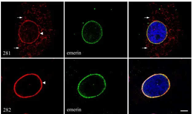
Endogenous human Sun1 was detected in human keratinocyte HaCaT cells by indirect immunofluorescence experiments using two different polyclonal antisera, which were kindly provided by Dr. Joseph Gotzmann (Biocenter, Vienna). One antibody is designed against the very N-terminus of Sun1 (termed 281; against polypeptides: MDFSRLHMYSPPQC), whereas the other one is directed against the C-terminus of Sun1 (termed 282; against polypeptides: RVDDPQDVFKPTTSR). Staining of both reagents indicates that Sun1 is localized largely to the nuclear envelope (Fig. 2.5, arrowheads) and colocalizes with the inner nuclear membrane protein emerin (Fig. 2.5 right panel). Thus, Sun1 is a nuclear membrane protein. Permeabilization studies employing digitonin revealed that Sun1 is an inner nuclear membrane protein (Padmakumar et al, 2005). As shown in Figure 2.5, the antiserum 282 stained only the nuclear envelope, whereas the antiserum 281 stained in addition also the cytoplasm (arrows). Therefore, for all other further studies we used preferentially the 282 antiserum.
Figure 2.4. Mouse Sun1 mRNA is broadly expresses in various tissues. Total RNA isolated from each tissue was subjected to RT-PCR amplification using the 5’NT/3’NT or 5’CT/3’CT primers, respectively.
All RT-PCR products were analyzed by agarose gel electrophoresis and visualized by ethidium bromide
staining. GAPDH was used as control.
281 emerin
emerin 282
2.1.5 Domain analysis of Sun1
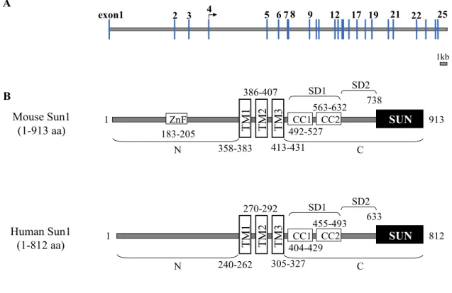
The mouse SUN1 gene is located in the mouse chromosomal locus 5G.2, spanning 49 kb and is encoded by 25 exons. A schematic representation of the intron and exon organization of mouse Sun1 is represented in Figure 2.6A. SUN1 codes for a 100 kDa protein (accession No.
BC048156) and is composed of 913 amino acids. The protein was predicted to have three putative transmembrane domains (358-383, 386-407 and 413-531 aa) located approximately in the middle of the protein, using the Tmpred software (Hofmann & Stoffel, 1993). The SMART program predicted a ZnF-C2H2 domain (183-205 aa) at the N-terminus of the protein. The C-terminus of the protein contains two coiled-coil domains (492-527 and 563- 632 aa). The last 175 amino acids form the evolutionarily conserved SUN domain (738-913
Figure 2.5. Endogenous human Sun1 localizes to the nuclear membrane in HaCaT cells. HaCaT cells were fixed with paraformaldehyde and stained with antibodies against the N-terminus (281) and the C-terminus of Sun1 (282), respectively. Emerin was stained by emerin specific antibodies and DAPI was used for nuclear staining. The secondary antibodies used for 281 and 282 were conjugated with Alexa 568 and for emerin were conjugated with Cy3. The cells were observed using the confocal microscope.
Bar=6 µm.
aa), which is highly homologous to C. elegans UNC-84. The region between the transmembrane and the SUN domain was divided into two subdomains, SD1 (432-632 aa) and SD2 (633-737 aa) for functional tests. SD1 contains the putative coiled-coils whereas SD2 does not contain any known structural features (Fig. 2.6B).
Mouse Sun1
(1-913 aa) TM2 TM 3
SUN
TM 1 CC1
CC2 ZnF
SD1 SD2
Human Sun1 (1-812 aa)
1 913
183-205
358-383 386-407
413-431 492-527
563-632 738
N C
TM2 TM3 SUN CC1
TM1 CC2
SD1 SD2
1 812
240-262 270-292
305-327 404-429
455-493 633
N C
exon1 4 6 7 8 9 12 17 19 21 22 25
3
2 5
A
B
1kb
Human Sun1 is a protein of 812 amino acids with a predicted mass of about 90 kDa. The domain structure of human Sun1 is identical to that of mouse, except that it lacks the
Figure 2.6. Genomic organization of mouse SUN1 and domain architecture of mouse and human Sun1.
(A) Stick diagram illustrating the genomic organization of the mouse SUN1 locus. Numbered vertical blue bars denote exons, yellow bars between the blue bars represent introns. The start codon is denoted by arrow.
The diagram is drawn to scale. Bar=1 kb (B) The domain architecture of mouse and human Sun1 were
predicted by the computer program SMART (Simple Modular ARchitecture Research Tool) and TMpred (a
transmembrane domain prediction software) program. Mouse Sun1 has three transmembrane domains in the
middle of the protein. The N-terminus of the protein has a ZnF-C2H2 domain and the C-terminus has two
coiled-coil domains. The C-terminal end of the protein is the evolutionarily homologous SUN domain. Human
Sun1 shares the same domain architecture with mouse Sun1 except for the ZnF domain. The numbers below
and above the schematic indicate amino acid positions of the Sun1 protein sequence.
proposed zinc-finger motif (Fig. 2.6B). The whole protein sequence of human Sun1 shares about 69 % identity and 77 % similarity to that of mouse Sun1.
2.1.6 Sun1 has three transmembrane domains
Tmpred software predicted three transmembrane domains for mouse Sun1. However,
other programmes like SMART revealed only two transmembrane domains, especially the
first transmembrane domain (358-383 aa) was ambiguous. In order to illuminate the real
transmembrane domain structure of Sun1, various constructs expressing EGFP fusion proteins
containing the entire Sun1 N-terminus and the first (Sun1-1TM-N) or the first two
transmembrane domains (Sun1-2TM-N) were made (Fig. 2.7A). Constructs were transiently
transfected into COS7 cells and the transfected cells were examined by direct
immunofluorescence. Sun1-2TM-N, which contains at least one real transmembrane domain,
was localized to the nuclear membrane in transfected cells as shown in Figure 2.7B lower
panel. Similarly to Sun1-2TM-N transfected cells, Sun1-1TM-N localized also to the nuclear
membrane (Fig. 2.7B higher panel). The nuclear envelope localization of Sun1-1TM-N and
Sun1-2TM-N are indicated by arrows. Thus, both fusion proteins have the ability to localize
to the nuclear envelope suggesting that the first transmembrane domain is indeed a
transmembrane domain. Haque et al. (2006) showed that the Sun1 N-terminus interacts with
nuclear lamin A in vitro. This combined with our data, which show that Sun1 has three
transmembrane domains and is able to span the inner nuclear membrane three times, suggests
that the Sun1 N-terminus resides in the nucleoplasm, whereas the C-terminus is situated in the
perinuclear space. Interestingly, a similar domain topology has been proposed for UNC-84
and matefin in C. elegans (Starr & Han, 2003; Fridkin et al., 2004) and Sun2 in human
(Hodzic et al., 2004).
2.1.7 Sun1 dynamics at the nuclear envelope
Sun1 is an inner nuclear membrane (INM) protein, and the N- and C-termini of Sun1 can be recruited to the NE, independently (Padmakumar et al., 2005). It has been proposed that integral INM proteins are retained at the INM by forming stable interactions with other INM proteins, the nuclear lamina or chromatin (Ohba et al., 2004). Therefore the N- and C-termini of Sun1 are likely to have distinct interactions at the nuclear envelope. Such interactions can be reflected by the kinetic properties of the protein. To investigate more accurately the kinetic properties of Sun1 full-length and the Sun1 N-and C-termini at the nuclear envelope, we performed inverse fluorescence recovery after photobleaching experiments (iFRAPs).
Figure 2.7. Sun1-1TM-N and Sun1-2TM-N localize to the nuclear envelope in COS7 cells. (A) Schematic GFP mouse Sun1 N-terminal constructs used in transfection experiments. Green oval indicates GFP and numbered boxes indicate transmembrane domains. (B) COS7 cells were transiently transfected with the appropriate plasmids and cells were subjected to direct fluorescence microscopy. DAPI was used to stain the nuclei. Images were taken with a confocal microscope. Bar =10 µm.
5µm
A
412 aa 384 aa
Sun1-N+2TM Sun1-N+1TM GFP
GFP 1 1
Sun1-1TM-N
Sun1-2TM-N
DAPI
DAPI
merge
merge
B
Sun1-1TM-N
Sun1-2TM-N
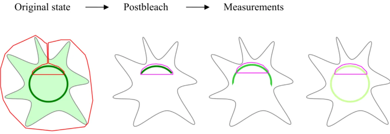
iFRAPs are more suitable to analyze binding interactions than classical FRAPs because the free pool of the tagged protein is bleached away, allowing precise measurement of the fluorescence changes of only the bound molecules. In an iFRAP, the entire cellular fluorescence is photobleached except for a small nuclear membrane region of interest containing the unbleached tagged proteins (Fig. 2.8). The dissociation of the tagged protein from the nuclear membrane is then reflected by the loss of fluorescence from this region over time monitored by confocal microscopy. The dissociation kinetics reflects the stability of the protein at the nuclear envelope.
To estimate the binding ability of distinct domains of Sun1, GFP-tagged Sun1 full-length and various N- and C-terminal polypeptides containing different functional domains (Fig. 2.9) were used in iFRAPs. As depicted in Figure 2.9, GFP-tagged Sun1 full-length, Sun1-2TM-N, Sun1-TM-C, Sun1-TM-SD1, 2 and Sun1-TM-CC-∆SUN were transiently transfected in HeLa cells. All fusion proteins are nuclear membrane associated. Figures 2.10A, 2.11A and C, 2.12A and C, show single iFRAP experiments for Sun1 full-length and for four structurally distinct Sun1 GFP-fusions with different dynamic behaviors. At least four similar experiments
Original state Postbleach Measurements
Figure 2.8. Presentation of the iFRAP technique. The scheme depicts the different stages of a typical
cell with nucleus (dark green circle) and cytoplasm (light green) in iFRAP. Bleached region is marked by
the red boundary. All of the fluorescence of the cell is bleached except for a small region of interest (pink
boundary). The postbleach images are analyzed to display the loss of fluorescence from the unbleached
region.
were performed for each GFP-tagged protein and were averaged to generate the mean fluorescence decays plotted in Figure 2.10B, 2.11B and D, 2.12B and D.
GFP-Sun1 full-length (1-913 aa)
GFP-Sun1-2TM-N (1-412 aa)
GFP-Sun1-TM-C
(358-913 aa) GFP
GFP-Sun1-TM-SD1, 2 GFP (358-737 aa)
TM2 TM3
GFP CC1 SUN
TM1 CC2 TM 2
GFP TM 1 TM2 TM3 CC1 SUN
TM1 CC2 TM2 TM3 CC1
TM1 CC2
GFP-Sun1-TM- Δ CC-SUN
(358-491, 633-913 aa) GFP
TM2 TM3 SUN
TM 1
ZnF
ZnF
SD1 SD2
2.1.7.1 iFRAPs of GFP-Sun1 full-length
Similar to endogenous Sun1 protein, GFP-Sun1 full-length mostly localized to the nuclear membrane in transfected cells. The whole cell except 1/3 of the nuclear envelope region was bleached. The fluorescence within this region of interest (ROI) was then followed every 3 minutes over a period of up to 10 hours. To calculate the loss of fluorescence attributed to the imaging process alone, the sum of pixel intensities was also calculated for the
Figure 2.9. Schematic of GFP-Sun1 and different N-/C- terminally truncated Sun1 constructs, which were employed in the iFRAP assays. The GFP part is indicated by green ovals. Sun1 amino acid residues fused to GFP are indicated by numbers. Domain labeling is shown as in Figure 2.6. The constructs were transfected into HeLa cells by FUGENE 6 to determine the dynamics of Sun1 at the nuclear envelope. GFP: green fluorescent protein; ZnF: zinc finger; TM: transmembrane domain; CC:
coiled-coil domain; SUN: Sad1/UNC-84 homology domain.
entire cell. This was used to normalize the fluorescence intensity for each ROI. The fluorescent signal of GFP-Sun1 full-length dropped very slowly, and had a residence time of more than 10 hours (Fig 2.10). This indicates that a large proportion of Sun1 at the nuclear membrane is immobile, at least within the time frame of these experiments.
Pre-bleach Post-bleach 1 h 33 m 4 h 40 m 7 h 32 m 10 h
0 2 4 6 8 10
No rm al ized fl uo re scen ce in te ns it y
Time (h) A
B
0 0.2 0.4 0.6 0.8 1.0 1.2
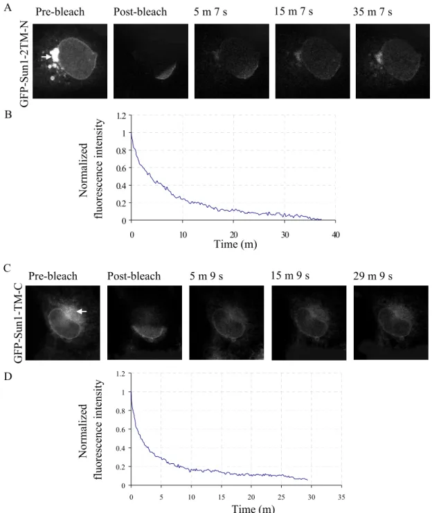
2.1.7.2 iFRAPs of GFP-Sun1-2TM-N and GFP-Sun1-TM-C
GFP-Sun1-2TM-N and GFP-Sun1-TM-C have the ability to localize to the nuclear envelope. In many cases we also observed substantial accumulation of the fusion proteins in the ER as shown in the pre-bleach images of Figure 2.11A and 2.11C (arrows). GFP-Sun1-
Figure 2.10. GFP-Sun1 full-length dynamics at the nuclear envelope. (A) HeLa cells expressing GFP- Sun1 were imaged before and after photobleaching of the entire nucleus, with the exception of a small region of the nuclear envelope. The loss of fluorescent signal was monitored using time-lapse microscopy.
(B) Corresponding plots of fluorescence decay kinetics from the unbleached region of iFRAP experiments
of GFP-Sun1. Six individually transfected cells were measured. The experiments were performed on a
customized LSM510 confocal microscope using a 40 x 1.3 NA oil objective and a completely open
pinhole.
2TM-N had a residence time of 35 min and GFP-Sun1-TM-C had a residence time of 29 min (Fig. 2.11). Thus, both constructs display a significantly reduced stability when compared to full-length Sun1. These results suggest that both N- and C- termini of Sun1 are required for the stability of Sun1 at the nuclear envelope.
0 0.2 0.4 0.6 0.8 1 1.2
0 10 20 30 40
Norm alize d fluo re sc ence in te nsit y
Pre-bleach Post-bleach 5 m 7 s 15 m 7 s 35 m 7 s
Time (m) A
B
No rmaliz ed fl uo rescen ce i ntensit y
0 0.2 0.4 0.6 0.8 1 1.2
0 5 10 15 20 25 30 35
Time (m)
Pre-bleach Post-bleach 5 m 9 s 15 m 9 s 29 m 9 s C
D
GFP-Sun 1- 2TM -N GF P- Su n1-TM -C
Figure 2.11. GFP-Sun1-2TM-N and GFP-Sun1-TM-C dynamics at the nuclear envelope. (A) and (C) Selected images of iFRAPs in GFP-Sun1-2TM-N and GFP-Sun1-TM-C transfected cells, respectively.
Arrows indicate the accumulation of the GFP-fusions in the ER. (B) and (D) Corresponding plots of
fluorescence decay kinetics from the unbleached region of iFRAP experiments of GFP-Sun1-2TM-N and
GFP-Sun1-TM-C. Four individual cells for each construct were analyzed.
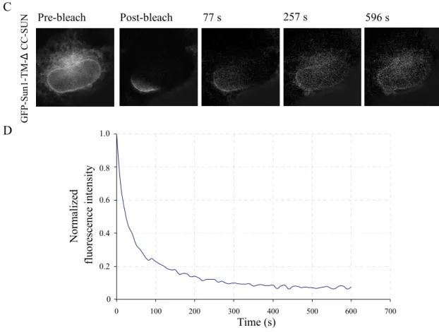
2.1.7.3 iFRAPs of GFP-Sun1-TM-SD1, 2 and GFP-Sun1-TM-∆CC-SUN
GFP-Sun1-TM-SD1, 2 and GFP-Sun1-TM-∆CC-SUN are clearly associated with the nuclear membrane and the ER membrane as represented in the pre-bleach images in Figure 2.12A and C. GFP-Sun1-2TM-SD1, 2 had a residence time of about 26 min (Fig. 2.12A and 2.12B), which is slightly faster than that of GFP-Sun1-TM-C. However, when iFRAP was applied to cells expressing GFP-Sun1-TM-∆CC-SUN, the major fraction of the fluorescence signal for GFP-Sun1-TM-∆CC-SUN dropped rapidly within 2 min and GFP-Sun1-TM-∆CC- SUN had a residence time of ~8-10 min (Fig. 2.12 C and D), which is much lower than that of GFP-Sun1-TM-C and GFP-Sun1-TM-SD1, 2. The fast drop-down of the fluorescence signal of GFP-Sun1-TM-C, GFP-Sun1-TM-SD1, 2 and GFP-Sun1-TM-∆CC-SUN within the first 10s after bleaching may be due to the migration of unbound GFP molecules in the unbleached ER region. GFP-Sun1-TM-SD1, 2 which contains the coiled-coil domains and lacks the SUN domain is more stable than GFP-Sun1-TM-∆CC-SUN which contains the SUN domain but lacks the coiled-coil domains, suggesting that the SUN domain of Sun1 may have a more
0 5 10 15 20 25 30
Nor m aliz ed fl uo re sc en ce in te ns it y
Time (m)
Pre-bleach Post-bleach 5 m 7 s 15 m 7 s 26 m 27 s
A
B
G F P- Su n1 -T M SD 1, 2
1.2
1.0
0.8
0.6
0.4
0.2
0
Norm alize d fl uo res cen ce in te ns ity
Time (s)
0 100 200 300 400 500 600 700
Pre-bleach Post-bleach 77 s 257 s 596 s
C
D
GFP-Sun1-TM-ΔCC-SUN
0 0.4 0.6 0.8 1.0
0.2
dynamic binding at the nuclear membrane in comparison to the N-terminal domain and the C- terminal coiled-coil domains. Moreover, the coiled-coil domains appear to be important for Sun1 NE dynamics suggesting that these domains may have additional binding partners at the nuclear envelope.
In summary, as shown in Table 1, GFP-Sun1 full-length has a residence time of more than 10 hours in iFRAPs, indicating that Sun1 is an immobile protein closely associated with the nuclear envelope. GFP-Sun1-2TM-N and GFP-Sun1-TM-C have residence times of 35 minutes and 29 minutes respectively, which are much more mobile than GFP-Sun1 full-length, suggesting that the stability of Sun1 at the nuclear envelope requires both Sun1 N- and C-
Figure 2.12. GFP-Sun1-TM-∆CC-SUN dynamics at the nuclear envelope. (A) and (C) Selected
images of iFRAPs of GFP-Sun1-TM-SD1, 2 and GFP-Sun1-TM-∆CC-SUN transfected cells,
respectively. (B) and (D) Corresponding plots of fluorescence decay kinetics from the unbleached region
of iFRAP experiments of GFP-Sun1-TM-SD1, 2 and GFP-Sun1-TM-∆CC-SUN. Four individual GFP-
Sun1-TM-SD1, 2 transfected cells and six GFP-Sun1-TM-∆CC-SUN transfected cells were investigated
by iFRAPs.
termini. GFP-Sun1-TM-SD1, 2, which contains the coiled-coil domains, has a residence time of 26 minutes that is slightly faster than GFP-Sun1-TM-C. However, GFP-Sun1-TM-∆CC- SUN, which contains only the SUN domain, migrated much faster than other GFP-Sun1 fusions and it has a residence time of 8-10 minutes. Thus, the Sun1 coiled-coil domains may be significant nuclear envelope retention domains.
Table 1. GFP-tagged Sun1 dynamics at the NE Sun1 fusion proteins Residence time in iFRAPs GFP-Sun1 full-length More than 10 hours
GFP-Sun1-2TM-N ~35 minutes
GFP-Sun1-TM-C ~29 minutes
GFP-Sun1-TM-SD1, 2 ~26 minutes
GFP-Sun1-TM-∆CC-SUN ~8-10 minutes
2.1.8 Sun1 interacts with chromatin
The iFRAPs on Sun1 full-length suggest that Sun1 is a stable integral protein at the nuclear envelope. Although Sun1 interacts with lamin A, the presence of lamin A/C is not required for Sun1 nuclear envelope localization (Haque et al., 2006). Therefore, Sun1 was proposed to bind to other nuclear factors. Many INM proteins like emerin, LAP2, MAN1 or lamin B receptor, interact with chromatin-associated proteins or bind directly to DNA, providing a membrane anchor important for chromatin organization (Wolff et al., 2001; Cai et al., 2001; Lin et al., 2000; Ye et al., 1997). Sun1 is also an INM protein with its N-terminus exposed to the nucleoplasm. Interestingly mouse Sun1 contains a predicted zinc finger DNA-
Table 1. Dynamics of GFP-tagged Sun1 fusions at the nuclear envelope. HeLa cells were transiently
transfected with GFP fusion constructs and examined 48 hours after transfection. iFRAPs were performed on
a customized LSM510 confocal microscope. Most cellular fluorescence was bleached using full laser power
and leaving only a fraction (typically 10−20%) of the nuclear envelope unbleached. Post-bleach images were
then acquired for up to 10 hours at regular time intervals, depending on the dynamics of the GFP fusion
proteins. In iFRAPs, GFP-Sun1 full-length has a residence time of more than 10 hours. GFP-Sun1-2TM-N,
GFP-Sun1-TM-C and GFP-Sun1-TMSD1, 2 reside at the NE ~35, ~29 and ~26 minutes, respectively. GFP-
Sun1-TM-∆CC-SUN has a residence time of 8-10 minutes.
binding motif. Therefore, a similar interaction between Sun1 and chromatin may exist. In order to test whether Sun1 interacts with chromatin, we carried out chromatin immunoprecipitation assays (ChIP). Formaldehyde cross-linked chromatin extracts from sonicated HaCaT cells were incubated with Sun1 antibodies (Sun1 281) and after immunoprecipitation, the proteins were removed by proteinase K treatment. The precipitated DNA was analyzed by PCR using a set of primers binding to the 5’ UTR of GAPDH.
Antibodies against RNA polymerase II were used as positive control and an unrelated antibody (negative IgG) was used as a negative control. As depicted in Figure 2.13, the DNA fragment was coimmunoprecipitated by Sun1 antibodies but not with the negative control IgG antibodies. Our results indicate thus, that Sun1 is able to interact with chromatin.
GAPDH
Inp ut DN A Po l II
Su n1 281
Ne gat ive Ig G
2.1.9 Sun1 oligomerizes via its coiled-coil domains
The results of the above iFRAP experiments suggested that the Sun1 coiled-coil domains may be a significant nuclear envelope retention domain. Coiled-coils have traditionally been recognized as an oligomerization unit in a large number of proteins (Burkhard et al., 2001).
Thus, one attractive mechanism would be that oligomerization of the GFP-fusion protein with the endogenous Sun1 leads to the retention of the GFP-fusion protein at the NE. To
Figure 2.13. Sun1 interacts with chromatin in HaCaT cells. HaCaT cell lysates were sheared by
sonication and used in chromatin immunoprecipitation assays in the presence of N-terminal Sun1
polyclonal antibodies (281). Primers binding to exon 1 of the GAPDH gene (for primer sequences please
see materials and methods) were designed and used for detection of immunoprecipitated DNA. An
unrelated antibody (negative IgG) was used for a control immunoprecipitation. An antibody against RNA
polymerase II (Pol II) was used as a positive control. Input DNA was isolated genomic DNA from the
sheared HaCaT cell lysates, which was used to test the designed primers.
investigate whether the predicted coiled-coils within the C-terminus of Sun1 are required for oligomerization, biochemical studies were performed.
The cDNA sequence encoding mouse Sun1 SD1, 2 (residues 432-737), which contains the Sun1 coiled-coil domains were amplified by PCR, and cloned into pGEX4T-1 vector and expressed in E. coli as GST fusion protein (GST-SD1, 2) and purified by affinity chromatography with Glutathione Sepharose 4B under native conditions. Thrombin digestion of GST-SD1, 2 generated the GST-free Sun1 SD1, 2 polypeptides (Fig. 2.14). The purified SD1, 2 was used for in vitro Native-PAGE and cross-linking assays.
97 kDa 66 kDa 45 kDa
30 kDa
1 2 3 4
GST-SD1, 2
SD1, 2
SDS-PAGE analysis of the purified SD1, 2 demonstrated that under reduced conditions the protein behaves as a monomer (32 kDa) (Fig. 2.15A). To examine whether SD1, 2 oligomerizes under native conditions, we performed Native-PAGE analysis, which is essentially used to determine native masses and oligomeric states. Purified SD1, 2 was mixed with 2 x native sample buffer and loaded into the native gel without heating. BSA was used as the standard. The gel was run at 4°C and subsequently stained with Coomassie Blue. By
Figure 2.14. Purification of mouse Sun1 SD1, 2 from E. coli. 1. Standard low molecular weight markers
2. Cell lysate of uninduced bacterial host DH5α 3. Cell lysate of IPTG induced bacteria, which expressed
GST-SD1, 2 fusion protein. 4. Purified SD1, 2 fraction after thrombin digestion. Samples were loaded on
a 12 % polyacrylamide gel and stained by Coomassie blue.
Native-PAGE we indeed observed a dimer band (64 kDa) and an additional tetramer band migrating as a 130 kDa protein (Fig. 2.15B).
B. Native-PAGE A. SDS-PAGE
97 kDa 66 kDa 45 kDa
30 kDa
monomer 66 kDa dimer
140 kDa 232 kDa
M SD1, 2 M BSA SD1, 2
tetramer
In addition to Native-PAGE analysis, we also performed chemical cross-linking experiments to provide additional evidence for the oligomerization of the Sun1 coiled-coil domains. The purified Sun1 SD1, 2 proteins were submitted to cross-linking using various concentrations of glutaraldehyde. The cross-linked samples were then analysed by SDS- PAGE. Without glutaraldehyde treatment, only the monomer band was observed. While the purified SD1, 2 proteins were treated with the cross-linking reagent, a dimer and tetramer bands were evident on the Coomassie stained gels. Furthermore, the relative amount of the monomer was reduced, indicating that the SD1, 2 fractions containing coiled-coils forms
Figure 2.15. The purified mouse Sun1 SD1, 2 forms dimers and tetramers under native conditions.
(A) Purified SD1, 2 recombinant protein was supplemented with SDS sample buffer containing β-
mercaptoethanol and subjected to SDS-PAGE using a 12% SDS-polyacrylamide gel. M, standard low
molecular weight marker proteins; SD1, 2, purified SD1, 2 fraction of mouse Sun1 under reduced
conditions. (B) Native–PAGE. Just before gel loading, the purified SD1, 2 was supplemented with blue
native sample buffer lacking SDS and β-mercaptoethanol. The samples were loaded on a 10% native-
polyacrylamide gel. After electrophoresis, the gel was visualized by Coomassie staining. M, native
molecular weight marker; SD1, 2, purified SD1, 2 fraction under native conditions. BSA was loaded as
control. BSA; Bovine serum albumin.
dimers or oligomers in vitro (Fig. 2.16A). In contrast, when we performed a similar assay using BSA as negative control, no higher mass polypeptides were detected in the samples treated with the glutaraldehyde cross-linking reagent (Fig. 2.16B).
M ar ke r 0 0. 00 1%
0. 00 5%
0. 01 % 0. 01 5%
0. 02 % 0. 03 %
97 kDa 66 kDa 45 kDa
30 kDa
monomer dimer
97 kDa 66 kDa 45 kDa
30 kDa B. BSA
M ar ke r 0 0. 00 1%
0. 01 % 0. 02 %
A. SD1, 2
tetramer
To further corroborate our in vitro results, we performed GST pull-down assays. For this we fused GST to the coiled-coil domains of Sun1 (GST-Sun1-SD1; 432-632 aa) to pull down ectopically expressed GFP-Sun1-SD1, 2 (432-737 aa) fusion proteins from COS7 cell lysates (Fig. 2.17A). The GFP-Sun1-SD1, 2 fusions lack the transmembrane domain and localize diffusely in the cytoplasm (data not shown). As anticipated GST-Sun1-SD1 was able to precipitate GFP-Sun1-SD1, 2 from COS7 cell lysates (Fig. 2.17C). In contrast to GST-Sun1- SD1, no interaction with GFP-Sun1-SD1, 2 was observed for GST alone (Fig. 2.17C). To confirm that coiled-coil domains indeed oligomerizes with itself but not with the SD2 domain, GFP-Sun1-SD2 (432-491, 633-737 aa) was constructed, expressed in COS7 cells and used in GST pull-down assays. As shown in Figure 2.17C, there is no interaction between GST-Sun1- SD1 and GFP-Sun1-SD2.
Figure 2.16. The Sun1 SD1, 2 oligomerizes when cross-linked with glutaraldehyde. (A) Equal amounts of the purified mouse Sun1 SD1, 2 were cross-linked with 0.001%-0.03% glutaraldehyde for 30 minutes at room temperature, respectively. Reactions were terminated by addition of SDS sample buffer.
The samples were loaded on a 12 % SDS gel and stained by Coomassie Blue. (B) As a negative control,
BSA was cross-linked with 0.001%-0.02% glutaraldehyde and examined by SDS-PAGE analysis.
GST-Sun1-SD1 (432-632 aa) GFP-Sun1-SD2
(432-491, 633-737 aa) GST
B Input
GFP-Sun1-SD1, 2- GFP-Sun1-SD2-
-GFP-Sun1-SD1, 2 C GST pull-down
IB: GST pAb IB: GFP mAb GFP-SD2 GFP-SD1, 2
GST GFP-SD2 GFP-SD1, 2
GST-SD1
-
+ +- -
+-
+-
+-
++
-
+-
A GFP-Sun1-SD1, 2
(432-737 aa)
GFPCC1 CC2
GFP
GST CC1 CC2
GST