Characterisation of Enaptin and Sun1, two novel mammalian nuclear envelope proteins
INAUGURAL-DISSERTATION zur
Erlangung des Doktorgrades
der Mathematisch-Naturwissenschaftlichen Fakultät der Universität zü Köln
vorgelegt von
Padmakumar Velayuthan Chellammal aus
Peruvilai, Indien
Köln, 2004
Referees/Berichterstatter: Prof. Dr. Angelika A. Noegel Prof. Dr. Karin Schnetz Date of oral examination: 02.07.2004
Tag der mündlichen Prüfung
The present research work was carried out under the supervision of Prof.
Dr. Angelika A. Noegel, in the Institute of Biochemistry I, Medical Faculty, University of Cologne, Cologne, Germany. From August 2001 to July 2004.
Diese Arbeit wurde von August 2001 bis Juli 2004 am Biochemischen Institut I der Medizinischen Fakultät der Universität zu Köln unter der Leitung von Prof. Dr. Angelika A. Noegel durchgeführt.
First of all, I would like to express my heartiest gratitude to Prof.Dr.Noegel for giving me an opportunity to work in her group with Enaptin. Her positive attitude, constant encouragement and sustained interest in my project proved essential for my successful PhD work.
I would like to thank Dr.Iakowos Karakesisoglou (Akis), our group leader for the excellent advice with my work, plentiful of encouragement and lots of motivation. He offered a hospitable environment in the lab with his friendly mannerisms and of course not to forget his sense of humour.
I would also like to thank Dr.Elena Korenbaum with whom I started my PhD in the first year. It was a wonderful first year with her.
It would be unfair if I don’t convey my thanks to Dr.Franciso Rivero, who helped me learn Confocal Microscopy and not the least, Spanish (all bad words).
I would also like to thank Jan Melichar and Martina Munck who helped me in the beginning with the administrative work and special thanks to Martina for teaching me many laboratory techniques, Cell Cuture to name one.
I would also like to thank Bettina Lauss, our secretary who made our life in Köln easy with her excellent administrative skills and promptness and Budi, our system administrator to lend his helping hand whenever I had a problem with my computer.
I would like to acknowledge my father, Velayuthan and mother, Chellammal who made enough sacrifices for me to be here today and also bear to be away from me for a long time.
It would be appropriate to note that it can’t be coincidental that the majority of successes in my PhD and my friendship with Yogi came in the final year.
My former and current lab mates, Thorsten Olski, who helped me understand more about the culture of the city of cologne, Sabu Abraham, who provided a pleasant and humorous atmosphere in the lab and whose friendship is invaluable, Thorsten Libotte and Hafida Zaim made my three years in the lab totally unforgettable. I would also like to convey my
special thanks to Wenschu Lu, who helped me in the Cell Culture with my knock-in experiments.
I would like to let Dhamu, Sunil, Bharathi, Soraya, Henning, Hameeda, Deen, Maria M, Michael, Sonia, Maria S, Rosi, Nandhu, Christoph, Berthold, Kathrin, Alex, Jessica, Patrick, Vasily, Carola and all the other members of the lab know that I am simply happy and privileged to have been one among all of you.
I would also like to take this opportunity to thank Dr.R.
Schröder, University hospital, Bonn for provoding me muscle section, Dr.Karin Niessen for giving ∝6β4 antibody, Dr.Mauch, Department of Dermatology, University of Cologne for offering me human skin sections, Dr. Josef Gotzmann, Biocenter, Vienna for giving Sun1 antibodies.
TABLE OF CONTENTS
1 Introduction
1.1 The cytoskeleton 1
1.2 The actin cytoskleleton 1
1.3 Actin binding proteins 2
1.4 Actin binding proteins related diseases 3
1.5 The nucleus and the nuclear envelope 4
1.6 Proteins of the nuclear envelope 6
1.7 Actin binding proteins in the nucleus and nuclear membrane 8 1.8 Nuclear migration and the involvement of actin binding proteins 9
1.9 Aim of the work 10
2 Materials and Methods
2.1 Materials
2.1.1 Enzymes, inhibitors and antibodies 12
2.1.2 Reagents 13
2.1.3 Kits 14
2.1.4 Bacterial host strains 15
2.1.5 Eukaryotic cells 15
2.1.6 Vectors 15
2.1.7 Oligonucleotides 15
2.1.8 Buffers and other solutions 16
2.1.9 Materials 17
2.1.10 Instruments 17
2.1.11 Computer programs 18
2.2 Molecular biological methods
2.2.1 Plasmid-DNA isolation from E. coli by alkaline lysis miniprep 18 2.2.2 Plasmid-DNA isolation with a kit from Macherey-Nagel
19
2.2.3 DNA agarose gel electrophoresis 19
2.2.4 Isolation of total RNA from mouse tissue with RNeasy mini/
midi kit 20 2.2.5 RNA agarose gel electrophoresis and northern blotting 20
2.2.6 Labelling of DNA probes 20
2.2.7 Hybridisation of labelled probes 21
2.2.8 Elution of DNA fragments from agarose gels 21 2.2.9 Measurement of DNA and RNA concentrations 21
2.2.10 Restriction digestion of DNA 21
2.2.11 Dephosphorylation of 5’-ends of linearised vectors 22
2.2.12 Creation of blunt ends 22
2.2.13 Ligation of vector and DNA-fragments 22
2.2.14 Polymerase chain reaction 22
2.2.15 Transformation of E.Coli cells with plasmid DNA 23
2.2.16 Correction of deletion in enaptin-165 by site directed
mutagenesis 23
2.3 Protein methods and immunofluorescence
2.3.1 Cloning of GFP fusion protein and yeast two hybrid cloning 24 2.3.2 Expression and purification of recombinant 6x-His tag protein 24
2.3.2.1 Expression 24
2.3.2.2 Purification by affinity chromatography 25 2.3.3 Affinity purification of polyclonal antibody 25 2.3.4 Extraction of protein homogenates from mouse tissues and
cell cultures 26
2.3.5 SDS-polyacrylamide electrophoresis (SDS-PAGE) 26
2.3.6 Western blotting 27
2.3.7 Cell fractionation 28
2.3.8 Cell culture methods 28
2.3.9 Immunofluorescence 28
2.3.10 Immunohistochemical staining of formalin-fixed paraffin-
embedded sections 29
2.3 Yeast two hybrid and GST pull down
2.4.1 Construction of yeast two hybrid and GST fusion proteins of Sun1 29
2.4.2 Yeast transformation 31
2.4.3 Yeast plasmid rescue 32
2.4.4 X-gal colony-lift filter assay 32
2.4.5 GST-pull down 32
2.5 Construction of GFP fusion proteins and transfection´ 33
2.6 Generation of knock in mouse
2.6.1 Target vector construction 34
2.6.2 Transfection and colony picking 35
2.6.3 Genomic DNA isolation 36
2.6.4Southern blotting (Southern, 1975) 36 2.6.5 Labeling of DNA probes and hybridization 37
3 Results
3 Characterisation, tissue distribution and subcellular localisation of Enaptin
3.1 Analysis of the Enaptin gene sequence 38 3.2 Isoform diversity and domain analysis of Enaptin 38 3.3 Multiple alignment of the actin binding domain of Enaptin with other related
ABDs 40
3.4 Analysis of the C-terminal transmembrane domain of Enaptin 42 3.5 Analysis of the spectrin repeats of Enaptin 42
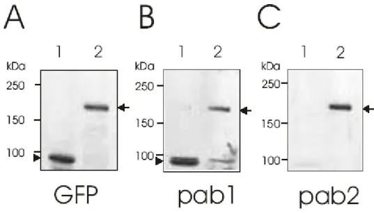
3.6 Generation of polyclonal antibodies 43
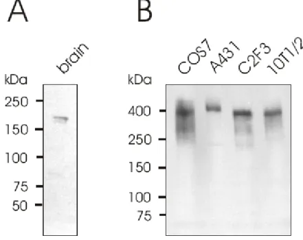
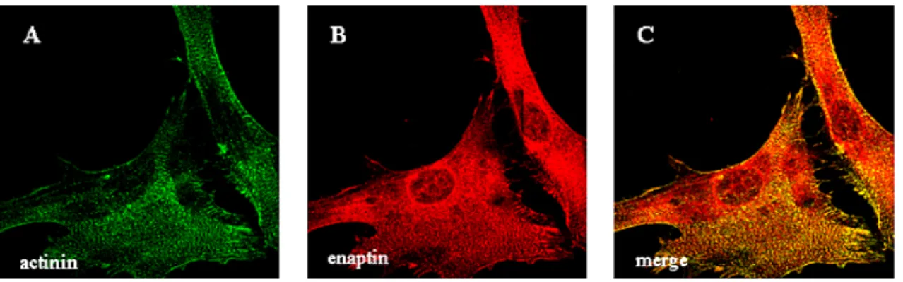
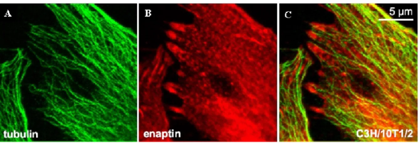
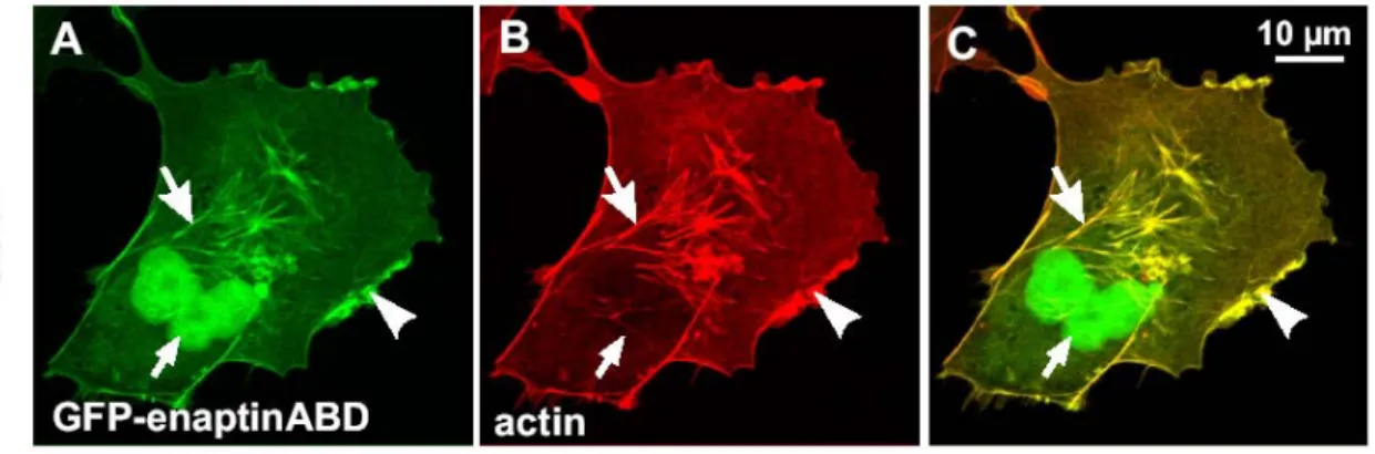
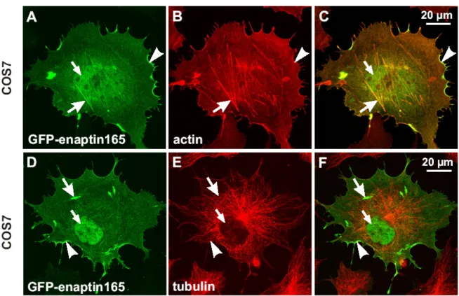
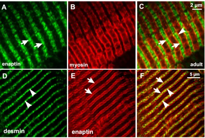
3.7 Expression of Enaptin in various cell lines and in brain 45 3.8 Immunofluorescence studies of Enaptin in various cell lines 46 3.9 Cell fractionation studies of fibroblasts cell lysates 48 3.10 Generation of various Enaptin GFP fusion proteins 49 3.11 Enaptin localisation in mouse skeletal muscle 51
3.12 Enaptin localisation in human skin 53
3.13 Influence of cytoskeletal drugs on the subcellular distribution of Enaptin 55 4 Characterisation, localisation and functional analysis of Sun1
4.1 The perinuclear region of Enaptin is highly conserved 56
4.2 SUN domain containing proteins 56
4.3 Sequence and domain analysis of Sun1 58
4.4 The perinuclear segment of Enaptin binds to the C-terminus of Sun1 in vivo
and the SUN domain is not required for this association 60 4.5 The perinuclear Enaptin amino acids bind to the C-terminus of Sun1 also
in vitro 61
4.6 A full-length GFP fusion protein of Sun1 localises to the nuclear membrane 62
4.7 Endogenous Sun1 is present in the nuclear membrane in HEK cells 62 4.8 Expression profile of Sun1 in COS7 and HEK cells 63 4.9 N- and C-terminal segments of Sun1 localise independently to the nuclear
membrane 64
4.10 Sun1 dimerises in vivo 67
4.11 The SUN domain is not required for the NE localisation of Sun1 68 4.12 Western blotting confirms the expression of all the GFP fusion proteins 71 4.13 GFP-CT+3TM behaves like a dominant negative of Sun1 and displaces
endogenous Sun1, NUANCE and emerin from the nuclear membrane 72 4.14 Overexpression of GFP-CT+3TM has no effect on lamin A/C or Lap2
localisation 74
4.15 Sun1 is an inner nuclear membrane protein 75
4.16 Sun1 localises to the nuclear envelope in a lamin A/C dependent manner 77 5 Generation of a knock-in mouse expressing GFP-tagged full-length
Enaptin without the transmembrane domain
5.1 Aim of the project 79
5.2 The knock out strategy 79
5.3 transfection and screening 81
6 Discussion
6.1 Enaptin, a novel NUANCE-like protein 83
6.2 Isoform diversity of Enaptin 84
6.3 Subcellular localisation of Enaptin 85
6.4 Tissue distribution of Enaptin 86
6.5 Sun1 is a novel nuclear envelope protein and binds to Enaptin 87 6.6 The N- and C-termini of Sun1 localise to the nuclear envelope independently 89
6.7 Sun1 is essential for the NE localisation Enaptin and NUANCE 90
6.8 Sun1 dimerises in vivo 90
5.9 Sun1 is an element of inner nuclear envelope and its localisation is
lamin A/C dependent 91
6.10 Enaptin, NUANCE, Sun1 and laminopathies 91
6.11 A model depicting the various interactions and findings 92 6.12 A hypothetical model depicting the involvement of Enaptin, NUANCE and
Sun1 in nuclear positioning and migration 93
6.13 Future perspectives 94
Summary 96
Zusammenfassung 97
7 Bibliography 98
Erklärung
Curriculum Vitae Lebenslauf
ABBREVIATIONS
Ade Adenine
AD Activating domain AP Alkaline phosphatase APS Ammonium persulphate 3-AT 3-amino 1,2,4-triazole BD Binding domain BSA Bovine serum albumin CH Calponin homology DEPC Diethylpyrocarbonate DMSO Dimethylsulfoxide
dNTP Deoxyribonucleotide triphosphate DTT 1,4-dithiothreitol
EDTA Ethylenediaminetetraacetic acid
EGTA Ethyleneglycol-bis (2-amino-ethylene) N,N,N,N-tetraacetic acid FITC Fluorescein-5-isothiocyanate
GFP Green Fluorescence Protein GST Glutathione S-transferase
His Histidine
HRP Horse radish peroxidase
HEPES N- (2-hydroxyethyl) piperazine-N-2-ethanesulphonic acid IPTG Iso-propylthio-galactopyranoside
IgG Immunoglobulin G Kb Kilobase pairs kDa KiloDalton
Leu Leucine
MOPS Morpholinopropanesulphonic acid Neo Neomycin cassette
mRNA messenger ribonucleic acid mAb Monoclonal Antibody
NP-40 Nonylphenylpolyethyleneglycol OD Optical density
PIPES Piperazine-N,N.-bis(2-ethanesulphonic acid) PMSF Phenylmethylsulphonylfluoride
PAGE Polyacrylamide gel electrophoresis rpm Rotations per minute
SDS Sodium dodecyl sulphate
TRITC Tetramethylrhodamine β isothiocyanate TAE Tris borate EDTA
Trp Tryptophan
WT Wild type allele
X-gal 5-bromo-4-chloro-3-indolyl-D-galactopyranoside
1 INTRODUCTION 1.1 The Cytoskeleton
The cytoskeleton is composed mainly of three types of filaments, microfilaments, microtubules and intermediate filaments. Microfilaments are fine, thread-like protein fibers, 7-9 nm in diameter. They are composed of a protein called actin, which is the most abundant cellular protein, often accounting 10 to 20 percent of the total cytoplasmic protein content.
Actin exists either as a globular monomer (called G-actin) or as a filament (designated F- actin), the latter formed by head-to-tail polymerisation of asymmetric monomers.
Microfilaments in association with the protein myosin are responsible for muscle contraction.
They can also carry out cellular movements including gliding, contraction, and cytokinesis.
Microtubules are cylindrical tubes, 20-25 nm in diameter. They are composed of alpha and beta tubulin. Microtubules act as a scaffold to determine cell shape and provide a set of
"tracks" for cell organelles and vesicles to move on. Microtubules also form the spindle fibers for separating chromosomes during mitosis. When arranged in geometric patterns inside flagella and cilia they are used for locomotion.
The intermediate filaments average 10 nm in diameter and thus are "intermediate" in size between actin filaments (8 nm) and microtubules (25 nm). There are five major types of intermediate filaments each constructed from one or more proteins characteristic of it. Despite their chemical diversity, intermediate filaments play similar roles in the cell, providing a supporting framework within the cell. For example, in epithelia the nucleus is held within the cell by a basketlike network of cytoplasmic intermediate filaments made of proteins called keratins, whereas lamins are nuclear proteins that line the nuclear membrane. Intermediate filaments (desmin) also anchor the thick and thin filaments of muscle cells in a fixed position and provide mechanical strength to the long axons found in some neurons (neurofilaments).
1.2 The Actin Cytoskeleton
Actin is a moderate sized protein consisting of approximately 375 residues, which is encoded by a large, highly conserved gene family. Some single-celled eukaryotes like yeasts have a single actin gene, whereas most organisms contain many actin genes. For example, mammals have six distinct actin isotypes (Vandekerckhove and Weber). Each actin molecule contains a Mg2+ ion complexed with either ATP or ADP. Thus there are four states of actin:
ATP-G-actin, ADP-G-actin, ATP-F-actin and ADP-F-actin. Two of these forms, ATP-G-actin
and ADP-F-actin predominate in a cell. The addition of ions, Mg2+, K+ or Na+ to a solution of G-actin will induce the polymerisation of G-actin into actin filaments. The process is also reversible: F-actin depolymerises into G-actin when the ionic strength of the solution is lowered. All subunits in a filament point towards the same end. Consequently, at one end of the filament, by convention designated plus or barbed end, the ATP-binding cleft of an actin subunit is exposed to the surrounding solution and at the opposite end, the minus or pointed end, the cleft contacts the neighbouring actin subunit. The actin cytoskeleton is organised into bundles and networks of filaments, which are the most common arrangements of actin filaments in a cell. Functionally, bundles and networks have identical roles in a cell: both provide a framework that supports the plasma membrane and, therefore, determines a cell’s shape. Structurally, bundles differ from networks mainly in the organisation of actin filaments. In bundles the actin filaments are closely packed in parallel arrays, whereas in a network the actin filaments crisscross, often at right angles, and are loosely packed. In all bundles and networks, the filaments are held together by actin cross-linking proteins. The length and flexibility of a cross-linking protein determine whether bundles or networks are formed.
1.3 Actin Binding Proteins
Actin binding proteins are classified according to their actin binding function. Actin filament severing proteins fragment filaments by mechanisms that do not require the hydrolysis of ATP. The purpose of this severing activity is probably to introduce a device whereby existing actin filament structures may be removed or remodeled to form other structures within the cell. So far, two major groups of actin severing proteins have been identified. The gelsolin group is the archetype of the group of actin binding proteins that sever and cap the fast growing barbed end of actin filaments and that initiate the polymerisation of new filaments by forming a nucleus (Yin et al., 1988; Weeds et al., 1993). The second group, the Actin depolymerising factor (ADF)/Cofilin group comprises low molecular weight actin filament severing proteins which in addition possess actin monomer binding activity.
Actin binding proteins grow by monomer addition exclusively at their ends, particularly barbed ends. Filament capping proteins like radixin (Funayama et al., 1991) and tensin (Davis et al., 1991) bind to the barbed ends of filaments in cells and are therefore essential for the control of actin polymerisation within cells or within local regions of individual cells. DNaseI (Podolski et al., 1988) and tropomodulin (Fowler et al., 1993) are actin binding proteins that bind to the pointed ends.
There is a well-documented abundance of non-filamentous actin in cells despite the intracellular conditions greatly favoring the formation of F-actin. A number of low molecular weight actin binding proteins have been identified that are thought to bind G-actin and thus directly sequester monomers from the filamentous pool. This pool of monomers is possibly drawn upon to support polymerisation when cells are stimulated, for examples by chemoattractants. Profilin (Southwick et al., 1990) is an example of an actin monomer binding protein, which has sequestering activity.
Actin bundling proteins are proteins, which are required to bundle actin filaments in two different ways. Any protein that binds actin at a site exposed on the filament, and also has the ability to self-associate, should bundle actin filaments. These proteins fall into a family sharing a common actin-binding domain (ABD) of the α-actinin type (Matsudaira, 1991).
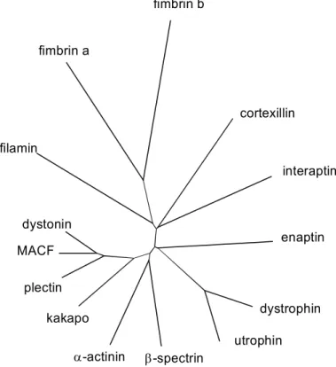
Sequence analysis revealed that these proteins share a 250-residue sequence, which can be divided into two homologous parts, each of them showing a significant similarity to the N- terminal part of the calponins, a family of proteins mainly involved in the regulation of smooth muscle contraction (Castresana & Saraste, 1995). The ABD composed of two calponin homology domains (CH domains) is found in proteins of the α-actinin superfamily with proteins such as β-spectrin, α-actinin, dystrophin, utrophin and filamin. Alternatively bundling proteins like fimbrin (T-plastin) (Namba et al., 1992) and ABP-50 (Demma et al., 1990) have two actin binding domains by which they bundle actin filaments. The monomeric bundling protein synapsin I has three actin binding sites (Südhof et al., 1989).
A distinct group of proteins is formed by the plakins, which have been shown to function as cytoskeleton linkers (Ruhrberg & Watt, 1997). These proteins are thought to crosslink the microfilaments with the microtubule and intermediate filament systems. Some of the plakins like the bullous phemphigoid antigen 1 (BPAG-1/dystonin), plectin and microtubule actin crosslinking factor (MACF) share an α-actinin type actin-binding domain at their N-terminus with the α-actinin superfamily. Plectin and BPAG-1/dystonin contain an additional intermediate-filament-binding domain (Wiche, 1998), and MACF has been shown to connect the actin cytoskeleton with the microtubule filaments (Leung et al., 1999).
1.4 Actin binding proteins related diseases
The critical role of actin-crosslinking proteins in maintaining cell structure and function was uncovered in studies on the Duchenne muscular dystrophy (DMD), a X-linked degenerative disorder of muscle. DMD affects about 1 in 3500 live born males. The progressive weakness of the striated muscles also leads to respiratory complications, which
mark the final stage of the disease before the end of the third decade of life (Moser, 1984).
Patients with DMD have a defect in the gene encoding the actin-crosslinking protein dystrophin (Ahn & Kunkel, 1993). Dystrophin crosslinks actin filaments into a supportive cortical network and attaches this network to a glycoprotein complex in the muscle cell membrane. The membrane complex is also connected with extracellular proteins. In patients lacking a functional dystrophin, the membrane of muscle cells is not supported by cortical actin and is easily damaged by stress of repeated muscle contraction. The actin-crosslinking protein spectrin mediates important structural properties of erythrocytes to make them robust in deformation so that they can squeeze through the small capillaries and sinusoids of the spleen. In spectrin-deficient erythrocytes deformability is reduced, which results in a spherical shape, lacking the central pallor associated with the biconcave cells. Hereditary spherocytosis, elliptocytosis and pyropoikilocytosis represent a group of disorders that are due to deficiency or dysfunction of spectrin and other proteins involved in the linkage of the cytoskeleton to the plasma membrane in red blood cells (Palek, 1987). Plectin and BPAG-1/dystonin have been shown to be important in the maintenance of the cytoskeletal integrity in several cell types (Brown et al., 1995; Dalpe et al., 1998). Mutations in the mouse BPAG-1/dystonin gene are responsible for the dystonia musculorum (dt) mutant phenotype, which is first recognizable between 7 and 10 days after birth and displays a progressive loss of limb coordination. The dt mice suffer from neurological, myelination and muscle abnormalities caused by intrinsic defects in sensory neurons, Schwann cells and skeletal muscle cells, respectively (Bernier et al., 1998). Similarly, targeted inactivation of the plectin gene in mice resulted in lethality two to three days after birth, probably a result of severe skin blistering caused by degeneration of keratinocytes (Andrä et al., 1997). Mutations in human plectin cause epidermolysis bullosa simplex which is associated with muscular dystrophy, an epidermal blister disease which is associated with a myopathy(McLean et al., 1996).
1.5 The nucleus and the nuclear envelope
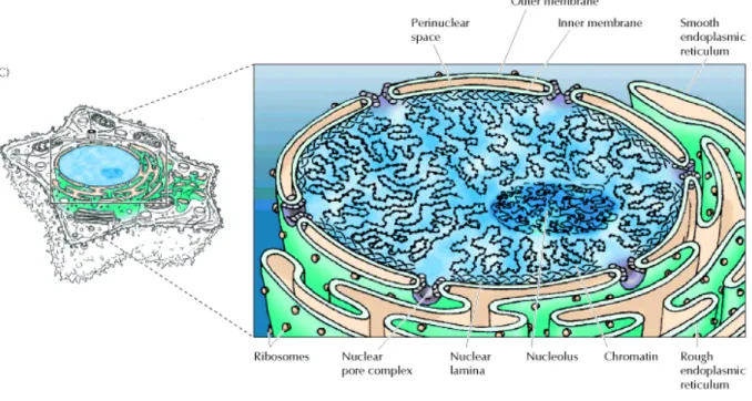
The nucleus is the control center and the hallmark of a eukaryotic cell. Usually the nucleus is round and is the largest organelle in the cell. It is surrounded by a membrane structure called nuclear envelope, which is similar to the cell membrane that encloses the entire cell. The nuclear envelope has a complex structure, consisting of two nuclear membranes, an underlying nuclear lamina and the nuclear pore complexes. The outer nuclear membrane is continuous with the endoplasmic reticulum, so the space between the inner and outer nuclear membranes is directly connected with the lumen of the endoplasmic reticulum.
In addition, the outer nuclear membrane is functionally similar to the membranes of the endoplasmic reticulum and has ribosomes bound to its cytoplasmic surface. In contrast, the inner nuclear membrane carries unique proteins that are specific to the nucleus.
Figure 1.1: A schematic diagram of the nucleus. The picture is taken from "The Cell, A molecular approach", Geoffrey M. Cooper., Second edition.
The critical function of the nuclear membranes is to act as a barrier that separates the contents of the nucleus from the cytoplasm. Like other cell membranes, the nuclear membranes are phospholipid bilayers, which are permeable only to small nonpolar molecules.
Other molecules are unable to diffuse through the phospholipid bilayer. The inner and outer nuclear membranes are joined at nuclear pore complexes, the sole channels through which small polar molecules and macromolecules are able to travel through the nuclear envelope.
The nuclear pore complex is a complicated structure that is responsible for the selective traffic of proteins and RNAs between the nucleus and the cytoplasm (Figure 1.1).
A unique feature of the nucleus is that in most eukaryotic organisms it disassembles and re-forms each time the cell divides. At the beginning of mitosis, the chromosomes condense, the nucleolus disappears, and the nuclear envelope breaks down, resulting in the release of most of the contents of the nucleus into the cytoplasm. At the end of mitosis, the process is reversed: The chromosomes decondense, and the nuclear envelopes re-form around the separated sets of daughter chromosomes and eventually fuse. Nuclear envelope
breakdown (NEBD) involves the depolymerisation of the lamina, the fragmentation and removal of the nuclear membranes from the chromatin, and the disassembly of the NPCs. The lamina in metazoans is composed of the intermediate filament-like proteins, called lamins, which connect with the NPCs and inner nuclear membrane to form a network underlying the nuclear envelope and extending into the nuclear interior. Here, the lamina can help to organize chromatin into functional domains and provide structure to the nucleus (Liu et al., 2000 and Wilson et al., 2001). The lamina, and by extension, chromatin, are attached to integral inner nuclear membrane proteins which, along with the integral pore membrane proteins, define the unique composition of the nuclear membranes (Worman and Courvalin, 2000).
There is a large body of evidence that many nuclear envelope-associated proteins are reversibly phosphorylated during mitosis, concomitant with their dramatic redistribution away from the vicinity of the nucleus. Initially, it was thought that these phosphorylation events promote NEBD, leading to the dispersal of the inner nuclear membrane into a discrete population of vesicles (Vigers and Lohka, 1991), but recent work has indicated that the nuclear envelope is not fated to vesiculate. Rather, mitosis involves the redistribution of the nuclear envelope membrane proteins into the ER. Although the ER-nuclear envelope membrane system is continuous, all membrane proteins do not normally freely diffuse within it. Instead, once synthesized, inner nuclear membrane proteins diffuse from the ER through the pores to the inner nuclear membrane where they become trapped, presumably by their interactions with the lamina, chromatin, and each other (Worman and Courvalin, 2000).
1.6 Proteins of the nuclear envelope
The major structural framework at the nuclear periphery is the nuclear lamina, whose core structure is formed by type V intermediate filament proteins, the lamins (Stuurman et al., 1998). Lamins assemble into a meshwork of tetragonally organised 10-nm filaments underneath the INM. The attachment of the lamins to the membrane involves several mechanisms, and depends also on the type of lamins. B type lamins, which are constitutively expressed in all somatic cells, contain a stable C-terminal farnesyl modification, which is important but not sufficient for targeting and anchoring B-type lamins to the membrane (Moir et al., 1995). Thus, interactions of B-type lamins with integral membrane proteins must also contribute to the assembly and stable association of lamin B filaments at the membrane (Hutchison et al., 2001). The best-known binding partners for B-type lamins in the INM are the lamin B receptor (LBR) (Worman et al., 1990) and LAP2 β (Furukawa et al., 1995). LBR
contains eight transmembrane domains, and interacts with B-type lamins both in vivo and in vitro (Simos et al., 1992; Ye et al., 1994). LAP2 β is the best-characterized membrane protein of the LAP2 family, which comprises up to six alternatively spliced mammalian isoforms named LAP2∝, β, γ, δ, ε and ζ (Harris et al., 1994 and Berger et al., 1996) and three Xenopus LAP2 isoforms (Lang et al., 1999; Gant et al., 1999). Except for LAP2 ∝ and LAP2 ζ, all mammalian LAP2 isoforms have a closely related N-terminal nucleoplasmic domain of variable length, plus a single membrane-spanning region and a short lumenal domain at their C-terminus (Dechat et al., 2000). LAP2 β has the longest nucleoplasmic N-terminal domain (408 residues). Due to alternative messenger RNA (mRNA) splicing, LAP2 ε, δ and γ lack stretches of 40, 72 and 109 amino acids, respectively, but are otherwise identical to LAP2 β.
LAP2 ζ is the smallest isoform of LAP2 β, and is missing ~ 190 residues of the nucleoplasmic domain as well as the transmembrane and lumenal regions. LAP2 ∝ is structurally and functionally a unique isoform; it shares only the N-terminal 187 residue ‘constant’ domain with all other LAP2 isoforms, and then contains a unique C-terminal domain of 506 residues with no transmembrane domain. LAP2 β, but not LAP2 ∝, interacts with lamin B in vitro (Foisner et al., 1993). The lamin B binding domain maps to 73 residues in the nucleoplasmic region (Furukawa et al., 1998), which are also present in the smaller isoforms LAP2 ε and δ, and are partly con-served in LAP2 γ. However, the lamin-binding activities of the smaller isoforms have not yet been demonstrated.
The most-studied A-type lamins are lamin A and its smaller splice variant, lamin C.
A-type lamins are only expressed in later stages of development (Moir et al., 1995; Cohen et al., 2001). Lamin A is transiently farnesylated, and lamin C is never farnesylated. Perhaps due to their lack of fatty acid modification, lamins A and C do not associate stably with membranes during mitosis. A-type lamin structures may also be less stable during interphase, or organized differently, because ectopic expression of headless lamin mutants in mammalian cells selectively mislocalises A-type lamins into intranuclear aggregates, whereas lamin B remains unchanged (Izumi et al., 2000; Dechat et al., 2000). The incorporation of mature, non-farnesylated lamins A and C into the lamina after mitosis might depend on B-type lamins (Stuurman et al., 1998) and specific interactions with membrane proteins at the INM. Integral membrane proteins that might link A-type lamins to the INM include three LAP1 proteins (A, B and C), which are alternatively spliced products of a gene unrelated to LAP2 (Martin et al., 1995), and emerin (Manilal et al., 1996), which shares an ~40 residue domain (LEM domain) with the LAP2 isoforms (Dechat et al., 2000; Lin et al., 2000). LAP1-A, LAP1-B and emerin all bind A-type lamins in vitro (Foisner et al., 1993; Clements et al., 2000), and the
localization of LAP1-C and emerin at the INM depends on A-type lamins (Powell et al., 1990;
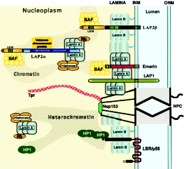
Sullivan et al., 1999). Thus, multiple interactions between lamins and INM proteins are probably required to form a stable peripheral lamina (Figure 1.2).
Figure 1.2: Proposed molecular interactions between components of the peripheral lamina, the nucleoskeleton and chromatin. INM, inner nuclear membrane, ONM, outer nuclear membrane and NPC, nuclear pore complex (taken from Vlcek et al., 2001).
1.7 Actin binding proteins in the nucleus and nuclear membrane
The discovery of actin and of numerous actin binding in the nucleus argues that not only is actin present in the nucleus but its polymerisation is controlled. The presence of some of the actin binding proteins in the nucleus might be irrelevant as many of them are small enough to enter the nucleus passively. Thymosin, a 5 kDa sequesterin protein and profilin, a 12 kDa actin sequestering and nucleotide exchange protein could be examples for this kind of passive diffusion into the nucleus. In the light of this objection, two types of nuclear actin binding proteins are of particular interest. The first class comprises proteins like c-Abl, a
tyrosine kinase involved in the cellular response to DNA damage, which was found to have both F- and G-actin binding domains and actin binding activity in vitro (Van Etten et al., 1994). The second class of protein comprises established actin binding proteins that are associated with the nucleus. An example of this class of proteins is the erythrocyte protein 4.1, which in red blood cells is thought to help to anchor the cytoskeleton to the membrane by promoting the interaction of spectrin with actin. In nucleated cells such as HeLa and MDCK cells, protein 4.1 has been reported as a component of the nuclear matrix (De Carcer et al., 1995; Correas et al., 1991). Spectrin has also been reported to be present in the nucleus (Bachs et al., 1990). The function of these proteins in the nucleus is not well understood, but recent data suggest that protein 4.1 plays a role in RNA splicing (Lallena et al., 1998).
Recently, a giant actin binding protein of 800 kDa called NUANCE (NUcleus and ActiN Connectind Element) was found to be associated with the nuclear membrane (Zhen et al., 2002).
1.8 Nuclear migration and the involvement of actin binding proteins
The nucleus and other organelles are in fact quite dynamic. They often migrate through the cytoplasmand then occupy specific locales, often far from the centerof the cell.
For example, in a newly fertilized zygote, pronucleimust first migrate towards one another, and then they migrate to a species-specific location before undergoing the first mitosis.
Nuclear positioning is also essential to a variety of polarized cells, such as intestinal brush- border cells and many secreting endocrine cells. In other cases, nuclear migration events reposition nuclei to distant regions of the cell. Examples include growing plantpollen tube cells (Hepler et al., 2001) and developing C. eleganshypodermal cells (Sulston et al., 1983).
Disruption of nuclearmigration in the cell bodies of the developing cerebral cortexleads to the human neurodevelopmental disease lissencephaly (Lambert de Rouvroit and Goffinet, 2001). Even in single celled organisms, such as budding and fission yeast, tight controls exist to positionthe nucleus correctly prior to cell division (Morris, 2000; Tran et al., 2001).
Nuclei must be carefully positioned throughout oogenesis and embryogenesis of Drosophila. During the cytoplasmic dumping stage of oogenesis, when the 15 nurse cells rapidly squeeze their cytoplasm into the oocyte through narrow ring canals, nuclei must remain anchored away from the ring canals. Normally, an array of striated actin bundles extends from the plasma membrane to nurse cell nuclei. These bundles shorten as dumping progresses and the nurse cells shrink (Guild et al., 1997). Mutations inthe actin-monomer- binding protein profilin or in the actin-filament-bundling proteins villin and fascin disrupt
these filaments, generatingfree-floating nuclei (Robinson and Cooley, 1997). These mutations block oocyte development because, as cytoplasmic dumping from the nurse cells begins, nuclei become physically stuck in ring canals. A second example is the early embryonic nuclear migrations towards the periphery of the syncytial blastoderm. The actin gel-like network around migrating nuclei depolymerizes. This has beenhypothesized to contribute to the force required for migration as nuclei passively `surf' the depolymerizing front (von Dassowand Schubiger, 1994).
Actin networks can also function actively to reposition nuclei. Chytilova et al. (2000) recently described a dramatic example. Actin depolymerising drugs completely abolished rapid, long-distance intracellular nuclear migration in Arabidopsis root hairs, whereas drugs that disrupted microtubules had no effect. Because of the speed and distance of the nuclear migrations in these cells, the actin network must be functioning actively to move nuclei, in contrast to the above examples of passive mechanisms. Budding yeast provides another example: both actin filaments and microtubules are required for proper localization of the nucleus and spindle at the bud neck to ensure normal cell division (Palmer et al., 1992;
Bloom, 2001).
ANC-1 is the C. elegans orthologue of the mammalian Enaptin, NUANCE proteins and mutations in anc-1 (nuclear anchorage defective) disrupt the positioning of nuclei and mitochondria in Caenorhabditis elegans (Starr et al., 2001). NUANCE is a huge human spectrin repeat containing protein having a functional ABD at its N-terminus and a single transmembrane domain at its C-terminus and is localised to the nuclear envelope (Zhen et al., 2000).
1.9 Aim of the work
The presence of Enaptin-165 and CPG2 (Nedivi et al., 1996), which are highly homologous to the N-terminus of NUANCE and are homologous to human Nesprin-1α, human Nesprin-1β, mouse Syne-1A, Syne-1B and human Myne-1 (Apel et al., 2000; Mislow et al., 2000; Zhang et al., 2000) which themselves are highly homologous to the C-terminus of NUANCE (Zhen et al., 2002) signalled the possible existence of Enaptin being a larger gene like NUANCE, with Enaptin-165, CPG2, Syne-1A, Syne-1B, Myne-1 and Nesprins being short alternatively spliced isoforms. To address this issue, antibodies for the ABD of Enaptin should be generated and used for immunofluorescence experiments to study the presence of Enaptin at the nuclear envelope in western blots and to detect the biggest transcript of Enaptin. The
antibodies will also be used to follow the distribution of Enaptin in various tissues. Yeast two hybrid and GST-pull down experiments will be used to study the binding partners of Enaptin especially to the evolutionally conserved regions like the region after the transmembrane domain. Mouse knock out targeting the transmembrane domain of Enaptin will be carried out to study the role of Enaptin in muscular dystrophy given the fact that Enaptin was shown to lamin A/C (Mislow et al., 2002), mutations in lamin A/C is known to cause muscular dystrophy.
MATERIALS AND METHODS 2.1 Materials
2.1.1 Enzymes, inhibitors and antibodies Enzymes for molecular biology
alkaline phosphatase Roche
DNase I (Desoxyribonuclease) Sigma
lysozyme Sigma
M-MLV reverse transcriptase Promega
restriction endonucleases Life Technologies
ribonuclease A Sigma
T4-DNA-ligase Life Technologies
Taq-DNA-polymerase Roche
Antibodies
primary antibodies:
mouse-anti-α-actinin Sigma
mouse-anti-desmin Sigma
mouse-anti-skeletal (Fast) myosin Sigma
mouse-anti-lamin A/C CHEMICON
mouse-anti-emerin NOVO Castra
mouse-anti-LAP2 Transduction
rabbit-anti-PDI Stressgen
secondary antibodies:
goat-anti-mouse-IgG, peroxidase-conjugated Sigma goat-anti-mouse-IgG, Cy3-conjugated Sigma goat-anti-mouse-IgG, alkaline phosphatase conjugated Sigma
goat-anti-mouse-IgG, Alexa 488 conjugated Molecular Probes goat-anti-rabbit-IgG,Alexa 568 conjugated Molecular Probes
Inhibitors
benzamidine Sigma
DEPC (Diethylpyrocarbonate) Sigma
PMSF (Phenylmethylsulfonylfluoride) Sigma ribonuclease-inhibitor (RNAsin) Promega
Complete Inhibitor-Cocktail Roche
Antibiotics
ampicillin Grünenthal
kanamycin Biochrom
penicillin/streptomycin Biochrom 2.1.2 Reagents
acrylamide National Diagnostics
agarose (electrophoresis grade) Life Technologies
acetone Riedel-de-Haen
Bacto-Agar, Bacto-Pepton, Bacto-Trypton Difco
BSA (bovine serum albumin) Roth
chloroform Riedel-de-Haen
calcium chloride Sigma
Coomassie-brilliant-blue R 250 Serva
p-cumaric acid Fluka
DMEM (Dulbecco´s Modified Eagle´s Medium) Biochrom
DMF (dimethylformamide) Riedel-de Haen
DMSO (dimethyl sulfoxide) Merck
DTT (1,4-dithiothreitol) Gerbu
EDTA ([ethylenedinitrilo]tetraacetic acid) Merck EGTA (ethylene-bis(oxyethylenenitrilo)tetraacetic acid) Sigma
ethanol Riedel-de-Haen
ethidium bromide Sigma
FCS (fetal calf serum) Biochrom,
fish gelatine Sigma
formamide Merck
formaldehyde Sigma
glycine Degussa
IPTG (isopropyl β-D-thiogalactopyranoside) Sigma
isopropanol Merck
β-mercaptoethanol Sigma
methanol Riedel-de-Haen
methylbenzoate Fluka
mineral oil Pharmacia
MOPS ([morpholino]propanesulfonic acid) Gerbu
Ni-NTA-agarose Qiagen
paraformaldehyde Sigma
RNase A Sigma
SDS (sodium dodecylsulfate) Serva
sodium azide Merck
TEMED (tetramethylethylenediamine) Merck Tris (hydroxymethyl)aminomethane Sigma Triton X-100 (t-octylphenoxypolyethoxyethanol) Merck X-Gal(5-bromo-4-chloro-3-indolyl-β-D-galactopyranoside)Roth
xylol Fluka
yeast extract Oxoid
Radionuclides
α-32P-desoxyadenosine-5‘-triphosphate (10 mCi/ml) Amersham α-32P-desoxycytosine-5‘-triphosphate (10 mCi/ml) Amersham
Reagents not listed above were purchased from Clontech, Fluka, Merck, Roth, Serva, Sigma, Promega and Riedel-de-Haen, respectively.
2.1.3 Kits
Nucleobond PC 500 Macherey-Nagel
NucleoSpin Extract 2 in 1 Macherey-Nagel
NucleoSpin Plus Macherey-Nagel
RNeasy midi kit Qiagen
pGEMT easy Cloning Kit Promega
Zero Blunt TOPO PCR Cloning Kit Invitrogen
EndoFree Plasmid Maxi kit Qiagen
2.1.4 Bacterial host strains E. coli M15
E. coli DH5α
2.1.5 Eukaryotic cells
C3H/10T1/2 mouse fibroblasts N2A mouse neuroblastoma cell line
COS-7 monkey SV40 transformed kidney cell line
human primary keratinocytes (kindly provided by Dr. I. Haase, Clinic of Dermatology, University of Cologne) C2F3 mouse myoblasts
MB50 human myoblast primary cell line 2.1.6 Vectors
pQE-30 Qiagen
pGEM-T Easy Promega
pCR-Blunt II-TOPO Invitrogen
pEGFP-C2 Clontech
pGBKT7 Clontech
pGADT7 Clontech
pBluescript Stratagen
pGEX4T1 Pharmacia
2.1.7 Oligonucleotides
Oligonucleotides for PCR (polymerase chain reaction) were purchased from MWG-Biotech AG (Ebersberg), Roth GmbH (Karlsruhe), Germany and metabion (Martinsried).
Oligonucleotides used to clone Enaptin-165 in pGEMTeasy vector
624fullGFP5’end
GGAATTCATGGCAACCTCCAGAGCATCTTC
624fullGFPC2-3’end
ACGCGTCGACTTAGAAGTGGTGAAGCACATACTCTTCTTCAAG
Oligonucleotides to correct the deletion after the ABD in the small isoform of Enaptin in the pGEMTeasy vetcor
del.for
5' ATTTTGAAGG AAACAAAAGT TTGGATAGAA C 3' del.rev
GTTCTATCCA AACTTTTGTT TCCTTCAAAA T
Oligonucleotides to clone the ABD of Enaptin into the yeast two hybrid vector 624fullGFP5’end
GGAATTCATGGCAACCTCCAGAGCATCTTC y2hABD3'
TGCGTCGACC TACTTGACTT CCATGAAGAG CTCCCTCC
2.1.8 Buffers and other solutions
Buffers and solutions not listed below are described in the methods section.
PBS (pH 7.2): 10x NCP-buffer (pH 8.0):
10 mM KCl 100 mM Tris/HCl
10 mM NaCl 1.5 M NaCl
16 mM Na2HPO4 5 ml Tween 20
32 mM KH2PO4 2.0 g sodium azide 10x MOPS (pH 7.0/ pH 8.0): PBG (pH 7.4):
20 mM MOPS 0.5% BSA
50 mM sodium acetate 0.045% fish gelatine 1 mM EDTA in 1x PBS
20x SSC: TE-Puffer (pH 8.0):
3 M NaCl 10 mM Tris/HCl (pH 8.0)
0.3 M sodium citrate 1 mM EDTA (pH 8.0, adjusted with NaOH) autoclaved
2.1.9 Materials
cryotubes, 1 ml Nunc
Eppendorf tubes, 1.5 ml and 2 ml Sarstedt
hybridization tubes Hybaid
3MM filters Whatmann
nitrocellulose, type BA85 Schleicher and Schüll
nylon membrane, Biodyne PALL
filter, sterile 0.45 µm and 0.2 µm Gelman Science
plastic cuvettes Greiner
quartz cuvettes Infrasil Hellma
Superdex75 PC3.2/30 Pharmacia Biotech
15 ml tubes, type 2095 Falcon
50 ml tubes, type 2070 Falcon
X-ray film X-omat AR-5 Kodak
2.1.10 Instruments
blotting chamber Trans-Blot SD Bio-Rad
centrifuges: Beckman Avanti J25 Beckman
Sorvall RC 5C plus Sorvall
Biotech fresco Heraeus Instruments
crosslinker UVC 500 Hoefer
pH-meter 766 Knick
heating blocks: type DIGI-Block JR neoLab
type thermomixer Eppendorf
hybridization oven Hybaid
incubator Lab-Therm Kühner
microscope: light microscope, Type DMI Leica
laser scan microscope Leica
Multiphor II/Immobiline focussing system Pharmacia Biotech
PCR-thermocycler MWG-Biotech pump system Biologic Workstation Bio-Rad
rotors: type JA-10 Beckman
typeJA-25.50 Beckman
SLA-1500 Sorvall
SLA-3000 Sorvall
SS-34 Sorvall
TLA 45 Beckmann
shaker 3015 GFL
lab-shaker Kühner
SMART-system Pharmacia Biotech
spectral photometer type Ultraspec 2000 Pharmacia Biotech
Ultra-Turrax IKA Labortechnique
ultracentrifuge Optima TLX Beckmann
UV-Monitor TFS-35 M Faust
UV-transilluminator MWG-Biotech
Vortex REAX top Heidolph
water bath GFL
2.1.11 Computer programs
For alignment analysis of cDNA sequences the GCG software package (University of Cologne) and the BLAST (NCBI) program were used. Protein sequences were aligned using the programs ClustalW and TreeView. For prediction of motif and pattern searches the ExPaSY (SIB) software package was used. Annealing temperatures of primers were calculated with the program “Primer Calculator” available in the Internet (http://www.williamstone.com).
2.2 Molecular biological methods
2.2.1 Plasmid-DNA isolation from E. coli by alkaline lysis miniprep
With this DNA isolation method plasmid DNA was prepared from small amounts of bacterial cultures. Bacteria were lysed by treatment with a solution containing sodium dodecylsulfate (1% SDS) and 0.5 M NaOH (SDS denatures bacterial proteins and NaOH
denatures chromosomal and plasmid DNA). The mixture was neutralized with potassium acetate, causing the plasmid DNA to reanneal rapidly. Most of the chromosomal DNA and bacterial proteins precipitate, as does SDS forming a complex with the potassium, and are removed by centrifugation. The reannealed plasmid DNA from the supernatant was concentrated by ethanol precipitation.
2.2.2 Plasmid-DNA isolation with a kit from Macherey-Nagel
NucleoSpin Plasmid is designed for the rapid, small-scale preparation of highly pure plasmid DNA (minipreps) and allows a purification of up to 40 µg per preparation of plasmid DNA.The principle of this plasmid-DNA purification kit is based on the alkaline lysis miniprep. Plasmid DNA was eluted under low ionic strength conditions with a slightly alkalibuffer. For higher amounts of plasmid DNA, the Nucleobond AX kit from Machery- Nagel was used. The plasmid DNA was used for sequencing and transfection of eukaryotic cells. The protocols were followed as described in the manufacturer’s manual.
2.2.3DNA agarose gel electrophoresis
10x DNA-loading buffer: 50X Tris acetate buffer (1000 ml) (pH:8.5) 40% sucrose, 0.5% SDS 242.2 g Tris0.25% bromophenol blue, in TE (pH 8.0) 57.5 mL acetic acid
100 ml of 0.5 M EDTA (pH:8.0, adjusted with NaOH)
Agarose gel electrophoresis was performed to analyse the length of DNA fragments after restriction enzyme digests and polymerase chain reactions (PCR), as well as for the purification of PCR products and DNA fragments. DNA fragments of different molecular weight show different electrophoretic mobility in an agarose gel matrix. Optimal separation results were obtained using 0.5-2% gels in TAE buffer at 10 V/cm. Horizontal gel electrophoresis apparatus of different sizes were used. Before loading the gel, the DNA sample was mixed with 1/10 volume of the 10x DNA-loading buffer. For visualization of the DNA fragments under UV-light, agarose gels were stained with 0.1µg/ml ethidium bromide.
In order to define the size of the DNA fragments, DNA molecular standard markers were also loaded onto the gel.
2.2.4Isolation of total RNA from mouse tissue with RNeasy Mini/Midi kit
Working with RNA always requires special precautions in order to prevent degradation by ubiquitous RNases, e.g. wearing gloves and using RNase-free water and material. The RNeasy technology combines the selective binding properties of a silica-gel- based membrane with centrifugation. A specialized high-salt buffer system allows up to 100 µg (mini) or 1 mg (midi) of RNA longer than 200 bases to absorb to the RNeasy silica-gel membrane. An appropriate amount of different mouse tissues was transferred into a lysis buffer containing guanidine isothiocyanate and β-mercaptoethanol followed by disruption and homogenization using a rotor homogenizer. After centrifugation the supernatant was transferred to a new tube and mixed with one volume of 70% ethanol. This mixture was loaded on the RNeasy spin column placed in a collection tube. After another centrifugation and discarding the flow through, the RNeasy column was treated with DNase I and washed with a washing buffer. To elute the RNA from the column an appropriate volume of RNase- free water was pipetted directly onto the spin-column membrane. The obtained RNA was used for cDNA synthesis by RT-PCR and for northern blot analysis. Exact compositions of the buffers used for RNA isolation are listed in the Qiagen RNeasy Handbook.
2.2.5 RNA agarose gel electrophoresis and northern blotting
RNA-buffer: RNA loading dye:
50% formamide 50% sucrose
6% formaldehyde 0.25% bromophenol blue
in MOPS buffer (pH 8.0; see 2.1.8) in RNase free water
The agarose gels were made under RNase free conditions and separation of RNA was performed overnight with 2 V/cm voltage. After washing the gels with water and equilibrating in a high salt solution (20x SSC), the RNA was transferred onto a nylon membrane overnight using the setup similar to the one for Southern blotting. Next day the membrane was washed in 2x SSC, briefly air-dried and the RNA was UV-crosslinked to the membrane.
2.2.6 Labeling of DNA probes
DNA probes used for radioactive labeling were obtained by PCR. About 25 ng DNA was denatured by heating for 5 minutes at 92°C and cooled immediately on ice. After adding random hexanucleotide primer, α-32P-dATP (50 µCi) and/or α-32P-dCTP, the reaction mix