identification and characterisation of the Spir actin nucleator - myosin V motor protein complex
DISSERTATION ZUR ERLANGUNG DES
DOKTORGRADES DER NATURWISSENSCHAFTEN (DR. RER. NAT.) DER FAKULTÄT FÜR BIOLOGIE UND VORKLINISCHE MEDIZIN
DER UNIVERSITÄT REGENSBURG
vorgelegt von Tobias Welz
aus Leipzig im Jahr
2016
Das Promotionsgesuch wurde eingereicht am:
05.12.2016
Die Arbeit wurde angeleitet von:
Prof. Dr. Eugen Kerkhoff
Unterschrift:
Ich, Tobias Welz, geboren am 23.07.1988 in Leipzig, erkläre hiermit, dass ich die vorliegende Arbeit ohne unzulässige Hilfe Dritter und ohne Benutzung anderer als der angegebenen Hilfsmittel angefertigt habe. Ergebnisse, Abbildungen und Beschreibungen, die im Rahmen einer Kollaboration entstanden sind, sind entsprechend gekennzeichnet.
Die aus anderen Quellen direkt oder indirekt übernommenen Daten und Konzepte sind unter Angabe der Quelle gekennzeichnet. Insbesondere habe ich nicht die entgeltliche Hilfe von Vermittlungs- bzw.
Beratungsdiensten (Promotionsberater oder andere Personen) in Anspruch genommen.
Die Arbeit wurde bisher weder im In- noch im Ausland in gleicher oder ähnlicher Form einer anderen Prüfungsbehörde vorgelegt.
_______________________________ _________________________________
Ort, Datum Tobias Welz
Table of contents
Abstract ... viii
Zusammenfassung ... ix
1 Introduction ... 1
1.1 Cell polarisation as a prerequisite for cellular and organismic function ... 1
1.2 Motor proteins as force generators for vesicle transport ... 3
1.2.1 Kinesins and dyneins transport cargo along microtubule networks
... 3
1.2.2 Myosin actin motor proteins
... 4
1.3 Class V myosin motor proteins ... 5
1.3.1 Vertebrate myosin V
... 6
1.3.2 Insect and yeast class V myosin motors
... 9
1.3.3 Regulation of myosin V activity and function
... 9
1.4 The Ras superfamily of small GTPases ... 11
1.4.1 The Ras superfamily includes five families of small GTPases
... 11
1.4.2 The Ras cycle
... 12
1.5 Rab family GTPases ... 13
1.5.1 Regulation of Rab GTPase activity and its impact on intracellular membrane cycling
... 14
1.5.2 Rab GTPases as master regulators of intracellular transport
... 15
1.5.3 Rab GTPases form complexes with motor proteins
... 17
1.6 The Rab11 GTPase ... 20
1.6.1 Regulation of Rab11 activity
... 22
1.6.2 Rab11 family-interacting proteins act as Rab11 specific adaptor proteins
... 23
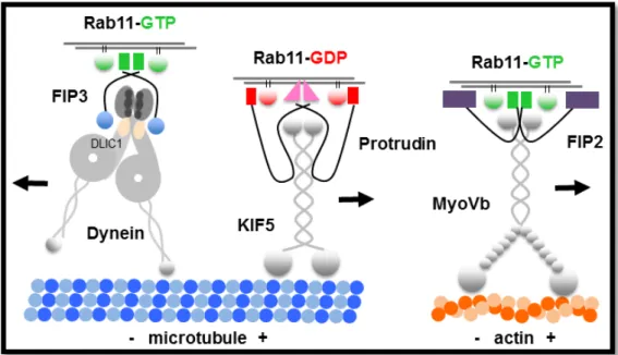
1.6.3 Rab11 motor protein complexes
... 23
1.7 Myosin motors and actin nucleators and elongators cooperate at intracellular membranes ... 26
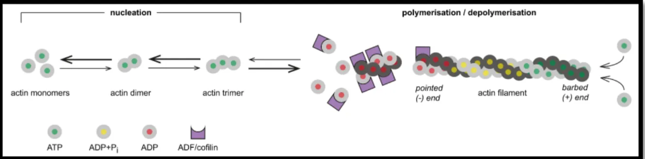
1.7.1 Actin nucleation factors
... 27
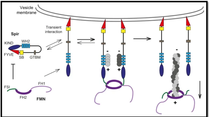
1.7.2 Spir actin nucleators
... 28
1.7.3 Cooperation of myosin motors and actin assembly factors in membrane trafficking
... 32
1.7.4 Overlapping functions of Rab11 GTPases and Spir actin nucleators in exocytosis and recycling
... 34
1.8 Aim of the thesis ... 34
2 Materials and methods ... 36
2.1 Multiple sequence alignments of Spir protein sequences ... 36
2.2 Agarose gel electrophoresis ... 36
2.3 Agarose gel clean-up ... 36
2.4 Measurement of DNA and RNA concentrations ... 37
2.5 PCR techniques ... 37
2.5.1 Amplification of cDNA fragments for cloning
... 37
2.5.2 Colony PCR from bacterial colonies
... 38
2.5.3 QuikChange PCR for site-directed mutagenesis
... 39
2.5.4 One-Step RT-PCR from RNA preparations
... 40
2.6 Cloning ... 40
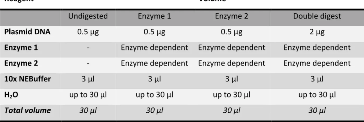
2.6.1 Restriction digest
... 40
2.6.2 Ligation
... 41
2.6.3 Transformation of Escherichia coli bacterial cells
... 42
2.6.4 Plasmid DNA extraction and purification from bacterial cells
... 42
2.6.5 Plasmid-Mini-Purification (MiniPrep) for control digests
... 42
2.6.6 Plasmid-Mini-Purification (MiniPrep) for sequencing
... 43
2.6.7 Plasmid-Maxi-Purification
... 43
2.6.8 Control digests
... 44
2.6.9 DNA Sequencing
... 44
2.7 Cell culture techniques ... 44
2.7.1 Thawing cells
... 45
2.7.2 Freezing cells
... 45
2.7.3 Poly-L-Lysine coating of 6-well plates
... 45
2.7.4 Seeding cells
... 46
2.7.5 Transfection of eukaryotic cells using Lipofectamine Reagent
... 46
2.8 Protein work ... 47
2.8.1 SDS-PAGE
... 47
2.8.2 Western blotting – Protein transfer
... 47
2.8.3 Western blotting - Ponceau S staining
... 47
2.8.4 Western blotting - Antibody treatment
... 47
2.8.5 Antibody protocols
... 48
2.8.6 Western blotting – Blot stripping
... 49
2.9 Production and purification of recombinant proteins in E.coli bacteria ... 49
2.9.1 Purification of GST-MyoVa/b-GTD
... 50
2.9.2 Purification of His6-mCherry-Spir-2-linker(LALA)
... 51
2.9.3 Purification of GST-Rab11a-Q70L
... 52
2.9.4 Purification of GST-Spir-2-GTBM-SB-FYVE
... 53
2.9.5 Bradford assay to determine protein concentrations
... 54
2.10 Co-immunoprecipitation ... 54
2.11 GST-pulldown from HEK293 lysates ... 55
2.12 GST-pulldown from mouse brain ... 56
2.13 GST-pulldown experiments with purified proteins ... 56
2.14 Quantitative GST-pulldown assays ... 57
2.15 Immunostaining ... 58
2.16 Colocalisation analysis ... 59
3 Results ... 60
3.1 Spir and MyoV proteins co-exist in a protein complex ... 62
3.2 Identification of a highly conserved sequence motif within the central Spir linker region ... 63
3.3 Spir/MyoV complex formation depends on the highly conserved sequence motif ... 67
3.4 Spir-2 and MyoV proteins interact directly in vitro ... 69
3.5 The Spir GTBM is necessary for colocalisation of Spir-2 and MyoV at vesicle membranes ... 71
3.6 Spir-2 and MyoV proteins directly interact at vesicle membranes ... 74
3.7 Crystal structure of the MyoVa-GTD:Spir-2-GTBM complex ... 77
3.8 Similarities and differences between Spir and MLPH binding to MyoVa-GTD ... 78
3.9 Spir proteins induce MyoVa targeting to vesicle membranes ... 81
3.10 A tripartite Spir:MyoV:Rab11 complex determines vesicle specificity ... 84
4 Discussion ... 89
4.1 Mechanisms for Spir/MLPH induced unfolding of MyoV motors ... 90
4.2 Rab11 by itself is not able to activate MyoV in vivo ... 94
4.3 Coordinated recruitment of Spir and MyoV proteins to vesicle surfaces ... 94
4.4 Nanotube formation as a potential mechanism for switching vesicle transport tracks ... 96
4.5. Diverse Rab/MyoV interactions suggest similar mechanisms for other transport processes . 100 4.6 The Spir/MyoV interaction in context of cellular processes ... 103
4.7 Conclusion ... 105
5 References ... 106
6 Supplement ... 123
6.1 Sequence related data... 123
6.2 Overview of cloning vectors ... 127
6.2.1 pcDNA3 (Invitrogen, ThermoFisher)
... 127
6.2.2 pAcGFP-C1 (TakaraBio/Clontech)
... 127
6.2.3 peGFP-C2 (Takara/Clontech)
... 128
6.2.4 pGEX-4T-1-NTEV (based on pGEX-4T-1, GE Healthcare Lifesciences)
... 128
6.2.5 pGEX-4T-3 (GE Healthcare Lifesciences)
... 129
6.2.6 pProEX-HTb
... 129
6.3 Expression vectors used in this thesis ... 130
6.3.1 Eukaryotic expression vectors
... 130
6.3.2 Bacterial expression vectors
... 132
6.4 Buffers, solutions and media ... 133
6.5 SDS polyacrylamide gels ... 137
6.6 Primer ... 137
6.7 Antibodies... 140
6.7.1 Primary antibodies
... 140
6.7.2 Secondary antibodies
... 140
6.8 Chemicals and reagents... 141
6.8.1 Restriction endonucleases
... 141
6.8.2 DNA polymerases
... 141
6.8.3 Enzymes
... 141
6.8.4 Chemicals
... 142
6.9 Cell culture media, reagents and supplements ... 144
6.10 Kits ... 144
6.11 Equipment ... 145
6.12 Disposable materials ... 146
7 List of abbreviations ... 147
8 List of figures ... 151
9 List of tables ... 153
10 Acknowledgements ... 154
Abstract
The establishment and maintenance of cell polarity constitutes the basis for the morphological and functional diversity of metazoan cells. Intracellular vesicle transport processes mediate the delivery of cell surface receptors, adhesion proteins and other components to the desired locales, in order to define the polarised higher eukaryotic nature of cells. Members of the Rab family of small GTPases thereby act as master regulators for the complex network of trafficking routes, as they specifically attach to intracellular membranes and recruit distinct effector proteins, including motor proteins, in order to facilitate directed transport along microtubule networks and actin tracks. The Rab11 GTPase is critical for exocytic and recycling pathways and has been shown before to recruit the processive actin motor protein myosin V (MyoV) to vesicle surfaces to drive specific cellular functions.
Furthermore, there is growing evidence for a coupling of actin assembly by actin nucleation and elongation factors, and myosin motor activity in eukaryotic cells. However, the mechanisms for recruitment of actin nucleators and motor proteins to specific membrane compartments remain unclear. By a set of protein interaction studies, this thesis unravelled a direct physical interaction of the Spir actin nucleators and myosin V motors. The interaction was shown to be mediated by the MyoV globular tail domain (GTD) and a newly identified highly conserved sequence motif (GTBM) in the central Spir linker region and the crystal structure of the Spir-2-GTBM:MyoVa-GTD complex was solved. By means of fluorescence microscopy and FLIM-FRET analysis, the Spir/MyoV interaction was observed in living cells at vesicle membranes as well, proving its biological significance. Further, a regulatory mechanism was revealed in which the Spir-2 protein is able to bind to the back-folded, auto-inhibited MyoV protein and targets it from a cytoplasmic state towards vesicle surfaces.
Spir proteins have been shown before to functionally overlap with Rab11 in exocytic and recycling pathways, although a direct Rab11/Spir interaction has never been revealed. The direct interaction of myosin V motors and Spir proteins identified here enables the formation of a tripartite Rab11:MyoV:Spir protein complex at vesicle surfaces in which the MyoV protein acts as a linker between Rab11 and Spir, as was shown by in vitro interaction studies and fluorescence microscopy.
The ternary complex architecture would explain how Rab11 vesicles support coordinated F-actin
nucleation and myosin force generation for vesicle transport and tethering. Considering the large
diversity of MyoV cooperation with Rab family members, the coordinated recruitment of actin
nucleators and actin motors to vesicle surfaces could provide a common mechanism to control force
generation and motility of vesicles and organelles in different cellular processes.
Zusammenfassung
Zur Gewährleistung der ordentlichen Entwicklung einer Zelle und deren Funktionen, und um deren Überleben zu sichern, ist die Ausbildung und Aufrechterhaltung einer Zellpolarität entscheidend.
Diese wird maßgeblich durch eine differentielle Zusammensetzung der Plasmamembran beeinflusst.
Für die Bereitstellung bestimmter Zelloberflächen-Rezeptoren, Anheftungsproteine und weiterer Membranbestandteile an den jeweiligen Membranbereichen sind intrazelluläre, Membranvesikel- getriebene Transportprozesse verantwortlich. Die kleinen Rab GTPasen werden als Hauptregulatoren der diversen, miteinander verknüpften Transportwege angesehen. Diese binden spezifisch an intrazelluläre Membranen und rekrutieren anschließend bestimmte Effektorproteine, wie beispielsweise Motorproteine, welche daraufhin den gerichteten Transport entlang von Mikrotubuli- Netzwerken und Aktinfasern umsetzen. Das Rab11 Protein ist hierbei speziell für den nach außen gerichteten, exozytotischen, und den wieder verwendenden, rückführenden Transport wichtig. In vorherigen Studien wurde bereits gezeigt, dass das Rab11 Protein in der Lage ist, das kontinuierlich laufende Aktin-Motorprotein Myosin V (MyoV) an die Vesikeloberfläche zu ziehen um damit bestimmte Zellfunktionen zu regulieren.
Des Weiteren gibt es vermehrt Hinweise darauf, dass an bestimmten Membrankompartimenten der Aufbau von Aktinfasern mittels Aktin-Nukleations- und Aktin-Polymerisierungs-Proteinen mit der Funktion von Motorproteinen verknüpft ist, wobei der genaue Mechanismus bisher unbekannt ist.
Mit Hilfe einer Reihe von Protein-Interaktions-Studien gelang es nun hier, eine direkte, unmittelbare Bindung des Aktin-Nukleations-Proteins Spir-2 an das Myosin V Motorprotein aufzudecken. An dieser direkten Bindung ist zum einen die kompakt gefaltete Endstruktur des MyoV Proteins (globular tail domain, GTD) und zum anderen ein hierbei neu entdecktes, hoch konserviertes Sequenzmotiv (globular tail domain binding motif, GTBM) in der Mitte des Spir Proteins beteiligt. Zudem wurde die Kristallstruktur für diesen Proteinkomplex gelöst. Mittels Fluoreszenz-Mikroskopie und FLIM-FRET Untersuchungen konnte die Interaktion von Spir und MyoV Proteinen auch an Membranvesikeln lebender Zellen beobachtet werden, wodurch letztendlich eine biologische Bedeutung dieser Komplexbildung abzuleiten ist. Außerdem wurde eine regulatorische Funktion des Spir-2 Proteins entdeckt, indem es in der Lage ist, an ein in sich gefaltetes und dadurch selbst-inaktiviertes MyoV- Dimer zu binden und es folglich aus dem Zytoplasma an die Oberfläche bestimmter Vesikel zu bringen.
Obwohl eine direkte Bindung von Spir Proteinen an Rab11 GTPasen nie gezeigt wurde, gibt es einen
funktionellen Zusammenhang beider Proteine im exozytotischen und rückführenden Transport. Die
im Rahmen dieser Arbeit identifizierte direkte Bindung von Myosin V Motorproteinen an Spir
Proteine ebnet allerdings den Weg für die Zusammensetzung eines aus drei Komponenten bestehenden Rab11:MyoV:Spir Proteinkomplex direkt an der Vesikeloberfläche. Wie mittels in vitro Interaktions-Studien und Fluoreszenz-Mikroskopie abgeleitet wurde, fungiert das MyoV Protein hierbei als ein verknüpfendes Protein, das die Funktionen von Rab11 und Spir miteinander verbindet.
Der Aufbau dieses dreiteiligen Komplexes würde die koordinierte Bildung von Aktinfasern und
Myosin-vermittelter Krafterzeugung erklären, die beide über das Rab11 Protein an die
Vesikelmembran lokalisiert werden und die letztendlich den Vesikeltransport und die
Vesikelanheftung steuern. Bedenkt man die Vielzahl möglicher Interaktionen von MyoV Proteinen
und Rab GTPasen für unterschiedliche Zwecke in verschiedenen Bereichen der Zelle, könnte die
genau koordinierte Verknüpfung von Aktin-Nukleatoren und Aktin-Motorproteinen an der
Vesikeloberfläche einen möglichen generellen Mechanismus der Krafterzeugung an Vesikeln und
anderen Zellorganellen darstellen, um folglich deren Bewegung während verschiedener zellulärer
Prozesse zu steuern.
1 Introduction
1.1 Cell polarisation as a prerequisite for cellular and organismic function
A common feature of eukaryotic cells is their polarised morphological and functional organisation.
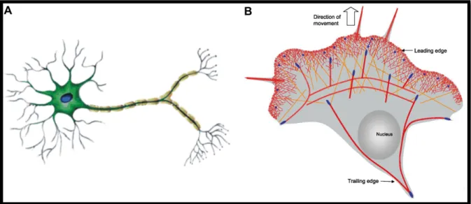
The establishment of cellular polarity is indispensable for developmental processes, such as asymmetric oocyte division, formation of epithelial tissues and the development of the central nervous system. Highly polarised cells undergo complex cellular rearrangements during their development in order to generate cells in which different cellular regions with different properties mediate different functions. Neuronal cells, being one of the most polarised cell types in vertebrate organisms, require a proper formation of morphological and thus functional polarity during development in order to fulfil their purpose to propagate excitation signals from the environment to the central nervous system and back to executing organs for reflexes and fine-tuned reactions (Figure 1A). Epithelial cells, lining the cavities and inner surfaces of organs and vessels in higher order organisms, demand a strong apical to basal polarity in order to fulfil their functions in secretion, absorption and transcellular transport, protection and sensing. Other cell types, such as immune cells, fibroblasts and, although not desired, cancer cells require fast and rather short-lasting polarisation processes as the base for cell migration (Figure 1B).
Figure 1 | Highly polarised eukaryotic cells. (A) Neuronal cells show a highly polarised morphology, forming branched dendrites for input and axons for output processing. The specified neuronal cell structure is required to enable efficient signal transduction within the nervous system. (B) Migrating cells adopt a highly polarised morphology, forming a leading edge with membrane protrusions and a retracting trailing edge. Both, persistent (A) and short-lasting or adaptive (B) cell polarisation, require the polarised expression of cell surface proteins mediated by intracellular transport. Images are adapted and modified from Chavarría and Cárdenas, 2013 (1) and Le Clainche and Carlier, 2008 (2).
The establishment of cell polarity is largely mediated by changes in the composition of the plasma membrane, therefore affecting how cells might act or react on their environment. Differential abundance of plasma membrane receptors, such as growth factor receptors, allows that one region of the cell is able to sense and respond to external signals in a different way than other regions of the cell do. The timely and spatially regulated presence (and absence) of cell adhesion proteins, such as integrin receptors, at the cell surface allows directed cell migration. Here, the adhesion receptors are constantly internalised at former adhesion sites and transported towards newly established adhesion sites. The insulin dependent insertion of glucose transporters provides the base for efficient glucose uptake from the intestinal tract and also other nutrients get internalised for breakdown and energy consumption. As another example, melanin, a natural pigment required for skin and hair colouration, needs to be transported from the centre towards the periphery of specialised cells, the melanocytes, to finally reach keratinocytes in the outer epidermal regions.
The cellular polarity is primarily mediated by intracellular transport processes in which highly sophisticated and highly regulated transport machineries deliver different cargoes, especially membrane vesicles, but also organelles, proteins, signalling peptides or RNAs to the demanding cellular regions. Different kinds of vesicle transport processes exist which are distinct in respect of contribution of specific proteins and co-factors, but which are also interconnected to generate a continuous flow of membrane trafficking inside the cell, thereby requiring complex regulatory mechanisms. Proteins considered for directed transport are newly synthesised at endoplasmatic reticulum (ER) associated ribosomes, delivered to the Golgi system where they undergo sorting for subsequent transport towards the desired target sites, for instance for exocytic transport through the trans-Golgi-network (TGN) towards the plasma membrane. Extracellular particles, nutrients, plasma membrane receptors but also other membrane components undergo endocytosis for sequential transport towards the cell interior and for subsequent degradation or reuse via recycling pathways. In this case, the endocytosed membrane components are sorted to specific membrane compartments, including the recycling endosome, from which they are transported back to the plasma membrane.
The aspects of intracellular vesicle transport are widely described by the so called highways and local
roads model (Figure 2) (3–6), which includes a fast and long-range transport mediated by a complex
and highly polarised microtubule network in order to quickly, but rather coarsely, transport cargo to
distinct cellular regions. Arrived there, a more slowly, but also more flexible cargo trafficking system
takes over which is mediated by the highly dynamic actin cytoskeleton that allows transport beyond
the microtubule network to precisely reach outlying regions of the cell and to provide the delivery at
the desired subcellular locales (7).
1.2 Motor proteins as force generators for vesicle transport
Microtubule networks and actin filaments, spanning the cell with different characteristics, serve as tracks for specific motor proteins which act as molecular motors that produce the force necessary for cargo movement. Motor proteins are ATPases, i.e. they hydrolyse ATP to produce energy and force, and are thus commonly referred to as mechanoenzymes (8). There are three major groups of motor proteins. Kinesin motors move along microtubules towards their plus ends, whereas dynein motors move in the opposite direction towards the microtubule minus end. The third major group comprises the myosin motor proteins which generate forces on actin filaments.
1.2.1 Kinesins and dyneins transport cargo along microtubule networks
There two major groups of microtubule associated motor proteins which are responsible for cargo movement. The kinesin motors comprise a large group of motor proteins, devided into 14 classes so far identified, including ~40 human genes (9–12). Kinesin motors move unidirectionally towards the microtubule plus end (13) with few exceptions (kinesins-14, e.g. Drosophila Ncd (nonclaret disjunctional), move minus end directed and kinesins-13 might not move at all) (9, 12). Most kinesins are heavy chain dimers and function as processive motors, i.e. they move continuously along microtubules due to their two motor heads binding in a sequential fashion to the microtubule tracks, finally allowing efficient cargo transport. One of those processive kinesins is the family of classical kinesins (kinesins-1). Kinesin-1 family members are capable to move over long distances with a velocity of ~0.8 µ𝑚/𝑠. Considering a step size of 8 nm (distance between two consecutive tubulin dimers), the motor takes a step every 10 ms (14, 15).
Dyneins belong to the AAA family of ATPases (ATPases Associated with Diverse Activities) and have a distinct evolutionary origin compared to kinesin and myosin motors (16). There are two classes of
Figure 2 | Schematic overview on intracellular vesicle transport processes. Membrane vesicles (green) containing proteins, membrane components and other material, are transported along microtubules (blue) for fast and long-range transport towards the cell periphery (microtubule plus end) and also towards the cell centre (microtubule minus end), as indicated by arrows. F-actin tracks (orange) are located in more peripheral regions of the cell and mediate rather slow but more flexible transport due to the highly dynamic character of actin filaments, in order to reach more outlying subcellular regions.
dyneins: the axonemal dynein and the cytoplasmic dynein (17). The axonemal dynein is important for the sliding of microtubules within the axonemes of cilia and flagella and thus for the beating movement of those. The cytoplasmic dynein motors transport diverse cargo towards the microtubule minus end and are thus involved in different cellular functions (18–21). Cytoplasmic dynein 1 transports cargo towards the cell centre and is essentially involved in mitosis, asymmetric cell division and developmental processes (22–31), whereas cytoplasmic dynein 2 has only transport functions within cilia (17). The cytoplasmic dynein comprises are large, 1.6 MDa motor complex formed by two heavy chains, including 6 AAA ATPase modules, and additional intermediate and light chains (16, 18). Not least owing their large size, dynein motor complexes are hard to handle in terms of recombinant expression and purification for subsequent analysis. In combination with the multiple ATPase domains within the heavy chain dimer the knowledge on the mechanism of dynein motors movement is limited compared to what is known for kinesin and myosin motors. Recent advances in the structural determination of dyneins allowed a better understanding of its motility mechanism, although it is still not completely clear how the specific minus end directed movement is mediated (17). A mechanism is proposed in which the two motor domains of the dynein dimer rather move in an inch-worm step manner, in which the leading head takes a step forward and is followed by the trailing head, in combination with other movement patterns, including eventual hand-over-hand steps, backward steps and steps with different step sizes (17, 32). By time-lapse fluorescence microscopy employing an intermediate chain-GFP fusion protein, the minus end directed movement velocities of cytoplasmic dynein have been measured ranging from ~1.4 to 2.8 µ𝑚/𝑠 (33).
In many cell types the microtubule network is organised by the microtubule-organising centre (MTOC), located next to the nucleus, leading to a polarised microtubule network in that the minus ends are trapped within the MTOC in the cell centre and the plus ends protrude towards the cell periphery. Therefore, kinesins are generally considered as motors driving outward directed movement towards the cell cortex, whereas dyneins are inward-directed motors to transport cargo towards the cell centre (13, 18).
1.2.2 Myosin actin motor proteins
Myosin motor proteins are molecular motors that associate with actin filaments. Myosin motors are
involved in a multitude of cellular and sub-cellular processes, including cell adhesion and migration,
cell division and transport of cargo, including vesicles, to drive endocytic and exocytic trafficking
events. Myosin motors are ATPases which hydrolyse ATP induced by actin filament binding in order
to generate forces which are subsequently used to either modulate the actin cytoskeleton leading to
morphological changes, or to move along actin tracks to transport cellular components.
The myosin superfamily consists of 35 classes (34) and 40 myosin genes have been identified in humans which are grouped into 13 classes. Myosin heavy chains are generally composed of three domains (35). A large motor domain, also referred to as head, is usually located at the N-terminus of the protein, harbours the ATPase activity and binds to F-actin. The motor is followed by a neck region containing a variable number of IQ motifs required for binding of calmodulin light chains or calmodulin related light chains. The neck region is also termed lever arm because of its structural rigidity that is utilized for the power stroke in consequence of conformational changes of the motor domain during the ATP hydrolysis cycle (see below) (36–39). The C-terminal part of myosin heavy chains comprises the tail domain which represents the highest diversity across different myosin classes in order to ensure the diversity of subcellular myosin functions.
The first myosin class discovered was the class II myosins, therefore named conventional myosins (40). All ensuing myosin classes discovered have been named accordingly unconventional myosins.
The most prominent member of conventional myosins is the sarcomeric myosin II which is primarily expressed in the striated skeletal muscle and cardiac muscle (8). Additionally, non-muscle myosin II and smooth muscle myosin II are found in vertebrates (8, 41). All class II myosins share the ability to form bipolar filaments of varying length wherein their motor heads are oriented in opposite directions to mediate the movement of complementary actin filaments towards each other. These characteristics enable sarcomeric myosin II to generate forces for muscle contraction by forming large bipolar filaments (8). Non-muscle myosin II forms shorter bipolar filaments and also generates forces on actin filaments, and is involved in cell adhesion and migration, protrusion formation and cytokinesis (8, 41–45). Myosin II family members also work to crosslink actin filaments (8).
1.3 Class V myosin motor proteins
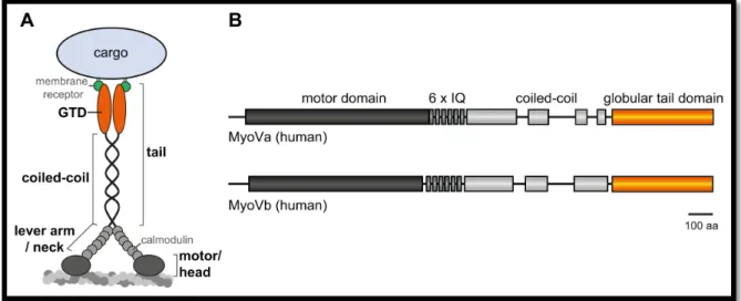
One member of the unconventional myosin motors comprises the class V myosins. In contrast to class II myosins which exert forces on actin to rather induce structural alterations, class V myosins work as molecular motors to transport a diversity of cargo along actin filaments (35, 46). The ability of class V myosins to function as a transporting motor is achieved by their specific structural organisation (Figure 3). Class V myosin proteins contain an N-terminal motor domain, an elongated neck with six IQ motifs and the C-terminal tail which is further subdivided into a coiled-coil region that mediates the dimerisation of two myosin V heavy chains and a globular tail domain (GTD) at the very C-terminus which serves as the cargo binding domain (8, 47). Myosin V motors move towards the actin filaments barbed end (plus end) and its gliding velocities are in the order of a magnitude slower than that of kinesins or dyneins and have been reported roughly between 0.2 and 0.8 µ𝑚/
𝑠𝑠𝑠 , although exceptions exist towards slower and faster movements (48–50).
Figure 3 | Structural organisation of class V myosins exemplified by the vertebrate myosin V (MyoV). (A) MyoV proteins contain an N-terminal motor domain (also called head) which binds to actin filaments and mediates the actin dependent ATPase activity. Six IQ motifs each bind calmodulin light chains and are also referred to as neck and forming the lever arm required for forward movement. The C-terminal tail is divided into a coiled-coil region which is periodically interrupted and required for heavy chain dimerisation, and the very C-terminal globular tail domain (GTD) which is the cargo binding domain, e.g. by binding to specific membrane receptors, and a major protein interaction site. (B) Schematic overview of the myosin Va and myosin Vb domain organisation. IQ, isoleucine/glutamine; GTD, globular tail domain; aa, amino acids.
1.3.1 Vertebrate myosin V
Class V myosins are conserved across almost all eukaryotic species (34). Three myosin V genes can be found in humans: MYO5A, MYO5B and MYO5C, encoding the myosin Va (MyoVa), myosin Vb (MyoVb) and myosin Vc (MyoVc) proteins, respectively (51–54). MyoVa and MyoVb proteins are expressed throughout the body (55) and can also be found in the brain, in contrast to myosin Vc (8, 54, 56–59). In the brain, MyoVb is strongly expressed in the hippocampus (57), whereas MyoVa is more widely distributed to other brain regions (58). Within neuronal cells, both isoforms are localised to dendritic spines and the postsynaptic densities (57, 60–62). Myosin Vc is highly abundant in epithelial and glandular tissues, such as pancreas, prostate, mammary, stomach, colon and lung, and is mainly expressed in epithelial cells (54). Different splice variants of all three MyoV isoforms exist, which are differentially expressed and which mediate specific interactions and functions (see below) (53, 55, 63–67). The vertebrate myosin Va protein has been first described in vitro to processively move along actin filaments, i.e. it moves processively as an isolated dimer on actin tracks in a step- wise, hand-over-hand fashion (8, 48, 68, 69). This characteristic is suggested to be demanded for an efficient actin dependent transport of cargo in living cells (69, 70). The vertebrate MyoV feature of processive movement is in contrast to other myosin classes and is even not conserved within the myosin V family (myosin Vc and Drosphila myosin V; see below).
The base for efficient MyoVa movement along F-actin is provided by its specific structural
organisation and adaptation. The myosin Va heavy chain dimer binds to actin filaments by its motor
domains forming a leading and a trailing head each time (Figure 4). At the point the trailing head
detaches from the filament, the leading head undergoes a slight conformational change, referred to as the power stroke, bringing the trailing head in front to become the new leading head, i.e. the motor takes one step (35, 71). The step size of myosin motors largely depends on the length of the neck region/lever arm. The two heads of the MyoVa dimer bind to the actin filament within a distance (step size) of 36 𝑛𝑚 which is facilitated by the elongated neck region containing six IQ motifs (69). In comparison, the step size of the conventional myosin II is only about 7 𝑛𝑚 due to the shorter neck (72). The large step size of myosin Va perfectly fits to the double helical actin filament organisation with consecutive actin monomers every 36 𝑛𝑚 on the short pitch helix. Thus, MyoVa is able to walk along actin filaments in a linear, step-like fashion instead of spiralling around the filament by following the long pitch helix, which would strongly impede transport through the cytoplasm (71, 73–77). Another structural feature facilitating continuous transport of cargo along actin tracks is the flexibility of the tail domain induced by interruptions in between the coiled-coil regions (78, 79). Finally, myosin Va proteins adapted to intracellular transport in terms of binding to specific cargoes in distinct ways therefore allowing to specifically switch between different cargoes for different transport routes. Although the cargo binding is mainly mediated by the globular tail domain which interacts with specific membrane associated proteins, such as Rab GTPases or their respective adaptors (80–84), the ability to select between different cargoes is further supported by other parts of the protein, for instance the flexible regions within the coiled-coil domains, and cell type specific alternative splicing events (66, 67, 80). The elongated neck of MyoVa also enables the motor to switch from one actin filament to another one allowing transport of cargo across a complex network of actin filaments or along branched filaments (6, 85).
The kinetic properties of myosin Va motors are another rationale for its processive movement (Figure
4). In general, there are four kinetic states of the motor domain influencing the actin binding capacity
and strength. The motor alone or the motor binding ADP shows high affinity for F-actin, whereas the
motor binding ADP and inorganic phosphate (P
i) or the motor binding ATP shows low affinity for F-
actin. As the hydrolysis of ATP to ADP and P
iand the subsequent release of P
iare rather fast
processes, the dissociation of ADP from the motor domain is considered to be the rate-limiting step
in the ATPase cycle of the myosin V motor domain (86, 87). Considerng that, the motor domain is
most of the cycle time tightly bound to the actin filament (it has a high duty ratio), which is reported
to be demanded for processive movement along actin tracks (8, 35, 68). As a final requirement for
the continuous movement, the ATP hydrolysis cycles within the two motor domains of the dimer
need to be tightly regulated in a way, that only one of the motors can detach from the filament at a
given time (68, 88–91).
Among the vertebrate class V myosins, myosin Vb is very similar to myosin Va in terms of structure and kinetics and is also reported to be a processive actin motor at single dimer level (48). The third vertebrate MyoV isoform, myosin Vc, is also similar in terms of structure (~50% overall identity to both MyoVa and MyoVb) (54), but has not been considered to be a processive motor as a single dimer. That is reasoned by the ADP release being not the rate-limiting step within the ATP hydrolysis cycle of myosin Vc (92, 93). Nevertheless, specific conditions are discussed in which myosin Vc might act as processive motor or that multiple MyoVc proteins assemble to a large motor complex to efficiently transport cargo (94).
Figure 4 | Processive movement of MyoVa along actin filaments. (1) Both motor heads of the MyoVa dimer bind ADP and are strongly attached to the actin track. (2) Due to an intramolecular strain, ADP is released first from the trailing head. (3) The trailing head binds ATP and subsequently dissociates from the actin track. (4) The still attached leading head undergoes a conformational change (power stroke) to bring the former trailing head in front to become the new leading head. ATP bound to the new leading head gets hydrolysed to ADP and Pi and by a thermally driven search the new leading head finds a binding site. (5) The new leading head binds to actin inducing the fast release of Pi followed by strong binding of the head to the actin track. The cycle is completed and the MyoVa dimer moved 36 nm (step size) towards the F-actin barbed end. Adapted and modified from Hammer and Sellers, 2012 (35).
1.3.2 Insect and yeast class V myosin motors
The Drosophila melanogaster myosin V (Dm-MyoV) is a well-characterised example for class V myosin members significantly differing from vertebrate MyoVa and MyoVb (49). As it is true for vertebrate MyoVc, the structural organisation of Dm-MyoV is similar to that of vertebrate MyoV proteins, besides their distinct evolutionary origin. Although the velocity of Dm-MyoV is nearly identical to that of vertebrate MyoV processive motors, major alterations can be found in respect to the kinetic cycle. Dm-MyoV is considered to be a low duty ratio myosin, i.e. the motor is most of the time of the ATP hydrolysis cycle detached from F-actin (in contrast to vertebrate MyoV), which prevents its function as a processive motor at the single molecule level. To overcome these intrinsic deficits, multiple motor proteins cluster at the potential cargo to form a processive motor unit which ultimately drives the processive transport (49).
In line with that, the two class V myosin members found in yeast, Myo2p and Myo4p, also act differently compared to vertebrate MyoV (95–97). Myo2p is structurally similar to vertebrate MyoV but does not function as a processive motor as a single dimeric molecule (95). Myo4p is even more different as it has a distinct structural organisation by forming a heterodimeric complex with the Swi5-dependent HO expression 3 protein (She3). Thus, Myo4p is a single-headed motor which does not move processively along F-actin alone but only by clustering into motor units, similar to Dm- MyoV (95, 97).
1.3.3 Regulation of myosin V activity and function
The property of myosin V motor proteins being processive motors on actin tracks demand regulatory mechanisms that prevent its activity and movement on F-actin when not required, e.g. when not attached to cargo. The full-length myosin V motor exists in two distinct states. There is an auto- inhibited, inactive state in which the MyoV dimer is back-folded in a way that its globular tail domains (GTD) bind to its motor domains (Figure 5) (98–100). In this state, the ATPase activity is inhibited and the motor binds only weakly to actin filaments (101, 102). Furthermore, the auto- inhibited motor shows a cytoplasmic localisation and is therefore not attached to vesicles or other cargo (103). Activatory mechanisms release the inhibiting GTD/motor domain interactions and enable MyoV to adopt an open and extended conformation that resembles the active state at which it binds to cargo and F-actin. Increased concentrations of intracellular Ca
2+ions induce the MyoV dimer opening and activate the ATPase activity in vitro (104). At the same time, excessive Ca
2+concentrations ultimately lead to the dissociation of calmodulin light chains from the neck region
thereby making it very flexible and thus not suited to drive the motor movement, which finally
results in the inability of MyoVa to move on F-actin tracks (102, 105, 106). The binding of cargo to the
globular tail domains might be ultimately the more physiologically relevant opening mechanism for
MyoV proteins. A perfect example in that respect is the melanophilin (MLPH or Slac-2a) protein (see below) which binds to the GTD of MyoVa, activates its ATPase activity and facilitates its movement on actin filaments in vitro (107, 108). Furthermore, it has been demonstrated very recently by means of electron microscopy, that MLPH is able to bind to the purified full-length back-folded MyoVa dimer and induces its opening into the extended conformation (109).
Besides the regulated opening and activation mechanisms for myosin V dimers, myosin motors also undergo a fine-tuned regulation at their active state, at least in some cases. Although, generally, myosin V proteins (e.g. the mammalian MyoVa and MyoVb) are processive actin-based motors as dimeric molecules, it has been repeatedly observed that myosin V proteins cluster at the cargo surface to form motor units (110–112), in a similar way as was described for the Drosophila MyoV or mammalian MyoVc. This mechanistic behaviour increases the run length of the motors on actin tracks but also reduces their velocities compared to single dimeric molecules (113–120). This phenomenon might be explained by a hindrance due to asynchronous movement of the different motors (119) and by a load dependence of the motor kinetics, meaning that the cargo itself can influence the motors walking behaviour, at least to some extent (88, 121–123). Even the lipid composition of the vesicular cargo can influence the myosin movement (50). A more fluid vesicle nature thereby induces walking velocities higher than those at the single dimeric molecule level even measuring up to velocities of unloaded motors. Contrary, more gel-like vesicle compositions reduced the walking velocity. These findings are further supported by a mechanism in which the melanophilin protein has also been demonstrated to slow myosin Va movement (~4-fold), but, on the other hand, increases the length of processive walks (~2-fold) compared to the single dimeric motor. Altogether, the time the motor is attached to actin tracks is significantly increased, therefore facilitating melanosome trafficking in melanocytes (124). A suitable explanation for these observations might be
Figure 5 | Model of the back-folded auto-inhibited state of the full-length myosin V motor. The two globular tail domains (GTD 1, 2) of the heavy chain dimer interact with the two motor domains to adopt a back-folded state in which the ATPase activity of the motor is inhibited and only weak F-actin binding is obtained. Increased concentrations of Ca2+ ions induce MyoV opening and ATPase activity of the motor but do not promote efficient processive movement due to the release of calmodulin from the neck region.
Cargo binding to the GTDs induces MyoV unfolding, activates ATPase activity and allows efficient MyoV driven cargo transport. The MyoVa-GTD Rab11 binding site is highlighted in blue. The model was generated from a partial electron microscopy model (PDB ID 2DFS) (99), mutational analysis data on residues participating in the GTD/motor interactions (109) and the crystal structures of the MyoVb GTD (210). Image provided by Olena Pylypenko.
that electrostatic interactions between MLPH and actin filaments make MLPH to act as a tether for the myosin Va motor to stay longer attached to the actin tracks.
Furthermore, an interconnectivity has been revealed in which motor proteins of different families cooperate to influence each other’s processivity (125). One example here is the cooperation of myosin V and kinesin family motors in the periphery-directed transport. In this process, myosin V performs a diffusional search along microtubules which in turn prolongs the run length of the kinesin motor attached to the same cargo. The same effects occur the other way around for myosin V motors on actin tracks. The proposed mechanism for a mutual run length enhancement might be explained (in similarity to the aforementioned MLPH effects on myosin Va processivity) by electrostatic interactions within the non-specific motor-cytoskeleton connections, which possibly have a tethering character that reduces the diffusion rate of the cytoskeleton specific motor.
1.4 The Ras superfamily of small GTPases
Kinesin, dynein and myosin motor proteins were introduced as the molecular motors that provide the forces required for movement of cargo on cytoskeletal tracks. The delivery of the highly diverse set of intracellular membrane vesicles to their respective destinations requires to be coordinated in a timely and spatially regulated manner. Members of the Rab family of small GTPases, being a family of the Ras superfamily of small GTPases, act here as master regulators as they control the membrane identity in order to provide the basis for a proper sequence of vesicle budding, movement and fusion by recruiting specific adaptor and effector proteins to intracellular membranes (126).
1.4.1 The Ras superfamily includes five families of small GTPases
The Ras superfamily comprises a very large and diverse set of small GTP binding proteins and is subdivided into five major families (127). The Rab (Ras-like in brain) family comprises the largest group of small GTPases (126, 127), next to the other four major families of Ras, Rho, Arf/Sar and Ran GTPases all of which serve different functions (127). Shortly, the Ras (Rat sarcoma) family members act as signalling molecules, integrating extracellular stimuli to activate intracellular signalling cascades (for instance the mitogen activated protein kinase cascade) to control gene transcription.
The most prominent members of the Ras family might be the oncogenic Ras proteins. Members of
the Rho (Ras homology) family are well known for their function in the regulation of actin dynamics
(128, 129), cell polarity (130), cell cycle progression and gene expression (127). The Ran (Ras-like
nuclear) family of small GTPases contains only one member in eukaryotic cells, except plants having
multiple members, and are involved in nuclear transport across the nuclear envelop (127). The Arf
(ADP-ribosylation factor) family is the most divergent family of the Ras superfamily and is also involved in vesicle transport (131).
The structural organisation that is shared by Ras superfamily small GTPases includes a large ( ~ 170 amino acids) G domain which mediates the GTP binding and hydrolysis, and the interaction with different effectors (131, 132). The G domain itself contains five G-boxes (G1 to G5) with conserved residues. The switch I and switch II regions as part of the G domain are particularly involved in effector binding and undergo a conformational change upon GTP binding, especially Switch I located around G2 (133).
1.4.2 The Ras cycle
A common feature of most Ras superfamily small GTPases is their function as molecular switches by alternating between two distinct conformational states: the GTP bound active state and the GDP bound inactive state. The conversion between the two states is mediated by a Ras GDP-GTP cycle (Figure 6). GDP in the inactive Ras protein is exchanged by GTP under support of guanine nucleotide exchange factors (GEFs) which facilitate the release of GDP and favour fast GTP loading due to the high abundance of GTP in the cytoplasm (~1 mM) (126). GTPase activating proteins (GAPs) stimulate the hydrolysis of GTP to GDP and inorganic phosphate (P
i) within the GTPase which is necessary given the relatively poor intrinsic GTP hydrolysis activity of the GTPase itself (132, 134). This is also the reason for the term GTPase being not completely precise, as GTP hydrolysis needs the support by GAPs, and therefore the term small G-protein might be more suited for these kinds of proteins.
However, to stay with the literature I will keep to the term small GTPase throughout this thesis.
Generally, GEFs and GAPs are essential factors determining small GTPase activity in time and space as they integrate a wide range of cellular inputs coming from different signal sources, such as binding of signalling proteins, second messengers like cAMP, or structural proteins (134).
Most family members of the Ras superfamily of small GTPases are specifically targeted to distinct
subcellular compartments, such as membrane vesicles or larger organelles, either by membrane-
targeting protein sequences or by post-translational lipid modifications inserting into the membrane
lipid bilayer (132). Members of the Ras, Rho and Rab families are typically modified via isoprenylation
at the C-terminus. Farnesyl, geranylgeranyl or palmitoyl lipid groups are here covalently linked to
cysteine residues of common sequence motifs, including the Caax (cysteine-aliphatic-aliphatic-
arbitrary) motif (131, 132, 135). Within the large and diverse family of Rab GTPases isoprenylation
motifs were found with C-terminal cysteine residues involving the motifs: xxxCC, xxCxC, xxCCx, xCCcc
and CCxxx which are modified by geranylgeranylation (135, 136). To ensure efficient isoprenylation,
members of the Rab family need the assistance of a helper protein, the Rab escort protein (REP)
(137). REP binds to newly synthesised Rab proteins and presents them to geranylgeranyl transferases, thus allowing efficient geranylgeranylation of both C-terminal cysteins (138–140). Arf family members are typically N-terminally myristoylated and Ran family members are not modified (131, 132, 141). In providing a strong and specific subcellular localisation of small GTPases to distinct vesicle pools and/or organelles, the post-translational modification is only one part of the truth; the target membrane composition, electrostatic properties of both membrane and protein, and the support by chaperones must act together in order to achieve selectivity and specificity in membrane targeting of Ras small GTPases (132).
The eventual presence of lipid modifications require a further regulatory feature for small GTPases involving the guanine nucleotide dissociation inhibitors (GDIs) (142). GDIs have a high affinity for the inactive, GDP bound and lipid modified GTPases and serve first, to prevent the release of GDP in order to stabilise the inactive form (143) and second, as a shield for the hydrophobic lipid modified part against the hydrophilic cytoplasm when being transported to destined locales (132, 144–149).
1.5 Rab family GTPases
The Rab family of small GTPases comprises the largest group within the Ras superfamily. Rab proteins are considered as being the master regulators of intracellular vesicle transport processes as they tightly coordinate all stages of a membrane vesicles lifecycle, including vesicle budding from the donor compartment, vesicle uncoating, vesicle transport, vesicle docking at the acceptor compartment and subsequent membrane fusion (Figure 7) (7, 126, 127). So far, 65 members of the Rab family have been identified in the human genome, which are divided into 9 functional groups.
The large diversity of this family is mainly explained by gene duplications during evolution. Each member of the Rab family is localised to specific intracellular membrane compartments (126, 127, 150–153). Rab proteins decide on the membrane identity and mediate their functions in membrane
Figure 6 | Activity cycle of Ras superfamily small GTPases. Ras small GTPases exist in to distinct states: the GDP bound inactive state (yellow) and GTP bound active state (red). Ras-GDP is bound by guanine nucleotide dissociation inhibitors (GDI, brown) to prevent GDP dissociation and shielding lipid modifications. Guanine nucleotide exchange factors (GEF, green) mediate the exchange of GDP by GTP inducing Ras GTPase activation and enabling its interaction with a wide range of effector and adaptor proteins. GTPase activating proteins (GAP, pink) support the intrinsic GTP hydrolysis activity of Ras proteins to hydrolyse GTP to GDP and inorganic phosphate (Pi), and finally inactivate the GTPase to complete the activity cycle.
transport by recruiting specific adaptor and effector proteins towards the membrane surface. To do so, they interact with a wide range of membrane associated proteins, such as vesicle coat proteins, motor proteins, tethering factors, signalling proteins and SNARE (SNAP (Soluble NSF Attachment Protein) Receptor) complex proteins (126).
1.5.1 Regulation of Rab GTPase activity and its impact on intracellular membrane cycling
The aforementioned cycling between the active, GTP bound and inactive, GDP bound Rab GTPase states induces major conformational changes in the switch I and switch II regions (154). Rab GEFs specifically recognise distinct residues in these switch regions to induce GDP release and GTP binding. Following the GTP-binding induced conformational change, adaptor and effector proteins bind to both switch and interswitch regions of the active Rab-GTP (155). GTP hydrolysis of Rab proteins is supported by Rab GAPs. Rab GAPs contain a conserved catalytic TBC domain (Tre2/Bub2/Cdc16) (156–158) promoting GTP hydrolysis by a dual arginine-glutamine-finger mechanism (157, 159, 160). There are at least 40 Rab GAPs in humans, suggesting specific interactions of individual GAPs with their respective Rab proteins (161, 162).
A well-regulated interplay between Rab GEFs and Rab GAPs wit h specific Rab GDIs, the Rab escort protein REP, and membrane bound GDI displacement factors (GDFs), last of which recognise cytoplasmic Rab-GDI complexes and induce GDI release, is proposed to be the driving force for specific targeting of distinct geranylgeranylated Rab GTPases to their desired membrane compartments, finally enabling the directed transport from donor to acceptor compartments (Figure 7) (126, 163). The whole membrane transfer procedure can be divided into five major steps (126):
vesicle budding and sorting of target proteins to the budding vesicle, potentially accompanied by
vesicle coating with stabilising coat proteins (i), vesicle uncoating (ii), vesicle transport through the
cytoplasm along cytoskeletal tracks (iii), vesicle tethering at target membrane compartments,
supported by tethering factors and SNARE complex proteins (iv), and vesicle fusion with the destined
compartment (v).
Figure 7 | Schematic overview on vesicle transport from a donor towards an acceptor membrane compartment and the involvement of Rab small GTPases. Vesicle budding starts from the donor membrane compartment. This step requires active, GTP bound Rab GTPases located at the future bud site and recruiting different adaptor and effector proteins.
Cargo destined to undergo trafficking is sorted into the vesicle bud. Potentially, vesicle formation involves clathrin coating of the membrane for clathrin mediated endocytosis.
Following contingent uncoating, membrane vesicles are transported in a Rab-GTP and motor protein dependent fashion along microtubule and actin tracks. When reaching the acceptor compartment, vesicle docking and subsequent vesicle fusion is facilitated by tethering factors located at the target membrane and SNARE proteins on both vesicle and target membrane. In succession of
vesicle fusion with the target Rab GAPs bind to Rab-GTP and support GTP hydrolysis to GDP and the release of inorganic phosphate (Pi), leading to Rab GTPase inactivation and subsequent Rab-GDP liberation from the membrane. Rab-GDIs bind to cytosolic Rab-GDP to prevent GDP dissociation and to shield hydrophobic lipid tails. Rab-GDP is recycled and eventually returned to membrane compartments. Here, Rab-GDFs target cytosolic Rab-GDP:GDI complexes, facilitate GDI release from Rab-GDP thus retargeting Rab proteins to membranes. Rab-GEFs mediate the exchange of GDP by GTP therefore activating Rab GTPases and start the cycle from the beginning. GAP, GTPase activating protein; GDI, guanine nucleotide dissociation inhibitor; GDF, GDI displacement factor; GEF, guanine nucleotide exchange factor.
1.5.2 Rab GTPases as master regulators of intracellular transport
Intracellular membrane trafficking comprises a large array of different types of membrane vesicles,
transported between different types of donor and acceptor compartments, organelles and other
cargo. Specific Rab GTPases are associated with distinct membraneous compartments and are
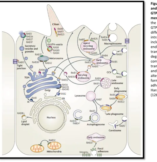
therefore involved in particular transport processes. These have been reviewed in detail by Harald
Stenmark (Figure 8) (126) and are shortly summarised here. Rab1 is involved in trafficking from the
endoplasmatic reticulum (ER) to the Golgi as it is localised to the ER and pre-Golgi intermediate
compartment (IC). Rab2 might have similar functions. Trafficking within the Golgi system is mediated
by Rab6, Rab33 and Rab40. Rab8 is a major regulator of exocytic transport from the trans-Golgi
network (TGN) to the plasma membrane, e.g. it supports the transport of GLUT4 vesicles, together
with Rab10 and Rab14. Different types of exocytic transport are further mediated by Rab3, Rab26,
Rab27 and Rab37. Rab27a is particularly important for the periphery directed transport of
melanosomes in melanocytes as an essential mechanism in skin and hair pigmentation, and Rab32
and Rab38 participate in the biogenesis of melanosomes, as do Rab7 and Rab8. Rab32 is furthermore
crucial for mitochondria fission. Rab22 is required for trafficking between the TGN and the early endosomes. Rab5 is potentially the most important regulator of endocytosis as it is localised to endocytic sites at the plasma membrane and to early endosomes and phagosomes as well. Rab5 functions in mediating endocytic events, for instance by inducing the formation of clathrin-coated pits and clathrin-mediated endocytosis, and macropinocytosis, and is also involved in subsequent endocytic pathways, such as the fusion of clathrin-coated vesicles with the early endosome and the maturation of early phagosomes, often together with other Rab proteins. Rab21, for instance, participates in integrin receptor re-uptake. Recycling pathways are used to return endocytosed receptors, signalling molecules and other membrane components back to the plasma membrane.
Fast recycling only involves the early endosome and is mediated by Rab4. In contrast, a more slowly recycling traverses the recycling endosome and requires function of Rab11 and Rab35. In this process also Rab15 plays a role in trafficking from the early endosome towards the recycling endosome.
Despite its role in exocytosis, Rab8 is also involved in endocytic and recycling events. Together with
the Arf family member Arf6, Rab8 induces macropinosome formation, subsequent tubulation of
those and final recycling of receptors, signalling molecules and membrane components (164). Rab7a
and Rab7b are structurally very similar but contribute to different endosomal transport steps (165,
166). Rab7a is localised to the early endosome and mediates transport towards the late endosome
and lysosome, therefore playing a role in protein degradation (165). In contrast, Rab7b is additionally
localised to the TGN and Golgi and rather mediates retrograde trafficking from the late endosome
towards the TGN (167). Similar functions are described for Rab9.
1.5.3 Rab GTPases form complexes with motor proteins
In order to achieve vesicle transport along cytoskeletal tracks, motor proteins need to associate with vesicle membranes in a highly specific and well-regulated fashion. Rab family GTPases specifically recruit motor proteins directly or indirectly to the respective vesicle surface. This mechanism is well described for class V myosin actin motors which have been shown to associate with Rab GTPases at vesicle membranes in both, in yeast and higher order eukaryotic cells. The yeast Rab GTPase Ypt11p has been shown to interact with the class V myosin member Myo2p in transporting the Golgi into a growing bud (168). Furthermore, a cooperation of Ypt11p and Myo2p has been implicated in the transport and inheritance of mitochondria (169, 170), and the yeast Rab GTPase Sec4p (the mammalian homolog is Rab8) has been demonstrated to recruit Myo2p to secretory vesicles (169, 171).
The well-studied role of Rab27a in melanosome transport in melanocytes is another example for the formation of Rab GTPase myosin V motor protein complexes (80, 124, 169, 172–174). Here, Rab27a forms a tripartite complex with myosin Va and the melanophilin protein (MLPH). MLPH functions to connect the myosin Va actin motor with the melanosome associated Rab27a small GTPase (83). The formation of this tripartite complex is essential for the transport of melanosomes to the periphery of
Figure 8 | Localisation and function of Rab GTPases at intracellular membranes. Members of the large family of Rab GTPases regulate different steps of intracellular trafficking, including exocytosis, endocytosis and recycling transport, pathways for degradation of cellular content, organelle transport and dynamics, and cell morphological alterations, such as cilia formation and cell adhesion. Adapted from Harald Stenmark, 2009 (126).
melanocytes for subsequent transfer to keratinocytes, an important process for skin and hair pigmentation (80, 175). Mutation of either of the complex partners underlies the manifestation of a disease called Griscelli syndrome (GS) (174, 176–180). There are three types of disease presentation all of which have in common a skin and hair hypopigmentation (176). Griscelli syndrome type 1 further includes severe neurological impairments and is caused by mutations in the MYO5A gene (176, 180). GS type 2 is based on mutations in the RAB27A gene and presents with misfunctions of the immune system and increased infection susceptibility (174, 176, 177). GS type 3 is related to mutations in the MLPH gene and here patients only show the phenotype of partial albinism (176, 178, 179).
The Myosin and Rab interacting protein (MyRIP or Slac-2c) is another Rab27a binding protein which has been shown to form a similar tripartite complex with myosin VIIa. The Drosophila myosin VIIa protein has been shown to function as a processive motor on actin filaments and the mammalian homolog is supposed to act in a similar way (181–183). Myosin VIIa plays a crucial role in the retina and in the inner ear as it is localised to photoreceptor cells and pigmented epithelial cells of the retina, and to the pericuticular necklace of sensory hair cells (181, 184–186), where it functions as a molecular transporter (187, 188). In particular, a Rab27a:MyRIP:myosin VIIa complex is required for melanosome motility in the retinal pigment epithelium (189). Mutations of the MYO7A gene have been shown to cause the human Usher syndrome type 1B (184). Usher syndrome type 1 is the most frequent cause for inherited deaf-blindness in humans (190), presenting sensorineural hearing loss, vestibular dysfunctions and early-onset retinis pigmentosa (184, 191), and underlies mutations in multiple loci leading to different disease subtypes (myosin VIIa is encoded by the USH1B locus) (184, 190).
This mechanism of differential targeting of distinct motor proteins to Rab27a vesicles by co- recruitment of specific adaptor proteins (MLPH vs. MyRIP) nicely shows the fine-tuned regulation of vesicle associated protein complex formation, depending on cell type and cargo. In accordance, Rab27a has been shown to functionally correlate with myosin Va in the transport of dense core secretory granules to the plasma membrane of pancreatic beta-cells (192). Here, Rab27a and myosin Va form a tripartite complex with the adaptors granuphilin-a, granuphilin-b and rabphilin-3A, respectively.
The small GTPase Rab3A, being the most abundant Rab family member in the brain (193), has been
shown to directly interact with myosin Va at synaptic vesicles to facilitate the transport of these
vesicles in neuronal cells (194), as has also been shown for Rab27b (195). Rab8a directly interacts
with myosin Vb at the aforementioned tubular recycling network containing Eps-15-homology
domain-containing proteins EHD1 and EHD3 (196). Rab8a, in conjunction with Rab11a, interacts with
myosin Vb in the regulation of enterocyte polarity by driving apical transport processes, and regulate
enterocytic microvilli growth (197). Mis-regulation of these Rab motor complexes has a major impact on the pathogenesis of microvillus inclusion disease, in which impaired interactions of MyoVb and Rab8a induce microvilli loss and uncoupling of MyoVb and Rab11a lead to microvilli inclusions. Rab8a also interacts with myosin Va in the insulin triggered transport of GLUT4 (glucose transporter type 4) vesicles towards the plasma membrane of rat muscle cells (198–200). A similar mechanism has been proposed for the cooperation of Rab10 and MyoVa in adipocytes (201). These studies nicely show the interconnection of stimuli induced intracellular signalling (e.g. phosphorylation events following insulin receptor activation) and vesicle transport processes (Rab GTPase activity). Rab8a is furthermore suggested to interact with the epithelial-related myosin Vc in the transferrin receptor transport in epithelial cells (54). Rab10, which is related to Rab8a and localises to the same tubular network (196), has also been shown to interact with myosin Va, Vb and Vc in vivo and is thus proposed to be involved in similar recycling functions (55, 202–206). Vertebrate myosin Vc directly interacts with Rab38 and the closely related Rab32 to cooperate in the biogenesis and secretion of melanosomes (207). Here, the myosin Vc motor is required to transport distinct melanosome biogenesis factors, such as the tyrosinase-related proteins Tyrp1 and Tyrp2, and the SNARE protein Vamp7, from the early and recycling endosomes towards maturing melanosomes. Interestingly, Rab32 and Rab38 interact specifically with MyoVc, but neither with MyoVa nor with MyoVb. In the following melanosome maturation process, MyoVc also interacts with Rab7a and Rab8a.
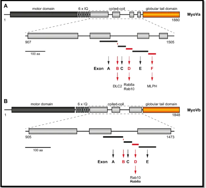
Alternatively spliced exons within the C-terminal regions between the neck and the globular tail domain in myosin Va, myosin Vb and myosin Vc specifically determine the interaction of the motor proteins with distinct Rab GTPases (Figure 9) (55). Myosin Va has six alternatively spliced exons:
exons A, B, C, D, E and F, whereas myosin Vb has only five as it misses exon F. Particularly, the three
exons B, D and F undergo alternative splicing in myosin Va (53, 63, 64). Exon F supports the
association of myosin Va with melanophilin to form a functional complex with Rab27a (80, 83, 208,
209). Exon B has been demonstrated to play a role in the interaction of myosin Va with dynein light
chain 2 (DLC2), which, however, has not been resembled for the exon B of myosin Vb (66, 67). Exon D
is required for MyoVa binding to Rab8a and Rab10. For MyoVb the exon D is also essential for Rab10
interaction, but, in contrast to MyoVa, inhibits binding to Rab8a (55). Rab11a interacts with both,
myosin Va and myosin Vb, in an exon independent fashion (55, 210, 211). Myosin Vc also expresses
an alternatively spliced exon F which determines its interaction with Rab32 and Rab38 (207).
Figure 9 | Schematic representation of alternatively spliced exons of mammalian MyoVa and MyoVb proteins.
Alternatively spliced exons are located within the coiled-coil regions of the MyoV-tail (exons A, B, C, D, E and F for MyoVa, and exons A, B, C, D and E for MyoVb). (A) Three exons are particularly subjected to alternative splicing in MyoVa: exons B, D and F (drawn in red). Exon B mediates interaction with dynein light chain 2 (DLC2). Exon D is essential for MyoVa interaction with Rab8a and Rab10. The melanocyte specific exon F is required for efficient interaction with melanophilin (MLPH). (B) Only two exons in particular undergo alternative splicing in MyoVb: exons B and D (drawn in red). A specific function for exon B has not been demonstrated so far. Exon D mediates MyoVb interaction with Rab10, similar to MyoVa, but, in contrast, inhibits its interaction with Rab8a, which binds to the same region in absence of exon D. There is no exon F present in MyoVb. Numbers on the protein domains indicate amino acids for mouse (Mm) MyoVa and human (Hs) MyoVb;
aa, amino acids.