Stable Immunosuppression Under Low-dose Tacrolimus Monotherapy is Dependent Upon
Immunological Regulation
DISSERTATION ZUR ERLANGUNG DES DOKTORGRADES DER NATURWISSENSCHAFTEN (DR. RER. NAT.) DER FAKULTÄT FÜR
BIOLOGIE UND VORKLINISCHE MEDIZIN DER UNIVERSITÄT REGENSBURG
vorgelegt von
Anja Nadine Kammler
aus
Straubing
im Jahr
2013
Das Promotionsgesuch wurde eingereicht am:
26.11.2013
Die Arbeit wurde angeleitet von:
Prof. Dr. Edward K. Geissler
Unterschrift:
Table of contents
ITable of contents
TABLE OF CONTENTS ... I
ABSTRACT... VI
ZUSAMMENFASSUNG ... VII
1 INTRODUCTION ... 1
1.1 Transplantation – an overview... 1
1.2 Terms in transplantation ... 1
1.3 Basic concepts ... 2
1.3.1 T cell activation... 2
1.3.1.1 TCR signalling ... 2
1.3.1.2 Costimulation... 3
1.3.2 T cell specification ... 3
1.4 Allorecognition... 4
1.4.1 Direct Allorecognition... 6
1.4.2 Indirect Allorecognition ... 7
1.4.3 Semi-direct Allorecognition... 8
1.5 Rejection... 9
1.5.1 Hyperacute rejection ... 9
1.5.2 Acute rejection ... 9
1.5.3 Chronic Rejection... 11
1.6 Regulatory immune cells in Transplantation... 12
1.6.1 Regulatory T cells ... 13
1.6.1.1 CD4+ regulatory T cells... 13
1.6.1.2 CD8+ regulatory T cells... 14
1.6.1.3 CD4-CD8- regulatory T cells ... 15
1.6.2 Regulatory B cells ... 15
1.6.3 Regulatory macrophages ... 15
1.6.4 Tolerogenic DCs ... 16
Table of contents
II1.6.5 Myeloid - derived suppressor cells (MDSCs) ... 16
1.7 Immunosuppressive treatment in Transplantation... 17
1.7.1 Overview ... 17
1.7.2 Calcineurin Inhibitors... 18
1.7.2.1 Cyclosporine (CsA) ... 18
1.7.2.2 Tacrolimus (FK-506) ... 19
1.7.3 CNI toxicity – a trade off?... 20
1.7.4 Pharmacokinetics ... 21
1.8 Tolerance - inducing strategies... 22
1.8.1 Costimulatory blockade with anti-CD154 in animal models ... 22
1.8.1.1 Effects of anti-CD154 ... 22
1.8.1.2 Combined treatment of anti-CD154 + DST ... 23
1.8.2 Clinically applied strategies ... 23
1.9 Tolerance – a balance?... 25
2 AIM ... 26
3 MATERIALS AND METHODS ... 27
3.1 Materials... 27
3.1.1 Instrumentation ... 27
3.1.2 Consumables ... 27
3.1.3 Operation consumables ... 28
3.1.4 Reagents ... 28
3.1.5 Kits ... 30
3.1.6 Antibodies ... 30
3.1.6.1 For injection ... 30
3.1.6.2 For Histology ... 30
3.1.6.3 For FACS ... 31
3.1.7 Buffers and solutions... 33
3.1.8 Primers ... 33
3.1.8.1 Housekeeping genes... 34
3.1.8.2 Genes of interest ... 34
3.1.9 Software ... 35
3.1.10 Mice... 35
Table of contents
III3.2 Methods ... 36
3.2.1 Methods involving mice... 36
3.2.1.1 Treatment of mice ... 36
3.2.1.2 Skin-Transplantation... 36
3.2.1.3 Graft monitoring ... 36
3.2.1.4 Donor specific transfusion ... 37
3.2.1.5 Retransplantation ... 37
3.2.1.6 Effector cells from sensitised mice ... 37
3.2.1.7 Transfer of LN cells ... 38
3.2.1.8 Splenectomy... 38
3.2.1.9 Thymectomy ... 38
3.2.1.10 Application of antibodies ... 38
3.2.1.11 Application of Tacrolimus therapy ... 39
3.2.1.12 Application of Diphtheria toxin ... 39
3.2.1.13 Toxicology ... 40
3.2.2 Molecular biology ... 40
3.2.2.1 RNA isolation ... 40
3.2.2.2 cDNA synthesis ... 40
3.2.2.3 Quantitative real-time PCR ... 41
3.2.2.4 IFNγ -ELISA... 43
3.2.3 Cell biological methods... 43
3.2.3.1 Determination of cell numbers... 43
3.2.3.2 Preparation of single cell suspension ... 43
3.2.3.3 MACS sorting ... 44
3.2.3.4 Suppression Assay ... 45
3.2.3.5 Suppression Assay – CFSE... 46
3.2.3.6 CFSE-labelling... 46
3.2.3.7 FACS staining... 46
3.2.3.8 Crossmatch – FACS... 48
3.2.4 Histology ... 49
3.2.4.1 Paraffin-embedded samples ... 49
3.2.4.2 Haematoxylin & Eosin staining ... 50
3.2.4.3 PAS (Periodic-Acid-Schiff)-reaction... 50
3.2.4.4 Masson-Trichrome Staining... 50
3.2.4.5 FoxP3 – Staining... 50
3.2.5 Statistics: ... 51
Table of contents
IV4 RESULTS... 52
4.1 Oral administration of Tacrolimus to mice... 52
4.2 Toxic effects of Tacrolimus administration in mice ... 53
4.3 Introducing Tacrolimus monotherapy into a skin transplantation model... 56
4.4 Defining a weak regulation-inducing therapy... 57
4.5 Combination of low-dose Tacrolimus therapy with a weak tolerance-inducing protocol . 59 4.6 Dose-dependent effect of Tacrolimus and two modes of action ... 61
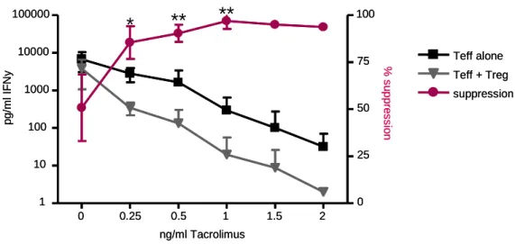
4.7 Tacrolimus in low-doses relatively enhances suppression by T regs ... 63
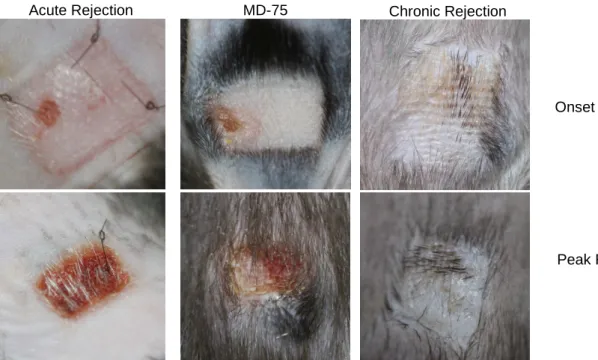
4.8 Allograft acceptance vs. chronic rejection ... 65
4.9 Absence of donor-specific antibodies in MD-75 mice... 68
4.10 Gene expression profiling in MD-75 mice with an allograft ... 70
4.10.1 Gene expression profile in skin grafts... 70
4.10.2 Gene expression profile in draining LN ... 73
4.11 A model of marginal states of allograft acceptance ... 74
4.11.1 Withdrawal of Immunosuppression leads to allograft rejection... 75
4.11.2 Enhancing the effector response leads to allograft rejection... 75
4.11.3 Disrupting regulation leads to allograft rejection... 77
4.12 Location of regulatory and effector cell populations... 81
4.12.1 Regulators and effectors in spleen and dLN ... 81
4.12.2 Regulators and effectors are also located in the graft ... 85
4.12.3 Analysis of myeloid cells in the graft... 91
4.13 The balance tips ... 94
4.14 Marginal states of allograft acceptance might be converted into operational tolerance 96 5 DISCUSSION ... 97
5.1 Synergistic effect of Tacrolimus and regulation ... 97
5.2 Breaking marginal states by disrupting regulation... 99
Table of contents
V5.3 Collapse of marginal states ... 101
5.4 Boost of marginal states ... 103
6 CONCLUSION & PERSPECTIVES ... 104
7 REFERENCE LIST... 105
8 APPENDIX ... 120
8.1 List of Figures ... 120
8.2 Supplementary figure... 122
8.3 Abbreviations... 123
9 ACKNOWLEDGEMENTS ... 127
Abstract
VIAbstract
Allograft acceptance in solid organ transplantation might not be determined by mechanisms unique to the tolerant state but rather by the balance between the effector and regulatory immune response. In consequence, this quantitative view of tolerance implies the existence of marginal states, wherein regulatory responses are just insufficient to prevent rejection, or in which regulatory responses are just sufficient to prevent rejection but are readily disturbed.
The presence of low-dose immunosuppression might be supportive in both scenarios and thus, allograft acceptance is promoted. This work aims to formally show that marginal states of allograft acceptance under low-dose immunosuppression exist and are dependent on regulation. Thus, a low-dose Tacrolimus monotherapy was combined with a weak regulation- inducing protocol in the fully mismatched BALB/c-to-C57BL/6 skin transplantation model.
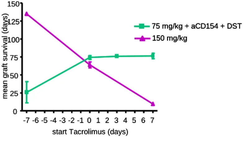
Orally administered Tacrolimus in doses of 150 mg per kg food was therapeutic and prevented allograft rejection when administered before or at the time of transplantation.
Tacrolimus at 75 mg/kg proved subtherapeutic when administered in monotherapy. The combination of costimulatory blockade with anti-CD154 antibody and a donor-specific transfusion (DST) led to moderate prolongation of allograft survival. The combination of anti- CD154 + DST and Tacrolimus at 75 mg/kg was not more effective than anti-CD154 + DST treatment alone, when Tacrolimus therapy was started 7 days prior to transplantation.
However, when Tacrolimus was introduced seven days post transplantation, a remarkable synergism between the induction therapy and the low-dose immunosuppression could be observed and allograft survival was significantly enhanced. This finding was supported by in vitro T reg suppression assays, where effector T cell division is additionally suppressed in the presence of low doses of Tacrolimus.
In line with our hypothesis, it was further demonstrated that in the model of low-dose Tacrolimus in combination with weak-regulation induction, allograft acceptance can be broken. This was done by 1) withdrawal of immunosuppression, 2) enhancing the effector response and 3) disrupting the regulatory response. Thus, it was proven that stable immunosuppression in marginal states of allograft acceptance depends upon the balance of regulatory and effector responses.
The findings of this work have far-reaching implications for patient management, interpretation of immunomonitoring studies and clinical tolerance-induction studies.
Zusammenfassung
VIIZusammenfassung
Die Akzeptanz eines Transplantats wird möglicherweise nicht durch einzigartige Toleranzmechanismen bestimmt, sondern vielmehr durch die Bilanz aus der Effektorantwort und der regulatorischen Immunantwort. Diese quantitative Ansicht der Toleranz umfasst auch die Existenz von Grenzfällen, wobei die regulatorische Immunantwort gerade nicht ausreicht, um die Transplantasabstoßung zu verhindern; oder wobei die regulatorische Immunantwort zwar gerade eben ausreicht, die Abstoßung zu verhindern, aber ohne weiteres gestört werden kann. Die Gegenwart von niedrig dosierter Immunsuppression kann in beiden Szenarien unterstützend wirken und dadurch möglicherweise die Akzeptanz des Transplantats vorantreiben. Mit dieser Arbeit soll formell gezeigt werden, dass diese Grenzfälle der Transplantatsakzeptanz unter Therapie mit niedrig dosierten Immunsuppressiva existieren und dass sie auf Regulation angewiesen sind. Dazu wurde eine niedrigdosierte Tacrolimus-Monotherapie mit einen schwach-regulationsinduzierendem Protokoll kombiniert und dies im BALB/c-auf-C57BL/6-Hauttransplantationsmodel mit vollständiger Gewebemerksmals-Inkompatibilität angewandt.
Oral verabreichtes Tacrolimus in Dosen zu 150 mg pro kg Futter hatte therapeutische Wirkung und verhinderte die Transplantatsabstoßung, wenn die Therapie vor, oder zum Zeitpunkt der Transplantation gestartet wurde. Als Monotherapeutikum hatte Tacrolimus in Dosen zu 75 mg pro kg Futter keine therapeutische Wirkung. Die Kombination aus Kostimulationsblockade mit dem anti-CD154 Antikörper und einer donorspezifischen Transfusion (DST) führte zu einer moderaten Verlängerung des Transplantatüberlebens. Die Kombination aus der Behandlung anti-CD154 + DST mit 75 mg/kg Tacrolimus zeigte sich nicht effektiver als die Behandlung mit anti-CD154 + DST alleine, wenn die Tacrolimustherapie sieben Tage vor der Transplantation gestartet wurde. Wenn hingegen die Tacrolimustherapie erst sieben Tage nach der Transplantation gestartet wurde, konnte ein bemerkenswerter Synergismus zwischen der regulationsinduzierenden Behandlung und der niedrig dosierten Immunsuppressionstherapie beobachtet werden, wobei das Transplantatüberleben signifikant verbessert wurde. Diese Erkenntnis wurde durch in vitro T reg Suppressionsassays bestätigt, wo eine zusätzliche Suppression der Effektor-T- Zellantwort in Anwesenheit von niedrig dosiertem Tacrolimus beobachtet wurde.
In Übereinstimmung mit unserer Hypothese konnten des Weiteren gezeigt werden, dass die Transplantatsakzeptanz im Modell der Kombination von niedrig dosiertem Tacrolimus mit einer schwachen Regulationsinduktion zerstört werden kann. Dies geschah durch: 1) Entzug der immunusppressiven Therapie, 2) Verstärkung der Effektorantwort oder 3) Abbruch der regulatorischen Immunantwort. Damit konnte gezeigt werden, dass die stabile
Zusammenfassung
VIIIImmunsuppression in oben beschriebenen Grenzfällen der Transplantatsakzeptanz von der Bilanz aus der Effektorantwort und der regulatorischen Immunantwort abhängt.
Die Erkenntnisse, die im Rahmen dieser Arbeit gewonnen wurden, haben weitreichende Auswirkungen auf das Patientenmanagement, die Interpretation von Studien zur Definierung von Biomarkern und klinischen Studien zur Induktion von Toleranz gegenüber dem Transplantat.
Introduction
11 Introduction
1.1 Transplantation – an overview
Almost 60 years ago, the first successful human kidney transplantation was performed in Boston by Joseph E. Murray and colleagues. This event was preceded by the work of half a century. Not only did the surgical techniques have to be established in order to transfer tissue or organs, but also unforeseen rejection of the grafts between different individuals had to be overcome. By transplanting between identical twins, Murray could bypass the latter problem. It was the work of several researchers that explained the rejection of grafts. Already in 1912, it was described by Georg Schöne that a second set skin transplant fails more rapidly than the original rejected one. James B. Murphy showed two years later that lymphoid cells were responsible for the destruction of (tumour -) grafts. The same conclusion was drawn by Leo Loeb 20 years later, based upon his rat skin transplant model [1]. Sir Peter B.
Medawar, the “father of transplantation” [2], showed in the mid-1940s with controlled and precise experiments on rabbits that skin graft rejection was an immunologic reaction [3].
Snell and Gorer identified the major histocompatibility complex (MHC), the genetically encoded information responsible for the graft rejection [4]. Strategies to reduce the recipients’ immune response were developed in the following years in order to overcome rejection. The combination of refined operation techniques and immunosuppressive treatment opened the door for transplantation as a widely spread therapy for organ failure and dysfunction.
Transplantation is the only curative therapy for end-stage organ failure. This includes end- stage heart failure [5], end-stage renal disease [6], end-stage liver disease [7] and diabetes with end-stage renal failure [8]. By end of June 2013, over 10,000 patients in Germany alone were registered on the Eurotransplant waiting list for solid organ transplantation. During the first half of the year 2013, a total of 1,622 solid organ transplants have been performed in German transplant centres [9]. Despite being a widely-spread live-saving therapy, long-term transplant outcomes are not satisfactory and transplantation remains an experimental field.
1.2 Terms in transplantation
Transplantation in general is the transfer of cells, tissue or organs from a donor to a recipient.
In autotransplantation, the donor himself is also the recipient; this for example may be the case in skin transplantation to treat burn. If the recipient is another individual than the donor, the term allotransplantation is used. Here we discriminate three different possibilities:
Introduction
2Syngeneic transplantation describes the transfer of cells, tissue or organs (then called syngraft or isograft) between genetically identical individuals, in humans, this only refers to transplantation between monozygotic twins. Allogeneic transplantation (of an allograft or homograft) is the transfer of cells, tissue or organs between genetically distinct members of the same species. Xenotransplantation is the term used for the transfer of a xenograft between members of two different species.
1.3 Basic concepts
1.3.1 T cell activation
1.3.1.1 TCR signalling
In the 1980’s, the structure of the antigen receptor on T cells was characterised [10,11]. The αβ-T cell receptor (TCR) consists of an α and a β chain, that form a heterodimer. The αβ heterodimer forms a TCR complex with the noncovalently associated CD3 and ζ proteins upon binding MHC–peptide complexes [12] (Figure 1).
ZAP70
NFAT AP1
DAG MAPK
IL-2 IL-2 TCR:CD3:ζ
complex
CN
NFkB PLC-γγγγ PLC-γγγγ CD4/
CD8
Lck
ERK PKC
Figure 1: Activation of T cells, adapted from Janeway’s Immunobiology [275]
The CD3 and ζ chains contain ITAMs (immunoglobulin receptor family tyrosine-based activation motif) that are essential for the intracellular signalling. The TCR complex clusters with a co-receptor (either CD4 or CD8), thus, the co-receptor associated protein tyrosine kinase Lck phosphorylates the ITAMs in the CD3 and ζ chains. This leads to binding and
Introduction
3activation of the intracellular protein tyrosine kinase ZAP-70, which phosphorylates several other cytoplasmic molecules, including LAT (Linker for Activation of T-Cells) and SLP-76 [13]. Thus, several signalling pathways are activated, such as MAP – kinase pathways, a PLCγ1-calcium - dependent pathway and a Diacylglycerol (DAG) – pathway. These pathways lead to activation of Extracellular-signal Regulated kinases (ERK) or Janus kinase (JAK), Calcineurin (CN) and enzyme protein kinase C (PKC), respectively. This leads to the activation of transcription factors such as NF-AT, NFκB or AP-1. These factors are responsible for the expression of genes required for proliferation, differentiation and effector functions of T cells [14,15].
1.3.1.2 Costimulation
For a functional T cell response, a second activation signal, besides TCR ligation, is necessary. This is transduced by so-called costimulatory molecules [15]. Costimulatory molecules can be grouped into the CD28/B7 family and the tumour necrosis factor (TNF) family. CD28 binds to the B7 molecules CD80 and CD86 on antigen – presenting cells (APCs) [16]. CD28 signalling pathways via phosphatidylinositol 3-kinase (PI3K) amplify TCR signalling pathways [15], and blocking the CD28 signalling whilst TCR signalling is present leads to anergy [17]. The inducible costimulatory molecule (ICOS) is a CD28 homolog that binds to B7h and seems to be important in effector cell differentiation [18]. The CD28/B7 family also includes negative costimulatory (i.e. coinhibitory) molecules such as CTLA and PD-L1. Both have been described as part of the suppressive mechanism of regulatory T cells [19,20]. Members of the TNF / TNFR – family of costimulatory molecules are, amongst others, CD40L (CD154), OX40, 4-1BB (CD137) and GITR. Since costimulatory molecules of the TNF – family are in general expressed upon activation, they may play a role in effector and memory responses rather than in naïve T cell responses [18]. Blockade of different costimulatory pathways in animal models of transplantation has been proven to be successful in prolongation of allograft survival [21]. Further, antibodies against costimulatory moleculed are in use or, considered to be, in the clinic.
1.3.2 T cell specification
The vast majority of T cells express the αβ-TCR. These cells comprise two lineages which are defined by their ability to bind distinct major histocompatibility complexes (MHC). They express either the MHC-class I – binding protein CD8 or the MHC-class II – binding protein CD4. With regard to their functional task within the immune response, they are also referred to as cytotoxic CD8+ cells, and helper or regulatory CD4+ cells [22]. A T cell that has not yet encountered antigen is called “naïve”; after activation, T cells proliferate and can differentiate into effector T cells. CD8+ cells can differentiate into cytotoxic lymphocytes (CTL) which
Introduction
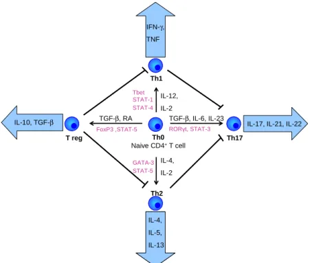
4mainly kill infected cells and contribute in allograft rejection. There are several distinct subsets of effector CD4+ cells, mainly TH1, TH2 and TH17 helper T cells and regulatory T cells (T reg). Differentiation into a distinct subset occurs in response to the present cytokine milieu and involves both transcriptional activation and epigenetic modification of target genes [23]
(Figure 2). Each of these subsets has a special cytokine profile and expresses specific transcription factors [24].
Th0 Naive CD4+T cell T reg
TGF-β, RA
Th17 TGF-β, IL-6, IL-23 Th1
IL-12, IL-2
Th2 IL-4, IL-2 Tbet STAT-1 STAT-4
GATA-3 STAT-5
FoxP3 ,STAT-5 RORγt, STAT-3
IL-10, TGF-β IL-17, IL-21, IL-22
IFN-γ, TNF
IL-4, IL-5, IL-13 IL-4, IL-5, IL-13
Figure 2 T cell polarisation
During the last years, there is increasing evidence that the differentiation into different T cell subsets is not terminal. In in vitro and in vivo studies, a T cell plasticity within and between the helper and regulatory cell subsets could be shown [23].
1.4 Allorecognition
The underlying genetic basis of graft rejection after allogeneic transplantation (or allotransplantation) was studied in the 1940s by Snell and Gorer. Using congenic inbred mouse strains, one region in the genome could be identified that was responsible for rejection of an allograft, the major histocompatibility locus, named H-2. Later it was found that the locus consists of multiple genes; therefore it was named major histocompatibility complex (MHC) [4]. The MHC in human was called human leukocyte antigen (HLA). The genes of the MHC code for the antigen-presenting MHC molecules, of which there are two classes: MHC class I and class II; respectively. There is also a third class of MHC genes
Introduction
5(MHC class III) that encode complement proteins or cytokines such as TNFα, but not all are involved in immune funtions. The MHC class I and II molecules serve the same process, which is antigen presentation to T cells. Without processed antigen being presented to them in MHC context (together with costimulation), T cells cannot be activated. MHC class I molecules present antigenic peptides of intracellular origin to CD8+ T cells, whereas CD4+ T cells recognize exogenous antigens presented on MHC class II molecules (Figure 3).
Ag ER
Cytosol
MHC Class I
ER
MHC Class II Ag
Figure 3 Major histocompatibility complex: antigen presentation, adapted from Janeway’s Immunobiology [275]
Every nucleated cell type expresses MHC class I molecules, but the expression of MHC class II molecules is generally limited to antigen-presenting cells (APC), though it can be induced in other cells such as endothelial cells or fibroblasts [25].
The ability of individual organisms to discriminate self- from non-self-antigens is known as allorecognition. It describes the process of recipient cells recognizing foreign antigen presented on a MHC, as it inevitably happens in allo- and xenotransplantation. During development in the thymus, T cells undergo positive and negative selection. T cells that bind with too high affinity to self-MHC, or do not bind to self-MHC will be deleted. The T cell repertoire is then tolerant towards self-antigens, but recognizes non-self antigens. So far, three different pathways of allorecognition have been described: 1) direct, 2) indirect and 3) semidirect allorecognition.
Introduction
61.4.1 Direct Allorecognition
The process of recipient T cells recognizing antigen presented via intact donor MHC on donor APC (here: dDC) is termed direct allorecognition (Figure 4).
dDC
T cell help
CD4
CD8
dDC
T cell help
CD4
CD8
Figure 4 Direct allorecognition
Two theories have been brought up to explain the underlying mechanisms of the interaction between host T cell receptor (TCR) and donor MHC: the “high determinant density” model and the model of “multiple binary complexes” [26,27]. Briefly, the former theory holds it that the T cells can directly recognize the allogeneic MHC itself and not only peptides bound to the MHC [26-28]. In consequence, the density of ligands for alloreactive T cells is very high in opposition to the density of peptide-specific ligands. Therefore, T cells with low-affinity receptors are also able to respond to foreign MHC, which leads to the high incidence of alloreactivity observed. The second theory of “multiple binary complexes” suggests that the complex of a variety of bound peptides together with an allogeneic MHC is recognized by alloreactive T cells. Subsequently, a single MHC molecule can stimulate many different alloreactive T cells [29,30]. Both theories are not mutually exclusive and may account for the high incidence (up to 7%) of alloreactive T cells described in the literature [31]. It has been proposed that in the case of structurally different MHC molecules between donor and recipient, the alloreactivity is directed against the MHC itself (“high determinant density”), whereas the alloreactivity against the peptides in an allogeneic MHC complex (“multiple binary complexes”) may be predominant when the MHC molecules do not differ substantially [32].
Experimental proof of the participation of direct allorecognition in rejection has been given by depleting the graft of donor APC prior to transplantation. This leads to prolonged allograft survival [33], yet is eventually not sufficient to prevent rejection (see below). Since the intact
Introduction
7donor APC that must be present in the graft and host to elicit direct allorecognition, will be eliminated by the host’s immune response, the contribution of this pathway is temporarily limited.
1.4.2 Indirect Allorecognition
T cells can also recognize donor histocompatibility antigen that is processed and presented by self-MHC (here: rDC) molecules; which is referred to as the indirect pathway of allorecognition (Figure 5).
dDC
rDC
Antigen
CD4 dDC
rDC
Antigen
CD4
Figure 5 Indirect allorecognition
The processing of peptides derived from donor MHC molecules is a common event [34,35].
This occurs when apoptotic donor cells are taken up by host antigen-presenting cells.
Additionally, the peptides can be shed from the surface of donor cells (here, a dDC is shown as an example). The existence of an indirect way of alloantigen presentation came into focus in a rat transplantation model. Here, after depletion of passenger donor APC in the graft, rejection did eventually occur [36]. The importance of this second pathway of allorecognition was demonstrated by Auchincloss et al. in a skin transplantation model. CD8+- depleted mice without MHC class I molecules were able to reject a MHC class II deficient skin graft via a CD4+ response. Since CD4+ cells do not interact with MHC class I molecules (the remaining MHC class in the graft), the donor antigens must have been presented by host MHC class II [37].
This aforementioned data proves the sufficiency of an indirect allorecognition to cause graft rejection in the absence of direct allorecognition. Host dendritic cells are constantly trafficking
Introduction
8in the body and also in the graft itself, which expresses donor MHC molecules. Therefore, indirect allorecognition that can occur every time after transplantation may mount an immune response leading to rejection. Thus, the indirect pathway is probably the dominant way of allorecognition in the long term.
1.4.3 Semi-direct Allorecognition
Experimental data indicated that T cells with indirect allospecifity can amplify or suppress T cells with direct allospecifity [38-40]. This phenomenon has been first explained by a four- cell, unlinked, model: CD4+ helper or suppressor T cells interact via the indirect pathway with recipient DC, whereas CD8+ cells directly recognize donor cells. Work of several groups showed the ability of DC to acquire intact MHC molecules from other cells in vitro, which was then further investigated by Herrera in vivo [41]. A third pathway of allorecognition was described then, the semi-direct allorecognition (Figure 6).
dDC
rDC Antigen
2
CD4 CD8
T cell help 1 dDC
rDC Antigen
2
CD4 CD8
T cell help 1
Figure 6 Semidirect allorecognition
Semi-direct allorecognition refers to direct pathway T cells recognizing intact, allogeneic MHC:peptide complexes that have been transferred from donor cells to recipients DC and are presented on their surface. Additionally, indirect pathway can recognize donor peptides that were internalised and are presented via the MHC class II on the same DC.
Introduction
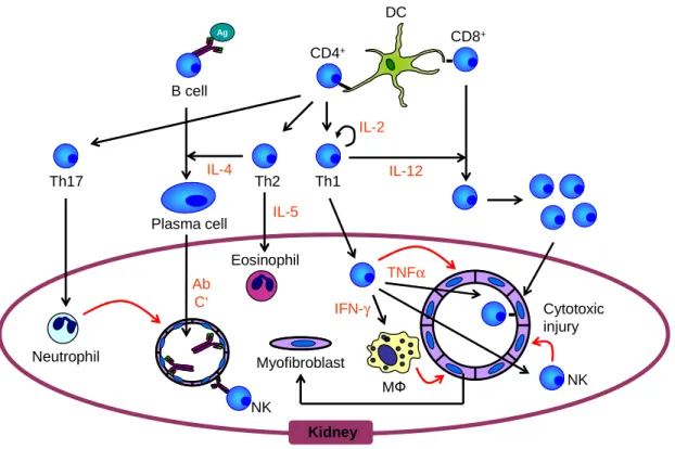
91.5 Rejection
Kidney
CD4+
DC
Th2 Plasma cell
Ag
B cell
IL-4
Cytotoxic injury CD8+
IL-12 IL-5
Eosinophil
IL-2
Th1
Ab C‘
NK
TNFα IFN-γ
MФ NK
Myofibroblast Th17
Neutrophil
Figure 7 Rejection of an allograft
1.5.1 Hyperacute rejection
Hyperacute graft rejection occurs usually within minutes after transplantation, when preformed recipient antibodies bind to donor endothelial ABO or major histocompatibility (MHC) antigens. This can activate the complement cascade or mediate antibody-dependent cellular toxicity (ADCC), leading to damage of endothelial cells, culminating in intra-vascular thrombosis and tissue injury [42,43]. Hyperacute rejection inevitably leads to allograft loss, but occurs nowadays rarely due to pre-transplant bloodtyping and T cell crossmatch testing [44]. Hyperacute rejection also prevents interspecies transplantation (Xenotransplantation).
1.5.2 Acute rejection
Acute rejection occurs usually within days or weeks after transplantation. Even when the recipient receives immunosuppressive treatment, acute rejection episodes can occur repeatedly. Acute rejection can be cellularly (lymphocytes) or humorally (antibody) mediated.
Every transplantation leads inevitably to tissue damage and thus to activation of the innate immune system. Innate immune cells such as neutrophils, macrophages and natural-killer cells (NK cells) express pattern recognition receptors (PRR) which recognize damage- associated molecular pattern molecules (DAMP) [45,46]. This antigen-independent
Introduction
10inflammatory response can promote further injury, e.g. caused by the production of tissue- damaging molecules such as reactive oxygen species (ROS) or nitric oxide (NO) [47]. The cells of the innate immune system produce cytokines, such as IFNγ, IL-6 or IL-12, leading to the activation of the adaptive immune system [48,49]. Work by Chalasani et al. indicated that the innate immune response towards the graft is necessary for an effective adaptive, antigen- dependent, immunity [50]. Dendritic cells (DCs) are considered to be the link between innate and adaptive immunity [51]. Immature DCs traffick through non-inflamed tissues, but exert immunogenic effects, when they receive a maturation signal. This can be provided by DAMPs following transplantation. Activated DCs migrate to the lymphoid tissue, presenting antigen to T cells, stimulating the adaptive antigen response [52]. There is also data demonstrating the influence of chemoattractants from neutrophils and macrophages on the optimal recruitment of T cells to the allograft [53].
Among the infiltrating cells in an acutely rejected allograft, T cells indeed build up the largest population [54,55]. Based mainly on experience from animal experiments, acute rejection is seen predominantly as a T cell mediated process. Athymic or neonatally thymectomized mice fail to reject MHC-mismatched skin grafts unless reconstituted with T cells from untreated syngeneic wildtype mice, after which the transplants will be rejected rapidly. T cells contribute to graft rejection in various ways after activation [56,57]. The release of proinflammatory cytokines (IL-1, IFNγ, TNF) triggers further graft infiltration by macrophages, monocytes, eosinophils and others, which promotes additional antigen-independent cytotoxicity. But also T cells, mostly MHC class I restricted CD8+ T cells, can secrete cytotoxic molecules thus inducing apoptosis of the target cells. Cytotoxic T cells lyse target cells via two distinct mechanisms, the perforin/granzyme pathway or the Fas/FasL pathway [58]. It could be shown that CD4+ T cells, but not CD8+ T cells are essentially required to initiate rejection of an allograft [59]. CD4+ T cells, mostly TH1 helper cells, mediate delayed- type hypersensitivity responses (DTH) in an antigen-specific manner [60].
Activated CD4+-T cells can also provide B cell help by cytokine production and expression of costimulatory molecules. B cells can act as APC for T cells, secrete inflammatory cytokines / chemokines and produce alloantibodies [61,62]. B cell infiltration was reported in acute rejection episodes of human kidney, liver and heart [61,63-65]. Studies on B cell-deficient µMT mice were performed, showing that B cells and antibodies are not required for acute rejection [66-68]. However, there is data indicating a significant role of B cells in acute rejection. Impaired indirect alloantigen presentation by B cells was followed by reduced antibody production and CD4+-T cell division in murine cardiac transplantation [61]. Depletion of B cells using the anti-CD19 or anti-CD20 antibodies aggravated or ameliorated rejection
Introduction
11depending on the organ transplanted and the intensity of the rejection [69]. Further, antibodies produced by B cells can induce complement and therefore lysis of graft cells. If the Fc receptor on NK cells or macrophages recognizes antibodies, this can lead to antibody mediated cellular cytotoxicity (ADCC) mounting in apoptosis of the graft cells. The aforementioned effector mechanisms of cytotoxicity, DTH, Lysis and ADCC will then lead to a rapid rejection of the transplanted tissue.
1.5.3 Chronic Rejection
Chronic rejection refers to chronic allograft injury mediated by immunologic factors in contrast to other mechanisms such as drugs, ageing or infection. The chronically rejecting organ displays vasculopathy with distinct histologic injury depending on the type of solid organ [70,71], leading to diminished function and eventually loss of the organ. In general, chronic rejection is influenced by both alloantigen-dependent and alloantigen-independent mechanisms [70]. A rat kidney-retransplantation model established by Tullius et al. [72]
demonstrated that early immunohistological changes in the chronic rejected allograft during the first 12 weeks are predominantly antigen-dependent and can be reversed by retransplantation back to the donor strain. In later retransplanted isografts, fibrotic injury continued to progress and even isografts did display immunohistological alterations similar to chronic rejection [73], indicating that late events in chronic rejection are antigen-independent.
Grafts undergoing chronic rejection display perivascular inflammation and fibrosis [74,75].
The pathognomonic lesion in chronic rejection is obliterative arteriopathy (OA), caused by fibrointimal hyperplasia [71]. The initial damage of endothelium and exposure of collagen causes repair mechanism involving fibrin and other clotting proteins and platelets [70,71,76].
Various factors such as platelet activating factors (PAF), platelet-derived growth factor (PDGF), tumour necrosis factor (TNF), leukotrienes and thromboxane are released, which can also lead to induced proliferation and migration of smooth muscle cells (SMC) [76-78].
The endothelial activation comes together with the upregulation of MHC II and adhesion molecules, supporting the infiltration of leukocytes [79-82]. The microscopic picture in the initial stage in rodents shows that monocytes/macrophages and T cells are predominant, but also eosinophils, plasma cells, DCs, B cells and mast cells were found [75,76,83-86]. T cells and macrophages build the arterial inflammatory response, with the lymphocytes attached to the intima and the macrophages permeating adventitia, media and intima of the vessel [71,79,86,87]. At this stage, cytokines such as tissue growth factor beta (TGF-β), interferon- γ (IFN γ), IL-1 and tumour necrosis factor (TNF) are expressed in the graft, as well as the chemoattractants RANTES (CCL5) and monocyte chemotactic protein-1 (MCP-1) [83,84,88].
This leads to further attraction of macrophages/monocytes and T cells, that in turn produce
Introduction
12more cytokines and chemoattractants. PDGF, released by endothelial cells, SMC, platelets and activated macrophages, and TNF, released predominantly by activated macrophages, stimulate the proliferation of SMC and their release of extracellular matrix proteins [76,89,90].
TGFβ activates extracellular matrix deposition and is expressed in grafts undergoing chronic rejection [70,91,92]. It has been demonstrated that TGFβ contributes to fibrosis, e.g. by upregulating connective tissue growth factor (CTGF) in fibroblasts and SMC, which has mitogenic effects on fibroblasts [93,94]. Häyry [95] communicates following hypothesis that is supported by many data including the above mentioned: SMC replicate in autocrine or paracrine response to the cytokines and growth factors. Extracellular matrix expressing metalloproteinases and proteolytic enzymes contribute to the migration of SMC to the intima, where they start remodelling the vascular wall. This leads to arterial narrowing and occlusion of small vessels, followed by damage of the surrounding tissue due to ischemia and fibrotic changes.
1.6 Regulatory immune cells in Transplantation
Whenever there is an activation of the immune system, i.e. an inflammatory process, there is also a regulatory response to control inflammation and thus prevent the host organism from damage. This is also true in transplantation. Indeed, the immune cell populations leading to rejection of an allograft also harbour regulatory cells that can suppress the effector response.
These specialised cells either underwent selection processes for regulatory function or were driven into a regulatory phenotype on site (of the allograft). Other mechanisms to regulate the immune response are: ignorance, anergy and deletion [96]. Ignorance simply refers to T cells ignoring antigen, either because T cells cannot enter the sites where the antigens are expressed (immuno-priviledged sites) or because the antigen signal does not overcome the threshold to lead to a T cell response. T cells can also be rendered anergic, i.e. non- responding to further stimulation. This happens when the TCR is stimulated without adequate costimulation and signalling through alternative receptors occurs. Deletion of T cells does not only take place in the thymus (central deletion), but also in the periphery.
Antigen-reactive T cells can be depleted by activation-induced cell death (AICD) upon restimulation of the TCR with signalling through other receptors such as Fas. This might occur in CD8+ T cells that are activated without CD4+ T cell – help [96,97].
In their ground-breaking publication in 1953, Billingham, Brent and Medawar already suggested that leukocytes able to suppress allospecific immune responses do exist [98].
Since then, different regulatory cell populations have been reported, such as regulatory T cells, B cells and macrophages, tolerogenic DCs and myeloid-derived suppressor cells
Introduction
13(MDSCs). The presence of these cells in a transplant recipient can promote acceptance of the allograft.
1.6.1 Regulatory T cells
Various T cell populations with regulatory function in transplantation have been discovered, including CD4+, CD8+ and CD4-CD8- regulatory T cells (Figure 8).
T reg CD4+CD25+FOXP3+
Naturally-occuring T reg
Th0 CD4+CD8-CD25-
Tc CD4-CD8+ CD25-
Dedicated suppressor cells Induced suppressor cells
CD4+CD25+FoxP3+ induced T reg
Tr1 cell
Th3 cell CD4-CD8-
DN T cell
CD8+CD28- Ts cell
Figure 8 Regulatory T cell subsets
1.6.1.1 CD4+ regulatory T cells
Naturally occurring, thymus-derived CD4+ regulatory T cells (T regs) form a self-renewing, actively dividing and differentiated population and maintain tolerance in the periphery, mainly against self-antigens [97]. They are characterised as CD4+CD25+FoxP3+ cells, as well as the population of induced T regs. In mice, FoxP3 is expressed exclusively on T cells and it is necessarily and sufficiently responsible for suppressive function in CD4+ T regs [99-101]. In addition, T regs constitutively express high levels of the costimulatory molecule glucocorticoid-induced tumor necrosis factor receptor (GITR) [102]. GITR is described to enhance proliferation of both T regs and effector T cells [103]. Altogether, induction of GITR signalling has been shown to abrogate T reg suppression [104].
FoxP3+ T regs highly express both PD-1 (programmed death receptor) and PD-L1 (programmed death receptor – ligand 1), a major coinhibitory receptor - ligand team [105].
PD-L1 was found to have a major role in the induction and maintenance of induced T regs, thus promoting tolerance [20]. Induced T regs are CD4+ T cells that inducibly express FoxP3 and differentiate after encountering antigen in a tolerogenic microenvironment, thus are converted from potential effector T cells. Both naturally occurring and induced T regs can
Introduction
14respond to alloantigens in a graft-protecting way. Thymus-derived T reg might be predominantly present in the initial period after transplantation, later, induced T regs probably play a more important role [106,107].
T regs (thymus-derived and induced) suppress proliferation and / or activation of naïve and memory CD4+ and CD8+ effector T cells, B cells, and the function of NK cells, macrophages and DCs [108,109]. They can exert their effects via cell-contact dependent mechanisms. Via binding of the cytotoxic T lymphocyte antigen 4 (CTLA4), a receptor constitutively expressed on T regs, to the costimulatory molecules CD80 and CD86 on DCs, their activity can be inhibited [110]. Further, this can induce the production of the enzyme indoleamine 2,3- dioxygenase (IDO) by DCs, which, due to tryptophan deprivation, leads to attenuated T cell proliferation [108]. T regs themselves produce cytokines such as IL-10 and TGFβ.
Interleukin-10 is an anti-inflammatory cytokine produced by many cells of the adaptive and innate immune system. In in vivo models of inflammatory bowel disease and transplantation, blockade or absence of IL-10 prevents T reg – mediated regulation [111,112]. TGFβ is a cytokine that is important for the development of induced T regs and in fact might be produced in part in an autocrine fashion [113]. It can be also expressed on the cell surface of activated T regs. TGFβ produced by T regs can inhibit the activation of effector T cells [114].
Another, more recently described, mechanism of suppression by T regs could be by IL-35 secretion [115]. However, the roles of these cytokines as suppressor mechanism is not completely clear, since in vitro data is often controversial [116]. Other mechanisms that have been described are cytotoxic activity of T regs via granzyme B and perforin [117] or apoptosis – inducement due to IL-2 depriviation [118].
In addition to T regs (thymus-derived and induced), other CD4+ regulatory T cells have been described, such as TR1 cells. These are distinct peripherally induced regulatory T cells that are negative for FoxP3 - expression, develop in response to IL-10 and can secrete IL-10 and TGFβ [119]. Further, TGFβ – producing TH3 cells have been described [116].
1.6.1.2 CD8+ regulatory T cells
CD8+CD28- have been described in human kidney transplant patients after leukocyte depletion treatment. They use cell contact mechanisms to inhibit T cell activation via APCs and seem to be a distinct subset from a IL-10-producing CD8+ T cell population [107]. The latter can be generated in vitro and can inhibit T cell responses through IL-10.
Introduction
151.6.1.3 CD4-CD8- regulatory T cells
Cells that express CD3 and the αβ-TCR, but neither CD4 nor CD8 (or the NK cell marker NK1.1) are so-called double-negative T cells. This immunosuppressive population has been first described in a mouse skin transplantation model, where graft survival could be enhanced. The suppressive mechanism shown was cell-contact - and Fas – dependent killing of CD8+ cytotoxicT cells [120]. This cell population has also been described in animal models of diabetes and graft-versus-host-disease and could be isolated from human blood.
In further experiments, double-negative regulatory T cells also had suppressive effects on CD4+ T cells, B cells, and APCs [121].
1.6.2 Regulatory B cells
B cells in transplantation may have more than an antibody-producing role. Regulatory B cells secreting IL-10 have been described as immunosuppressive in models of autoimmunity such as experimental autoimmune encephalomyelitis (EAE), IBD, arthritis and diabetes [122].
Further, they were also detectable in human patients [123]. B regs have been described to induce populations of regulatory T cells in animal models of colitis and EAE [124]. CD40 and CD80/86 engagement is necessary for the establishment and/or function of B regs [125]. In a mouse transplantation model, hindered IL-10 production of regulatory B cells does not interfere with tolerance induction. It rather is suggested to be dependent on direct interaction between B cells and target cells [126]. In human kidney transplant patients after CD52 – (lymphocyte / monocyte) depletion, the repopulating B cells had B reg and transitional B cell phenotypes [127]. Transitional B cells are poor costimulators and thus may lead to T cell unresponsiveness [128]. Interestingly, the presence of naïve and transitional B cells after transplantation is associated with a positive outcome and a B cell gene signature was described in immunosuppressive-free patients with maintained graft function (operationally tolerant). Such B cell related gene markers were Cd20, Ms4-a1 and Fcrl1 [129,130].
1.6.3 Regulatory macrophages
Macrophages are activated quickly upon tissue damage as it occurs in transplantation, as already mentioned earlier. But macrophages do not only promote graft damage, they can also contribute in wound healing. Macrophages are often classified into two groups, the classically activated M1 – macrophages and the alternatively activated M2 – macrophages [131]. Further, regulatory macrophages have been described. Genome microarray studies on M regs induced in vitro from mouse and human monocytes show that these macrophages have a gene expression profile different from M1 and M2 polarised macrophages
Introduction
16[132](Hutchinson, unpublished data). Markers for these mouse M regs are typical macrophage – markers such as CD11b, F4/80, CD68 and CD14. Further these M regs express only intermediate levels of MHC II and low levels of costimulatory molecules CD80, CD86 and CD40. Further, they express PD-L1 and CD11c [132]. A variety of different stimuli has been shown to induce suppressive function of macrophages. Amongst these are M-CSF, IL-10, vitamin D, IFNγ, immune complexes and repetitive TLR stimulation, reviewed in [133].
Thus, no unique phenotype can be described for suppressor macrophages. IL-10 secretion may be one mode of action of regulatory macrophages [134]. Further, production of iNOS in mouse M regs or IDO in human M regs, has been described [132](Hutchinson, unpublished data). By these means, M regs may directly inhibit activation and proliferation of effector T cells. In addition, this leads to a microenvironment that can promote induction of regulatory T cells (Walter, unpublished data). M regs have already been used as cell therapy in kidney transplant patients [135], and interestingly, these two patients are maintained on additional immunosuppressive therapy at unexpectedly low doses.
1.6.4 Tolerogenic DCs
Mature dendritic cells can efficiently activate T cells and improve memory T cell responses.
In steady state conditions, DC found in the peripheral lymphoid tissue are not fully mature. In order to achieve immunity, the antigen needs to be coadministered with a maturation stimulus [136]. If the antigen is delivered without a maturation signal, the immature DCs will engage T cells, but this lead to unresponsiveness [137]. It has also been shown that injection of ex vivo antigen.pulsed DC under the absence of maturation signals leads to downregulation of the immune response and induction of T regs [138]. Further, DCs might promote tolerance in response to tolerogenic signals such as IL-10 and TGFβ or to signals coming from T regs [109,139]. As already mentioned previously, IDO is one of the mechanism by which tolerogenic DCs can suppress T cell responses [139]. In addition, tolerogenic DCs inhibit T cells via IL-10 or heme oxygenase 1 (HO-1) [140,141]. Immature myeloid tolerogenic DCs can promote allograft acceptance in solid organ transplantations [142]. It has also been described that the population of plasmacytoid DCs (pDCs) which express more PD-L1 correlate with increased numbers of T regs in liver transplant patients [143]. This induction of T regs by pDCs has also been observed in animal models of transplantation [144].
1.6.5 Myeloid - derived suppressor cells (MDSCs)
MDSCs are a heterogeneous population of myeloid progenitor cells present in tissues during inflammation. They were first described in cancer patients, and now their
Introduction
17immunosuppressive function has been acknowledged in other diseases and transplantation [145]. Several subsets of MDSCs have been defined in both human and mouse. Common phenotypical markers of mouse MDSC subsets are expression of CD11b and Gr1 [146].
Activated T cells, stromal cells and, in cancer, tumour cells produce factors such as macrophage–colony stimulating factor (M-CSF), granulocyte-macrophage-CSF (GM-CSF), IL-6 or prostaglandins that regulate expansion and activation of MDSCs [146]. Upon activation, MDSCs can inhibit T and B cell responses by production of iNOS and arginase 1 [147,148]. Release of reactive oxygen species (ROS) is also part of the suppressive function of MDSCs [145]. In a murine skin transplantation model, MDSCs producing IL-10 and HO-1 did prolong allograft survival [149]. It has been shown that MDSCs can induce T regs [150]
and in murine islet transplantation, this enhancement is mediated by expression of PD-L1 [151].
1.7 Immunosuppressive treatment in Transplantation
1.7.1 Overview
The first drugs successfully used to prevent acute rejection in transplantation between non- identical individuals were steroids (cortisone) and Azathioprine, a chemotherapy drug found to be effective in kidney transplantation in the early 1960s [152]. Azathioprine inhibits de novo purine synthesis and has an anti-proliferative effect on T and B lymphocytes [153]. The immunosuppressive therapy in transplantation could be expanded years later when the calcineurin-inhibitor Cyclosporine was introduced in the clinic in 1978 [154]. Thus, the one year survival time of an allograft increased dramatically [155]. In the following, more immunosuppressive drugs have been introduced into transplantation. In 1982, the type 2 isoform inosine 5'-monophosphate dehydrogenase (IMPDH) inhibitor mycophenolate mofetil (MMF) was developed. Studies showed that MMF is, in contrast to Azathioprine, more lymphocyte-specific and more effective in preventing graft rejection [156,157]. Thus, and because it is effective in combination with other immunosuppressants, MMF has largely replaced Azathioprine in the clinic [158].
In 1986, a new calcineurin-inhibitor (CNI) called Tacrolimus, was discovered and found to be more potent than Cyclosporine. The mechanisms of action of CNI will be discussed below. In 1989, the immunosuppressive properties of Rapamycin (Sirolimus), a microbial product with structural similarity to Tacrolimus, were further tested in transplantation models [159].
Rapamycin binds the same protein as Tacrolimus, but does not inhibit calcineurin. It acts on the mammalian target of Rapamycin (mTOR), thus leads to cell-cycle arrest in T cells [160].
Introduction
18In general, immunosuppressive therapy includes glucocorticoids and small-molecule immunosuppressive drugs as the drugs mentioned above. A third group are protein immunosuppressive drugs including fusion proteins such as CTLA-4-Ig, depleting antibodies and non-depleting antibodies [158].
Antibodies as induction therapy have been used since the early 1980’s. Anti-Thymocyte globulin (ATG) and Campath-1H (Alemtuzumab) are widely used antibodies depleting T and B cells (and the latter to a lesser extend NK cells, monocytes and macrophages). Basiliximab is a non-depleting anti-IL2R antibody inhibiting lymphocyte proliferation. Further protein immunsuppressive drugs are developed (e.g. non-depleting CD40L antibodies ASKP1240 or 4D11) or in use (e.g. CTLA4-Ig) for blockade of the costimulatory CD40/CD40L or the CD28/CD80/CD86 pathways [21].
The common therapy protocol in transplantation includes an antibody such as Basiliximab with higher doses of CNI in the induction phase with an additional anti-proliferative drug (MMF) and tapered steroids. The maintenance phase then is based mostly on the CNI, with possible addition of MMF or Rapamycin to reduce CNI doses and toxicity [161,162]. Since CNIs as Cyclosporine and Tacrolimus are the basis of current standard immunosuppressive therapy, they will be described in more detail [162].
1.7.2 Calcineurin Inhibitors
1.7.2.1 Cyclosporine (CsA)
Cyclosporine is a fungal metabolite discovered in a screening program for immunosuppressive agents in 1972 [154]. Cyclosporine is a calcineurin-inhibitor that inhibits T cell proliferation and was introduced in the clinic by Sir Roy Calne six years later [163].
Since then, it has been used in transplantation and is in use until now. Its mechanism of action will be described below in context with another CNI.
Introduction
191.7.2.2 Tacrolimus (FK-506)
Tacrolimus is a macrolide lactone (C44H69NO12) that could first be isolated from Streptomyces tsukubaensis – cultures in Japan in the mid-1980’s (Figure 9) [164].
Figure 9 Chemical structure of Tacrolimus. Source: www.medlibrary.com
Various animal studies followed to further evaluate the immunosuppressive and anti- lymphocytic effects and soon, Tacrolimus was given to acutely rejecting transplant patients as “rescue” therapy. In 1990, a liver transplant study started, using Tacrolimus as first-line therapy [165]. Subsequently, Tacrolimus has been widely used in solid organ and bone marrow transplantation. The drug has been described as being up to 100-fold more potent in in vitro suppression assays than the CNI Cyclosporine [166]. Further, it was shown that Tacrolimus has suppressive effects on T cells without affecting myeloid cells at the same concentrations [167].
Activation of a T cell via engagement of the TCR results in activation of the calcium – calcineurin - NF-AT – pathway. Once Tacrolimus has entered the cell, it binds to the abundant FK506-binding protein FKBP-12, which is a cytosolic immunophilin. The FKBP- FK506 complex then competitively binds to calcineurin, a Ca2+ / calmodulin-dependent proteine phosphatase enzyme [168], and thus the calcium-dependent signal transduction pathway in T cells is interrupted (see Figure 10). Without Calcineurin, the cytosolic subunit of the nuclear factor of activated T cells (NF-ATc) will not be dephosporylated, thus the translocation to the nucleus is blocked. Therefore, NF-ATc cannot form a complex with the nuclear component of the nuclear factor of activated T cells (NF-ATn), which is necessary for promoter-binding of the IL-2 gene und subsequent production of IL-2 [169], a crucial cytokine for T cell activation. Also, further genes regulated through NF-AT are affected by calcineurin-
Introduction
20inhibitors, such as IL-4, IFNγ or Fas-ligand [170]. By inhibition of calcineurin with Tacrolimus, the activation, differentiation and proliferation of naïve and memory effector CD4+ and CD8+ T cells is effectively suppressed [171,172]. In its mode of action, Cyclosporine is similar to Tacrolimus. The correspondent immunophilin for Cyclosporine is Cyclophilin A, the formed complex can also bind calcineurin with the above described consequences. In comparator studies, evidence was found that Tacrolimus is superior to Cyclosporine treatment regarding acute rejection episodes and graft loss [173,174].
NF-ATn
NF-ATc P
NF-ATc P
IL-2 gene Calcineurin
FK506FK506 FKBP
FK506 FKBP
Dephosphorylation FK506
Nucleus
T cell cytoplasm IL-2
TCR complex Ca2+
Ca2+
Calmodulin Ca2+
NF-ATn
NF-ATc P
NF-ATc P
IL-2 gene Calcineurin
FK506FK506 FKBP
FK506 FKBP FK506 FKBP
Dephosphorylation FK506
Nucleus
T cell cytoplasm IL-2
TCR complex Ca2+
Ca2+
Calmodulin Ca2+
Figure 10 Effector mechanims of Tacrolimus
1.7.3 CNI toxicity – a trade off?
The short-term graft survival could be strikingly improved by the use of calcineurin inhibitors.
Yet, the long-term outcome did not change much [175], due to further problems arising by numerous adverse drug-related effects. The toxic effects of both Tacrolimus and Cyclosporine are described similar: Nephrotoxicity and chronic kidney damage, neurotoxicity, disturbances of glucose metabolism and susceptibility to malignancy have been associated with both treatments [176,177]. Both MMF and Rapamycin in combination alone or together with either Tacrolimus or Cyclosporine were subject of various studies in order to spare / minimise the CNI doses. Late conversion from CNI-MMF treatment to a combination of MMF and Rapamycin did not improve renal function, in fact it was harmful to kidney transplant patients with already impaired renal function. An early conversion results only in an initial
Introduction
21better renal function. Additionally, also Rapamycin has adverse effects such as proteinuria, bone marrow suppression and, of note after an operative procedure, impaired wound healing [178]. In the Efficacy Limiting Toxicity Elimination (ELITE)-Symphony study [161], graft survival and acute rejection episodes with de novo Rapamycin in combination with MMF were worse than with the Tacrolimus-MMF treatment. Further, treatment with low-dose Tacrolimus (3-7 ng/ml) in combination with MMF had the best outcome (renal function and graft survival) compared to normal or low-dose Cyclosporine in combination with MMF [178].
Disregarding the low-dose use, the general toxicity profiles of Tacrolimus, Cyclosporine and Rapamycin were found to be retained, but by minimising the doses of CNI in stable renal transplant patients, impaired renal function can be improved [179,180].
1.7.4 Pharmacokinetics
Immunosuppressive drugs have variable pharmacokinetics in the individual patient. Thus, drug monitoring is important to achieve optimal efficient dosages to exert therapeutic effects with minimised side effects. A method used widely in the clinic is the measurement of trough levels (C0), i.e. the concentration immediately before intake of a new dose of the administered drug.
0 1 2 3 4 5 6 7 8 9 10 11 12 hours
Concentration [ng/ml]
Dose Dose
Peak (Cmax)
Trough (C0) AUC
0 1 2 3 4 5 6 7 8 9 10 11 12 hours
Concentration [ng/ml]
Dose Dose
Peak (Cmax)
Trough (C0) AUC
Figure 11 Concepts in drug monitoring (compare [[107,181])
After the intake, there is an initial absorption phase where drug levels then reach a peak (maximum concentration Cmax) until the concentration then falls off to Cmin. The total drug exposure between two doses is the area under the concentration-time curve (AUC). To determine the AUC, the drug concentration should be measured at several different time points to create a 12 hour pharmacokinetic profile [181]. This is not feasible in the clinic for every patient. Thus, measurement of Tacrolimus C0 - trough levels, often reported as correlated with AUC [182-185], was recommended in 2009 by the KDIGO clinical practice guideline [186]. Nonetheless, there are studies reporting that other time points such as C2 or C4 correlate better with the AUC [182,183], though the relevance of this in regard to acute rejection episodes remains unclear. Important for the clinic is the fact that the differences in the maximum concentration Cmax do not affect graft survival time, if the AUC stays the same
![Figure 1: Activation of T cells, adapted from Janeway’s Immunobiology [275]](https://thumb-eu.123doks.com/thumbv2/1library_info/4645695.1608027/12.892.321.574.598.995/figure-activation-t-cells-adapted-janeway-s-immunobiology.webp)
![Figure 3 Major histocompatibility complex: antigen presentation, adapted from Janeway’s Immunobiology [275]](https://thumb-eu.123doks.com/thumbv2/1library_info/4645695.1608027/15.892.244.657.306.670/figure-histocompatibility-complex-antigen-presentation-adapted-janeway-immunobiology.webp)