• Keine Ergebnisse gefunden

The Control of Lung Branching Morphogenesis

N/A
N/A
Protected

Academic year: 2021

Aktie "The Control of Lung Branching Morphogenesis"

Copied!
44
0
0

Wird geladen.... (Jetzt Volltext ansehen)

Volltext

(1)

Research Collection

Working Paper

The Control of Lung Branching Morphogenesis

Author(s):

Iber, Dagmar Publication Date:

2020-12-22 Permanent Link:

https://doi.org/10.3929/ethz-b-000460510

Rights / License:

Creative Commons Attribution 4.0 International

This page was generated automatically upon download from the ETH Zurich Research Collection. For more information please consult the Terms of use.

ETH Library

(2)

The Control of Lung Branching Morphogenesis

Dagmar Iber

1,2,*

Affiliations:

1

Department of Biosystems, Science and Engineering (D-BSSE), ETH Zurich, Mattenstraße 26, 4058 Basel, Switzerland

2

Swiss Institute of Bioinformatics (SIB), Mattenstraße 26, 4058 Basel, Switzerland

* Corresponding author: Dagmar Iber dagmar.iber@bsse.ethz.ch

Keywords: Lung Branching Morphogenesis, ligand-receptor based Turing Mechanism, Epithelial

Tube, Directional Growth, Computational Model, Shear Stress, Cell-based Tissue Simulations

(3)

Branching morphogenesis generates epithelial trees which facilitate gas exchange, filtering, as well as secretion processes with their large surface to volume ratio. In this review, we focus on the developmental mechanisms that control the early stages of lung branching morphogenesis.

Lung branching morphogenesis involves the stereotypic, recurrent definition of new branch

points, subsequent epithelial budding, and lung tube elongation. We discuss current models and

experimental evidence for each of these steps. Finally, we discuss the role of the mesenchyme in

determining the organ-specific shape.

(4)

Introduction

Reptiles, birds, and mammals all rely on their lungs to breathe. Gas exchange is highly efficient within the limited space of the thoracic cavity because the tree-like airway system greatly enlarges the contact surface between blood vessels and airways (Fig. 1A). The typical human lung tree undergoes 23 generations of dichotomous branching (Weibel and Gomez, 1962). The first 16 branch generations are conducting airways (bronchi and bronchioles) and thus serve a structural function in creating the branched architecture. At the distal end of each conducting airway, an acinus forms. Acini are the functional units of the lung, and are composed of a small tree of gas exchanging airways (respiratory bronchioles, alveolar ducts, and alveolar sacs) built from the remaining 7 branch generations

(Schittny, 2017). The resulting gas-exchanging surface area of 40-130 square meters is more than 250-times larger than that of a spherical organ with the same volume (Glenny, 2011; Weibel and Gomez, 1962). The combination of a huge surface area and a very thin air-blood barrier maximises gas exchange. Narrow tubes would minimize the dead volume of the conducting airways, but only at the price of increased resistance to ventilation. The optimal design of the conducting airways tree is a fractal-like architecture, where the branch length and diameter shrink by a constant factor, 2

−1 3

≈ 0.8, in each branch generation (Fig. 1B) (Mauroy et al., 2004; Wilson, 1967). The ratio between the branch length and diameter is conserved between branch generations, but differs between species (Fig. 1C) (Nelson et al., 1990; West et al., 1986). In humans, branches are 3-fold longer than wide (Nelson et al., 1990). As a result of dichotomous branching , the particular shrinkage factor, and the conserved relationship between branch length and width, the combined volume of all branches in a branch generation is conserved. All branch volumes together add up to a total of 5-6 liters in humans.

The branching architecture of the human lung is highly conserved, with airway branch variants found

in less than 30% of a multi-ethnic population (Smith et al., 2018). The observed airway variants were

associated with higher odds of chronic obstructive pulmonary disease (COPD) and chronic bronchitis,

and could be linked to single-nucleotide polymorphisms (SNPs) in introns of the gene for Fibroblastic

(5)

growth factor (FGF)10 (Smith et al., 2018), a key regulatory factor in lung branching morphogenesis (Bellusci et al., 1997; De Moerlooze et al., 2000; Peters et al., 1994; Weaver et al., 2000). Similarly, in mice, the sequence of branching events is highly conserved, with only few deviations between littermates (Blanc et al., 2012; Metzger et al., 2008; Short et al., 2013), suggesting that lung branching morphogenesis is not left to chance. So, how does the lung tree emerge during development?

Lung Specification

The lung develops from the ventral anterior foregut, adjacent to the heart (Fig. 2) (Cardoso and Lu, 2006; Herriges and Morrisey, 2014). The particular position where the lung develops along the foregut appears to be set by cardiac mesoderm-derived fibroblastic growth factor (FGF)1 and FGF2, which segregate a common pool of foregut endoderm cells into lung, liver or pancreatic fields in a dose-dependent manner (Serls et al., 2005). Compared to the thyroid and pancreas, the lung field requires particularly high FGF concentrations (Serls et al., 2005). In the absence of cardiac mesoderm or FGF1/FGF2, the endoderm adopts a default pancreatic fate (Deutsch et al., 2001; Rossi et al., 2001;

Serls et al., 2005). The specification of the lung field also depends on canonical WNT2/2b and retinoic acid (RA) signaling (Goss et al., 2009; Que et al., 2006). The homeobox transcription factor Nkx2-1 is the earliest marker of the developing lung field, but the two endodermal lung buds emerge independent of NKX2.1 around embryonic day (E)9.5 (Kimura et al., 1996; Minoo et al., 1999). They subsequently invade the adjacent mesoderm, and elongate to form the primary buds of the left and right lung. Even though FGF10 promotes Nkx2-1 and inhibits Sox2 expression (Que et al., 2007), FGF10 is only required for the formation of the primary buds, but not for the induction of the lung field or the formation of the trachea ; the ERK(extracellular signal-regulated

kinases)/MAPK(mitogen-activated protein kinases) pathway is not required for the formation of the primary buds (Boucherat et al., 2014; De Moerlooze et al., 2000; Hubert et al., 2018; Min et al., 1998;

Sekine et al., 1999). The receptor FGFR2 is expressed in the foregut endoderm, while Fgf10

(6)

expression is restricted to the adjacent mesoderm. WNT2/2b signaling enhances mesodermal Fgf10 expression (Goss et al., 2011; Goss et al., 2009), and WNT signalling can also upregulate endodermal Fgfr2b expression (Kadzik et al., 2014). Bone Morphogenetic Protein (BMP) signaling counteracts lung bud formation, and conditional removal of the BMP type I receptor genes Bmpr1a and Bmpr1b, which are normally upregulated in the lung field, results in ectopic primary lung buds that can undergo branching morphogenesis (Domyan et al., 2011; Ikonomou et al., 2020). Additional removal of the gene for the FGFR2 receptor suppresses the formation of ectopic buds. Sonic Hedgehog (SHH) is not required for the formation of the primary buds, but trachea or lung primordia are not observed in Gli2/Gli3 double mutants (Litingtung et al., 1998; Motoyama et al., 1998; Pepicelli et al., 1998).

The specification of the lung field, the emergence of the primary buds, and the separation of the trachea from the future esophagus are thus independent developmental processes that are controlled by distinct regulatory networks.

Trachea Development

Between E10 and E11.5, the trachea separates from the dorsal foregut, the future esophagus (Fig. 2).

The border between the two tissue types is defined by mutual repression between Nkx2.1, which is restricted to the ventral foregut endoderm, and Sox2, which is expressed in the dorsal foregut endoderm (Que et al., 2007). BMP signaling represses Sox2 and thereby enables trachea formation (Domyan et al., 2011), while RA signaling induces Nkx2.1 expression and is required for tracheal–

esophageal separation (Que et al., 2006). The separation of trachea and esophagus requires SHH, which acts downstream of Nkx2.1 and Sox2 (Ioannides et al., 2010; Litingtung et al., 1998; Minoo et al., 1999; Motoyama et al., 1998; Pepicelli et al., 1998; Que et al., 2007).

The airways are stabilized by cartilage rings and airway smooth muscles that emerge in

complementary domains around the airway epithelium and ensure the passage of airflow to the lungs

(Hines et al., 2013; Miller et al., 2004; Park et al., 2010; Pepicelli et al., 1998). There are two

(7)

symmetry breaks, one along the dorsal-ventral axis, giving rise to the distinct domains of smooth muscle and cartilage, and one along the proximal-distal axis in form of the periodic cartilage ring pattern. The dorsal-ventral split is maintained by the mutual inhibition between NKX2.1 and SOX2 (Que et al., 2007). Nkx2.1 null mice upregulate Sox2 and form a continuous ring of smooth muscle and no cartilage rings (Minoo et al., 1999; Que et al., 2007; Yuan et al., 2000). Later interference with cartilage or smooth muscle formation alters the spatial domain of the remaining tissue type in the bronchi, but not in the trachea (Hines et al., 2013).

So, how does the spatial restriction of epithelial transcription factors result in mesenchymal patterning? NKX2.1 directly positively regulates expression of Wnt7b in the tracheal epithelium (Kuwahara et al., 2020), and WNT ligands from the tracheal epithelium and mesodermal canonical WNT signalling are required for tracheal mesoderm and cartilage formation (Kishimoto et al., 2020;

Snowball et al., 2015). Additional factors must promote WNT production as the tracheal mesoderm is specified also independent of Nkx2.1 (Kishimoto et al., 2020). Together with SOX2, NKX2.1 also upregulates Shh expression (Kuwahara et al., 2020). Sox2 and Shh expression are both higher in the dorsal half (Que et al., 2009). SHH is necessary for smooth muscle formation in the dorsal tracheal mesenchyme (Litingtung et al., 1998; Pepicelli et al., 1998), and for the expression of Sox9 and type II collagen (Col2a1) in the ventral tracheal mesenchyme (Miller et al., 2004; Park et al., 2010). Type II collagen and SOX9 are essential for the differentiation of condensed mesenchymal cells into

chondrocytes and subsequent formation of cartilage (Barbieri et al., 2003). Even though Shh expression is higher in the dorsal epithelium, overexpression of Shh does not change the relative cartilage and smooth muscle domains (Sala et al., 2011). Mesenchymal ERK/MAPK signalling further enhances Sox9 expression, and mesenchymal loss of Mek function results in near-complete loss of the C-shaped cartilage rings along the entire length of the upper airways (Boucherat et al., 2014).

So, how do the cartilage rings assume their periodic patterns? Shh, its transcriptional targets Sox9 and

type II collagen (Col2a1), as well as active mesenchymal ERK show first signs of periodicity around

(8)

E13 (Elluru et al., 2009; Hines et al., 2013; Miller et al., 2004; Park et al., 2010; Sala et al., 2011;

Turcatel et al., 2013; Yoshida et al., 2020; Young et al., 2020). Shh expression becomes periodic only in the ventral, but not in the dorsal epithelium (Sala et al., 2011). Active mesenchymal ERK, Sox9 and Col2a1 are restricted to the same subdomains in the ventral mesenchyme because active ERK drives

Sox9 expression, and SOX9 directly regulates Col2a1 expression (Boucherat et al., 2014; Rockich et al., 2013; Yoshida et al., 2020). It is not clear which FGF ligand/receptor pair drives the periodic activation of mesenchymal MAPK/ERK. One candidate is FGF18, which is expressed in the tracheal mesenchyme, signals via MAPK/ERK, and drives Sox9 expression (Elluru et al., 2009). However, there is no tracheal phenotype in Fgf18 null mice (Usui et al., 2004). SHH appears to engage in a positive feedback with FGF10 in that SHH represses Fgf10 expression in the mesenchyme (Abler et al., 2009; Bellusci et al., 1997; Park et al., 1998), while FGF10 represses Shh expression in the tracheal epithelium (Sala et al., 2011), potentially by repressing Sox2, which enhances Shh expression (Que et al., 2009; Que et al., 2007). Fgf10 expression is restricted to the ventral mesenchyme, and weak uniform expression becomes first visible at E12.5 (Sala et al., 2011). From E14.5, Fgf10 expression becomes restricted to the ventral mesenchyme in between the cartilage rings (Sala et al., 2011). SHH and FGF10 can, in principle, both give rise to periodic patterns via a Turing mechanism (Kurics et al., 2014; Menshykau et al., 2012). The regulatory interactions between SHH and FGF10 (Sala et al., 2011) would then explain why cartilage rings become disorganised in Shh and Fgf10 null mice (Miller et al., 2004; Pepicelli et al., 1998; Sala et al., 2011). Consistent with a Turing

mechanism, the spacing between cartilage rings increases when trachea explants are cultured with an ERK inhibitor for two days from E13.5 (Young et al., 2020). However, Shh expression still becomes periodic in Fgf10 null mice, if with one day delay (Sala et al., 2011), and epithelial MAPK/ERK is not required for cartilage ring formation (Boucherat et al., 2014). Further redundant regulatory

interactions must exist that result in the periodic patterns. These could be provided by other FGF-

receptor pairs or by BMP signalling, which, in principle, can result in Turing patterns (Badugu et al.,

2012; Kurics et al., 2014), and which controls ERK activity as well as Sox9 and Sox2 expression

(Boucherat et al., 2014; Domyan et al., 2011; Park et al., 2010).

(9)

Lung Branching Morphogenesis

Between E10.5 and E16.5, a highly regulated and repetitive combination of branching and elongation generates the arborized airway network, in a process referred to as branching morphogenesis. Three dominating modes of branching generate the airway tree: lateral domain branching, planar

bifurcations, and orthogonal bifurcations of consecutive branching events (Metzger et al., 2008).

During domain branching, the epithelium of an existing branch bulges out to form a new branch.

Bifurcations emerge when the tip splits either in plane with a previous branching event or orthogonal to the previous branching direction. In the mouse, acini form on average after 13-17 branch

generations of a total of 27 branch generations, but can also be observed already after 6 branch generations (Madl et al., 2010; Schittny, 2017). The proximal endoderm, which gives rise to the conducting airways with airway neuroendocrine cells, secretory cells, ciliated cells and mucosal cells, is marked by Sox2 expression, while the distal endoderm, which gives rise to alveolar epithelial cells, is marked by the combined expression of Sox9 and Id2 (Herriges and Morrisey, 2014). WNT/- catenin and BMP signaling are required to establish the distinct proximal-distal cell fates.

While the esophagus forms a multilayered epithelium, the lung epithelium forms a single layer

epithelium, which is pseudostratified in the proximal airways and single simplified in the distal

airways. In Nkx2.1 null mice, the lung endoderm is multilayered and fails to branch after formation of

the primary buds (Que et al., 2007). In addition to Shh, the expression of alpha-integrins and collagen

type IV is strongly reduced or absent (Yuan et al., 2000). Interestingly, conditional disruption of Itgb1

in the lung epithelium, and thus removal of the major isoform of the eight beta integrin subunits that

forms 12 of the 24 known integrin a/b heterodimers, results in a multilayered lung epithelium that can

no longer branch (Chen and Krasnow, 2012). In the mutant, the mitotic spindle orientation is no

longer mostly parallel to the luminal surface, and apical markers are observed also in the basal-most

layer. The epithelial architecture is thus important for branching morphogenesis.

(10)

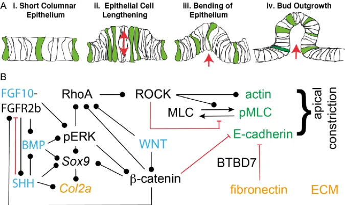
In the following, we will discuss the regulatory mechanism that control distinct hallmarks of the branching process: the stereotypic definition of branch points, the coordination of lung growth and branching, the biased elongation of lung tubes, and the shape of lung bud tips.

Definition of Branch Points

As the lung tubes are growing out, new buds emerge either along the stalk or by bifurcation of the tip (Fig. 3A). The sequence by which these branch points appear is highly conserved between embryos (Blanc et al., 2012; Metzger et al., 2008; Short et al., 2013). Several models have been put forward to explain how branch points are defined in the growing lung, as reviewed before (Iber and Menshykau, 2013). FGF10 signalling via the ERK/MAPK pathway is central to the control of lung branching morphogenesis as it is both necessary and sufficient to induce a lung branch (Fig. 3B) (Bellusci et al., 1997; Boucherat et al., 2014; De Moerlooze et al., 2000; Peters et al., 1994; Weaver et al., 2000).

Fgf10 is expressed in the submesothelial mesenchyme, while expression of its receptor, FGFR2b, and thereby FGF10 signalling, is restricted to the epithelium. FGF10 signalling induces the expression of Sonic Hedgehog (Shh) in the epithelium, which in turn diffuses into the mesenchyme and represses Fgf10 (Abler et al., 2009; Bellusci et al., 1997; Park et al., 1998). It has been proposed that because of this negative feedback, Fgf10 expression levels are lower, the smaller the distance between the

mesothelium and the epithelium (Bellusci et al., 1997; Hirashima and Iwasa, 2009). As branches grow

out, this distance becomes smaller, and the lower Fgf10 expression in front of the bud could then

result in bifurcating outgrowth of branches. According to an alternative model, the epithelium directly

recognizes the distance to the mesothelium via the local amplitude or the steepness FGF10 gradient at

the epithelial-mesenchyme border (Clement et al., 2012). Finally, it has been proposed that a protein

that inhibits branching is secreted from the epithelium and the epithelial geometry results in the

observed signalling pattern and points of outgrowth (Gleghorn et al., 2012). This protein would be

SHH or TGF-beta (Gleghorn et al., 2012). None of the above mechanisms would be able to explain

branching in mutant lung buds where Fgf10 is expressed throughout the mesenchyme (Volckaert et

(11)

al., 2013) or in mesenchyme-free explant cultures where recombinant FGF10 is added uniformly (Bellusci et al., 1997; Ohtsuka et al., 2001; Park et al., 1998).

We have shown that the interaction of the FGF10 ligand with its receptor FGFR2b, and the interaction of SHH with its receptor PTCH can both give rise to ligand-receptor based Turing patterns (Fig. 3C) (Kurics et al., 2014; Menshykau et al., 2014; Menshykau et al., 2012). Turing mechanisms enable the deterministic, stereotyped formation of patterns from noisy initial conditions (Turing, 1952). The ligand-receptor based Turing mechanism is based on the well-known Schnakenberg equations, where the two equations describe the dynamics of the receptor (R) and ligand (L),

𝜕𝑅

𝜕𝑡 = ∆𝑅 + 𝛾(𝑎 − 𝑅 + 𝑅

2

𝐿)

𝜕𝐿

𝜕𝑡

= 𝑑∆𝐿 + 𝛾(𝑏 − 𝑅

2

𝐿).

To generate Turing patterns, the ligand must diffuse faster than the receptors (d > 1), which is indeed the case because the receptors diffuse more slowly within the cell membrane than the ligands in the extracellular space. A key aspect of the ligand-receptor based Turing mechanism is that ligands and receptors must create multimeric ligand-receptor complexes (Kurics et al., 2014). FGF10 is indeed a dimer, and SHH a multimer, such that one ligand can engage multiple receptors (Ibrahimi et al., 2005;

Zeng et al., 2001). The binding of ligand and receptor must result in the removal of the ligand and the upregulation of the receptors on the cell surface, as is the case for SHH/PTCH (Ingham and

McMahon, 2001), and likely also for FGF10/FGFR2b as FGF10 upregulates -catenin signalling,

which in turn upregulates FfgR2b expression (Estival et al., 1996; Jones et al., 2018; Kadzik et al.,

2014). Interestingly, FGF7, which binds to the same receptor as FGF10 but triggers its removal rather

than recycling to the membrane, blocks branching morphogenesis (Francavilla et al., 2013). Finally,

in the default ligand-receptor based Turing model, receptor and ligands are produced at constant rates,

a and b respectively. But this is not a necessary condition, and feedbacks that modulate the production

rate, can lead to additional important properties, as discussed below.

(12)

The ligand-receptor based Turing mechanism explains how FGF10 signalling concentrates at newly forming branch tips (Fig. 4D, E), and produces signalling patterns that are consistent with the observed branching patterns in the wildtype and all known relevant mutants (Cellière et al., 2012;

Menshykau et al., 2014; Menshykau et al., 2012). In particular, the Turing mechanism reproduces the larger spacing between branch points in hypomorphic mutants that express Fgf10 at lower levels (Mailleux et al., 2005), and explains how branching patterns can still arise when Fgf10 is expressed homogenously throughout the lung mesenchyme (Cellière et al., 2012; Menshykau et al., 2014;

Menshykau et al., 2012; Volckaert et al., 2013). When put in proximity, lung buds compete for FGF ligand (Miura and Shiota, 2002). This depletion is consistent with a ligand-receptor based Turing mechanism, and particular relevant in kidney branching morphogenesis (Menshykau et al., 2019). We went on to quantitatively evaluate both the ligand-receptor based Turing models and the other

candidate models. Given that both FGF10 and SHH are both necessary and sufficient to induce the formation of new branches during lung development (Bellusci et al., 1997; De Moerlooze et al., 2000;

Litingtung et al., 1998; Pepicelli et al., 1998; Peters et al., 1994; Weaver et al., 2000), we evaluated the different models by comparing the predicted pattern of ligand-receptor signalling to the measured embryonic growth fields on 3D shapes of developing embryonic lung buds (Fig. 3E) (Menshykau et al., 2014). We found that only the ligand-receptor based Turing models, but none of the other proposed models succeeded in reproducing the observed growth fields. The ligand-receptor based Turing mechanism not only predicts the branch points, but can also guide the outgrowth of branches as observed in embryonic lungs (Menshykau et al., 2014). Here, we solved the Turing mechanism on an embryonic geometry and grew and deformed the epithelium according to the signalling strength predicted by the Turing mechanism, i.e. we applied a velocity field normal to the epithelial boundary whose strength was proportional to the local intensity of the local ligand-receptor complex pattern (Menshykau et al., 2014). The Turing mechanism, but none of the alternative mechanism, succeeded in giving rise to a stereotyped branched structure (Menshykau et al., 2014). We compared the

simulation output to the branching patterns observed with lung explants that were cultured for 48-72h,

and the simulations with the ligand-receptor-based Turing mechanism recapitulated the observed

(13)

branching pattern of lung buds very well. Based on all these studies, we conclude that the Turing mechanism, but none of the other proposed mechanisms, quantitatively recapitulates the lung branching patterns both in wildtype and perturbed signalling conditions.

The Turing mechanism on its own has several limitations. For one, many different patterns can emerge with the same parameter set (Fig. 3F) (Murray, 1993), such that branching sequences would vary between embryos. This is not the case for the ligand-receptor based Turing mechanism because the ligands and receptors are expressed in two different domains in the lung, the mesenchyme and the epithelium (Fig. 3B). As a result, the tissue geometry creates a pre-pattern that biases the Turing pattern to the same pattern in all embryos (Fig. 3G) (Menshykau et al., 2014). When ligands and receptors are both expressed in the kidney epithelium, branching morphogenesis still proceeds, but branching patterns indeed differ between embryos (Shakya et al., 2005).

A second problem concerns the evolution of Turing mechanisms. The parameter space that supports Turing patterns, the so-called Turing space, is small for classical Turing mechanism (Fig. 3H) (Murray, 1982). This would make it unlikely for nature to hit on the relevant parameter space and to retain it during evolution. The confinement of receptors to single cell surfaces greatly enlarges the Turing space (Fig. 3H) (Kurics et al., 2014). Receptor clustering (Fig. 3I) and regulatory interactions between Turing modules (Fig. 3J,K), as they exist between SHH and FGF10, further enlarge the Turing space such that the evolution of this patterning mechanism is highly plausible.

As the lung grows, new branch points emerge at a set distance from previous branch points in Turing models. Turing mechanisms thus provide a direct link between growth and branching. Indeed, faster growth is typically accompanied by enhanced branching and vice versa (Table 1). Enhanced

branching can also result from a smaller distance between branch points, which can come about from changes in the reaction or diffusion rates. Many perturbations that affect the growth rate also affect these Turing parameters. Thus, inactivation of any of the key genes for lung branching

morphogenesis, Shh, Fgf10, and FgfR2b impairs embryonic growth (Chiang et al., 1996; De

(14)

Moerlooze et al., 2000; Min et al., 1998; Sekine et al., 1999), and the FGF receptor inhibitor SU5402 and Smoothened agonist (SAG) reduce both growth and branching in lung explants (Goodwin et al., 2019; Runser et al., 2020). Conditional removal of the WNT receptor Frizzled in the lung epithelium reduces the growth rate and branching, even though Fgf10 expression is increased in the mesenchyme (Kadzik et al., 2014). This is likely due to the reduction in FgfR2 and Shh expression in the lung epithelium. WNT/-catenin signaling enhances Sox9 expression (Ustiyan et al., 2016), and both, upregulation and inactivation of epithelial Sox9, results in smaller lungs with reduced branching (Chang et al., 2013; Rockich et al., 2013).

The growth speed can be increased also mechanically by an increase in luminal pressure, as

generated either by tracheal occlusion or with a microfluidic chamber (Nelson et al., 2017; Unbekandt et al., 2008). Increased growth and branching, however, are still linked to increased Fgf10 expression and are not observed in cauterized lung explants from FgfR2b null mice (Unbekandt et al., 2008).

Removal of Adamts18, which encodes a secreted matrix metalloproteinase (MMP) that degrades the extracellular matrix (ECM), results in shorter primary airways and a proportional reduction in branching without affecting the expression of Shh, Bmp4, or Fgf10 (Rutledge et al., 2019).

Interestingly, heterozygous Adamts18 mutants have larger lungs with more branches. Disrupting the actomyosin cytoskeleton or the basement membrane impairs both growth and branching (Moore et al., 2005; Relan and Schuger, 1999).

There are also examples where growth and branching are not linked. In all those cases, the bud tips,

and sometimes also the tubes, are dilated. Thus, E11.5 lung explants that are treated with EGF or TGF

become larger, but have fewer branches with dilated buds (Ganser et al., 1991). The dilation and

branching defect can be prevented by co-culture with Tissue inhibitors of metalloproteinases

(TIMPs), which regulate ECM degradation by MMPs. Likewise, addition of collagenase inhibits

branching and dilates tips. Similarly, E12 lungs in Erk null mice have the same size, but fewer

branches with dilated tips (Miettinen et al., 1997). Expression of a hyperactive KRas allele in the lung

epithelium results in larger lungs with wider tubes and cystic tips, but unchanged number of branches

(15)

(Chang et al., 2013), though it is unclear whether the length of branches is increased compared to the wildtype, or only their width (Tang et al., 2011). In all these cases, branching appears to be impaired because of changes in the epithelial structure and the ECM. This shows that in addition to the chemical definition of new branch points, the structure of the epithelium and the ECM must be such that bud formation is possible, as will be discussed in more detail below.

The potential of Turing mechanisms to explain lung branching morphogenesis was recognised early on, and several other Turing mechanisms have been proposed to explain lung branching

morphogenesis (Guo et al., 2014; Meinhardt, 1976; Warburton et al., 2003; Xu et al., 2017; Zhu and Yang, 2018). Together, these mathematical studies demonstrate that important hallmarks of branching morphogenesis can be recapitulated by Turing mechanisms. To test a Turing mechanism, it is

important to identify the Turing components and check that the mechanism is consistent with all reported genetic and pharmacological perturbations. To date, this has only been done with the ligand- receptor-based Turing mechanism for the lung (Cellière et al., 2012; Menshykau et al., 2014;

Menshykau et al., 2012). Even though the qualitative and quantitative explanation of a wide range of observations provides important support, pattern likeness alone is not sufficient proof that branching morphogenesis is controlled by a Turing mechanism. Ultimately, all parameter values would have to be measured to prove the existence of a Turing pattern, but this is so far impossible in biological tissue.

In addition to signalling mechanisms, mechanical mechanisms have been proposed (Kim et al., 2015;

Lubkin and Murray, 1995; Wan et al., 2008). However, these would result in random, rather than

stereotyped branching patterns. Finally, differential proliferation in the epithelial and mesenchymal

layers has been suggested to result in budding via epithelial buckling (Varner et al., 2015). The

distance between buds would then depend on the thickness and the mechanical properties of the

layers. The distance between buds in the mesenchyme-free epithelial cultures was indeed found to

depend both on the Matrigel concentration and on the FGF1 concentration. In terms of the ligand-

receptor based Turing mechanism, this observation could, however, also be explained with a change

(16)

in the interaction of FGF1 with the extracellular matrix (ECM) and its diffusivity at different Matrigel concentrations (Makarenkova et al., 2009; Miura and Shiota, 2002). The reported shorter distance between branch points at higher FGF1 concentrations is consistent with the ligand-receptor based Turing mechanism.

Formation of a Bud

Once the position of a new branch point has been specified, how does the epithelial bud form? Since buds can form also in mesenchyme-free cultures, the epithelium itself must be able to generate buds.

Even though growth factor signaling concentrates at the branch point (Fig. 3E) (Liu et al., 2004;

Runser et al., 2020), a localized increase in the proliferation rate is not required for bud formation (Kim et al., 2013; Mollard and Dziadek, 1998; Nogawa et al., 1998). The first sign of a nascent bud is an epithelial thickening that results from the lengthening of the cells along the apical–basal axis (Fig.

4A) (Fumoto et al., 2017; Kadzik et al., 2014). The epithelial sheet then bends and creates a bud.

Given the geometry of the buds, the apical surface area is smaller than the basal surface area (Fumoto et al., 2017; Kadzik et al., 2014; Kim et al., 2013). Such cell shapes are characteristic for apical constriction (Martin and Goldstein, 2014), and simulations demonstrate that apical constriction in combination with proliferation can generate buds (Fumoto et al., 2017; Kim et al., 2013). The

mechanistic details of apical constriction and cell lengthening are still under debate, but the molecular motor non-muscle myosin II has long been implicated in generating the force that drives apical constriction by contracting actin filament (F-actin) networks that are linked to the apical cell-cell adhesion belt via α-catenin and E-cadherin, as well as other protein complexes (Martin and Goldstein, 2014). The extracellular domain of E-cadherin mediates cell-cell adhesion.

Myosin motor activity is regulated by phosphorylation of the myosin regulatory light chain (MLC)

(Fig. 4B), and both actin and phosphorylated myosin light chain (pMLC) have been reported to

accumulate at the apical surface of lung buds (Fumoto et al., 2017; Kadzik et al., 2014; Kim et al.,

(17)

2013; Rockich et al., 2013). Conditional removal of the WNT receptor Frizzled from the lung epithelium interferes with cell lengthening and concomitant epithelial thickening and bud formation (Kadzik et al., 2014). The effects of the Frizzled mutation can be counteracted with calpeptin, a RhoA activator, which activates Rho kinase (ROCK), which in turn phosphorylates MLC, inhibits the myosin phosphatase, and blocks the depolymerisation of actin filaments. Inhibitors of actin and myosin activation impair not only branching, but also growth (Kadzik et al., 2014; Moore et al., 2005).

Ectopic expression of E-cadherin impairs budding of E11.5 mesenchyme free lung epithelium (Liu et al., 2008). Fibronectin supports lung branching morphogenesis by inducing Btbd7, which regulates the expression of Snail2 and E-cadherin, and more generally epithelial motility (Onodera et al., 2010;

Sakai et al., 2003). Fibronectin and BTBD7 induce labile cell-cell adhesion and loss of E-cadherin from cell boundaries. Blocking FGF10 signalling or disrupting the downstream mitogen-activated protein kinase (MAPK) p38 increases expression of E-cadherin (Jones et al., 2018; Liu et al., 2008).

Deletion of Mek1/2 in the ureteric bud epithelium leads to accumulation of E-cadherin also on the baso-lateral cell surfaces and a failure to form new buds, though branches continue to elongate (Ihermann-Hella et al., 2014). Branching is impaired also in the lung, but the impact on E-cadherin has not been documented (Boucherat et al., 2014). The -catenin agonist CHIR enhances apical constriction and budding in E11.5 mesenchyme-free epithelial lungs cultured at low FGF10 concentrations (Fumoto et al., 2017). Biochemical studies suggest that E-cadherin can bind both α- catenin and β-catenin, but not at the same time (Martin and Goldstein, 2014). WNT signalling thus impacts on bud formation both via canonical (β-catenin) and non-canonical (Rho pathway) signalling.

The wrapping of airway smooth muscles has been suggested as additional driver of bud formation

(Goodwin et al., 2019), but mutant studies show that airway smooth muscles are dispensable for

branching morphogenesis (Young et al., 2020), and buds emerge already long before airway smooth

muscles first emerge at E11.5 (Hines et al., 2013; Tang et al., 2011).

(18)

Epithelial Tube Elongation

Once formed, epithelial tubes initially preferentially elongate (Fig. 5A), and only later switch to isotropic outgrowth, first in the trachea (from E12.5), and later in the most proximal bronchus (from E13.5) (Kishimoto et al., 2018; Runser et al., 2020; Tang et al., 2011; Tang et al., 2018). While proliferation is elevated in the tips of extending branches (Kim et al., 2013; Nogawa et al., 1998), biased outgrowth is not due to tip elongation. Rather, the proliferating stalks show biased elongation (Tang et al., 2011). Cell shape and cell division are biased in the direction of lung outgrowth (Fig. 5B) (Tang et al., 2011; Tang et al., 2018; Yates et al., 2010). Cells have long been known to divide

through their mass point and perpendicular to their longest axis, an observation termed Hertwig's rule, or long-axis rule (Fig. 5C) (Hertwig, 1884). In the lung, the mitotic spindle becomes biased once the cell aspect ratio at interphase is higher than 1.53 (Tang et al., 2018). Computational modelling shows that such a bias in cell shape and cell division can, in principle, result in biased outgrowth (Runser et al., 2020; Stopka et al., 2019; Tang et al., 2011). So, what leads to this bias in cell shape?

The planar cell polarity (PCP) pathway generates polarised patterns in epithelial fields and plays an

important role in regulating the mitotic spindle angle distribution in other organs like the kidney

(Ciruna et al., 2006; Gong et al., 2004; Saburi et al., 2008). However, airway shape and spindle

orientation are normal in E11.5 lungs from Van Gogh-like (Vangl)2

Lp

homozygotes (Tang et al.,

2011), though the number of epithelial buds is lower and the bud width is wider after 48 hours of

culture (Yates et al., 2010). Independent of whether the PCP pathway is involved, an additional

mechanism would still be required to set the direction of polarisation along the tube. In principle, such

a bias could originate from a polarization along the tube, from a mechanical constraint that limits

expansion in the circumferential direction, or from a pulling force at the tip or inside the tube in form

of flow-induced shear stress (Fig. 5D).

(19)

While FGF10 can serve as chemoattractant for explant lungs (Fig. 5E) (Park et al., 1998), branching morphogenesis and biased tube elongation are still observed when FGF10 is added homogenously to explant cultures (Bellusci et al., 1997; Ohtsuka et al., 2001; Park et al., 1998; Runser et al., 2020).

Moreover, lung epithelial tubes continue to elongate in Fgf10 and Fgfr2 conditional mutants and when an FGF receptor inhibitor is added homogenously to explant cultures (Abler et al., 2009; Runser et al., 2020). Accordingly, neither FGF gradients nor focused FGF signalling at the epithelial tips are required for biased epithelial outgrowth (Fig. 5E). There is also no evidence for epithelial leader cells with actin-rich protrusions (Fig. 5F), as previously observed in the Drosophila trachea and the mammalian kidney (Chi et al., 2009; Ghabrial and Krasnow, 2006). Also a pulling force from the tip mesenchyme is not necessary as the mesenchyme is not required for biased epithelial outgrowth (Runser et al., 2020).

Circumferential mechanical constraints have been proposed to drive epithelial tube elongation in the mammary gland (Fig. 5G) (Paine et al., 2016). Lungs do not have a myoepithelium like mammary glands, but are lined by airway smooth muscles, which result in peristaltic fluid movement from E12.5 (Jesudason et al., 2006). However, lung branches are thinner rather than wider when

inactivation of Myocardin prevents the formation of airway smooth muscles (Young et al., 2020), and biased lung tube elongation is strongest before airway smooth muscles become detectable at E11.5 (Hines et al., 2013; Tang et al., 2011), and is observed also in the absence of mesenchyme (Runser et al., 2020). Furthermore, cell-based simulations show that constricting forces that generate the observed bias in outgrowth, result in a much lower cell shape bias than what is observed in the embryonic lung epithelium (Stopka et al., 2019). Constricting forces are therefore unlikely to drive the elongating outgrowth of lung tubes.

MMPs degrade ECM throughout embryogenesis and thereby enable the invasion of the epithelial bud

into the mesenchyme. The basement membrane, composed of nidogen, collagen, fibronectin, and

laminin, is thinner at the lung tips (Fig. 5H) (Mollard and Dziadek, 1998; Moore et al., 2005), and

epithelial tube elongation sensitively depends on the level of ECM turn-over. Thus, removal of the

(20)

secreted MMP Adamts18 enhances epithelial growth in heterozygous mutants and lowers growth in homozygous mutants (Rutledge et al., 2019). Timp3 mutants have lower fibronectin and E-cadherin levels, lower epithelial proliferation rates, and fewer branches in the lung (Gill et al., 2003; Gill et al., 2006). Low concentrations of the MMP inhibitor GM60001 can rescue the Timp3 phenotype, while high concentrations completely block MMP activity, preventing the digestion of the ECM ahead of the lung bud, and therefore invasion of the bud into the mesenchyme. It is currently not known to what extent the ECM biases the outgrowth of epithelial tubes, but it would have to do so without exerting a constricting force.

Finally, fluid flow could bias the direction of outgrowth (Fig. 5I). A tangential apical force, as would result from shear stress, can indeed result in both the reported bias in cell shape and elongating outgrowth in cell-based simulations (Runser et al., 2020). Flow-induced shear stress does not deform cells directly, but cells sense shear stress with their cilium and respond actively (Weinbaum et al., 2011). For the observed narrow luminal space (Runser et al., 2020), the reported flow rates (𝑉̇ = 420 𝜇𝑚

3

/𝑠 (George et al., 2015), and the 10-times higher viscosity of the luminal fluid compared to water (Bokka et al., 2015), the expected shear stress level (0.5-1 Pa) in the lung buds is within the range that cells can, in principle, sense (10

-3

- 5 Pa) (Nauli et al., 2003)(Flitney et al., 2009;

Molladavoodi et al., 2017; Resnick and Hopfer, 2007; Weinbaum et al., 2011). The primary cilium, which is required to sense shear stress, is lost in KRas

G12D

mutants (Tape et al., 2016). Consistent with a role of shear stress in driving elongating outgrowth, the bias in cell division and epithelial outgrowth is lost in embryonic lungs that express the constitutively active form KRas

G12D

in the epithelium (Shh

cre/+;

KRas

LSL-G12D/+

) (Tang et al., 2011).

In later developmental stages, the closed epithelial lung tubes open up (Runser et al., 2020). Given the larger tube diameter, shear stress levels are expected to drop below a level that cells can sense.

Consistent with this, tube outgrowth becomes isotropic in the trachea from E12.5, and in the left

bronchus from E13.5 (Kishimoto et al., 2018; Runser et al., 2020). Blockage of FGF10 signalling

(21)

results in the collapse of bronchi, but how this affects tube outgrowth has not been studied (Jones et al., 2018). Similarly, fluid flow in the lung tubes has so far only been studied from E12.5, while biased outgrowth is observed between E10.5 and E13.5 in the left bronchus. It will be important to measure flow rates also in the earlier stages of lung development, and to understand how cell signalling, fluid dynamics, and mechanical effects integrate to define airway shape.

Lung Bud Shape

A wide range of mutations and pharmacological reagents affect the shape of the distal lung tips (Table 1). Most notably, addition of FGF7 or FGF9 to lung explants results in inflated epithelial tips, while lung explants that are treated with the FGF receptor inhibitor SU5402 have narrower tips (Cardoso et al., 1997; del Moral et al., 2006; Runser et al., 2020; White et al., 2006). The epithelium in the inflated FGF9-treated tips tends to be thinner (del Moral et al., 2006). We have seen above that EGF, TGF, hyperactive KRas, and loss of Erk all result in dilated tips, and that Tissue inhibitors of

metalloproteinases (TIMPs), which regulate ECM degradation by MMPs, can counteract the TGF effect (Ganser et al., 1991; Miettinen et al., 1997) (Tang et al., 2011). Hyperactive KRas inhibits WNT/-catenin signaling, and WNT/-catenin signaling upregulates FGFR2b in the lung epithelium (Chang et al., 2013; Kadzik et al., 2014). Hyperactive KRas, WNT/-catenin signaling, and FGFR2b all support Sox9 expression (Chang et al., 2013; Kadzik et al., 2014; Ustiyan et al., 2016). Conditional removal of the WNT receptor Frizzled, and both upregulation and removal of Sox9 expression in the lung epithelium result in larger, rounder tips with open, cyst-like structures (Kadzik et al., 2014;

Rockich et al., 2013). SOX9 affects mainly the cytoskeleton and the extracellular matrix (Rockich et

al., 2013). In scratch assays, epithelial cells with disrupted Sox9 expression are less motile. While

actin remains distributed normally inside the epithelial cells, there is a reduction in cell-cell adhesion

and stabilized tubulin becomes disrupted on the basal side of Sox9 negative epithelia. Moreover,

laminin deposition becomes affected when Sox9 is removed or overexpressed, and laminin affects the

cell shape and polarity in the lung epithelium (Chang et al., 2013) (Nguyen and Senior, 2006; Schuger

(22)

et al., 1997). SOX9 directly regulates transcription of Type 2 collagen (Col2a1), and treatment of lung explants with collagenase results in cyst-like shapes (Chang et al., 2013; Miura and Shiota, 2002).

Buds are also widened when lung explants are cultured with cytochalasin D, a drug that disrupts actin microfilament integrity, fasudil, which inhibits Rho kinase (ROCK), or ML7 or ML9, which inhibits myosin light chain kinase (Kadzik et al., 2014; Moore et al., 2005). The tip shape thus depends on the mechanical state of both the epithelial cells and the extracellular matrix.

The Mesenchyme defines the Shape of the Epithelial Tree

The shape of epithelial trees differs profoundly between organs. The shape of a branched tree is determined by the distance between branch points, the branch angle, curvature, and aspect ratio.

Recombination experiments demonstrate that the mesenchyme strongly affects these parameters.

Thus, the epithelial tree that emerges when a ureteric bud is cultured with lung mesenchyme resembles more a lung epithelial tree than that of a kidney (Lin et al., 2003). The growth factors FGF10 and GDNF, that define the branch points in the lung and kidney respectively, are expressed in the lung and kidney mesenchyme (Bellusci et al., 1997; Moore et al., 1996; Pepicelli et al., 1997;

Pichel et al., 1996), but beyond this, the mesenchymal factors that determine organ shape are largely elusive. Interestingly, only the bronchial mesenchyme can support lung branching morphogenesis.

Mesenchyme from other sources, including the salivary gland, kidney, pancreas, stomach, intestine, dermis, and somites, can induce the formation of a single bud, but cannot support several rounds of branching (Spooner and Wessells, 1970), unless it is renewed (Lawson, 1983). In the lung, the

expansion of the mesenchyme and epithelium may be linked via an epithelial-mesenchymal feed-back

between FGF and SHH signalling. FGF9 and SHH both increase mesenchymal proliferation though in

different parts of the mesenchyme, and the FGF receptor inhibitor SU5402 reduces mesenchymal

proliferation (White et al., 2006). Several other morphogens promote epithelial and/or mesenchymal

proliferation, including BMP and canonical WNT signalling (Volckaert and De Langhe, 2015).

(23)

Concluding Remarks

There are still many open questions regarding lung branching morphogenesis. Are branch points really defined by a ligand-receptor based Turing mechanism? How does localized FGF10/ERK signalling induce bud formation? When do epithelial cells start to secrete fluid into the lung lumen and thereby create a flow that cells can sense? Are there other mechanisms that bias the outgrowth of lung tubes? What role does the ECM play, and how does the mesenchyme define the specific organ shapes? And how does the final fractal lung tree emerge during development? Unlike the final lung tree, the developing lung tree does not have a fractal architecture. As imaging, genetic and

computational techniques become ever more powerful, these questions are now in reach to be answered.

Acknowledgments

I thank members of the CoBi group for discussions.

(24)

REFERENCES

Abler, L. L., Mansour, S. L. and Sun, X. (2009). Conditional gene inactivation reveals roles for Fgf10 and Fgfr2 in establishing a normal pattern of epithelial branching in the mouse lung. Dev Dyn 238, 1999-2013.

Ali, M. (2010). Pulmonary Drug Delivery. In Handbook of Non-Invasive Drug Delivery Systems (ed. V. S. Kulkarni), pp. 209-246: Elsevier.

Badugu, A., Kraemer, C., Germann, P., Menshykau, D. and Iber, D. (2012). Digit patterning during limb development as a result of the BMP-receptor interaction. Sci Rep 2, 991.

Barbieri, O., Astigiano, S., Morini, M., Tavella, S., Schito, A., Corsi, A., Di Martino, D., Bianco, P., Cancedda, R. and Garofalo, S. (2003). Depletion of cartilage collagen fibrils in mice carrying a dominant negative Col2a1 transgene affects chondrocyte differentiation. American journal of physiology. Cell physiology 285, C1504-1512.

Bellusci, S., Grindley, J., Emoto, H., Itoh, N. and Hogan, B. L. (1997). Fibroblast growth factor 10 (FGF10) and branching morphogenesis in the embryonic mouse lung.

Development 124, 4867-4878.

Blanc, P., Coste, K., Pouchin, P., Azaïs, J.-M., Blanchon, L., Gallot, D. and Sapin, V. (2012). A role for mesenchyme dynamics in mouse lung branching morphogenesis. PLoS ONE 7, e41643.

Bokka, K. K., Jesudason, E. C., Lozoya, O. A., Guilak, F., Warburton, D. and Lubkin, S. R.

(2015). Morphogenetic Implications of Peristalsis-Driven Fluid Flow in the Embryonic Lung. PLoS One 10, e0132015.

Boucherat, O., Nadeau, V., Berube-Simard, F. A., Charron, J. and Jeannotte, L. (2014).

Crucial requirement of ERK/MAPK signaling in respiratory tract development.

Development 141, 3197-3211.

Cardoso, W. V., Itoh, A., Nogawa, H., Mason, I. and Brody, J. S. (1997). FGF-1 and FGF-7 induce distinct patterns of growth and differentiation in embryonic lung epithelium.

Developmental Dynamics 208, 398-405.

Cardoso, W. V. and Lu, J. (2006). Regulation of early lung morphogenesis: questions, facts and controversies. Development 133, 1611-1624.

Cellière, G., Menshykau, D. and Iber, D. (2012). Simulations demonstrate a simple network to be sufficient to control branch point selection, smooth muscle and vasculature formation during lung branching morphogenesis. Biol Open 1, 775-788.

Chang, D. R., Martinez Alanis, D., Miller, R. K., Ji, H., Akiyama, H., McCrea, P. D. and Chen, J. (2013). Lung epithelial branching program antagonizes alveolar differentiation.

Proc Natl Acad Sci U S A 110, 18042-18051.

Chen, J. and Krasnow, M. A. (2012). Integrin Beta 1 suppresses multilayering of a simple epithelium. PLoS One 7, e52886.

Chi, X., Michos, O., Shakya, R., Riccio, P., Enomoto, H., Licht, J. D., Asai, N., Takahashi, M., Ohgami, N., Kato, M., et al. (2009). Ret-dependent cell rearrangements in the Wolffian duct epithelium initiate ureteric bud morphogenesis. Dev Cell 17, 199-209.

Chiang, C., Litingtung, Y., Lee, E., Young, K. E., Corden, J. L., Westphal, H. and Beachy, P. A.

(1996). Cyclopia and defective axial patterning in mice lacking Sonic hedgehog gene function. Nature 383, 407-413.

Ciruna, B., Jenny, A., Lee, D., Mlodzik, M. and Schier, A. F. (2006). Planar cell polarity

signalling couples cell division and morphogenesis during neurulation. Nature 439,

220-224.

(25)

Clement, R., Douady, S. and Mauroy, B. (2012). Branching geometry induced by lung self- regulated growth. Phys Biol 9, 066006.

De Moerlooze, L., Spencer-Dene, B., Revest, J. M., Hajihosseini, M., Rosewell, I. and Dickson, C. (2000). An important role for the IIIb isoform of fibroblast growth factor receptor 2 (FGFR2) in mesenchymal-epithelial signalling during mouse

organogenesis. Development 127, 483-492.

del Moral, P. M., De Langhe, S. P., Sala, F. G., Veltmaat, J. M., Tefft, D., Wang, K.,

Warburton, D. and Bellusci, S. (2006). Differential role of FGF9 on epithelium and mesenchyme in mouse embryonic lung. Dev Biol 293, 77-89.

Deutsch, G., Jung, J. N., Zheng, M. H., Lora, J. and Zaret, K. S. (2001). A bipotential precursor population for pancreas and liver within the embryonic endoderm.

Development 128, 871-881.

Domyan, E. T., Ferretti, E., Throckmorton, K., Mishina, Y., Nicolis, S. K. and Sun, X. (2011).

Signaling through BMP receptors promotes respiratory identity in the foregut via repression of Sox2. Development 138, 971-981.

Elluru, R. G., Thompson, F. and Reece, A. (2009). Fibroblast growth factor 18 gives growth and directional cues to airway cartilage. Laryngoscope 119, 1153-1165.

Estival, A., Monzat, V., Miquel, K., Gaubert, F., Hollande, E., Korc, M., Vaysse, N. and Clemente, F. (1996). Differential regulation of fibroblast growth factor (FGF) receptor-1 mRNA and protein by two molecular forms of basic FGF - Modulation of FGFR-1 mRNA stability. Journal of Biological Chemistry 271, 5663-5670.

Flitney, E. W., Kuczmarski, E. R., Adam, S. A. and Goldman, R. D. (2009). Insights into the mechanical properties of epithelial cells: the effects of shear stress on the assembly and remodeling of keratin intermediate filaments. FASEB J 23, 2110-2119.

Francavilla, C., Rigbolt, K. T., Emdal, K. B., Carraro, G., Vernet, E., Bekker-Jensen, D. B., Streicher, W., Wikstrom, M., Sundstrom, M., Bellusci, S., et al. (2013). Functional proteomics defines the molecular switch underlying FGF receptor trafficking and cellular outputs. Mol Cell 51, 707-722.

Fumoto, K., Takigawa-Imamura, H., Sumiyama, K., Kaneiwa, T. and Kikuchi, A. (2017).

Modulation of apical constriction by Wnt signaling is required for lung epithelial shape transition. Development 144, 151-162.

Ganser, G. L., Stricklin, G. P. and Matrisian, L. M. (1991). EGF and TGF alpha influence in vitro lung development by the induction of matrix-degrading metalloproteinases. Int J Dev Biol 35, 453-461.

George, U. Z., Bokka, K. K., Warburton, D. and Lubkin, S. R. (2015). Quantifying stretch and secretion in the embryonic lung: Implications for morphogenesis. Mech Dev 138 Pt 3, 356-363.

Ghabrial, A. S. and Krasnow, M. A. (2006). Social interactions among epithelial cells during tracheal branching morphogenesis. Nature 441, 746-749.

Gill, S. E., Pape, M. C., Khokha, R., Watson, A. J. and Leco, K. J. (2003). A null mutation for Tissue Inhibitor of Metalloproteinases-3 (Timp-3) impairs murine bronchiole branching morphogenesis. Developmental Biology 261, 313-323.

Gill, S. E., Pape, M. C. and Leco, K. J. (2006). Tissue inhibitor of metalloproteinases 3 regulates extracellular matrix--cell signaling during bronchiole branching morphogenesis. Dev Biol 298, 540-554.

Gleghorn, J. P., Kwak, J., Pavlovich, A. L. and Nelson, C. M. (2012). Inhibitory morphogens

and monopodial branching of the embryonic chicken lung. Dev Dyn 241, 852-862.

(26)

Glenny, R. W. (2011). Emergence of matched airway and vascular trees from fractal rules. J Appl Physiol 110, 1119-1129.

Gong, Y., Mo, C. and Fraser, S. E. (2004). Planar cell polarity signalling controls cell division orientation during zebrafish gastrulation. Nature 430, 689-693.

Goodwin, K., Mao, S., Guyomar, T., Miller, E., Radisky, D. C., Kosmrlj, A. and Nelson, C. M.

(2019). Smooth muscle differentiation shapes domain branches during mouse lung development. Development 146.

Goss, A. M., Tian, Y., Cheng, L., Yang, J., Zhou, D., Cohen, E. D. and Morrisey, E. E. (2011).

Wnt2 signaling is necessary and sufficient to activate the airway smooth muscle program in the lung by regulating myocardin/Mrtf-B and Fgf10 expression. Dev Biol 356, 541-552.

Goss, A. M., Tian, Y., Tsukiyama, T., Cohen, E. D., Zhou, D., Lu, M. M., Yamaguchi, T. P. and Morrisey, E. E. (2009). Wnt2/2b and beta-catenin signaling are necessary and

sufficient to specify lung progenitors in the foregut. Dev Cell 17, 290-298.

Guo, Y., Chen, T. H., Zeng, X., Warburton, D., Bostrom, K. I., Ho, C. M., Zhao, X. and Garfinkel, A. (2014). Branching patterns emerge in a mathematical model of the dynamics of lung development. The Journal of physiology 592, 313-324.

Haefeli-Bleuer, B. and Weibel, E. R. (1988). Morphometry of the human pulmonary acinus.

Anat Rec 220, 401-414.

Herriges, M. and Morrisey, E. E. (2014). Lung development: orchestrating the generation and regeneration of a complex organ. Development 141, 502-513.

Hertwig, O. (1884). Das Problem der Befruchtung und der Isotropie des Eies. Eine Theorie der Vererbung. Jenaische Zeitschrift fur Naturwissenschaft. 18, 274.

Hines, E. A., Jones, M. K., Verheyden, J. M., Harvey, J. F. and Sun, X. (2013). Establishment of smooth muscle and cartilage juxtaposition in the developing mouse upper

airways. Proc Natl Acad Sci U S A 110, 19444-19449.

Hirashima, T. and Iwasa, Y. (2009). Mechanisms for split localization of Fgf10 expression in early lung development. Developmental Dynamics 238, 2813-2822.

Hubert, F., Payan, S. M. and Rochais, F. (2018). FGF10 Signaling in Heart Development, Homeostasis, Disease and Repair. Frontiers in genetics 9, 599.

Iber, D. and Menshykau, D. (2013). The control of branching morphogenesis. Open biology 3, 130088-130088.

Ibrahimi, O. A., Yeh, B. K., Eliseenkova, A. V., Zhang, F., Olsen, S. K., Igarashi, M.,

Aaronson, S. A., Linhardt, R. J. and Mohammadi, M. (2005). Analysis of mutations in fibroblast growth factor (FGF) and a pathogenic mutation in FGF receptor (FGFR) provides direct evidence for the symmetric two-end model for FGFR dimerization.

Mol Cell Biol 25, 671-684.

Ihermann-Hella, A., Lume, M., Miinalainen, I. J., Pirttiniemi, A., Gui, Y., Peranen, J., Charron, J., Saarma, M., Costantini, F. and Kuure, S. (2014). Mitogen-activated protein kinase (MAPK) pathway regulates branching by remodeling epithelial cell adhesion. PLoS Genet 10, e1004193.

Ikonomou, L., Herriges, M. J., Lewandowski, S. L., Marsland, R., 3rd, Villacorta-Martin, C., Caballero, I. S., Frank, D. B., Sanghrajka, R. M., Dame, K., Kandula, M. M., et al.

(2020). The in vivo genetic program of murine primordial lung epithelial progenitors.

Nat Commun 11, 635.

Ingham, P. W. and McMahon, A. P. (2001). Hedgehog signaling in animal development:

paradigms and principles. Genes Dev 15, 3059-3087.

(27)

Ioannides, A. S., Massa, V., Ferraro, E., Cecconi, F., Spitz, L., Henderson, D. J. and Copp, A.

J. (2010). Foregut separation and tracheo-oesophageal malformations: the role of tracheal outgrowth, dorso-ventral patterning and programmed cell death. Dev Biol 337, 351-362.

Jesudason, E. C., Smith, N. P., Connell, M. G., Spiller, D. G., White, M. R., Fernig, D. G. and Losty, P. D. (2006). Peristalsis of airway smooth muscle is developmentally regulated and uncoupled from hypoplastic lung growth. Am J Physiol Lung Cell Mol Physiol 291, L559-565.

Jones, M. R., Dilai, S., Lingampally, A., Chao, C. M., Danopoulos, S., Carraro, G.,

Mukhametshina, R., Wilhelm, J., Baumgart-Vogt, E., Al Alam, D., et al. (2018). A Comprehensive Analysis of Fibroblast Growth Factor Receptor 2b Signaling on Epithelial Tip Progenitor Cells During Early Mouse Lung Branching Morphogenesis.

Frontiers in genetics 9, 746.

Kadzik, R. S., Cohen, E. D., Morley, M. P., Stewart, K. M., Lu, M. M. and Morrisey, E. E.

(2014). Wnt ligand/Frizzled 2 receptor signaling regulates tube shape and branch- point formation in the lung through control of epithelial cell shape. Proceedings of the National Academy of Sciences of the United States of America 111, 12444-12449.

Kadzik, R. S. and Morrisey, E. E. (2012). Directing lung endoderm differentiation in pluripotent stem cells. Cell Stem Cell 10, 355-361.

Kim, H. Y., Pang, M. F., Varner, V. D., Kojima, L., Miller, E., Radisky, D. C. and Nelson, C. M.

(2015). Localized Smooth Muscle Differentiation Is Essential for Epithelial Bifurcation during Branching Morphogenesis of the Mammalian Lung. Dev Cell 34, 719-726.

Kim, H. Y., Varner, V. D. and Nelson, C. M. (2013). Apical constriction initiates new bud formation during monopodial branching of the embryonic chicken lung.

Development 140, 3146-3155.

Kimura, S., Hara, Y., Pineau, T., Fernandez-Salguero, P., Fox, C. H., Ward, J. M. and Gonzalez, F. J. (1996). The T/ebp null mouse: thyroid-specific enhancer-binding protein is essential for the organogenesis of the thyroid, lung, ventral forebrain, and pituitary. Genes Dev 10, 60-69.

Kishimoto, K., Furukawa, K. T., Luz-Madrigal, A., Yamaoka, A., Matsuoka, C., Habu, M., Alev, C., Zorn, A. M. and Morimoto, M. (2020). Bidirectional Wnt signaling between endoderm and mesoderm confers tracheal identity in mouse and human cells. Nat Commun 11, 4159.

Kishimoto, K., Tamura, M., Nishita, M., Minami, Y., Yamaoka, A., Abe, T., Shigeta, M. and Morimoto, M. (2018). Synchronized mesenchymal cell polarization and

differentiation shape the formation of the murine trachea and esophagus. Nat Commun 9, 2816.

Kurics, T., Menshykau, D. and Iber, D. (2014). Feedback, receptor clustering, and receptor restriction to single cells yield large Turing spaces for ligand-receptor-based Turing models. Phys Rev E Stat Nonlin Soft Matter Phys 90, 022716.

Kuwahara, A., Lewis, A. E., Coombes, C., Leung, F. S., Percharde, M. and Bush, J. O. (2020).

Delineating the early transcriptional specification of the mammalian trachea and esophagus. eLife 9.

Lawson, K. A. (1983). Stage specificity in the mesenchyme requirement of rodent lung

epithelium in vitro: a matter of growth control? J Embryol Exp Morphol 74, 183-206.

(28)

Lin, Y., Zhang, S., Tuukkanen, J., Peltoketo, H., Pihlajaniemi, T. and Vainio, S. (2003).

Patterning parameters associated with the branching of the ureteric bud regulated by epithelial-mesenchymal interactions. Int J Dev Biol 47, 3-13.

Litingtung, Y., Lei, L., Westphal, H. and Chiang, C. (1998). Sonic hedgehog is essential to foregut development. Nat Genet 20, 58-61.

Liu, Y., Martinez, L., Ebine, K. and Abe, M. K. (2008). Role for mitogen-activated protein kinase p38 alpha in lung epithelial branching morphogenesis. Dev Biol 314, 224-235.

Liu, Y., Stein, E., Oliver, T., Li, Y., Brunken, W. J., Koch, M., Tessier-Lavigne, M. and Hogan, B. L. (2004). Novel role for Netrins in regulating epithelial behavior during lung branching morphogenesis. Curr Biol 14, 897-905.

Lubkin, S. R. and Murray, J. D. (1995). A mechanism for early branching in lung morphogenesis. J Math Biol 34, 77-94.

Madl, P., Hofmann, W., Oldham, M. J. and Asgharian, B. (2010). Stochastic morphometric model of the BALB/c mouse lung. Anat Rec (Hoboken) 293, 1766-1775.

Mailleux, A. A., Kelly, R., Veltmaat, J. M., De Langhe, S. P., Zaffran, S., Thiery, J. P. and Bellusci, S. (2005). Fgf10 expression identifies parabronchial smooth muscle cell progenitors and is required for their entry into the smooth muscle cell lineage.

Development 132, 2157-2166.

Makarenkova, H. P., Hoffman, M. P., Beenken, A., Eliseenkova, A. V., Meech, R., Tsau, C., Patel, V. N., Lang, R. A. and Mohammadi, M. (2009). Differential interactions of FGFs with heparan sulfate control gradient formation and branching morphogenesis.

Science signaling 2, ra55.

Martin, A. C. and Goldstein, B. (2014). Apical constriction: themes and variations on a cellular mechanism driving morphogenesis. Development 141, 1987-1998.

Mauroy, B., Filoche, M., Weibel, E. R. and Sapoval, B. (2004). An optimal bronchial tree may be dangerous. Nature 427, 633-636.

Meinhardt, H. (1976). Morphogenesis of lines and nets. Differentiation 6, 117-123.

Menshykau, D., Blanc, P., Unal, E., Sapin, V. and Iber, D. (2014). An interplay of geometry and signaling enables robust lung branching morphogenesis. Development 141, 4526-4536.

Menshykau, D., Kraemer, C. and Iber, D. (2012). Branch mode selection during early lung development. PLoS Comput Biol 8, e1002377.

Menshykau, D., Michos, O., Lang, C., Conrad, L., McMahon, A. P. and Iber, D. (2019).

Image-based modeling of kidney branching morphogenesis reveals GDNF-RET based Turing-type mechanism and pattern-modulating WNT11 feedback. Nat Commun 10, 239.

Metzger, R. J., Klein, O. D., Martin, G. R. and Krasnow, M. A. (2008). The branching programme of mouse lung development. Nature 453, 745-750.

Miettinen, P. J., Warburton, D., Bu, D., Zhao, J. S., Berger, J. E., Minoo, P., Koivisto, T., Allen, L., Dobbs, L., Werb, Z., et al. (1997). Impaired lung branching morphogenesis in the absence of functional EGF receptor. Dev Biol 186, 224-236.

Miller, L. A., Wert, S. E., Clark, J. C., Xu, Y., Perl, A. K. and Whitsett, J. A. (2004). Role of Sonic hedgehog in patterning of tracheal-bronchial cartilage and the peripheral lung.

Dev Dyn 231, 57-71.

Min, H., Danilenko, D. M., Scully, S. A., Bolon, B., Ring, B. D., Tarpley, J. E., DeRose, M. and

Simonet, W. S. (1998). Fgf-10 is required for both limb and lung development and

(29)

exhibits striking functional similarity to Drosophila branchless. Genes Dev 12, 3156- 3161.

Minoo, P., Su, G., Drum, H., Bringas, P. and Kimura, S. (1999). Defects in tracheoesophageal and lung morphogenesis in Nkx2.1(-/-) mouse embryos. Dev Biol 209, 60-71.

Miura, T. and Shiota, K. (2002). Depletion of FGF acts as a lateral inhibitory factor in lung branching morphogenesis in vitro. Mechanisms of Development 116, 29-38.

Molladavoodi, S., Robichaud, M., Wulff, D. and Gorbet, M. (2017). Corneal epithelial cells exposed to shear stress show altered cytoskeleton and migratory behaviour. PLoS One 12, e0178981.

Mollard, R. and Dziadek, M. (1998). A correlation between epithelial proliferation rates, basement membrane component localization patterns, and morphogenetic potential in the embryonic mouse lung. American journal of respiratory cell and molecular biology 19, 71-82.

Moore, K. A., Polte, T., Huang, S., Shi, B., Alsberg, E., Sunday, M. E. and Ingber, D. E.

(2005). Control of basement membrane remodeling and epithelial branching morphogenesis in embryonic lung by Rho and cytoskeletal tension. Dev Dyn 232, 268-281.

Moore, M. W., Klein, R. D., Fariñas, I., Sauer, H., Armanini, M., Phillips, H., Reichardt, L. F., Ryan, A. M., Carver-Moore, K. and Rosenthal, A. (1996). Renal and neuronal

abnormalities in mice lacking GDNF. Nature 382, 76-79.

Motoyama, J., Liu, J., Mo, R., Ding, Q., Post, M. and Hui, C. C. (1998). Essential function of Gli2 and Gli3 in the formation of lung, trachea and oesophagus. Nat Genet 20, 54-57.

Murray, J. (1993). Mathematical Biology.

Murray, J. D. (1982). Parameter space for turing instability in reaction diffusion mechanisms: a comparison of models. J Theor Biol 98, 143-163.

Nelson, C. M., Gleghorn, J. P., Pang, M. F., Jaslove, J. M., Goodwin, K., Varner, V. D., Miller, E., Radisky, D. C. and Stone, H. A. (2017). Microfluidic chest cavities reveal that transmural pressure controls the rate of lung development. Development 144, 4328- 4335.

Nelson, T. R., West, B. J. and Goldberger, A. L. (1990). The fractal lung: universal and species-related scaling patterns. Experientia 46, 251-254.

Nguyen, N. M. and Senior, R. M. (2006). Laminin isoforms and lung development: all isoforms are not equal. Dev Biol 294, 271-279.

Nogawa, H., Morita, K. and Cardoso, W. V. (1998). Bud formation precedes the appearance of differential cell proliferation during branching morphogenesis of mouse lung epithelium in vitro. Dev Dyn 213, 228-235.

Ohtsuka, N., Urase, K., Momoi, T. and Nogawa, H. (2001). Induction of bud formation of embryonic mouse tracheal epithelium by fibroblast growth factor plus transferrin in mesenchyme-free culture. Dev Dyn 222, 263-272.

Onodera, T., Sakai, T., Hsu, J. C., Matsumoto, K., Chiorini, J. A. and Yamada, K. M. (2010).

Btbd7 regulates epithelial cell dynamics and branching morphogenesis. Science 329, 562-565.

Paine, I., Chauviere, A., Landua, J., Sreekumar, A., Cristini, V., Rosen, J. and Lewis, M. T.

(2016). A Geometrically-Constrained Mathematical Model of Mammary Gland Ductal

Elongation Reveals Novel Cellular Dynamics within the Terminal End Bud. PLoS

Comput Biol 12, e1004839.

Abbildung

Table 1: Reagents that impact lung branching morphogenesis
Figure 1: The Adult Lung Tree
Figure 2: Control of Lung Development  The lung develops from the ventral anterior  foregut endoderm, adjacent to the cardiac  mesoderm
Figure 3: Control of Lung Branching Morphogenesis by a ligand-receptor based Turing  mechanism
+3

Referenzen

ÄHNLICHE DOKUMENTE

Correction factor F 3 − circle versus normalized wafer diameter d / s for (a) an in -line and (b) a square 4P probe array on a finite circular slice (s is the probe spacing for

Previous work described the functional requirement of the T-box (Tbx) transcription factor genes Tbx2-Tbx5, in the development of the pulmonary mesenchyme [6-10], of which the

Evolutionarily Singular Strategies and the Adaptive Growth and Branching of the Evolutionary Tree.. IIASA Working

• «Progress of ASTEC Code Validation on Circuit Thermal-hydraulics And Core Degradation», presented at the last ERMSAR ERMSAR meeting (SARNET European Review Meeting on

Moreover, by peribronchial lymph nodes (LNs), lung, and bronchoalveolar space sampling following RSV infection, we assessed the potential contributors of the host neonatal T

To further elucidate whether elevated BAFF levels are associated with certain diseases in humans, a sensitive ELISA for hBAFF was developed and sera from patients suffering

To measure TCR ligand affinities and thymic selection capacities of wildtype, α-CPM mutant and TM control thymocytes, the different T1-TCR transgenes were bred to Rag2

16 shows computations of cell voltage with constant exchange current density as well as concentration dependent exchange current density, for slow ( D f A = 1) and fast ( D f A