PATTERNING THE RETINA OF
DROSOPHILA MELANOGASTER FOR COLOR AND POLARIZED LIGHT VISION
Inaugural-Dissertation Zur Erlangung des Doktorgrades
Der Mathematisch-Naturwissenschaftlichen Fakultät Der Universität zu Köln
vorgelegt von Mathias F. Wernet
Aus Strasbourg
Köln
2004
Berichterstatter: Prof. Dr. Diethard Tautz
Prof. Dr. Siegfried Roth
Tag der mündlichen Prüfung: 5. 1. 2005
TEILPUBLIKATIONEN
Wernet, MF, Mazzoni, EO, Duncan, DM, Celik, A, Duncan, I, and Desplan, C.
Stochastic expression of the Drosophila Dioxin receptor Spineless creates the retinal mosaic required for color vision, in preparation.
Wernet, MF and Desplan, C. Building a retinal mosaic: cell-fate decision in the fly eye. Trends Cell Biol. 2004 Oct; 14(10):576-84.
Wernet MF, Labhart T, Baumann F, Mazzoni EO, Pichaud F, Desplan C.
Homothorax switches function of Drosophila photoreceptors from color to polarized light sensors. Cell. 2003 Oct 31;115(3):267-79.
Tahayato A, Sonneville R, Pichaud F, Wernet MF, Papatsenko D, Beaufils P, Cook T, Desplan C. Otd/Crx, a dual regulator for the specification of ommatidia subtypes in the Drosophila retina. Dev Cell. 2003 Sep;5(3):391-402.
Mollereau B, Wernet MF, Beaufils P, Killian D, Pichaud F, Kühnlein R, Desplan
C. A green fluorescent protein enhancer trap screen in Drosophila photoreceptor
cells. Mech Dev. 2000 May;93(1-2):151-60.
I. ABSTRACT (English and German) 1
II. INTRODUCTION
II.1 Retinal mosaics in humans and flies 7 II.2 The Drosophila compund eye 9 II.3 The Drosophila ommatidium: outer photoreceptors 10 II.4 The Drosophila retinal mosaic: inner photoreceptors 12 II.5 Visualization of p and y ommatidial subtypes 13 II.6 Maturation and re-organization of pupal ommatidia 15 II.7 Specification of adult inner PRs: the role of spalt 17 II.8 Distinguishing between R7 and R8 cell fates 18 II.9 Dorso-ventral development of the Drosophila eye 20 II.10 The current model for ommatidial subtype specification 23 II.11 GFP in living flies: the GAL4 enhancer trap screen 25 III. RESULTS
III.1 The GAL4 enhancer trap screen 29
III.2 Specification of the polarization sensitive ommatidia in the ‘dorsal rim area’
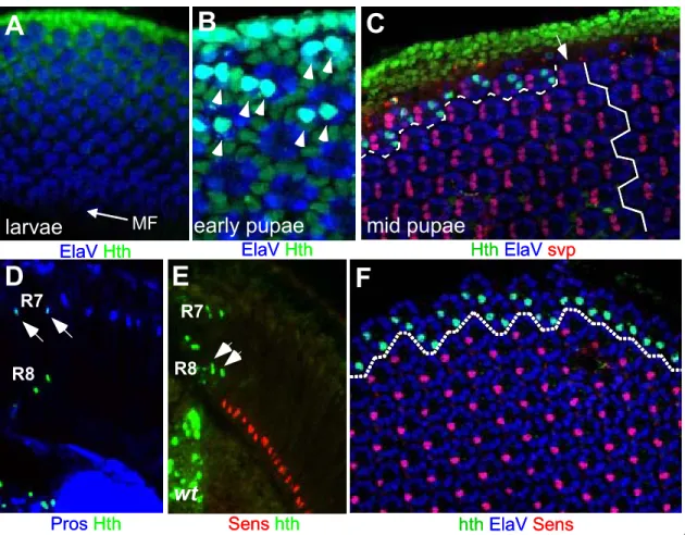
III.2.1. A GAL4 enhancer trap insertion in homothorax 33
III.2.2 Inner photoreceptors in the DRA express Homothorax 35
III.2.3 Molecular characterization of the DRA 38
III.2.4 Development of the DRA 41
III.2.5 Genetic manipulation of the DRA: cell fate decisions at the dorsal rim 43
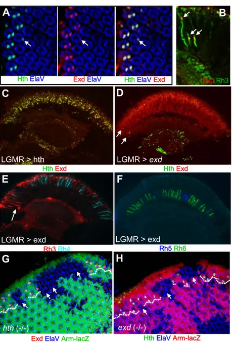
III.2.6 Extradenticle co-localizes with Homothorax during DRA development 48
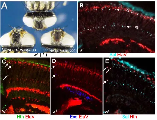
III.2.7 Expression of Hth/Exd is conserved between Musca and Drosophila 52
III.2.8 Homothorax is sufficient to induce the DRA fate in inner PRs 54
III.2.9 Mutual exclusion between Sens expression and DRA development 57
III.2.10 Transcriptional activity of Hth is required for DRA development 61
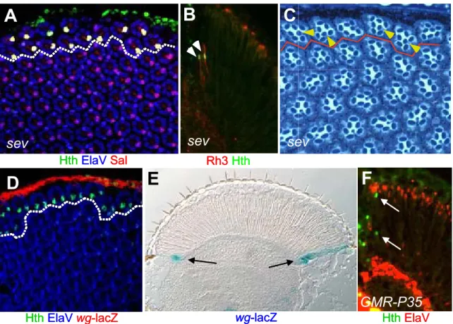
III.2.11 Only inner PRs are competent to become DRA: the role of spalt 65
III.2.14 Orthodenticle is required downstream of hth in DRA development 73 III.2.15 The dorsal selector genes are not necessary for DRA formation 78 III.2.16 The IRO-C complex is sufficient to induce DRA formation ventrally 82 III.2.17 Expansion of the DRA in optomotorblind Quadroon mutants 85 III.2.18 optomotorblind is not required for DRA development 88 III.2.19 Interaction of IRO-C and optomotorblind Quadroon mutants 92 III.2.20 The DRA develops in response to wingless signaling 95 III.2.21 IRO-C and optomotorblind expression in the expanded DRA 98 III.2.22 DRA development requires unusual wg signal transduction 100 III.2.23 Removing Hth function in the expanded DRA: odd coupled ommatidia 103 III.2.24 Homothorax, IRO-C and Wingless interact to form the DRA 107
III.3 Generating the ommatidial mosaic required for color vision:
Specification of pale and yellow ommatidia
III.3.1 spineless mutants show a dramatic opsin phenotype in R7 cells 109 III.3.2 spineless mutants show a less dramatic opsin phenotype in R8 cells 113 III.3.3 spineless is required in R7 cells 116 III.3.4 Spineless is specifically expressed in a large subset of R7 cells 118 III.3.5 Photoreceptor cell fates get specified correctly in spineless mutants 122 III.3.6 spineless is sufficient to induce the yellow R7 fate 124 III.3.7 spineless does not depend on spalt 128 III.3.8 Spineless over-expression does not alter photoreceptor cell fates 130 III.3.9 Genetic manipulation of the pale and yellow ommatidia 133 III.3.10 Spineless acts in a limited window of time 136 III.3.11 The PAS HLH dimerization partner Tango is not required in the eye 140 III.3.12 Antagonism between spineless and homothorax 144 III.3.13 Analysis of the spineless ‘eye enhancer’ using Bioinformatics 146 III.3.14 JAK/STAT signaling and ommatidial subtype specification 149
III.3.15 The wingless pathway antagonizes spineless function 152
III.3.16 Notch signaling might induce formation of y ommatidia 156
III.3.19 Activated Ras induces both rh3 and rh4 in sevenless mutants 167 III.3.20 The Notch rerceptor might not be necessary for retinal patterning 171
IV. DISCUSSION
IV.1 Homothorax provides new insights into PR development 175 IV.2 DRA ommatidia as a model system for wingless pathway activity 180 IV.3 The localized specification strategy of DRA ommatidia 185 IV.4 Loss of Homothorax and odd-coupled ommatidia 187 IV.5 Different default opsins in R7 and R8: spalt gain-of-function 191 IV.6 Spineless provides new insights into the formation of a retinal mosaic 194 IV.7 Transcriptional control of R7 opsin expression in color ommatidia 197 IV.8 Ommatidial subtype specification as a transcriptional model system 201 IV.9 Instruction of opsin expression in R8 204 IV.10 The stochastic specification strategy for color ommatidia 209 IV.11 Retinal patterning in Drosophila: combination of two strategies 211 IV.12 Similarities to retinal patterning in vertebrates 216
V. MATERIAL & METHODS 219
VI. REFERENCES 231
VII. ACKNOWLEDGEMENTS 245
VIII. APPENDIX 247
Fig II.1 Retinal mosaics in humans and flies 8
Fig II.2 The Drosophila compund eye 9
Fig II.3 The Drosophila ommatidium: outer photoreceptors 11 Fig II.4 The Drosophila retinal mosaic: inner photoreceptors 13 Fig II.5 Visualization of p and y ommatidial subtypes 14 Fig II.6 Maturation and re-organization of pupal ommatidia 16 Fig II.7 Specification of adult inner PRs: the role of spalt 17 Fig II.8 Distinguishing between R7 and R8 cell fates 20 Fig II.9 Dorso-ventral development of the Drosophila eye 21 Fig II.10 The current model for ommatidial subtype specification 24 Fig II.11 GFP in living flies: the GAL4 enhancer trap screen 26
Fig III.2.1 A GAL4 enhancer trap insertion in homothorax 33 Fig III.2.2 Inner photoreceptors in the DRA express Homothorax 35 Fig III.2.3 Molecular characterization of the DRA 38
Fig III.2.4 Development of the DRA 41
Fig III.2.5 Genetic manipulation of the DRA: cell fate decisions at the dorsal rim 43
Fig III.2.6 Extradenticle co-localizes with Homothorax during DRA development 47
Fig III.2.7 Expression of Hth/Exd is conserved between Musca and Drosophila 51
Fig III.2.8 Homothorax is sufficient to induce the DRA fate in inner PRs 53
Fig III.2.9 Mutual exclusion between Sens expression and DRA development 57
Fig III.2.10 Tanscriptional activity of Hth is required for DRA development 60
Fig III.2.11 Ony inner PRs are competent to become DRA: the role of spalt 64
Fig III.2.12 Loss of Homothorax results in loss of the Dorsal Rim Area 68
Fig III.2.13 The DRA forms normally in orthodenticle and prospero mutants 72
Fig III.2.14 Orthodenticle is required downstream of hth in DRA development 76
Fig III.2.15 The dorsal selector genes are not necessary for DRA formation 80
Fig III.2.18 optomotorblind is not required for DRA development 91 Fig III.2.19 Interaction of IRO-C and optomotorblind Quadroon mutants 94 Fig III.2.20 The DRA develops in response to wingless signaling 97 Fig III.2.21 IRO-C and optomotorblind expression in the expanded DRA 100 Fig III.2.22 DRA development requires unusual wg signal transduction 102 Fig III.2.23 Removing Hth function in the expanded DRA: odd coupled ommatidia 105 Fig III.2.24 Homothorax, IRO-C and Wingless interact to form the DRA 107
Fig III.3.1 spineless mutants show a dramatic opsin phenotype in R7 cells 110 Fig III.3.2 spineless mutants show a less dramatic opsin phenotype in R8 cells 114
Fig III.3.3 spineless is required in R7 cells 117
Fig III.3.4 Spineless is specifically expressed in a large subset of R7 cells 120 Fig III.3.5 Photoreceptor cell fates get specified correctly in spineless mutants 123 Fig III.3.6 spineless is sufficient to induce the yellow R7 fate 126
Fig III.3.7 spineless does not depend on spalt 129
Fig III.3.8 Spineless over-expression does not alter photoreceptor cell fates 132
Fig III.3.9 Genetic manipulation of the pale and yellow ommatidia 135
Fig III.3.10 Spineless acts in a limited window of time 138
Fig III.3.11 The PAS HLH dimerization partner Tango is not required in the eye 141
Fig III.3.12 Antagonism between spineless and homothorax 145
Fig III.3.13 Analysis of the spineless ‘eye enhancer’ using Bioinformatics 147
Fig III.3.14 JAK/STAT signaling and ommatidial subtype specification 150
Fig III.3.15 The wingless pathway antagonizes spineless function 153
Fig III.3.16 Notch signaling might induce formation of y ommatidia 158
Fig III.3.17 The activated Notch receptor induces yR7 specification 161
Fig III.3.18 Activated Notch specifically induces rh4 in sevenless mutants 165
Fig III.3.19 Activated Ras induces both rh3 and rh4 in sevenless mutants 168
Fig III.3.20 The Notch rerceptor might not be necessary for retinal patterning 172
Fig IV.3 The localized specification strategy of DRA ommatidia 185 Fig IV.4 Loss of Homothorax and odd-coupled ommatidia 189 Fig IV.5 Different default opsins in R7 and R8: spalt gain-of-function 192 Fig IV.6 Spineless provides new insights into the formation of a retinal mosaic 195 Fig IV.7 Transcriptional control of R7 opsin expression in color ommatidia 199
Fig IV.9 Instruction of opsin expression in R8 206
Fig IV.10 The stochastic specification strategy for color ommatidia 210 Fig IV.11 Retinal patterning in Drosophila: combination of two strategies 213
Fig V.1 Map of the P-Element pGawB 221
Fig V.2 The pGawB /UAS-GFP crossing scheme 222
Fig V.3 Ligation of ss
eye-GAL4 227
Fig V.4 Inverse PCR rescue of genomic DNA flanking a P-element 230
LIST OF TABLES
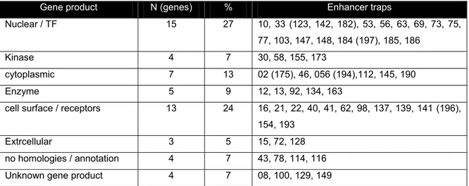
Table III.1 enhancer trap lines retained from the pGawB/UAS-GFP screen. 30 Table III.2 Screen results: targeted genes grouped into functional categories 31 Table III.3 Putative transcription factor binding sites in the ss eye enhancer 148
Table V.1 Fly strains used 219
Table V.2 Antibodies used 224
I. ABSTRACT
Across the animal kingdom, specialized sensory epithelia are used for photoreception, allowing individuals to interact with their environment based on visual cues. Generally, neuronal photoreceptor cells (PRs) are organized in the retina, a specialized part of body tissue exposed to the outside world, and transform the energy of incoming electromagnetic radiation into neuronal excitation. This process depends on the large family of opsin proteins which are required in PRs of all animal species. This lead to the theory, that the very divergent eye structures may share a common ancestor, although they most likely arose several times independently during evolution. PRs transmit their electrical excitation to higher order neurons, which are organized in the brain of the animal. How the brain then integrates the incoming signals from a multitude of PRs to reproduce a reliable representation of the world remains one of the central questions of neurobiology.
Most animals can extract different kinds of visual information from their
environment. Besides detecting the shape and movement of objects, additional
qualities like color or degree of polarization can also be distinguished. In most
cases, different classes of PRs are used for each of these visual tasks. For
instance, color discrimination is achieved by comparing the outputs of PRs
having different spectral sensitivity, as they express different opsin molecules. In
humans, three different subclasses of so-called cone PRs, are specialized to
absorb light of either short, medium or long wavelengths, corresponding to blue,
green or red colors, respectively. Loss of any one of these PR classes leads to a
dramatic impairment in the ability to discriminate between colors. Cones are most
highly concentrated in the center of the retina (fovea), where the three
subclasses form a random mosaic. Much remains to be understood about how
different PR subtypes choose expression of their opsin and how they distribute in
the retina.
The developing eye of the fruitfly Drosophila melanogaster was used here as a model system to investigate both nature and regulation of the different strategies involved in retinal patterning. The adult Drosophila eye consists of
~800 stereotypical unit eyes (ommatidia), each containing exactly 8 PRs (R1- R8). The six ‘outer PRs’ (R1-R6) are molecularly identical in all ommtidia as they always express the same opsin. They form a separate visual system contributing to the detection of shapes and motion. The morphological and molecular differences between inner PRs (R7 and R8) from different ommatidia leads to the formation of a retinal mosaic in Drosophila. Three ommatidial subtypes can be distinguished: while the ommatidia of the ‘dorsal rim area’ (DRA) are always found precisely localized in the dorsal periphery, the remaining ‘pale’ and yellow’
ommatidia are distributed stochastically through the rest of the retina. Only DRA ommatidia can be identified based on morphologic criteria, as these ommatidia form a polarizing filter which the fly uses to measure e-vector orientation of polarized sunlight for navigational purposes. The remaining two ommatidial subtypes are believed to serve color discrimination. They can only be identified based on the combination of opsins their inner PRs express. In order to identify genes and pathways involved in generating the retinal mosaic in Drosophila, a GAL4 enhancer trap screen was performed. Genes exhibiting expression patterns similar to inner PR opsins were analyzed genetically.
The homeodomain transcription factor Homothorax (Hth) was identified as
the key regulator of DRA specification. Hth is both necessary and sufficient for
the formation of the polarization sensors. During pupal development, positional
information provided by the diffusible morphogen Wingless (Wg), as well as the
dorsal selector genes of the Iroquois complex (IRO-C) and the gene
optomotorblind (omb) get integrated, leading to the specific induction of Hth
expression in inner PRs of prospective DRA ommatidia. In contrast to this
localized specification approach, stochastic expression of the Drosophila
arylhydrocarbon receptor Spineless (Ss) in a large subset of pupal R7 cells is
responsible for the specification of color ommtidia. Ss is both necessary and
sufficient to induce the ‘yellow’ R7 fate (yR7). Ss was therefore identified as the
key effector of a stochastic specification approach. How stochastic expression of Ss is regulated, remains obscure. However, an activating effect of the Notch (N) pathway on yR7 specification indicates that retinal patterning in Drosophila might combine the inductive effects of both wg and N signaling once again during pupal development.
Further investigation of the regulatory relationship between Hth and Ss (or
Wg and N) will provide a better understanding how retinal patterning contributes
to the integration of different kinds of visual information.
I. ZUSAMMENFASSUNG
Das Tierreich bietet eine Vielzahl von Beispielen, wie es spezialisiserte Sinnesepithelien einem Individuum erlauben, aufgrund optischer Eindrücke mit seiner Umgebung zu interagieren. Neuronale Photorezeptorzellen (PRs) sind grundsätzlich in der Retina organisiert - einem spezialisierten Teil der Körperoberfläche, welcher der Aussenwelt ausgesetzt ist - und wandeln die Energie der einfallenden elektromagnetischen Strahlung in neuronale Erregung um. Dieser Prozess involviert die grosse Familie der Opsinproteine, welche in den PRs aller Lebewesen benötigt werden. Dies begründete die Theorie, dass sich die sehr vielgestaltigen Augenstrukturen auf einen gemeinsamen Vorfahren zurück führen lassen, obwohl sie im Laufe der Evolution aller Wahrscheinlichkeit nach mehrfach unabhängig voneinander entstanden. PRs übertragen ihre elektrische Erregung auf Neuronen höherer Ordnung, welche im Gehirn des Tieres organisiert sind. Wie das Gehirn dann die eintreffenden Signale einer Vielzahl von PRs integriert, um eine zuverlässige Reproduktion der Welt zu erzeugen, bleibt eine der zentralen Fragen der Neurobiologie.
Die meisten Tiere können verschiedene Arten visueller Information von
ihrer Umgebung ableiten. Neben der Detektion von Form und Bewegung von
Objekten, können auch zusätzliche Qualitäten wie Farbe und Polarisierungsgrad
unterschieden werden. In den meisten Fällen werden unterschiedliche Klassen
von PRs für jede visuelle Augabe eingesetzt. Das Unterscheiden von Farben,
zum Beispiel, wird durch Vergleich der Ausgangssignale von PRs verschiedener
spektraler Sensitvität erreicht, da diese unterschiedliche Opsinmoleküle
exprimieren. Beim Menschen sind drei unterschiedliche Subklassen der
sogenannten Zapfen darauf spezialisiert, entweder Licht kurzer, mittlerer oder
langer Wellenlänge zu absorbieren, was blauer, grüner oder roter Farbe
entspricht. Verlust einer dieser PR Subklassen führt zu einer dramatischen
Einschränkung der Fähigkeit, Farben unterscheiden zu können. Zapfen treten in höchster Konzentration in der Fovea (dem ‘gelben Fleck’) auf, wo die drei Subklassen ein zufälliges Mosaik bilden. Bisher ist nicht klar, wie unterschiedliche PR Unterarten die Expression ihres Opsins wählen, oder wie sie sich in der Retina verteilen.
Das sich entwickelnde Auge der Fruchtfliege Drosophila melanogaster wurde hier benutzt, um die Art, sowie die Regulation der unterschiedlichen Strategien zu untersuchen, welche in der retinalen Musterbildung involviert sind.
Das adulte Auge von Drosophila besteht aus ca. 800 Komplexaugen (Ommatidien), von welchen jedes genau acht PRs enthält (R1 bis R8). Die sechs
‘äusseren PRs’ (R1 bis R6) sind molekular identisch in allen Ommatidien, da sie immer das selbe Opsin exprimieren. Sie bilden ein abgesondertes visuelles System, welches zur Wahrnehmung von Formen und Bewegung beiträgt. Die morphologischen, sowie molekularen Unterschiede zwischen den ‘inneren PRs’
(R7 und R8) unterschiedlicher Ommatidien führen zur Bildung eines retinalen Mosaiks in Drosophila. Drei Subtypen von Ommatidien können unterschieden werden: Während die Ommatidien der ‘dorsal rim area’ (DRA, dorsale Randregion) immer präzise lokalisiert, in der dorsalen Peripherie angetroffen werden, sind die verbleibenden ‘pale’ (‘blass’) und ‘yellow’ (‘gelb’) Ommatidien stochastisch über den Rest der Retina verteilt. Lediglich DRA Ommatidien können aufgrund morphologischer Kriterien identifiziert werden, da diese Ommatidien einen Polarisationsfilter bilden, welchen die Fliege zu Navigationszwecken zur Bestimmung der Orientierung des e-Vektors polarisierten Sonnenlichts benutzt. Es wird angenommen, dass die beiden verbleibenden Ommatidien-Subtypen der Unterscheidung von Farben dienen.
Sie können nur aufgrund der Opsine, welche ihre inneren PRs exprimieren, identifiziert werden. Um Gene und Signalwege zu identifizieren, welche bei der Bildung des retinalen Mosaiks in Drosophila eine Rolle spielen, wurde ein GAL4
‘enhancer trap screen’ durchgeführt. Gene, welche Expressionsmuster
vorweisen die denen der inner PR Opsinen ähneln, wurden weiter genetisch
untersucht.
Der Homeodomänen-Transkriptionsfaktor Homothorax (Hth) wurde identifiziert als der zentrale Regulator der Spezifikation von DRA Ommatidien.
Hth is sowohl notwendig als auch hinreichend für die Bildung der Polarisations- Sensoren. Während des Puppenstadiums werden verschiedene Aspekte räumlicher Information, welche vom diffundierenden Morphogen Wingless (Wg), den dorsalen Selektor-Genen des Iroquois Komplexes (IRO-C) und dem Gen optomotorblind (omb) bereit gestellt werden, in einer Weise integriert so dass Hth spezifisch in den inneren PRs der sich entwickelnden DRA Ommatidien exprimiert wird. Im Gegensatz zu diesem lokalisierten Spezifikationsansatz ist die stochastische Expression des Drosophila Arylhydrocarbonrezeptors Spineless (Ss) in einer grossen Subpopulation pupaler R7 Zellen zuständig für die Spezifikation jener Ommatidien, welche dem Farbensehen dienen. Ss ist sowohl notwendig als auch hinreichend für die Spezifikation von ‘yellow’ R7 Zellen (yR7).
Ss wurde somit als zentraler Effektor eines stochastischen Spezifikationsansatzes identifiziert. Wie die stochastische Expression von Ss reguliert wird, bleibt ein Rätsel. Die aktivierende Wirkung des Signaltransduktionsweges um den Notch Rezeptor (N) auf die Spezifikation von yR7 Zellen birgt nichtsdestotrotz den Hinweis, dass retinale Musterbildung in Drosophila die induktiven Effekte der wg und N Signalwege während des Puppenstadiums aufs Neue kombiniert.
Eine weitere Untersuchung der regulativen Zusammenhänge zwischen
Hth und Ss (oder Wg und N) wird ein besseres Verständnis davon ermöglichen,
wie retinale Musterbildung zur Integration verschiedener Arten von visueller
Information beiträgt.
II. INTRODUCTION
Despite the broad range of eye structures across the animal kingdom, all visual systems use similar cellular mechanisms to respond to environmental cues. For instance, all animals use related opsin proteins in their photoreceptor cells (PRs) to capture photons (for review: Arendt and Wittbrodt, 2001). In addition, the eyes of most animals can be used to perform two distinct visual tasks: They not only form images of the surrounding environment, but they can also detect the 'quality' of the visual stimulus, e.g. color or skylight polarization, through the use of specialized PR subclasses. These PR subclasses exhibit important morphological and molecular differences as well as characteristic distribution patterns through the retina in order to maximize the amount of information extracted from the environment. Emerging data indicate that retinal patterning includes a series of highly coordinated and organized processes.
Several recent works in the fly eye have begun to identify many of the factors involved, and interestingly, similar patterning events occur in the vertebrate retina that are sometimes regulated by orthologous factors. These data further imply that the vertebrate single lens eye and the insect compound eye use similar strategies to achieve their function and to control the development of the retina.
Rather than reflecting common ancestry of the visual systems, this might represent convergent mechanisms used to control opsin expression in different PR subtypes and may provide insight into understanding how the complexity of the retina is created and maintained.
1. Retinal mosaics in humans and flies
Humans use rod PRs (‘rods’) for detecting objects under low-light
conditions, while the cone PRs (‘cones’) participate in color discrimination as well
as high resolution vision. To serve these purposes most efficiently, the different
subclasses of cones (called S, M and L, indicating their maximal sensitivity to
short, medium or long wavelengths) are highly concentrated in the center of the
retina, the fovea. Interestingly, their distribution there appears to be stochastic,
resulting in a cone mosaic that can be visualized in vivo (Fig 1A). This allows the fovea to serve as the color and high acuity center for the eye. Rods, on the other hand, are concentrated towards the periphery of the eye which specializes in shape and motion vision under low light conditions.
A B
A B
Even species as distantly related to humans as flies share important similarities in the organization of their retina. For instance, specialized groups of PRs are used to discriminate between colors (in analogy to cones), whereas other PRs have been optimized for the detection of shapes and for motion detection (in analogy to rods). As in the human retina, the different fly PRs also exhibit specific distribution (Fig 1B). For instance, despite the dramatic differences in retinal organization, both fly and human color PR subtypes show a similar random distribution through the retina or the fovea, repectively. Additionally, another group of fly PRs is highly concentrated in a certain part of the retina, thereby forming a specialized eye region similar to the human fovea. The retinal mosaic of the fruitfly therefore represents an attractive model system for the study of both stochastic and localized specification events occurring during retinal patterning (for review: Wernet and Desplan, 2004).
Fig II.1 Retinal mosaics in humans and flies
(A) Pseudocolor image of the trichromatic cone mosaic from a living human retina. Blue, green and red colors represent the S, M, and L cones, respectively.
(B) Visualization of the ommatidial mosaic in the housefly, Musca domestica, using epifluorescence and water immersion microscopy.
2. The Drosophila compound eye
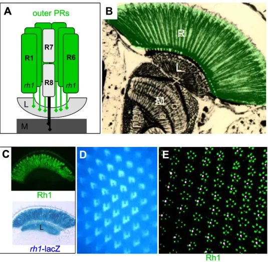
The Drosophila eye consists of ~800 stereotypical unit eyes (ommatidia), each containing 8 light-sensing PRs (called R1-8) as well as accessory cells involved in forming the lens or in shielding PRs from light coming from other ommatidia (Fig 2A, B and C; for review: Hardie, 1985). The light gathering membranes (rhabdomeres) of the six outer PRs (R1-R6) are organized in a chiral trapezoid. The center of each ommatidial trapezoid is occupied by the two inner PRs, R7 and R8. The rhabdomere diameter of these two inner PRs is significantly reduced as compared to outer PRs and they span only half of the
Fig II.2 The Drosophila compound eye
(A) Scanning electron micrograph of an adult Drosophila eye, composed of ~800 unit eyes (ommatidia).
(B) Cross section through an adult Drosophila ommatidium. The light gathering structures (rhabdomeres) of seven photoreceptors are visible (R8 is below focal plane). The Rhabdomere diameter is larger for outer PRs (R1-R6) which are aligned as a chiral trapezoid. R7 has a smaller rhabdomere diameter and is located in the center of the trapezoid.
(C) Schematic representation of an adult ommatidium. In the center of the ommatidium, R7 (pink) is located distally on top of R8 (blue) in the same path of light. Outer PRs (grey) span the entire retina from the apical to the basal side. Pigment cells Green and red) shield the ommatidium from light received by neighboring ommatidia, while cone cells (yellow) secrete the lens.
(D) PRs get specified during third instar larval stages from a pool of undifferentiated cells. Top: third instar larval eye-antennal disc (anterior to the left). Bottom: posterior to the morphogenetic furrow (MF), neuronal PR cells get recruited sequentially. First R8 (blue), then R2+R5, then R3+R4, then R1+R6 and finally R7.
R1 R2 R3 R4 R5
R6 R7
R7
R8 R1-6
A B C
D
MF
R2,R5R8 +R3,R4+R1,R6 +R7
R1 R2 R3 R4 R5
R6 R7
R7
R8 R1-6
A B C
D
MF
R2,R5R8 +R3,R4+R1,R6 +R7
retina, with the R7 rhabdomere located distally on top of that of R8: they are therefore in the same path of light, providing the ideal configuration to compare their outputs. This is absolutely required for the two functions of inner PRs, color vision and detection of the vector of polarized light. During third instar larval life, the eight Drosophila PRs of each ommatidium (R1-R8) are selected from an undifferentiated pool of cells (Fig 2D; for review: Brennan and Moses, 2000).
Through a process that is now fairly well understood, the interplay of the Notch, EGFR and Sevenless signaling pathways at the ‘morphogenetic furrow’ (MF) leads to a sequential recruitment of PRs into evenly spaced clusters. R8 is the first PR to be determined. This ‘founder cell’ then recruits all six outer PRs in a pair-wise fashion (first R2 and R5, then R3 and R4, finally R1 and R6). R7 is the last PR to be recruited (Freeman, 1996; Wolff, 1993).
3. The Drosophila ommatidium: outer photoreceptors
According to their morphology, axonal projections and opsin expression,
the fly PRs of every adult ommatidium can be grouped into two functional
categories: The outer PRs are the fly equivalent of the vertebrate rods and are
involved in motion detection and image formation (Fig 3A). Computation of their
outputs begins in the first optic lobe of the fly, the lamina (L), where the outer
PRs project their axons. Inner PRs project to the second optic lobe, the medulla
(M), where the neuronal processing both color and polarized light vision begins
(for review: Meinertzhagen and Hanson, 1993; Morante and Desplan, 2004). The
outer PRs have been shown to be both molecularly and morphologically identical
in all ommatidia. They capture photons with high efficiency, due to the expression
of their broad spectrum rhodopsin Rh1, as well as the large diameter of their
rhabdomeres which extend from the basal to the apical side of the retina (Zuker
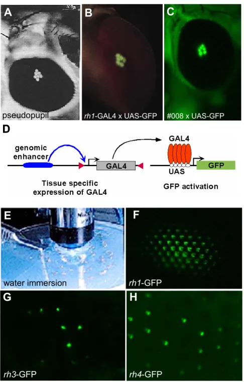
et al., 1985); Fig 3B). Different techniques have been used to characterize the
adult Drosophila visual system and the first PR subclass to be studied
molecularly in detail was the outer PR system. Cloning of the rh1/ninaE gene
allowed the visualization of Rh1 expression using antibodies against the Rh1
protein (Fig 3C, top). Furthermore, axon projections of the outer
M
R1 R6
R7
R8 L
rh1 rh1
A B
R L M
rh1-lacZ Rh1
L
Rh1 outer PRs
C D E
M
R1 R6
R7
R8 L
rh1 rh1
M
R1 R6
R7
R8 L
rh1 rh1
A B
R L M
rh1-lacZ Rh1
L
Rh1 outer PRs
C D E
PR axons to the lamina could be visualized by fusing the ninaE promoter to reporter genes like lacZ (Sheng et al., 1997); Fig 3C, bottom). These new histological techniques proved to be significantly more reliable than the previously developed water immersion microscopy, under which auto- fluorescence of Rh1 could be observed in outer PRs (Fig 3D). The use of this technique was limited due to the rapid bleaching of the visual pigment (Pichaud and Desplan, 2001). Finally, visualization of the ommatidal mosaic was revolutionized by the development of multi-colored fluorescent antibodies as well as whole mount and thin section techniques (Fig 3E).
Fig II.3 The Drosophila ommatidium: outer photoreceptors
(A) Schematic representation of an adult ommatidium. The outer PRs R1-R6 are identical in all ommatidia.. They express the broad band opsin Rh1 (ninaE) and project to the first layer of the optic lobe, the lamina (L).
(B) Silver-stained section through the Drosophila visual system. Outer PRs are highlightened in green. Their rhabdomeres span the entire retina. L=lamina; M=medulla.
(C)-(E) Expression of rh1 in outer PRs. Top: Frozen sections (10 µm) through adult heads from wildtype flies stained with an antibody against Rh1 (green). Bottom: Expression of rh1-lacZ visualized on frozen sections, using X-Gal. βGal activity is detectable in the entire retina as well as in axonal projections to the lamina (L). (D) Visualization of Rh1 using autofluorescence under UV illumination. (E) Thin section (1 µm) through through an adult wildtype eye double labeled with Anti-Rh1 (green) and an antibody labeling a subset of inner PRs (pink).
4. The Drosophila retinal mosaic: inner photoreceptors
Although the general external morphology of the fly eye does not indicate heterogeneity among ommatidia, three ommatidial subtypes have been described in Drosophila (Fig 4A). In all three cases, molecular and sometimes morphological features of the inner PRs have been used to categorize the ommatidial subtypes. Detailed morphological analysis of adult eyes from different fly species has revealed a first subset of ommatidia always found in one or two rows at the dorsal rim of the fly eye, called the ‘dorsal rim area’ (DRA, shown in pink in Fig 4A; (Hardie, 1984; Wada, 1971; Wada, 1974). These ommatidia exhibit an enlarged rhabdomere diameter as well as specialized rhabdomeric microvilli, making them strongly polarization sensitive (Fig 4B and C). It is believed that DRA ommatidia are used to improve navigation by measuring the oscillation plane of polarized skylight (Wolf et al., 1980)for review: Labhart and Meyer, 1999; Labhart and Meyer, 2002). The remaining two ommatidial subtypes have first been characterized in elegant studies by Franceschini and Kirschfeld (Franceschini et al., 1981). Fluoroscopy revealed the existence of two separate classes of ommatidia interspersed randomly within the fly retina: inner PRs appeared either pale (p) or yellow (y), with 30% being p and the remaining 70%
being y. Based on their different spectral sensitivities, p and y ommatidia were proposed to contribute to the discrimination between different colors.
Approximately 25 years later, the cloning of the rhodopsin genes (rh) from Drosophila has provided a molecular basis for all three ommatidial subtypes.
Inner PRs in DRA ommatidia were found to be monochromatic as they express the UV opsin Rh3 in both R7 and R8 (Fig 4D left; (Fortini and Rubin, 1990;
Fortini and Rubin, 1991; Fryxell and Meyerowitz, 1987; Fryxell and Meyerowitz,
1991). Outside of the DRA, ommatidia with monochromatic inner PRs have never
been reported, suggesting that this situation developed to specifically avoid
confusion between color and polarization. The p ommatidia were found to always
contain the UV-sensitive Rh3 in R7 and the blue-sensitive Rh5 in R8 (middle). A
different UV-sensitive Rh4, was found in the R7 of y ommatidia, which always
express the green-sensitive Rh6 in R8 (Chou et al., 1996; Huber et al., 1997;
Papatsenko et al., 1997). Therefore, expression of the rhodopsin genes is always coupled between R7 and R8, leading to the formation of only two ommatidial subtypes (p and y) in the main part of the retina (for review: (Cook and Desplan, 2001). It is believed that the differences in opsin expression play a crucial role for the fly’s ability to discriminate between colors, with the p ommatidia discriminating among shorter wavelengths (UV to blue) while the y ommatidia are specialized in the perception of longer wavelengths, reaching into the green part of the spectrum. Interestingly, the human cones, which express blue-, red- or green-specific opsins are also distributed stochastically in the fovea, but there is no tight coupling of the opsin expression between different cells (for review:
Nathans, 1999). Taken together, the retinal mosaic of the fruitfly is composed of three ommatidial subtypes, which are found either localized (DRA) or randomly distributed (p and y) throughout the retina (Fig 4E).
5. Visualization of the p and y ommatidial subtypes
Cloning of the genes encoding the 4 Drosophila inner PR Rhodopsins (rh3-rh6) allowed a more precise characterization of p and y ommatidia (Chou et
dorsal
ventral
‘pale’
~ 30%
Rh3 / Rh5
‘yellow’
~ 70%
Rh4 / Rh6
R1 R6
R7
R8
R1 R6
R7
R8
M
R1 R6
Dorsal Rim Area (DRA) Rh3 / Rh3
R8 R7
L eq
A B C
D E
Non-DRA DRA
dorsal
ventral
‘pale’
~ 30%
Rh3 / Rh5
‘yellow’
~ 70%
Rh4 / Rh6
R1 R6
R7
R8
R1 R6
R7
R8
M
R1 R6
Dorsal Rim Area (DRA) Rh3 / Rh3
R8 R7
L eq
A B C
D E
Non-DRA DRA
Fig 4. The Drosophila retinal mosaic: inner photoreceptors
(A) Three ommatidial subtypes are distributed throughout the Drosophila retina. Scanning electron micrograph illustrating the fact that, although they look identical from this point of view, ommatidia fall into three categories.
(B)+(C) DRA ommatidia were identified morphologically. The vast majority of ommatidia manifests a small inner PR rhabdomere diameter. (C) In the one or two dorsal-most rows of ommatidia, the ‘dorsal rim area’ ( DRA), however, inner PRs R7 and R8 have a dramatically enlarged rhabdomere diameter.
(D) Based on opsin expression and rhabdomere morphology, three ommatidial subtypes can be distinguished. Inner PRs of ommatidia located in the DRA always both express the UV-specific opsin Rh3 (left). So-called ‘pale’ (p) ommatidia always express Rh3 in R7 and Rh5 in R8 cells (middle) whereas ‘yellow’ (y) ommatidia express Rh4 in R7 and Rh6 in R8 cells (right).
(E) DRA ommatidia are always found in 1-2 rows at the dorsal periphery of the adult retina, whereas p (~30%) and y (~70%) ommatidia are distributed randomly through the retina (eq = equator).
al., 1996; Chou et al., 1999; Fryxell and Meyerowitz, 1987; Huber et al., 1997;
Montell et al., 1987; Papatsenko et al., 1997). Fusion of the rh3 and rh4 promoters with lacZ allowed the visualization of the p and y subtypes in R7 cells (Fig 5A). Expression of these reporter transgenes was shown to faithfully re- produce the expression pattern of the endogenous opsin proteins: expression of rh3-lacZ was restricted to the DRA inner PRs (arrow) as well as to the pR7 cells, whereas rh4-lacZ expression is specific to the yR7 cells. Due to the cytoplasmic localization of the βGal protein, these transgenes have also been used to
rh3-lacZ rh4-lacZ rh5-lacZ rh6-lacZ
Rh3 Rh4
Rh3 Rh4
Rh5 Rh6
Rh5 Rh6
A
B
C
D
E
F
L L L L
M M M M
rh3-lacZ rh4-lacZ rh5-lacZ rh6-lacZ
Rh3 Rh4
Rh3 Rh4
Rh5 Rh6
Rh5 Rh6
A
B
C
D
E
F
L L L L
M M M M
Fig II.5 Visualization of the p and y ommatidial subtypes
(A)-(C) Visualization o p and y subtypes in R7 cells. Frozen sections through an adult head from wildtype flies carrying the opsin reporter constructs rh3-lacZ (left) and rh4-lacZ (right). βGal activity is detectable in the DRA (rh3, arrow) and in subsets of R7 cells with cell bodies in the distal half of the retina and projections to the medulla (M). (B) Frozen section double labeled with antibodies against Rh3 (red) and Rh4 (cyan) allows simultaneous visualization of both R7 subtypes. pR7 and yR7 are non- overlapping. (C) Whole mounted retina double labeled with Anti-Rh3 and Anti-Rh4 reveales ~70 of yR7 and ~30% of pR7.
(D)-(F) Visualization o p and y subtypes in R7 cells. Opsin reporter constructs rh5-lacZ (left) and rh6-lacZ (right)are expressed in subsets of R8 cells with cell bodies in the proximal half of the retina and projections to the medulla (M). (E) Double labeled with antibodies against Rh5 (blue) and Rh6 (green) allows simultaneous visualization of both R8 subtypes. (F) Whole mounted retina double labeled with Anti-Rh5 and Anti-Rh6 reveales ~70 of yR8 and ~30% of pR8.
visualize the axonal projections of inner PRs to the medulla (M). Furthermore, antibodies raised against the Rh3 and Rh4 proteins have been used on frozen sections to simultaneously visualize exclusive expression of these opsins in pR7 and yR7 cells, respectively (Fig 5B). Recently, this technique has been applied to whole mounted retinas from adult flies (Cook et al., 2003), thereby specifically visualizing the ommatidial mosaic in R7 cells (Fig 5C).
Similar techniques have recently been applied to visualize p and y subtypes in the R8 cells. Fusions of the rh5 and rh6 promoters with lacZ were used to specifically visualize pR8 and yR8 cells and their axonal projections to the medulla (Fig 5D). Antibodies against Rh5 and Rh6 were used on frozen sections to visualize exclusion between both subsets in R8 cells (Fig 5E). Finally, the ommatidial mosaic in R8 cells has only recently been visualized by applying these antibodies on whole mounted adult retinas (Fig 5F).
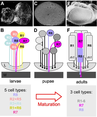
6. Maturation and re-organization of pupal ommatidia
During third instar larval life, the eight Drosophila PRs of each
ommatidium (R1-R8) are selected from an undifferentiated pool of cells (Fig
6A+B; for review: Brennan and Moses, 2000). Based on their order of
recruitment and the combination of transcription factors that they express, the
eight larval PRs represent at least 5 different cells types (R8, R2+R5, R3+R4,
R1+R6, and R7). It should be noted that the larval R3 and R4 cells can also be
viewed as individual cell types as they respond differently to the positional
information that establishes the chirality of the ommatidium in a process called
planar polarity (see below; for review: Tree et al., 2002). During the next four
days of pupal development, the PRs undergo dramatic morphological changes,
with the formation of the rhabdomeres as well as the onset of opsin expression
(Fig 6C+D). At the end, only three functional classes of PRs can be distinguished
in the adult ommatidium: the outer PRs R1-R6 and the two inner PRs R7 and R8
(Fig E+F; for review: Wolff, 1993). The outer PRs have now become virtually
identical. They all express the same opsin gene (rh1/ninaE) and their
rhabdomere morphology as well as axon projection pattern allows the fly to use
3 cell types:
R1-6 R7 R8 5 cell types :
R8 R2+R5 R3+R4 R1+R6
R7
R44
2 R6 R8 1R7 R2 R1 R3
R5
M L
R1 R6
R7
rh1 R8 rh1
Maturation
larvae pupae adults
A
B
C
D
E
F
R7 R8
3 cell types:
R1-6 R7 R8 5 cell types :
R8 R2+R5 R3+R4 R1+R6
R7
R44
2 R6 R8 1R7 R2 R1 R3
R5
M L
R1 R6
R7
rh1 R8 rh1
Maturation
larvae pupae adults
A
B
C
D
E
F
R7 R8
this group of PRs as a separate visual system for shape and motion vision (Lee et al., 2001). The two inner PRs, which, interestingly were the first (R8) and the last (R7) PR to be recruited into the ommatidium, are now grouped together to form the second visual system. They have become morphologically similar, with both rhabdomeres spanning opposite halves of the retina (R8 proximally and R7 distally). Both inner PRs from a given ommatidium always project to the same position in the medulla. However, terminations of R7 and R8 are found at slightly different layers (with R8 terminating before R7; for review: Clandinin and Zipursky, 2002). Adult R7 and R8 cells therefore provide the optimal configuration for comparing stimuli, both by working together in the same optical
Fig II.6 Maturation and re-organization of pupal ommatidia
(A)+(B) Third instar larval PRs develop in the eye imaginal disc, posterior to the morphogenetic furrow. They can be grouped into at least 5 categories, based on the order of their recruitment and the combination of transcription factors they express. R7 and R8 are already distinguishable based on their axon projection to the same layer of the brain, the medulla (M)..
(C)+(D) The pupal retina starts to re-organize. Approximately 48 hrs later, the disc has everted and a flat retina is visible. PRs start to move apart. Opsin expression and rhabdomere formation are about to begin.
(D)+(E) The adult retina: another 2-3 days later, the re-organization is complete. The eye and the head capsule are fused. PRs have developed elongated rhabdomeres and R7 has moved on top of R8. Outer PRs express rh1, whereas inner PR opsin expression and rhabdomere morphology dictate the subtype of the ommatidium.
path and by allowing further processing in the brain. Taken together, all pupal ommatidia have to re-organize their PRs by grouping them into the two functional categories represented in the adult eye.
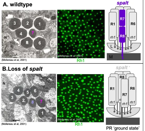
7. Specification of inner PRs: the role of spalt
An important first step towards understanding ommatidial maturation came with the description of the role of the spalt gene complex, which encodes two homologous zinc finger transcription factors (Kuhnlein et al., 1994). The spalt genes are specifically expressed in R7 and R8 (Fig 7A) and loss of spalt leads
M
R1 R6
R7
R8
spalt
L
rh1 rh1
R1 R6
R8
PR ‘ground state’
spalt
-rh1 rh1 rh1 rh1 R7 Rh1
Rh1 6
4
3 2 1
7
1
3 5 6
8 7 5
2 4
A. wildtype
B.Loss of spalt
(Mollereau et al, 2001)
(Mollereau et al, 2001)
(Mollereau et al, 2001)
(Mollereau et al, 2001)
M
R1 R6
R7
R8
spalt
L
rh1 rh1
M
R1 R6
R7
R8
spalt
L
rh1 rh1
R1 R6
R8
PR ‘ground state’
spalt
-rh1 rh1 rh1 rh1 R7 Rh1
Rh1 6
4
3 2 1
7
1
3 5 6
8 7 5
2 4
A. wildtype
B.Loss of spalt
(Mollereau et al, 2001)
(Mollereau et al, 2001)
(Mollereau et al, 2001)
(Mollereau et al, 2001)
Fig II.7 Specification of inner PRs: the role of spalt
(A) Spalt is expressed specifically in inner PRs R7 and R8, in the wildtype (purple).
(B) In spalt mutants, inner PRs get mis-specified. Electron microscopy revealed loss of the typical inner PR rhabdomeres with small diameter, in sal (-/-) eyes. (compare to A,left). (B) Antibody staining on thin sections revealed that all 8 PRs express Rh1 (green) in the absence of Sal (compare to A, center: Only six outer PRs per ommatidium express Rh1, in the wildtype). Right:
schematic representation of the spalt (-/-) phenotype: Both molecular as well as morphological features of inner PRs are lost.
Instead, these PRs gain all characteristica of outer PRs, except their axon projection pattern: R7 and R8 still project to the medulla (m), whereas R1-R6 terminate in the lamina (L). This indicates that, in the absence of Spalt, inner PRs got originally specified correctly, but then failed to maturate into adult inner PRs. Instead, they seem to be stuck in an outer-PR-like PR ‘ground state’.
to a loss of inner PR characteristics, both morphologically and molecularly, i.e.
loss of Rh3, Rh4, Rh5 and Rh6. Instead, R7 and R8 gain outer PR markers, like rhabdomere morphology and rh1 expression (Fig 7B). However, the axonal projections of these transformed inner PRs to the medulla are maintained (Domingos et al., 2004; Mollereau et al., 2001). It was therefore concluded that, in spalt mutants, inner PRs are initially properly specified but then lose their identity and instead terminally differentiate into outer PRs. Spalt, is therefore necessary to distinguish differentiating inner PRs from an otherwise outer PR-like
‘ground state’ toward which all PRs tend to develop (right). This provided a molecular basis to the existence of two overlapping visual systems in the adult.
This is also particularly interesting as it might provide a simple explanation of how the originally very divergent outer PRs R1-R6 adopt their uniform cell fate simply by being denied further differentiation signals like spalt expression. The presence of the two distinct genetic programs of specification, followed by differentiation, might illustrate the dual function of Drosophila PRs: They are first specified as neurons that must find their appropriate target in the optic lobes.
They subsequently differentiate as light sensing cells. In contrast, the vertebrate retina has two cell types to perform these roles: rods and cones to detect light, and retinal ganglion cells to project out of the retina into the brain.
8. Distinguishing between R7 and R8 cell fates
Both inner PRs require spalt to adopt their appropriate cell fate (Domingos et al., 2004). Nevertheless, R7 or R8 represent different PRs, both morphologically (position within the retina) and molecularly (different rhodopsins), and other factors are therefore necessary to further distinguish between the two inner PR cell fates. By screening for factors that bind to conserved sequences in the Drosophila opsin promoters, the gene prospero was recently shown to be necessary for distinguishing R7 from the R8 cell fate (Cook et al., 2003).
Prospero is a homeodomain transcription factor that is known to be important for
peripheral nervous system development as well as asymmetric cell division
(Kauffmann et al., 1996). In the adult eye, Prospero is expressed specifically in
the R7 cell in response to the signaling pathways responsible for R7 specification, Notch, EGFR and Sevenless (Fig 8A, left and center). It has been shown that Prospero binds to conserved sequences in the R8 opsin promoters (rh5 and rh6), thereby leading to their repression in R7. Loss of prospero indeed leads to a de-repression of rh5 and rh6 in adult R7 cells, creating a second R8- like cell per ommatidium (R8*, right). Interestingly, loss of prospero also results in repression of the proper R7 rhodopsins, most likely to avoid co-expression of opsin genes, a situation that is generally not observed in sensory receptors (for review: Celik et al., in press). These observations suggested that prospero is able to act on the generic inner PR fate and to push cells toward and R7 fate and away from an R8 fate. However, although R7 cells mis-express R8 rhodopsins in prospero mutants, they do not gain all R8 markers and their rhabdomeres are still positioned correctly in the distal part of the retina. This observation can be interpreted as a reversion of R7 back to a generic inner PR fate, which favors expression of R8 rhodopsins. This suggests that other genes are necessary to fully push the generic inner PR fate toward the R8 cell type.
One gene necessary for both specification and maturation of R8 cells encodes the Zn finger transcription factor Sensless (sens; (Nolo et al., 2000).
Sens had been shown to be specifically expressed in R8 cells, throughout eye
development (Fig 8B): PRs get mis-specified in sens mutants: Although R8 cells
are originally specified correctly, they then develop into outer PRs and loose their
inner PR identity (Frankfort et al., 2001). Consequently, R7 cells are not recruited
in sens (-/-) mutants, due to the loss of the R8-specific Boss signal required for
Sevenless receptor activation in potential R7 cell precursors (Cooper and Bray,
2000; Reinke and Zipursky, 1988; Tomlinson and Struhl, 2001). Fewer PRs are
therefore counted in ommatidia lacking Sens function (right). Sens has recently
been shown to be sufficient for activation of rh6 expression when ectopically
expressed in developing PRs, further suggesting that Sens is both necessary
and sufficient to induce R8 cell maturation (Domingos et al., 2004). Finally,
expression of both Pros and Sens has been shown to be lost in sal
M
R1 R6
R7
R8
prospero
R1 R8* R6 R8
prospero -
‘generic inner PR fate’
L
rh1 rh1 rh1 rh1
M
R1 R6
R7
R8
senseless senseless –
L
rh1 rh1
rh1 R8
0rh1 R7
R8
R7
R8
Pros Sal
Sens Sal
A. Specification of R7
Loss of R8 (and R7)
B. Specification of R8
rh1
M
R1 R6
R7
R8
prospero
R1 R8* R6 R8
prospero -
‘generic inner PR fate’
L
rh1 rh1 rh1 rh1
M
R1 R6
R7
R8
senseless senseless –
L
rh1 rh1
rh1 R8
0rh1 R7
R8 R7
R8
R7
R8
Pros Sal
Sens Sal
A. Specification of R7
Loss of R8 (and R7)
B. Specification of R8
rh1
(-/-) PRs (Domingos et al., 2004). This suggested that inner PRs undergo a series of consecutive determination steps, by gaining expression of different combinations of transcription factors. It appears therefore that R7 cells (Sal + Pros) and R8 cells (Sal + Sens) are the products of such a combinatorial code.
9. Dorso-ventral development of the Drosophila eye
All developing Drosophila ommatidia rotate by 90 degrees. Interestingly, the clusters rotate into opposite directions in the ventral and dorsal half of the eye
Fig II.8 Distinguishing between R7 and R8 cell fates
(A) The transcription factor Prospero (Pros) is expressed specifically in R7 cells, as seen on frozen sections double labeled for Pros (red) and Sal (green). Only R7 cells co-stain for Pros and Sal (top arrow). Loss of Pros results in R7 cells losing their typical adult characteristica (opsin expression, nuclear position - not shown); instead, these cells now resemble R8 cells (R8*; bottom, right). This suggests that Pros is necessary to distinguish the fate of R7 cells from an R8-like ‘generic inner PR fate’, in particular by repressing R8 opsin expression.
(B) The transcription factor Senseless (Sens) is expressed specifically in R8 cells, as seen on frozen sections double labeled for Sens (blue) and Sal(green). Only R8 cells co-stain for Sens and Sal (bottom arrow). Loss of Sens results in the R8 cell developing into an outer PR (R0); consequently, R7 cells never get specified due to the lack of the Boss signal.
(Fig 9A; (Cooper and Bray, 1999); for review: Strutt and Strutt, 2003). In the adult eye, two compartments can therefore be distinguished within which all chiral ommatidia point towards the nearest pole (Fig 9B; (Tomlinson and Struhl, 1999). Ommatidia of the dorsal and ventral compartments meet at a sharp
Fz
A B
C D
E F
IroC-lacZ
d
v MF
Tomlinson
& Struhl, 1999
(Cavodeassi et al, 1999)
(Irvine, 1999) Cooper
& Bray 1999
(Cooper & Bray 1999) (Brodsky & Steller, 1996)
Fz
A B
C D
E F
IroC-lacZ
d
v MF
Tomlinson
& Struhl, 1999
(Cavodeassi et al, 1999)
(Irvine, 1999) Cooper
& Bray 1999
(Cooper & Bray 1999) (Brodsky & Steller, 1996)
Fig II.9 Dorso-ventral development of the Drosophila eye
(A)+(B) Larval ommtidia rotate by 90 degrees. Dorsal and ventral ommatidia rotate in opposite directions (blue arrows). The two ommatidial forms meet at the ventral midline, the equator (Eq). (B) Left: Dorsal ommatidia point with R3 towards the dorsal pole (up), while ventral ommatidia point with R3 towrds the ventral pole (bottom). Dorsal ommatidia (red) and ventral ommatidia (blue) have opposite chirality. Right: In the wildtype, all ommmtidia on either side of the equat have the same chirality.
(C) Expression of the IRO-C complex is specific to the dorsal compartment. Thord instar larval eye disc from flies carrying a lacZ enhancer trap in IRO-C. βGal activity is specific to the dorsal half of the developing eye (d=dorsal, v=ventral, MF=morphogenetic furrow).
(D)+(E) The IRO-C complex is essential for generating the equator. IRO-C contributes to generating Notch (N) pathway activity a the equator, by restricting fringe (fng) expression to the ventral eye. N activity is crucial for establishment of ommatidial polarity as well as eye growth. The wingless (wg) pathway represses growth from the poles (DL=Delta, SER=Serrate). (E) Ectopic equators form at the boundary of IRO-C (-/-) clones.
(F) The N pathway serves as an amplification system in R4, to read small local differences in the Wg morphogen gradient (=
Frizzled/Fz receptor activity gradient). N responsive genes (mδ) get specifically activated in R4 (dsh=disheveled).
boundary at the midline of the adult eye, called the equator (Eq). Very early during eye development, the undifferentiated cells of the Drosophila eye imaginal disc anterior to the morphogenetic furrow are subdivided into dorsal and ventral compartments. The dorsal selector genes araucan (ara), caupolican (caup), and mirror (mirr) encode homologous homeodomain transcription factors and form the Iroquois complex (IRO-C; for review, see (Cavodeassi et al., 2001). During early larval stages these genes become specifically expressed in the territory that will give rise to the dorsal eye and head capsule (Fig 9C; (Brodsky and Steller, 1996). The IRO-C complex is activated very early during eye development by the diffusible morphogen Wingless (wg; (Lee and Treisman, 2001). At this stage, the most important function of IRO-C is the repression of the gene fringe, whose expression is thereby limited to the ventral eye tissue (Fig 9D left; for review:
Irvine, 1999). This was shown to be crucial for the generation of localized Notch (N) pathway activation at the developing equator, through a complicated interplay of the proteins Fringe, Serrate and Delta (center). N activity at the equator is essential for both growth of the eye as well as for the establishment of ommatidial polarity (right; (Cho and Choi, 1998). At later stages, wg expression becomes restricted to the peripodial membrane at the dorsal and ventral poles of the imaginal disc, from where the Wg morphogen is believed to form two gradients towards the equator (Heberlein et al., 1998; Treisman and Rubin, 1995). The IRO-C complex is therefore particularly important for dorsoventral development and ectopic equators form at the border of clones of eye tissue lacking IRO-C function (Fig 9E; (Cavodeassi et al., 1999; McNeill et al., 1997).
The interplay of the N and wg pathways during the establishment of ommatidial
polarity has been studied in great detail. In this context, the two outer PRs R3
and R4 proved to be a particularly interesting model system, as their position
within the ommatidium determines its chirality. Using sophisticated Drosophila
genetics, the N pathway was shown to serve as an amplification system in R4
that allows the decoding of tiny local differences in the Wg gradient over the
distance of only one cell diameter (Fig 9F). Important wg pathway components
like the receptor Frizzled and the effectors Armadillo, Disheveled and TCF were
shown to be required in this process, as well as the N pathway components mδ and suppressor of Hairless / su(H) (Cooper and Bray, 1999); for review: Irvine, 1999). Mutations in these genes will be used in this report, to test a possible role of the N and wg pathways during ommatidial subtype specification.
10. The current model for ommatidial subtype specification
Outside of the DRA, p and y ommatidia are found in a ratio of 30:70, distributed randomly through the fly retina (Chou et al., 1996; Fortini and Rubin, 1990; Franceschini et al., 1981). Although an elegant model has been proposed to explain the distribution of M and L cones in humans (Nathans, 1999), it is still not clear how stochastic choices are made between different PR cell fates in humans, or in flies. Nevertheless, two simple mutant backgrounds were used in Drosophila to build a mechanistic model describing the instructive signals specifying p and y ommatidia. In the absence of R7 cells (sevenless), R8 cells always express the yR8 opsin rh6 (Banerjee et al., 1987; Hafen et al., 1987;
Tomlinson et al., 1987). It was therefore proposed that rh6 represents the
‘ground state opsin’ expressed in R8 cells (Fig 10A; (Chou et al., 1999).
Furthermore, a signal from those R7 cells that have chosen to express the p opsin rh3 (pR7) is necessary for R8 to acquire the p fate (rh5). Chou and colleagues (1999) have further elaborated on this model; they generated adult ommatidia lacking R8 cells and found that both rh3 and rh4 were expressed randomly in the R7 cells of these retinas, suggesting that R8 is not necessary for the stochastic choice to occur in R7 cells. Similarly, when several R7 cells were induced within one ommatidium (seven-up), these extra R7 were found to choose randomly between the p and y fates (Chou et al., 1999; Mlodzik et al., 1990); Fig 10B). The model for stochastic specification of p and y ommatidia that was drawn from these experiments can therefore be divided into two steps (Fig 10C).
First, the stochastic, but biased choice between p and y fates appears to be
made in R7 cells (left), which then impose the corresponding fate onto the
underlying R8 cells (right; (Chou et al., 1999). The result is the fly color vision
system: a mosaic of two ommatidial subsets with the R8 cells exhibiting highest
spectral sensitivity in the blue (p) or green (y) part of the spectrum, comparing their inputs with UV-sensitive R7 cells.
R 1
R 6 R8
R 1
R 6 R7
R8
B. extra R7 cells A. no R7 cells
R7
R8 R7
R8
choice
y p
R7
R8 R7
R8
instruction
y p
1st step: choice in R7 2nd step: instruction of R8
pR7 yR7 rh3-GFP Rh1
Rh1
7 7 7
7
7 7 7 7
7 7
7 7
svp(-/-) 7 7 svp(+/-)
2 1
5
6 2
5 4
2 1
5 6 2
5 4
C. Model: specification of color ommatidia
Rh3 Rh4 Rh3 Rh4 Rh6 Phall
wt sev sev
Feiler et al, 1992 F.Pichaud
‘R8 ground state’
seven up (-/-)
R 1
R 6 R8 R 1
R 6 R8
R 1
R 6 R7
R8 R 1
R 6 R7
R8
B. extra R7 cells A. no R7 cells
R7
R8 R7
R8
choice
y p
R7
R8 R7
R8
instruction
y p
1st step: choice in R7 2nd step: instruction of R8
pR7 yR7 rh3-GFP Rh1
Rh1
7 7 7
7
7 7 7 7
7 7
7 7
svp(-/-) 7 7 svp(+/-)
2 1
5
6 2
5 4
2 1
5 6 2
5 4
C. Model: specification of color ommatidia
Rh3 Rh4 Rh3 Rh4 Rh6 Phall
wt sev sev
Feiler et al, 1992 F.Pichaud