Research Collection
Journal Article
Generation of integration-free induced pluripotent stem cells from healthy individuals
Author(s):
Yde Ohki, Cristine M; Grossmann, Leoni; Döring, Christian; Hoffmann, Per; Herms, Stefan; Werling, Anna M.; Walitza, Susanne; Grünblatt, Edna
Publication Date:
2021-05
Permanent Link:
https://doi.org/10.3929/ethz-b-000474257
Originally published in:
Stem Cell Research 53, http://doi.org/10.1016/j.scr.2021.102269
Rights / License:
Creative Commons Attribution-NonCommercial-NoDerivatives 4.0 International
This page was generated automatically upon download from the ETH Zurich Research Collection. For more information please consult the Terms of use.
ETH Library
Available online 24 February 2021
1873-5061/© 2021 The Author(s). Published by Elsevier B.V. This is an open access article under the CC BY-NC-ND license
(http://creativecommons.org/licenses/by-nc-nd/4.0/).
Lab Resource: Multiple Cell Lines
Generation of integration-free induced pluripotent stem cells from healthy individuals
Cristine Marie Yde Ohki
a,1, Leoni Grossmann
a,1, Christian D oring ¨
a, Per Hoffmann
b,c, Stefan Herms
b,c, Anna Maria Werling
a, Susanne Walitza
a,d,e, Edna Grünblatt
a,d,e,*aDepartment of Child and Adolescent Psychiatry and Psychotherapy, Psychiatric Hospital, University of Zurich, Zurich, Switzerland
bHuman Genomics Research Group, Department of Biomedicine, University of Basel, 4031 Basel, Switzerland
cInstitute of Human Genetics, University of Bonn, School of Medicine & University Hospital Bonn, 53105 Bonn, Germany
dNeuroscience Center Zurich, University of Zurich and the ETH Zurich, Zurich, Switzerland
eZurich Center for Integrative Human Physiology, University of Zurich, Switzerland
A B S T R A C T
Ten human induced pluripotent stem cell (iPSC) lines have been derived from five healthy controls matched to a study including Attention-Deficit Hyperactivity Disorder patients (ADHD). Both female and male children and adolescents aged 6–18 years were recruited. Isolated keratinocyte cells from the participants were reprogrammed into iPSCs using non-integrating Sendai virus to deliver the reprogramming factors Oct3/4, Sox2, Klf4 and cMyc.
1. Resource table:
Unique stem cell lines
identifier TMPi001-A
TMPi001-B TMPi002-A TMPi002-B TMPi003-A TMPi003-B TMPi004-A TMPi004-B TMPi005-A TMPi005-B Alternative names of stem
cell lines KO-001 c6 (TMPi001-A) KO-001 c9 (TMPi001-B) KO-003 c14 (TMPi002-A) KO-003 c20 (TMPi002-B) KO-005 c12 (TMPi003-A) KO-005 c13 (TMPi003-B) KO-008 c13 (TMPi004-A) KO-008 c44 (TMPi004-B) KO-011 c6 (TMPi005-A) KO-011 c10 (TMPi005-B)
Institution Psychiatric University Hospital Zurich, Department of Child and Adolescent Psychiatry and Psychotherapy, University of Zurich.
Contact information of
distributor Prof. Dr. Edna Grünblatt (edna.gruenblatt@kjpd.uzh.
ch)
(continued on next column)
(continued)
Type of cell lines iPSC
Origin Human
Cell Source Keratinocytes
Clonality Clonal
Method of reprogramming Sendai virus transduction
Multiline rationale Control and disease pair (Grossmann et al., 2021) Gene modification NO
Type of modification N/A
Associated disease Healthy controls
Gene/locus N/A
Method of modification N/A Name of transgene or
resistance N/A
Inducible/constitutive
system N/A
Date archived/stock date March 2020 Cell line repository/bank N/A
Ethical approval Cantonal Ethics Committee (BASEC-Nr.-2016-00101 &
BASEC-Nr.-201700825)
2. Resource utility
Induced pluripotent stem cells (iPSCs) are a powerful tool in disease modelling by maintaining the genetic background from somatic cells of
* Corresponding author at: Department of Child and Adolescent Psychiatry & Psychotherapy, Psychiatric University Hospital Zurich, University of Zurich, Wagistrasse 12, 8952 Schlieren, Switzerland.
E-mail address: edna.gruenblatt@kjpd.uzh.ch (E. Grünblatt).
1 Both authors contributed equally.
Contents lists available at ScienceDirect
Stem Cell Research
journal homepage: www.elsevier.com/locate/scr
https://doi.org/10.1016/j.scr.2021.102269
Received 8 February 2021; Received in revised form 8 February 2021; Accepted 18 February 2021
Stem Cell Research 53 (2021) 102269
2 individuals. Molecular and cellular analysis using iPSC-derived neural cells allows to elucidate differences between patients (e.g. ADHD) and healthy controls after iPSC generation and characterization to guarantee viable and trustworthy research.
3. Resource details
ADHD is one of the most complex and heterogeneous neuro- developmental disorders, with a worldwide prevalence of over 5% and a high heritability rate of 80% (Polanczyk et al., 2007). Disease modelling through iPSCs derived from ADHD patients is crucial to understand the underlying molecular mechanisms involved in ADHD and establishing matching control lines is equally important for such research develop- ment. In this paper, iPSC lines from five healthy controls were generated using the non-integrative Sendai virus (SeV) transduction. These control cell lines match the ADHD cell lines reported by Grossmann and col- leagues (Grossmann et al., 2021).
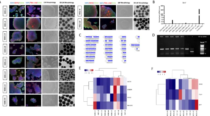
Keratinocytes from plucked hair were used as primary source for reprogramming (Table 1). The characterization of the derived iPSC lines is summarized in Table 2. By phase contrast microscopy, iPSC lines showed formation of well-delimited colonies, formed by small round cells with high nucleus:cytoplasm ratio (Fig. 1A, LM: light microscopy, scale bar: 200 µm). To biologically characterize the lines as pluripotent cells, gene and protein expression of pluripotency markers was also assessed through quantitative real-time PCR (qRT-PCR) and immuno- cytochemistry assays, respectively. All cell lines exhibited OCT4, SOX2, SSEA4 and TRA-1-60 expression in the latter (Fig. 1A, LM: light micro- scopy, scale bar: 200 µm) and positive expression of LIN28A, NANOG, OCT4 and SOX2 in the former (Fig. 1E). Additionally, possible myco- plasma contamination was assessed in a conventional PCR and demon- strated negative detection. The 1.2% agarose gel was cropped to separate controls from ADHD lines tested at the same time (Fig. 1B). All iPSCs were analysed regarding their potential to become embryoid bodies (EBs) (Fig. 1A, LM: light microscopy, scale bar: 200 µm) and their Table 1
Summary of lines.
iPSC line names Abbreviation in figures Gender Age Ethnicity Genotype of locus Disease
TMPi001-A KO-001 c6 Male 15 Caucasian N/A Healthy controls
TMPi001-B KO-001 c9 Male 15 Caucasian N/A Healthy controls
TMPi002-A KO-003 c14 Female 10 Caucasian N/A Healthy controls
TMPi002-B KO-003 c20 Female 10 Caucasian N/A Healthy controls
TMPi003-A KO-005 c12 Male 16 Caucasian N/A Healthy controls
TMPi003-B KO-005 c13 Male 16 Caucasian N/A Healthy controls
TMPi004-A KO-008 c13 Female 8 Caucasian N/A Healthy controls
TMPi004-B KO-008 c44 Female 8 Caucasian N/A Healthy controls
TMPi005-A KO-011 c6 Male 16 Caucasian N/A Healthy controls
TMPi005-B KO-011 c10 Male 16 Caucasian N/A Healthy controls
Table 2
Characterization and validation.
Classification Test Result Data
Morphology Light microscopy Normal morphology in which iPSCs show round morphology,
aggregation in colonies with well-delimited borders Fig. 1, panel A Phenotype Qualitative analysis by immunocytochemistry Positive protein expression of pluripotency markers (OCT4,
SOX2, SSEA4, and TRA-1-60) Fig. 1, panel A
Quantitative analysis Positive expression of pluripotency genes (LIN28, NANOG,
OCT4 and SOX2) Fig. 1, panel E
Genotype Genetic integrity analysis between saliva and iPSC DNA
CNVs using genome-wide association array KO-001 c6: 46XY KO-001 c9: 46XY KO-003 c14: 46XX KO-003 c20: 46XX KO-005 c12: 46XY KO-005 c13: 46XY KO-008 c13: 46XX KO-008 c44: 46XX KO-011 c6: 46XY KO-011 c10: 46XY
Fig. 1, panel C and supplementary
Identity Infinium global screening array (Illumina) N/A N/A
N/A Submitted in archive
with journal Mutation analysis (IF
APPLICABLE) Sequencing N/A N/A
Southern Blot OR WGS N/A N/A
Microbiology and
virology Mycoplasma Negative Fig. 1, item B
Differentiation potential Embryoid body Embryoid body formation from all cell lines with positive expression of AFP (endoderm), SOX2 (ectoderm) and FLK1 (mesoderm)
Fig. 1, panels A and F Donor screening
(OPTIONAL) HIV 1 +2 Hepatitis B, Hepatitis C N/A N/A
Genotype additional info
(OPTIONAL) Saliva genotyping DNA analysis Not shown but available
with author
HLA tissue typing N/A N/A
C.M. Yde Ohki et al.
potential to differentiate into the three germ layers (ectoderm, meso- dermal and endoderm) by expression analysis of specific markers (Fig. 1F). Regular karyograms without significant genomic aberrations were also obtained to prove that the reprogramming process has not compromised our study (Fig. 1C shows K001 i6; see supplementary file for all lines). Possible remaining SeV was verified by qRT-PCR and demonstrated that the only cell line with minimal SeV traces (relative expression =0.22%) was K003 c14, compared to the positive control (P1 iPSC) (relative expression =100%) (Fig. 1D). The negative control (control iPSC) showed no expression, as expected (Fig. 1D).
4. Materials and methods 4.1. Subject recruitment
Two female and three male healthy individuals have been recruited by the Department of Child and Adolescent Psychiatry and Psycho- therapy, University Hospital of Psychiatry Zurich, University of Zurich, to be part of a disease modelling study of ADHD (Grossmann et al., 2021). In contrast with ADHD patients, healthy controls did not show any psychiatric disorder, according to ICD-10 or DSM-5 (see details under supplementary material).
4.2. Reprogramming
Patient-specific keratinocytes were used as primary source for iPSCs generation by Sendai virus transduction (CytoTuneTM-iPS 2.0 kit by Invitrogen – Thermo Fisher Scientific), carrying polycistronic Klf4-Oct3/
4-Sox2 (KOS), cMyc and Klf4. The reprogramming protocol was per- formed according to Re et al. (2018).
Generated iPSCs were cultivated on Vitronectin-coated wells in E8 medium and passaged every 3–4 days at a 1:3 ratio by Versene (Gibco).
The iPSCs were expanded until at least passage 10 for quality control (QC).
4.3. Immunocytochemistry
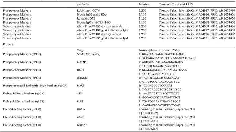
Protein expression of OCT4, SSEA4, TRA-1-60 and SOX2 was assessed by the Pluripotent Stem Cell 4-Marker Immunocytochemistry Kit (InvitrogenTM), according to the manufacturer’s instructions. Anti- bodies used are listed in Table 3.
4.4. Real-time quantitative PCR analysis
Expression of pluripotency genes was determined by qRT-PCR using the QuantiFast® SYBR® Green PCR kit (Qiagen) from cDNA. RNA was extracted from iPSCs using the RNeasy® Plus Mini kit (Qiagen) and reverse transcription from 500 ng RNA into cDNA was performed by iScriptTM cDNA Synthesis Kit (Bio-Rad). cDNA production and amplifi- cation of genes of interest (GOIs) and reference genes (RGs) were per- formed on the CFX384 thermal cycler using the primers detailed in Table 3. PCR efficiency was calculated by LinRegPCR (version 2020.0.0.3), while normalization of GOIs’ mRNA levels in relation to RGs was accomplished by Biogazelle qBasePLUS2 software (version 2.3) (ACTB and HMBS for iPSCs, ACTB and GAPDH for EBs). The heatmap plot was generated by the package heatmap3 in RStudio (version 1.1.423).
Fig. 1.Quality control of generated induced pluripotent stem cells (iPSCs) from 10 control lines. A) Light microscopy images showing morphology of all iPSCs (first column) and positive expression of pluripotent markers (OCT4, SSEA4, TRA-1-60 and SOX2) by immunocytochemistry (second and third column). Embryoid Bodies (EBs) morphology is also displayed (fourth column). B) Absence of mycoplasma in the supernatant of all iPSCs was demonstrated in an 1.2% agarose gel following DNA amplification by conventional PCR. C) Possible genetic aberrations caused by the reprogramming process were absent, as represented by the karyotype from KO-001 c6 (TMPi001-A) (for all lines see supplementary material). D) Sendai virus traces in culture were assessed by quantitative real time PCR (qRT-PCR) and compared to a positive control (P1 iPSC). E) Hierarchical gene expression analysis of pluripotency genes between iPSC lines, assessed by qRT-PCR. F) Hierarchical gene expression of endodermal (AFP), mesodermal (FLK1) and ectodermal (SOX2) markers in EBs, assessed by qRT-PCR.
Stem Cell Research 53 (2021) 102269
4 4.5. Detection of SeV genome
Presence of possible remaining SeV was tested in culture by using SeV primers in qRT-PCR (Table 3). RNA from an iPSC at passage one and a commercial human episomal iPSC line (A18945, Thermo Fisher Sci- entific) were used as positive and negative controls, respectively.
4.6. Mycoplasma testing
Possible mycoplasma contamination was assessed in the supernatant of iPSCs at passage eleven and above by the LookOut Mycoplasma PCR Detection Kit (Sigma-Aldrich), according to manufacturer’s instructions.
C1000TM/CFX96TM Thermal Cycler was used for DNA amplification and amplicons were loaded on a 1.2% agarose gel containing HDGreen Plus (INTAS, Germany) and run at 100 V for 30 min. The negative internal control sample showed a band at 480 bp, while positive bands were expected at 260 bp.
4.7. Generation of embryoid bodies (EBs)
EBs were formed after iPSC culture in AggreWells with E8/PVA medium for 48 h (Lin and Chen, 2014). RNA extraction was then per- formed and 1 μg RNA was used for expression analysis of specific markers (Table 3).
4.8. Genotyping analysis
DNA from saliva and iPSCs were individually extracted with the GeneFixTM saliva-Prep DNA kit (Isohelix) and DNeasy® Blood & Tissue kit (Qiagen), respectively. Samples were sent for genotyping with Infinium Global Screening Array (GSA, Illumina). Analysis and com- parison between the genotypes were made with the Genome Viewer
function from the GenomeStudio software (version 2.0).
Declaration of competing interest
The authors declare that they have no known competing financial interests or personal relationships that could have appeared to influence the work reported in this paper.
Acknowledgments
These cell lines belong to a research study supported by the PUK Forschungsfonds Nr. 8702 “Fonds für wissenschaftliche Zwecke im Interesse der Heilung von psychiatrischen Krankheiten”.
Appendix A. Supplementary data
Supplementary data to this article can be found online at https://doi.
org/10.1016/j.scr.2021.102269.
References
Grossmann, L., Yde Ohki, C.M., D¨oring, C., Hoffmann, P., Herms, P., Werling, A.M., Walitza, S., Grünblatt, E., 2021. Generation of integration-free induced pluripotent stem cell lines from four pediatric ADHD patients. Stem Cell Res. https://doi.org/
10.1016/j.scr.2021.102268. In press.
Lin, Y., Chen, G., 2014. Embryoid body formation from human pluripotent stem cells in chemically defined E8 media. Harvard Stem Cell Institute, StemBook [Internet], pp.
1–4.
Polanczyk, G., et al., 2007. The Worldwide Prevalence of ADHD: A Systematic Review and Metaregression Analysis. Am. J. Psychiatry 7.
Re, S., et al., 2018. Improved generation of induced pluripotent stem cells from hair derived keratinocytes – a tool to study neurodevelopmental disorders as ADHD.
Front. Cell. Neurosci. 12, 321. https://doi.org/10.3389/fncel.2018.00321.
Table 3 Reagents details.
Antibodies used for immunocytochemistry/flow-citometry
Antibody Dilution Company Cat # and RRID
Pluripotency Markers Rabbit anti-OCT4 1:200 Thermo Fisher Scientific Cat# A24867, RRID: AB_2650999
Pluripotency Markers Mouse IgG3 anti-SSEA4 1:100 Thermo Fisher Scientific Cat# A24866, RRID: AB_2651001
Pluripotency Markers Rat anti-SOX2 1:100 Thermo Fisher Scientific Cat# A24759, RRID: AB_2651000
Pluripotency Markers Mouse IgM anti-TRA-1-60 1:100 Thermo Fisher Scientific Cat# A24868, RRID: AB_2651002 Secondary antibodies Alexa Fluor™ 555 donkey anti-rabbit 1:250 Thermo Fisher Scientific Cat# A24869, RRID: AB_2651006 Secondary antibodies Alexa Fluor™ 488 goat anti-mouse IgG3 1:250 Thermo Fisher Scientific Cat# A24877, RRID: AB_2651008 Secondary antibodies Alexa Fluor™ 488 donkey anti-rat 1:250 Thermo Fisher Scientific Cat# A24876, RRID: AB_2651007 Secondary antibodies Alexa Fluor™ 555 goat anti-mouse IgM 1:250 Thermo Fisher Scientific Cat# A24871, RRID: AB_2651009 Primers
Target Forward/Reverse primer (5′-3′)
Pluripotency Markers (qPCR) Sendai Virus (SeV) F: GGATCACTAGGTGATATCGAGC
R: ACCAGACAAGAGTTTAAGAGTATGTATC
Pluripotency Markers (qPCR) LIN28A F: AGCGCAGATCAAAAGGAGACA
R: CCTCTCGAAAGTAGGTTGGCT
Pluripotency Markers (qPCR) OCT4 F: GGAGGAAGCTGACAACAATGAAA
R: GGCCTGCACGAGGGTTT
Pluripotency Markers (qPCR) NANOG F: TACCTCAGCCTCCAGCAGAT
R: CTTCTGCGTCACACCATTGC
Pluripotency and Embryoid Body Markers (qPCR) SOX2 F: TGCGAGCGCTGCACAT
R: TCATGAGCGTCTTGGTTTTCC
Embryoid Body Markers (qPCR) AFP F: AAATGCGTTTCTCGTTGCTT
R: GCCACAGGCCAATAGTTTGT
Embryoid Body Markers (qPCR) FLK1 F: TGATCGGAAATGACACTGGA
R: CACGACTCCATGTTGGTCAC
House-Keeping Genes (qPCR) HMBS According to manufacturer Qiagen 249,900
(QT00014462)
House-Keeping Genes (qPCR) ACTB According to manufacturer Qiagen 249,900
(QT00095431)
House-Keeping Genes (qPCR) GAPDH According to manufacturer Qiagen 249,900
(QT00079247)
C.M. Yde Ohki et al.