Research Collection
Journal Article
Generation of integration-free induced pluripotent stem cell lines from four pediatric ADHD patients
Author(s):
Grossmann, Leoni; Yde Ohko, Cristine M.; Döring, Christian; Hoffmann, Per; Herms, Stefan; Werling, Anna M.; Walitza, Susanne; Grünblatt, Edna
Publication Date:
2021-05
Permanent Link:
https://doi.org/10.3929/ethz-b-000473499
Originally published in:
Stem Cell Research 53, http://doi.org/10.1016/j.scr.2021.102268
Rights / License:
Creative Commons Attribution-NonCommercial-NoDerivatives 4.0 International
This page was generated automatically upon download from the ETH Zurich Research Collection. For more information please consult the Terms of use.
ETH Library
Stem Cell Research 53 (2021) 102268
Available online 24 February 2021
1873-5061/© 2021 The Author(s). Published by Elsevier B.V. This is an open access article under the CC BY-NC-ND license
(http://creativecommons.org/licenses/by-nc-nd/4.0/).
Lab Resource: Multiple Cell Lines
Generation of integration-free induced pluripotent stem cell lines from four pediatric ADHD patients
Leoni Grossmann
a,1, Cristine Marie Yde Ohki
a,1, Christian D oring ¨
a, Per Hoffmann
b,c, Stefan Herms
b,c, Anna Maria Werling
a, Susanne Walitza
a,d,e, Edna Grünblatt
a,d,e,*aDepartment of Child and Adolescent Psychiatry and Psychotherapy, Psychiatric University Hospital Zurich, University of Zurich, Zurich, Switzerland
bHuman Genomics Research Group, Department of Biomedicine, University of Basel, 4031 Basel, Switzerland
cInstitute of Human Genetics, University of Bonn, School of Medicine & University Hospital Bonn, 53105 Bonn, Germany
dNeuroscience Center Zurich, University of Zurich and the ETH Zurich, Zurich, Switzerland
eZurich Center for Integrative Human Physiology, University of Zurich, Switzerland
A B S T R A C T
Human induced pluripotent stem cell (iPSC) lines have been derived from four male patients with childhood attention-deficit hyperactivity disorder (ADHD).
Children and adolescents between the ages 6 and 18 suffering from ADHD were recruited for this work. Isolated keratinocytes or peripheral blood mononuclear cells from the participants were reprogrammed into iPSCs using non-integrating Sendai virus to deliver the reprogramming factors Oct3/4, Sox2, Klf4 and c-Myc.
Resource Table:
Unique stem cell lines
identifier TMPi006-A
TMPi006-B TMPi007-A TMPi007-B TMPi008-A TMPi009-A TMPi009-B Alternative names of stem
cell lines MR001 c3 (TMPi006-A) MR001 c15 (TMPi006-B) MR010 c3 (TMPi007-A) MR010 c18 (TMPi007-B) MR013 c3 (TMPi008-A) MR014 c12.1.1 (TMPi009-A) MR014 c27 (TMPi009-B)
Institution Psychiatric University Hospital Zurich, Department of Child and Adolescent Psychiatry and Psychotherapy, University of Zurich
Contact information of
distributor Prof. Dr. Edna Grünblatt (edna.gruenblatt@kjpd.uzh.
Type of cell lines ch) iPSC
Origin Human
Cell Source Keratinocytes and peripheral blood mononuclear cells
Clonality Clonal
Method of reprogramming Sendai virus
Multiline rationale Control and disease pair (Yde Ohki et al., 2021) Gene modification NO
(continued on next column)
(continued)
Type of modification N/A
Associated disease Attention-Deficit Hyperactivity Disorder (ADHD)
Gene/locus N/A
Method of modification N/A Name of transgene or
resistance N/A
Inducible/constitutive
system N/A
Date archived/stock date March 2020 Cell line repository/bank N/A
Ethical approval Cantonal Ethics Committee (BASEC-Nr.-2016–00101 &
BASEC-Nr.-201700825)
1. Resource utility
iPSC lines derived from patients with ADHD are a useful tool to create a model of the disorder. Characterized iPSCs and derived neuronal stem cells or neurons from patients can be compared to healthy control lines to study various molecular pathways and thereby fill the gap of knowledge behind the cellular and molecular origin of ADHD.
* Corresponding author at: Department of Child and Adolescent Psychiatry & Psychotherapy, Psychiatric University Hospital Zurich, University of Zurich, Wagistrasse 12, 8952 Schlieren, Switzerland.
E-mail address: edna.gruenblatt@kjpd.uzh.ch (E. Grünblatt).
1 Both authors contributed equally.
Contents lists available at ScienceDirect
Stem Cell Research
journal homepage: www.elsevier.com/locate/scr
https://doi.org/10.1016/j.scr.2021.102268
Received 8 February 2021; Accepted 18 February 2021
Stem Cell Research 53 (2021) 102268
2 2. Resource details
ADHD is a heterogeneous childhood developmental disorder with a high prevalence of 5% in children and adolescents (American Psychi- atric Association, 2013). In the present study, seven iPSC lines were established from four ADHD patients using a Sendai virus reprogram- ming kit (Invitrogen – Thermo Fisher Scientific) to deliver the four Yamanaka reprogramming factors.
Keratinocytes and peripheral blood mononuclear cells (PBMCs) from ADHD patients were reprogrammed into seven different iPSC lines using Sendai virus (Table 1). The characterization of the derived iPSC lines is summarized in Table 2. All derived clones displayed typical iPSC morphology: The border of the colonies was well defined and the col- onies themselves were composed of small cells with prominent nuclei
(Fig. 1A, LM: light microscopy, scale bar: 200 µm). The expression of key pluripotency markers OCT4, SSEA4, TRA-1-60 and SOX2 was confirmed in all cell lines by immunocytochemistry proofing their pluripotent potential (Fig. 1A, scale bar: 200 µm). All cell lines were tested for mycoplasma contamination. PCR products generated with a myco- plasma detection kit were run on a 1.2% agarose gel (Fig. 1B). The negative internal control sample showed a distinct control band at 480 base pair indicating a successful PCR reaction. The positive control showed a band at 260 base pair. No mycoplasma contamination was detected. The cell lines had a normal karyotype without any gross genomic aberrations (Fig. 1C represents MR001 c3, see all lines in Supplementary). Sendai virus trace testing was performed using quan- titative real-time PCR (qRT-PCR). The positive control, P1 iPSC, showed high expression of SeV (relative expression =100%) and the negative control, control iPSC, no expression (Fig. 1D). Minimal residues of Sendai virus (relative expression =0.44%) were detected in cell line MR001 c3 (Fig. 1D). In addition to immunocytochemistry, qRT-PCR was used to reveal the pluripotency potential of the cells. The activation of pluripotent genes such as NANOG, OCT4, LIN28A and SOX2 was confirmed in all cell lines (Fig. 1E). The formation of embryoid bodies (EBs) assessed the differentiation potential of the iPSC lines (Fig. 1A, LM:
light microscopy, scale bar: 200 µm). The formation of all three germ layers was confirmed by the expression of endodermal, mesodermal and ectodermal markers using qRT-PCR (Fig. 1F).
3. Materials and methods 3.1. Subject recruitment
Four male children and adolescents with ADHD were recruited by experienced clinicians of the Department of Child and Adolescent Psy- chiatry and Psychotherapy, University Hospital of Psychiatry Zurich.
ADHD of the patients fulfilled the criteria according to DSM-5 as well as ICD-10 (World Health Organization, 1992; American Psychiatric Asso- ciation, 2013). Standardized clinical interviews have been performed and subjects suffering from a severe psychiatric or neurological comorbidities were excluded to accomplish the utmost homogeneity of the ADHD core symptoms (see Supplementary). Furthermore, the ge- netic background of each participant was analysed using a polygenic risk score (PRS) analysis. The PRS, a weighted sum of the number of risk alleles a patient carries for a specific disease, was calculated to assess the genetic load using the most recent meta-analysis identifying the first genome-wide significant loci for ADHD association study (GWAS) by Demontis et al. (2019).
3.2. Reprogramming
iPSCs were generated from either patient derived keratinocytes or PBMCs using a Sendai virus CytoTuneTM-iPS 2.0 kit (Invitrogen – Thermo Fisher Scientific) carrying three non-integrative viral vectors containing polycistronic Klf4-Oct3/4-Sox2 (KOS), c-Myc and Klf4. Ker- atinocyte culture and reprogramming was initiated three days prior to viral transduction and the reprogramming was performed in accordance with a previously established protocol (Re et al., 2018). PBMC culture and reprogramming was initiated four days before viral transduction.
Table 2
Characterization and validation.
Classification Test Result Data
Morphology Photography Normal Fig. 1 panel A
Phenotype Qualitative analysis
(Immunocytochemistry) Expression of pluripotency markers:
OCT4, SSEA4, Tra-1–60 and SOX2
Fig. 1 panel A
Quantitative analysis
(qRT-PCR) Expression of
pluripotent genes: NANOG, OCT4, LIN28A, SOX2
Fig. 1 panel E
Genotype Genetic integrity analysis comparing saliva and iPSC DNA CNVs using genome-wide association array
All lines: 46,
XY Fig. 1 panel C
and Supplementary Identity Infinium Global
Screening Array (Illumina)
N/A N/A
N/A Submitted in
archive with journal Mutation
analysis (IF APPLICABLE)
Sequencing N/A N/A
Southern Blot OR WGS N/A N/A
Microbiology
and virology Mycoplasma Negative Fig. 1 panel B Differentiation
potential Embryoid body formation Successful EB generation and expression of ectodermal (SOX2), mesodermal (FLK1) and endodermal (AFP) marker
Fig. 1 panel A and F
Donor screening (OPTIONAL)
HIV 1 +2 Hepatitis B,
Hepatitis C N/A N/A
Genotype additional info (OPTIONAL)
Saliva genotyping DNA analysis Not shown but available with author
HLA tissue typing N/A N/A
Table 1
Summary of lines.
iPSC line names Abbreviation in figures Gender Age Ethnicity Genotype of locus Disease Primary material
TMPi006-A MR001 c3 Male 15 Caucasian N/A ADHD Keratinocytes
TMPi006-B MR001 c15 Male 15 Caucasian N/A ADHD Keratinocytes
TMPi007-A MR010 c3 Male 9 Caucasian N/A ADHD Keratinocytes
TMPi007-B MR010 c18 Male 9 Caucasian N/A ADHD Keratinocytes
TMPi008-A MR013 c3 Male 16 Caucasian N/A ADHD PBMCs
TMPi009-A MR014 c12.1.1 Male 13 Caucasian N/A ADHD PBMCs
TMPi009-B MR014 c27 Male 13 Caucasian N/A ADHD PBMCs
L. Grossmann et al.
The reprogramming was performed according to manufacturer’s in- structions. Derived iPSCs were cultured under serum- and feeder-free conditions on Vitronectin (Gibco) in E8 medium supplemented with 10 mM Y-27362 (StemcellTM). The iPSCs were passaged every three to four days in a 1:3 splitting ratio using Versene Solution (Gibco). Quality control was performed on all cell lines after reaching passage 11.
3.3. Embryoid body formation
iPSCs were cultured in AggreWells with E8/PVA medium for two days to form EBs according to a protocol by Lin and Chen (2014).
3.4. Immunocytochemistry
Expression of SSEA4, OCT4, SOX2 and TRA-1-60 in iPSCs was assessed with the Pluripotent Stem Cell 4-Marker Immunocytochemistry Kit (Invitrogen™) following manufacturer’s instructions (Table 3).
3.5. Real-time quantitative PCR analysis
RNA was extracted from iPSCs using the RNeasy® Plus Mini kit (Qiagen) according to manufacturer’s instructions. 500 ng RNA per iPSC sample, respectively 1 µg RNA per EB sample, were reverse transcribed into cDNA using the iScript™ cDNA Synthesis Kit (Bio-Rad) with a C1000™/CFX96™ thermal cycler. Gene expression profiles were assessed using the QuantiFast® SYBR® Green PCR kit (Qiagen). Each sample was run in triplicates. Genes of interest (GOI) and reference genes (RG) were amplified on the CFX384 thermal cycler using the primers detailed in Table 3. LinRegPCR (version 2020.0.0.3) was used to calculate the PCR efficiency and Biogazelle qBasePLUS2 (version 2.3) to normalize the mRNA levels of GOI against the RGs (ACTB and HMBS for iPSCs, ACTB and GAPDH for EBs). A heatmap plot was generated in RStudio (version 1.1.423) with the package heatmap3.
3.6. Detection of SeV genome
iPSC lines were tested for Sendai virus residues by qRT-PCR, as described above, using a primer for SeV amplification (Table 3). RNA from a passage one iPSC was used as positive control. Negative control RNA was obtained from a commercial human episomal iPSC line (A18945, Thermo Fisher Scientific, derived from CD34 +cord blood using a three-plasmid, seven-factor EBNA-based episomal system).
3.7. Mycoplasma contamination testing
Absence of mycoplasma was detected by the LookOut Mycoplasma PCR Detection Kit (Sigma-Aldrich – Merck) following manufacturer’s instructions. PCR was run on a C1000™/CFX96™ Thermal Cycler. The products were loaded on a 1.2% agarose gel containing HDGreen Plus (INTAS, Germany) and run at 100 V for 30 min. The bands were visu- alized in the Bio-Rad ChemiDoc™ XRSC System using Image Lab™ (Bio- Rad, version 6.0.0).
3.8. Genotyping analysis
Salivary and iPSC DNA were extracted using the GeneFix™ saliva- Prep DNA kit (Isohelix), respectively the DNeasy® Blood & Tissue kit (Qiagen), and sent for genotyping with Infinium Global Screening Array (GSA, Illumina). The GWAS data by Demontis et al. (2019) was used to assess the PRS of each patient, while genomic alterations were assessed comparing genotypes originating from saliva to the clone’s DNA. This was conducted with the Genome Viewer function from the GenomeS- tudio software (version 2.0).
Declaration of competing interest
The authors declare that they have no known competing financial interests or personal relationships that could have appeared to influence the work reported in this paper.
Fig. 1.
Stem Cell Research 53 (2021) 102268
4 Acknowledgment
This study was supported by the PUK Forschungsfonds Nr. 8702
“Fonds für wissenschaftliche Zwecke im Interesse der Heilung von psy- chiatrischen Krankheiten”.
Appendix A. Supplementary data
Supplementary data to this article can be found online at https://doi.
org/10.1016/j.scr.2021.102268.
References
American Psychiatric Association, 2013. ‘Diagnostic and Statistical Manual of Mental Disorders (DSM-5)’, Washington, DC: American Psychiatric Publishing. DOI:
10.1176/appi.books.9780890425596.
Demontis, D., et al., 2019. Discovery of the first genome-wide significant risk loci for attention deficit/hyperactivity disorder. Nature Genet. 51 (1), 63–75. https://doi.
org/10.1038/s41588-018-0269-7.
Lin, Y., Chen, G., 2014. Embryoid body formation from human pluripotent stem cells in chemically defined E8 media. Harvard Stem Cell Institute StemBook [Internet] 1–4.
Re, S., et al., 2018. Improved generation of induced pluripotent stem cells from hair derived keratinocytes – a tool to study neurodevelopmental disorders as ADHD.
Front. Cell. Neurosci. 12, 321. https://doi.org/10.3389/fncel.2018.00321.
World Health Organization, 1992. ‘The ICD-10 Classification of Mental and Behavioural Disorders: Clinical Descriptions and Diagnostic Guidelines’, Geneva: World Health Organization.
Yde Ohki, Cristine Marie, D¨oring, Christian, Hoffmann, Per, Herms, Stefan, Grossmann, Leoni, Werling, Anna Maria, Walitza, Susanne, Grünblatt, Edna, 2021.
Generation of integration-free induced pluripotent stem cells from healthy individuals. Stem Cell Res. https://doi.org/10.1016/j.scr.2021.102269. In press.
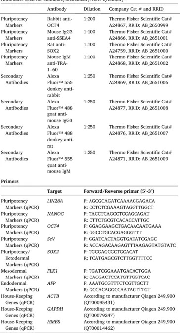
Table 3 Reagents details.
Antibodies used for immunocytochemistry/flow-cytometry
Antibody Dilution Company Cat # and RRID Pluripotency
Markers Rabbit anti-
OCT4 1:200 Thermo Fisher Scientific Cat#
A24867, RRID: AB_2650999 Pluripotency
Markers Mouse IgG3
anti-SSEA4 1:100 Thermo Fisher Scientific Cat#
A24866, RRID: AB_2651001 Pluripotency
Markers Rat anti-
SOX2 1:100 Thermo Fisher Scientific Cat#
A24759, RRID: AB_2651000 Pluripotency
Markers Mouse IgM anti-TRA- 1–60
1:100 Thermo Fisher Scientific Cat#
A24868, RRID: AB_2651002 Secondary
Antibodies Alexa Fluor™ 555 donkey anti- rabbit
1:250 Thermo Fisher Scientific Cat#
A24869, RRID: AB_2651006
Secondary
Antibodies Alexa Fluor™ 488 goat anti- mouse IgG3
1:250 Thermo Fisher Scientific Cat#
A24877, RRID: AB_2651008
Secondary
Antibodies Alexa Fluor™ 488 donkey anti- rat
1:250 Thermo Fisher Scientific Cat#
A24876, RRID: AB_2651007
Secondary
Antibodies Alexa Fluor™ 555 goat anti- mouse IgM
1:250 Thermo Fisher Scientific Cat#
A24871, RRID: AB_2651009
Primers
Target Forward/Reverse primer (5′-3′) Pluripotency
Markers (qPCR) LIN28A F: AGCGCAGATCAAAAGGAGACA R: CCTCTCGAAAGTAGGTTGGCT Pluripotency
Markers (qPCR) NANOG F: TACCTCAGCCTCCAGCAGAT R: CTTCTGCGTCACACCATTGC Pluripotency
Markers (qPCR) OCT4 F: GGAGGAAGCTGACAACAATGAAA R: GGCCTGCACGAGGGTTT Pluripotency
Markers (qPCR) SeV F: GGATCACTAGGTGATATCGAGC R: ACCAGACAAGAGTTTAAGAGTATGTATC Pluripotency/
Ectodermal Markers (qPCR)
SOX2 F: TGCGAGCGCTGCACAT
R: TCATGAGCGTCTTGGTTTTCC Mesodermal
Markers (qPCR) FLK1 F: TGATCGGAAATGACACTGGA R: CACGACTCCATGTTGGTCAC Endodermal
Markers (qPCR) AFP F: AAATGCGTTTCTCGTTGCTT R: GCCACAGGCCAATAGTTTGT House-Keeping
Genes (qPCR) ACTB According to manufacturer Qiagen 249,900 (QT00095431)
House-Keeping
Genes (qPCR) GAPDH According to manufacturer Qiagen 249,900 (QT00079247)
House-Keeping
Genes (qPCR) HMBS According to manufacturer Qiagen 249,900 (QT00014462)
L. Grossmann et al.