Functional and molecular analysis of cardiomyocytes derived from reprogrammed
pluripotent cells and embryonic stem cells
Inaugural-Dissertation Zur
Erlangung des Doktorgrades
der Mathematisch-Naturwissenschaftlichen Fakultät der Universität zu Köln
Vorgelegt von
Azra Fatima
Köln, Germany, 2011
Die Untersuchungen zur vorliegenden Arbeit wurden in der Zeit von September 2006 bis Mai 2011 am Institut für Neurophysiologie der Medizinischen Fakultät der Universität zu Köln unter der Leitung von Dr. Dr. Tomo Šarić durchgeführt.
This study was undertaken under the guidance of Dr. Dr. Tomo Šarić in the period between September 2006 and May 2011 at the Institute for Neurophysiology of the Medical faculty of the University of Cologne.
Vorsitzende des Promotionsausschusses: Prof. Dr. Angelika A. Noegel
Erstgutachter: Prof. Dr. Guenter Plickert
Zweitgutachter: Prof. Dr. Peter Kloppenburg
Date of Oral Exam 05.12.2011
DANKSAGUNGEN (ACKNOWLEDGEMENTS)
All praises and thanks for Almighty ALLAH who is the ultimate source of all knowledge to mankind, and who made me reach at the present pedestal of knowledge with quality of doing something adventurous, novel, thrilling and path bearing. All respects are for the Holy Prophet Muhammad (Peace be upon him) who is the symbol of guidance, fountain of knowledge, and made mankind to get out of depths of darkness.
I take this opportunity to express my gratitude to my mentor Dr. Dr. Tomo Šarić for his kind supervision, constructive criticism, valuable suggestions, sympathetic attitude, freedom of work, and ever inspiring guidance throughout the progress of this work and also for critically reviewing my work. It would not have been possible to make an interesting piece of research without his constant motivation throughout my graduate studies. His critical assessment and scientific demands has always kept me going.
I am greatly indebted to our Institute Director Prof. Jürgen Hescheler for providing excellent research facilities all the way.
Heartiest thanks to Prof. Guenter Plickert, and Prof. Peter Kloppenburg for providing their valuable time in reviewing my dissertation. I am extremely thankful to and Prof. Angelika A. Noegel for accepting to chair my thesis defense and to Dr.
Jens Schulze for accepting to assess my oral examination.
Special thanks to all my colleagues and friends with a special mention of Kurt, Markus, Filomain, Guoxing, Manoj, Narges, Vera, Birte, Dina, Matthias, Lukas and many others for sharing the enthusiasm of a lively work atmosphere. I deeply cherish the unconditional help and support of Devi, Naidu, Fakhera, Huamin, Katja, Marialice and Joiya through the early days of my Ph.D. Cordial thanks to all technical and official staff at the Institute specially Rebecca, Nadin, Evmorphia, Frau. Böttinger, Suzanne Wood and computational staff Mr. Stassen and Mr. Döweling.
At this point I remember my former mentor Dr. Gopal Pande from CCMB, India, who has always motivated me for an ambitious scientific career. The cooperation of our active collaborators has helped me in achieving my goals in this work.
I am forever indebted to my family, my mummy, my brothers, my sister and my little niece for their prayers, understanding and encouragement. My husband’s love and scientific cooperation is always cherished.
I thank the BMBF for financial support and the Graduate School of Biological Sciences, Köln for giving me opportunity and providing me with a wonderful thesis committee.
This thesis is dedicated to my father Mr. Mohammed Osman Siddiqui who has always been my inspiration. I lost him in my journey but will always cherish his inspirational words.
List of Publications
Highly purified cardiomyocytes generated by drug selection from murine induced pluripotent stem cells and embryonic stem cells are functionally and transcriptionally indistinguishable
Azra Fatima, Filomain Nguemo, Tobias Hannes, Guoxing Xu, Markus Khalil, Alexey Kuzmenkin Andrea Gaarz, Sabine Classen, Eugen Kolossov, Michael Reppel, Joachim L. Schultze, Jürgen Hescheler, Tomo Šarić . (Manuscript prepared).
In vitro modeling of ryanodine receptor 2 dysfunction using induced pluripotent stem cells
Azra Fatima, Guoxing Xu, Shao Kaifeng, Symeon Papadopoulos, Martin Lehmann, Juan-Jose Arnaiz Cot, Angelo O. Rosa, Filomain Nguemo, Sven Dittmann, Matthias Matzkies, Susannah L. Stone, Matthias Linke, Ulrich Zechner, Vera Beyer, Hans- Christian Hennies, Stephan Rosenkranz, Baerbel Klauke, Martin Farr, Abdul S.
Parwani, Wilhelm Haverkamp, Lars Cleemann, Martin Morad, Hendrik Milting, Jürgen Hescheler and Tomo Šarić. (In press)
Dual-color photoactivation localization microscopy of cardiomyopathy associated desmin mutants
Andreas Brodehl, Per Niklas Hedde, Mareike Dieding, Azra Fatima, Volker Walhorn, Susan Gayda, Tomo Šarić, Bärbel Klauke, Jan Gummert, Dario Anselmetti, Mike Heilemann, Gerd Ulrich Nienhaus, Hendrik Milting. (Under revision)
1. Comparison of contractile behaviour of native murine ventricular tissue and cardiomyocytes derived from embryonic or induced pluripotent stem cells.
Xi J, Khalil M, Shishechian N, Hannes T, Pfannkuche K, Liang H, Fatima A, Haustein M, Suhr F, Bloch W, Reppel M, Sarić T, Wernig M, Jaenisch R, Brockmeier K, Hescheler J, Pillekamp F. FASEB J. 2010 Aug; 24(8):2739-51.
2. Initial colony morphology-based selection for iPS cells derived from adult fibroblasts is substantially improved by temporary UTF1-based selection.
Pfannkuche K, Fatima A, Gupta MK, Dieterich R, Hescheler J. PLoS One. 2010 Mar 8; 5(3):e9580.
3. Functional characterization of cardiomyocytes derived from murine induced pluripotent stem cells in vitro.
Kuzmenkin A, Liang H, Xu G, Pfannkuche K, Eichhorn H, Fatima A, Luo H, Saric T, Wernig M, Jaenisch R, Hescheler J. FASEB J. 2009 Dec; 23(12):4168-80.
4. Cardiac myocytes derived from murine reprogrammed fibroblasts: intact hormonal regulation, cardiac ion channel expression and development of contractility.
Pfannkuche K, Liang H, Hannes T, Xi J, Fatima A, Nguemo F, Matzkies M, Wernig M, Jaenisch R, Pillekamp F, Halbach M, Schunkert H, Sarić T, Hescheler J, Reppel M.
Cell Physiol Biochem. 2009; 24(1-2):73-86.
5. Proteomic Analysis Of The “Side Population” (SP) Cells From Murine Bone Marrow
Ravipati Satyavani, Azra Fatima, Curam S. Sundaram, Charumathi Anabalagan, CV Saritha, G Srinivas, Aleem Ahmed Khan, CM Habibullah and Gopal Pande
Journal of Proteomics & Bioinformatics
http://scitechnol.com/journals/journal_fulltext.php?id=236&jid=34
Book Chapter
Embryonic Stem Cells, Cardiomyoplasty, and the Risk of Teratoma Formation Tomo Šarić, Lukas P. Frenzel, Azra Fatima, Manoj K. Gupta andJürgen Hescheler Trends in Stem Cell Biology and Technology 2009, 229-260.
CONTENTS
ABBREVIATIONS ABSTRACT
ZUSAMMENFASSUNG
1 INTRODUCTION________________________________________________ - 14 -
1.1 Pluripotent stem cells __________________________________________________ - 14 -
1.2 Developmental potential of pluripotent stem cells___________________________ - 16 - 1.3 Nuclear reprogramming________________________________________________ - 17 -
1.4 Strategies of nuclear reprogramming _____________________________________ - 18 - 1.4.1 Reprogramming by somatic cell nuclear transfer (SCNT)________________________ - 18 -
1.4.2 Reprogramming by fusion of somatic cells____________________________________ - 20 - 1.4.3 Nuclear reprogramming by cell extracts ______________________________________ - 23 - 1.4.4 Reprogramming by transcription-factor transduction____________________________ - 24 - 1.5 Advancement of methods for iPS cell generation ___________________________ - 26 - 1.5.1 Factor delivery into target cells______________________________________________ - 26 - 1.5.2 Integration-free iPS cells ___________________________________________________ - 28 - 1.5.3 Identification of iPS cell colonies_____________________________________________ - 31 -
1.6 Direct reprogramming: Conversion of one somatic-cell type to another ________ - 32 - 1.7 Epigenetics of reprogrammed cells _______________________________________ - 35 - 1.8 Differences of iPS cells and ES cells ______________________________________ - 37 -
1.9 Cardiac differentiation of pluripotent stem cells ____________________________ - 38 - 1.9.1 Purification of cardiomyocytes from differentiating pluripotent stem cell cultures____ - 40 - 1.10 Potential application of research on cardiomyocytes ________________________ - 41 - 1.10.1 A model system for study of embryonic development___________________________ - 41 - 1.10.2 Regenerative medicine_____________________________________________________ - 45 -
1.10.3 Drug discovery____________________________________________________________ - 47 - 1.10.4 Gene targeting to create reporter lines and disease models_____________________ - 49 - 2 AIM____________________________________________________________ - 51 - 3 MATERIALS AND METHODS ______________________________________ 52
3.1 Materials_______________________________________________________________ 52 3.1.1 Cell culture reagents__________________________________________________________52 3.1.2 Consumables________________________________________________________________53 3.1.3 Enzymes, Nucleotides and Markers_____________________________________________54 3.1.4 Kits_________________________________________________________________________54
3.1.5 Laboratory apparatus_________________________________________________________55 3.1.6 PCR primers used for SNP genotyping__________________________________________55 3.1.7 PCR primers used for gene expression analyses _________________________________57 3.1.8 Primary and secondary antibodies______________________________________________58 3.1.9 Buffers and Solutions_________________________________________________________59 3.1.10 Sterilization of Solutions and Equipments________________________________________60 3.2 Methods _______________________________________________________________ 61 3.2.1 Molecular Biology Methods____________________________________________________61 3.2.2 Cell Culture Techniques_______________________________________________________69 3.2.3 Immunoassaying, microscopy and flow cytometry_________________________________75 4 RESULTS ________________________________________________________ 77
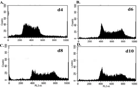
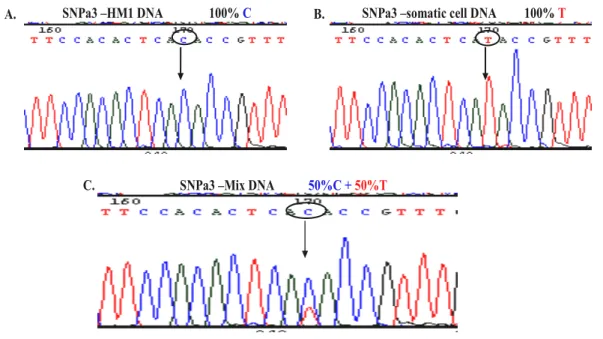
4.1 Reprogramming by fusion ________________________________________________ 77 4.1.1 Generation of fusion hybrid clones___________________________________________77 4.1.2 Ploidy of fusion hybrid cells____________________________________________________79 4.1.3 Confirmation of tetraploid character of fusion hybrid clones by SNP genotyping_______80 4.1.4 Expression of both parental MHC class I haplotypes on fusion hybrid cells ___________83 4.1.5 Expression of somatic CD markers on fusion hybrid cells __________________________87 4.1.6 Cardiac differentiation of fusion hybrid clones ____________________________________88 4.1.7 Electrophysiological properties of fusion hybrid-derived cardiomyocytes______________89
4.2 Advancement of methods for iPS cell generation _____________________________ 94 4.2.1 Instability of the morphology-based selected iPS cell line TiB7-4____________________94 4.2.2 UTF1-Neo selection of pluripotent iPS cell sub clones from TiB7.4 __________________94 4.2.3 Analysis of TiB7-4 sub clones derived from TiB7-4 with SV40-Neo selection__________97 4.3 Generation of transgenic iPS cells to allow purification of cardiomyocytes________ 99 4.3.1 Transgenic iPS cells retain their pluripotential characteristics______________________100 4.3.2 Transgenic iPS cells efficiently form EBs with a kinetic similar to that of ES cells_____101 4.3.3 Drug selection yields highly purified iPS-CMs ___________________________________102 4.3.4 Virally-encoded Oct4 is incompletely silenced in iPS-CMs_________________________106 4.3.5 Structure of cardiomyocytes derived from iPS and ES cells________________________108
4.3.6 Cardiomyocytes derived from iPS cells are not fully mature _______________________109 4.3.7 Intact β-adrenergic and muscarinic signalling in drug-selected iPS-CMs____________109 4.3.8 Functional voltage-gated ion channels in iPS-CMs_______________________________110 4.4 Global transcriptional profiling of iPS and ES cell-derived cardiomyocytes ______ 112 4.4.1 iPS-CMs and ES-CMs are transcriptionally highly similar__________________________112
4.4.2 Enriched functional categories in purified cardiac clusters_________________________115 5 D ISCUSSION____________________________________________________ 118 6 REFERENCES___________________________________________________ 126 7 ERKLÄRUNG____________________________________________________ 139 8 LEBENSLAUF___________________________________________________ 140
ABBREVIATIONS
AP Action potentials
BMP4 Bone morphogenetic protein 4
CCs Cardiac clusters
CMs Cardiomyocytes
cTnT cardiac troponin T
EBs Embryoid bodies
ES cells Embryonic stem cells
FH Fusion hybrid
GFP Green fluorescent protein
HAT Hypoxanthine, aminopterin and thymidines
ICM Inner cell mass
IFNγ Interferon gamma iPS Induced pluripotent stem
LIF Leukemia inhibitory factor MEFs Mouse embryonic fibroblasts
MHC Major histocompatibility complex MLC Myosin light chain
PEG Polyethylene glycol
PI Propidium iodide
SCNT Somatic cell nuclear transfer
SNP Single-nucleotide polymorphism SSEA1 Stage Specific Embryonic Antigen 1
TTF Tail tip fibroblasts
UTF Undifferentiated transcription factor1 α-MHC Alpha myosin heavy chain
α-PIG Vector in which puromycin N-acetyltransferase (PAC) and enhanced green fluorescent protein (eGFP) linked with the internal ribosomal entry site (IRES) are expressed under the control of α-myosin heavy chain (MHC) promoter
β-MHC β-myosin heavy chain
ABSTRACT
Pluripotent stem cells can be obtained from embryonic stage or generated by in vitro reprogramming of terminally differentiated somatic cells. Such reprogrammed cells possess similar developmental potential as embryonic stem (ES) cells and therefore are an indispensible source of diverse cell types, including cardiomyocytes (CMs), for basic research. CMs derived from ES cells and reprogrammed cells can provide possibilities for study of development, differentiation processes, stem cell malignancy and genetic diseases in vitro. Such studies can allow for development of novel compounds for drug discovery and toxicity testing which might help to develop more efficient and safer drugs
The present thesis is based on analyzing the impact of switching the cell fate on the differentiated state of reprogrammed cells using cardiomyocytes as a model. Detailed structural molecular and functional characterization of CMs derived from ES cells and reprogrammed cells like fusion hybrid (FH) cells and induced pluripotent stem (iPS) cells was performed.
Somatic cells (spleenocytes and bone marrow cells) were fused with ES cells to generate FH cells. The formed hybrid cells were morphologically similar to pluripotent ES cells and retained a tetraploid genome through many passages.
Spontaneous differentiation led to formation of embryoid bodies (EBs) with appearance of beating areas representing differentiation to cardiac lineage. The EBs derived from FH cells also retained tetraploid genome and expressed major histocompatibility (MHC) class I molecules of both fusing partners. FH derived CM expressed typical cardiac structural proteins and intact β-adrenergic and muscarinic signaling receptors. Atrial, ventricular and pacemaker cardiac subtypes could be found in FH-CMs. Thus, CMs derived from tetraploid FH cells appear to be structurally and functionally intact.
Ongoing research proved iPS cell technology to be more robust reprogramming strategy. In order to obtain homogenous population of CMs for further studies an iPS cell line TiB7.4 was genetically manipulated to allow for antibiotic-based purification of cardiomyocytes after spontaneous differentiation. We generated highly purified CMs from transgenic murine iPS and ES cell lines expressing puromycin N-
acetyltransferase and EGFP under the control of α-myosin heavy chain promoter. iPS and ES cells differentiated into CMs at comparable efficiencies yielding highly purified CMs after drug selection. Purified iPS- and ES-CMs exhibited indistinguishable structural properties, similarly responded to pharmacological agents, expressed functional voltage-gated sodium, calcium and potassium channels and possessed comparable current densities. Global transcriptional profile and gene ontology signature of transgenic iPS-CMs were very similar to that of ES-CMs but clearly distinct from fibroblasts that were used to generate iPS cells and differentiated cells in iPS or ES cell-derived embryoid bodies. After puromycin selection, iPS-CMs did not contain any residual pluripotent cells nor formed teratoma in immunodeficient mice.
Therefore, cell fate switching brought about by reprogramming does not affect the structural, molecular and functional characteristics of cardiac derivatives of the reprogrammed cells.
ZUSAMMENFASSUNG
Pluripotente Stammzellen können aus Embryonen gewonnen oder durch Reprogrammierung vollständig differenzierter somatischer Zellen in vitro generiert werden. Diese reprogrammierten Zellen besitzen ein ähnliches Differenzierungspotential wie embryonale Stammzellen (ES) und sind somit eine vielversprechende Quelle unterschiedlicher Zelltypen, wie Kardiomyozyten (KM), für die Grundlagenforschung. Die aus ES Zellen und reprogrammierten Zellen differenzierte KM, ermöglichen Untersuchungen von Entwicklungsprozessen, Mechanismen genetischer Krankheiten des Herzens, Entwicklung neuer Medikamente und Bestimmung ihrer Toxizität.
Die hier vorliegende Arbeit basiert auf der Analyse des Einflusses von Zellschicksalsänderungen auf den differenzierten Zustand von reprogrammierten Zellen.
Dabei wurde eine detaillierte strukturelle, molekulare und funktionale Charakterisierung der KM durchgeführt, die aus ES Zellen und reprogrammierten Zellen, wie Fusionshybrodoma (FH) Zellen und induzierten pluripotenten (iPS) Zellen, gewonnen wurden.
Die FH Zellen wurden durch Fusion somatischer Zellen der Milz oder des Knochenmarks mit ES Zellen generiert. Die erhaltenen Hybridzellen waren morphologisch pluripotent, aber behielten ein tetraploides Genom über mehrere Passagen. Spontane Differenzierung dieser Zellen führte zur Formation von
`embryoid bodies’ (EB), welche schlagende Areale und somit die Differenzierung zur kardialen Linie aufwiesen. Die EB der FH Zellen behielten ebenfalls ein tetraploides Genom und exprimierten MHC Klasse I Moleküle beider Fusionspartner.
Kardiomyozyten aus FH Zellen zeigten typische kardiale Strukturproteine auf und exprimierten intakte β-adrenerge und muskarine Signalwege. Obwohl die KM von tetraploiden FH Zellen generiert wurden, waren sie strukturell und funktionell normal.
Die gegenwärtige Forschung bewies, dass die iPS-Technologie die robustere Reprogrammierungsstrategie ist. Um KM besser zu analysieren, wurde eine transgene iPS Zelllinie generiert, welche Puromyzin-N-Acetyltransferase und EGFP unter der Kontrolle des Promoters für die schwere Kette des α-Myosin Gens exprimiert. Diese
Zelllinie ermöglichte die Aufreinigung einer homogenen Population der KM und somit die Analyse ihrer physiologischer und transkriptioneller Eigenschaften unter kontrollierten Bedingungen. Transgene iPS- und ES-Zellen differenzierten zu KM mit einer vergleichbaren Effizienz und lieferten nach Antibiotikaselektion hoch reine KM.
Gereinigte iPS- und ES-KM zeigten ähnliche strukturelle Eigenschaften, expremierten funktionelle spannungskontrollierte Natrium-, Kalzium- und Kaliumkanäle und besaßen eine vergleichbare elektrische Stromdichte. Globale Transkriptionsprofile und Genontologiesignaturen transgener iPS- und von ES-KM waren auch vergleichbar und unterschieden sich deutlich von den Fibroblasten, welche zur Generierung der iPS Zellen verwendet wurden. Nach Puromyzinselektion enthielten die iPS-KM weder verbliebene pluripotente Stammzellen noch bildeten sie Teratome in immundefizienten Mäusen.
Somit beeinflusst die Veränderung des Zellschicksals mittels Reporgrammierung weder die strukturellen noch molekularen oder funktionellen Charkateristika der von ihnen abgeleiteten kardialen Zellen.
1 INTRODUCTION
Development of a new organism begins with fertilization of an oocyte by the sperm leading to the formation of single cell called zygote. The zygote undergoes several rounds of cleavage to form a ball of cells called morula. Each cell in morula is called blastomere. The characteristic of the zygote and blastomere is a state of totipotency which marks their ability to give rise to all the different cells of an adult organism including the extra-embryonic membranes. The cells of the morula undergo more divisions to form a blastocyst, a hollow ball of cells in which the outer cells become trophoblast and develop into placenta. Some cells trapped in the interior of the blastocyst, termed the inner cell mass (ICM); generate the epiblast and the hyphoblast.
The epiblast and hyphoblast will form the embryo and the yolk sac, respectively. The cells of the ICM are termed pluripotent as they are able to form each of the three germ layers; the endoderm, the ectoderm and the mesoderm. These germ layers form distinct lineages of terminally differentiated post-mitotic cells, which contribute to specific organ function. As a fertilized egg develops into an adult organism, specialized cells are formed by a one-way process, and they become increasingly, and normally irreversibly, committed to their fate.
1.1 Pluripotent stem cells
Pluripotency is the potential of stem cells to give rise to any cell of the embryo proper. The study of pluripotent stem cells began with the study of teratocarcinomas (Evans and Kaufman 1981), which contain a haphazard array of almost any somatic cell type found in the developing embryo. The stem cells of these tumours are embryonal carcinoma (EC) cells, which express characteristics including a developmental potential similar to those of the ICM of the early embryo (Na et al.
2010). Mouse Embryonic stem (ES) cells were first pluripotent cells derived from the ICM of mouse blastocyst-stage embryos (Evans and Kaufman 1981). Human ES (hES) cells were isolated from the ICM of human blastocysts, generated by in vitro fertilization (IVF)-produced embryos that were no longer designated for clinical use, and donated by individuals after informed consent (Thomson et al. 1998).The importance of ES cells to modern biology and medicine derives from two unique distinguishing characteristics. First, they have the ability to be maintained in an
undifferentiated state (self renewal) indefinitely in culture. Second, they are pluripotent, possessing the capacity to generate every cell type in the body (Fig.1).
The pluripotent nature of mouse ES cells was formally demonstrated by their ability to contribute to all tissues of adult mice, including the germ-line, following their injection into host blastocyst (Bradley et al. 1984). ES cells are of great interest for both basic science and medicine. Studies on ES cells can provide information about the complex events that occur during embryonic development by identifying how undifferentiated stem cells become the differentiated cells that form the tissues and organs. ES cells provide opportunities for basic research on developmental gene regulation through functional genomics, signaling, molecular mechanisms of disease, drug screening and toxicological testing. As gene modulation techniques, including gene targeting, are well established in ES cells, it is relatively easy to identify a role of a specific gene in the development of a cell lineage (Keller et al. 1993).
Figure 1. Pluripotent stem cells, isolated from the ICM in the blastocyst, have the ability to give rise to all types of cells in the human body, but not the placenta and other supporting tissues. www.innovitaresearch.org/news/res/04121501_01.jpg
1.2 Developmental potential of pluripotent stem cells
In addition to their developmental potential in vivo, ES cells display a remarkable capacity to form differentiated cell types in vitro (Keller 1995; Smith 2001).Studies during the past 20 years have led to the development of appropriate culture conditions and protocols for the generation of a broad spectrum of lineages. Because of their ability to give rise to multiple lineages, ES cells open exciting new opportunities to model embryonic development in vitro for studying the events regulating the earliest stages of lineage induction and specification. Comparable studies are difficult in the mouse embryo and impossible in the human embryo.
When injected into immuno-compromised animals, pluripotent cells form teratomas composed of multiple differentiated tissues in all three germ layers: ectoderm, mesoderm, and endoderm including the germ line of chimeric animals. Tetraploid complementation assay test is the most stringent test for the pluripotency of ES cells.
In this test a tetraploid embryo, incapable of developing beyond blastocyst stage, is fused with diploid ES cells. The resulting embryo develops normally wherein the fetus is exclusively derived from ES cells and the extra embryonic tissues develop exclusively from tetraploid embryo. In addition to these the in vivo tests, ES cells are able to differentiate into multiple cell types in vitro. When removed from the factors that maintain them as stem cells, ES cells will differentiate and, under appropriate conditions, generate progeny consisting of derivatives of the three embryonic germ layers: mesoderm, endoderm, and ectoderm ES cells can be differentiated in the form of three-dimensional aggregates, known as embryoid bodies (EBs), containing various cell types like cardiac myocytes, neuronal cells, erythrocytes, melanocytes, and others (Doetschman et al. 1985; Yamane et al. 1999; Lee et al. 2000).
The mouse ES cell differentiation system can be regarded as an unlimited source of cells and tissues for a broad spectrum of research. To date, mouse ES cells have been successfully differentiated into hematopoietic precursors (Potocnik et al. 1997), CMs (Yamashita et al. 2000), neural precursors (Nakano et al. 1996; Brustle et al. 1997;
Garrington et al. 2000), insulin-producing cells (Klug et al. 1996), and mast cells (Tsai et al. 2000) and have been transplanted successfully into recipient animals. In
vitro differentiation of ES cells has been used in basic science to study gene expression during development of specific cell types.
Studies of pluripotent mouse ES cells have led to in vitro models of cardiomyocyte differentiation. Much of what we know about the differentiation of pluripotent ES cells to CMs in vitro has been learned from studies with mouse ES cells.
Morphological, electrophysiological, and molecular techniques indicate that the in vitro differentiation process recapitulates the developmental pattern of early cardiogenesis, and genetic studies have shown how signaling molecules, transcription factors, ECM components, and calcium-handling proteins affect this process.
Genomic studies, coupled with in vitro differentiation, have also led to the identification of developmentally regulated genes (gene trapping) and to the identification of differentiation-responsive genes.
The potential use of ES cells for research is challenged by ethical considerations and more so due to imposition of strict regulations. Although ES cells are regarded as a
‘gold standard’ for pluripotency, there is a need for alternate sources of pluripotent cells which can be easily accessible and possess the same developmental potential as ES cells.
1.3 Nuclear reprogramming
Embryonic development is a unidirectional process characterized by constant loss of developmental potential with progress in time. Cells progress from totipotency to pluripotency, multipotency and then finally commit to differentiated cell fates.
Genomic content of totipotent zygote is identical to that of its pluripotent progeny and terminally differentiated somatic cells. This content remains unchanged throughout development. Thus the difference in developmental potential of these cell types is imposed by a cell specific transcriptional state that is ultimately dependent on epigenetic changes of genome. The cell fate of a defined specialized cell type can be reversed, returning the cell to an embryonic state. Reprogramming is a term used to describe the process that reverts nuclear gene expression of fully differentiated somatic cells to a pluripotent state. This process is of interest for three reasons.
Understanding of reprogramming process can help us to understand how cell differentiation and specialized gene expression are normally maintained. Nuclear
reprogramming enables the culture of lines of cells from diseased tissues, and hence allows us to analyze the nature of the disease and to screen for therapeutic drugs. It can open several new opportunities of in vitro disease models to help in understanding disease mechanisms thus leading to devising new strategies for treatment. Nuclear reprogramming could represent a first major step in cell-replacement therapy, providing replacement of heart, pancreas, or other types of cells from the skin of the same individual.
1.4 Strategies of nuclear reprogramming
1.4.1 Reprogramming by somatic cell nuclear transfer (SCNT)
Nuclear reprogramming is initiated when a nucleus from a differentiated somatic cell is transplanted into an enucleated oocyte leading to the generation of a live offspring, which is a genetically identical clone of the original somatic cell. Such nuclear- transfer experiments, also known as cloning, have shown definitively that all of the genes required to create an entire organism are present in the nucleus of the specialized cell and can be activated on exposure to reprogramming factors present in the oocyte. Briggs and King (Briggs and King 1952) first succeeded in producing normal swimming tadpoles of Rana pipiens by transplanting the nuclei of embryo (blastula) cells into enucleated eggs. Similar experiments were carried out with enucleated eggs of Xenopus laevis (Gurdon and Uehlinger 1966), which were transplanted with the nuclei of intestinal epithelium of feeding tadpoles to obtain entirely normal and fertile male and female frogs. The major conclusion from these and subsequent findings was that development imposes reversible epigenetic rather than irreversible genetic changes on the genome during cellular differentiation. The first successful cloning of a higher animal using SCNT was demonstrated by the cloning of normal adult sheep (Dolly) by transplanting the nuclei of cultured mammary gland cells derived from an adult sheep to enucleated sheep eggs (Wilmut et al. 1997). Cloning experiments have been successfully performed with different kinds of terminally differentiated cells, such as T lymphocytes (Hochedlinger and Jaenisch 2002) post-mitotic olfactory neurons (Eggan et al. 2004), natural killer T cells (Inoue et al. 2005), peripheral blood granulocytes, and malignant melanoma (Hochedlinger, 2004). In addition to sheep and mice, a wide range of species have
now been successfully cloned using SCNT, ranging from domesticated animals such as dogs and goats, and their hybrids such as mules, to wild animals such as African wildcats and wolves (Gomez et al. 2004). Extinct animals could also be successfully cloned by transplantation of nuclei from frozen cells into enucleated oocytes a decade after tissue freezing (Wakayama et al. 2008). However, most cloned animals with apparently normal gross anatomy can have numerous abnormalities. Common abnormalities include aberrant gene expression in embryos, telomere elongation, obesity in adults, impaired immune systems and, often, increased cancer susceptibility and premature death (Gomez et al. 2004)
SCNT ES cells have been derived from an immunodeficient mouse and then used in vitro to correct the original genetic defect by homologous recombination.
Subsequently, the repaired SCNT ES cells were differentiated into hematopoietic stem cells and transplanted back to the immunodeficient donor mouse to restore lymphopoiesis (Rideout et al. 2002). Although no human SCNT ES cell lines have been created yet, the successful recent generation of SCNT ES cell lines in primates (Byrne et al. 2007) suggests that therapeutic cloning in humans may be feasible in the future. Fig. 2 shows the process of the derivation of ES cells from a blastocyst derived from oocyte which has received nucleus from somatic cell.
To increase the frequency of cloned animals various technical modifications have been tested in mice, including attempts at chemically activating oocytes to make them more responsive, changing the time of enucleation, inhibiting cytokinesis and using cell fusion instead of nuclear injection, but these alterations have led to only modest results of 1-3% increase (Thuan et al. 2010).
Figure 2. Schematic representation of the technique of somatic cell nuclear transfer (SCNT). Yamanaka and Blau; Nature 465, 704–712.
The need for a large number of donated human oocytes, ethical concerns and extremely low cloning efficiency by SCNT in mammals render this approach impractical. Nuclear transfers between different strains or subspecies are just as successful as those within a species; however, eggs produced by transfers between very different species such as human, mouse, cow, or pig generally die before the 32- cell stage (Tecirlioglu et al. 2006). So far, there is no confirmed evidence that proliferating ES cells can be obtained from such distant combinations, including human nuclei in monkey cytoplasm.
1.4.2 Reprogramming by fusion of somatic cells
Cell fusion involves fusing two or more cell types to form a single entity. The possibility that two different cell types can fuse, known as heterotypic fusion, and form a somatic cell hybrid was initially suggested back in 1965 (Harris and Watkins 1965). The hybrid cells were called ‘heterokaryons’. Cell-cell fusion between pluripotent teratocarcinoma and differentiated thymocyte cells resulted in hybrid cells maintaining the potential for unlimited self renewal and differentiation into a variety of cell types. The authors had hypothesized that the teratocarcinoma might lose pluripotency by fusion with differentiated somatic cells, but instead, the hybrid cells obtained pluripotency and resembled EC cell morphology without expressing a tissue specific gene such as Thy (Miller and Ruddle 1976; Miller and Ruddle 1977).
Following this initial study, many studies have shown that various somatic cells can be reprogrammed by fusing with pluripotent stem cells like ES, EG, or EC cells (Matveeva et al. 1998; Tada et al. 2001; Flasza et al. 2003; Cowan et al. 2005).Cell fusion also was suggested as a mechanism for somatic cell plasticity. Phenotype and potency of somatic cells (bone marrow cells and brain cell) were changed by spontaneous cell fusion with ES cells after co-culture with ES cells (Terada et al., 2002, Ying et al., 2002). The hybrid cells that showed over-diploid DNA content expressed pluripotency related genes and could differentiate into all three embryonic germ layer in vivo and in vitro.
EC cells are another source of pluripotent cells that can reprogram somatic cells by fusion. Since EC cells share many of the key pluripotent characteristics with ES therefore EC cells can provide a readily amenable alternative source for
reprogramming (Do et al., 2009a; Flasza et al., 2003; Mise et al., 1996). Moreover, mouse EC cells can reprogram human somatic cells into pluripotent state, indicating that reprogramming factors can cross-act through another species (Flasza et al., 2003).
The fusion hybrid cells present pluripotential characteristics, such as inactivation of tissue-specific genes, reactivation of pluripotency related genes, differentiation potential to all three germ layers, and a specific epigenetic state corresponding to the pluripotent cells (Do et al., 2006). The differentiated state of somatic cells could also be altered by fusion with another type of somatic cell, suggesting that cellular factors between the two different types of cells dynamically interact and might be responsible for the plasticity and re-establishment of new characteristics. However, the fusion hybrid cells are not identical to the pluripotent fusion partner cells.
When somatic cells acquire pluripotency through cell-cell fusion with pluripotent stem cells, the reprogrammed hybrid cells express pluripotency-related genes but did not express tissue-specific genes. The ‘memory’ of somatic cells is almost like a dogma considered to be erased by fusion with pluripotent cells during fusion-induced pluripotential reprogramming. Fusion-induced reprogramming was thought to be a unidirectional process resulting in an ES cell phenotype without other viable cell states (Silva et al., 2006). But Do et al in 2009 demonstrated that pluripotent stem cells also could acquire some characteristics of differentiated cells indicating that the reprogramming direction in pluripotent hybrid cells is not solely unidirectional, and some genes could be reprogrammed opposite to that of the pluripotent fusion partner (Do et al. 2009).
Under differentiation-inducing conditions, fusion hybrid cells exhibit similar differentiation potential of their pluripotent fusion partner, and are not preferentially committed to the lineage of the somatic cells that had been fused with the pluripotent cells. Inability for neural differentiation of the F9- neural stem cells (NSC) fusion hybrid cells demonstrated that NSCs lose their memory and adopt the similar differentiation potential of F9 cells. Therefore, the differentiation potential of fusion hybrid cells is contingent on the type of pluripotent fusion partner cells, and the resulting hybrid cells have the same potential as the pluripotent fusion partner cells.
Pluripotent cells reprogram somatic cells and may induce erasure of somatic cell memory by fusion.
Figure 3. Schematic representation of reprogramming brought about fusion of somatic cells with ES cells leading to formation of fusion hybrids. Yamanaka and Blau; Nature 465,704-712.
Major disadvantage of this approach of reprogramming is that it leads to tetraploid cells or aneuploid cells (Fig. 3). Variable chromosome number ranging from 40-85 was observed in hybrids between mouse spleenocytes and murine ES cells due to extensive loss or gain of individual chromosomes. This indicates that spontaneous segregation leading to diploid genome in the hybrid is not achieved (Matveeva et al.
1998).
Experiments into cell fusion mediated by PEG have provided important information about the molecules and pathways that have been implicated in the regulation of reprogramming. Specifically, it has been seen that over-expression of the embryonic factor Nanog in ES cells, or Sall4 in mouse embryonic fibroblasts (MEFs), can increase the number of reprogrammed somatic cells after fusion (Wong et al. 2008;
Silva et al. 2009). On the other hand, the periodic activation of the Wnt/β-catenin signaling pathway in ES cells and the activation of Akt signaling remarkably enhance cell-fusion-mediated reprogramming (Lluis, 2008; Nakamura, 2008). Interestingly, in ES cells over-expression of Nanog, and at the same time activation of the Wnt/β- catenin pathway, can enhance the reprogramming efficiency of somatic cells even further after fusion (Lluis and Cosma 2009).The method of cell fusion might be regarded superior to SCNT, as it is technically less challenging and does not require the use of oocytes and pre-implantation embryos.
Fusion hybrid cells can be used to study gene expression, basics of cell division, and transformation of normal cells to malignant cells, obtain viral replication, chromosome or gene mapping, production of monoclonal antibodies and as a tool for investigating the mechanism of cellular plasticity.
1.4.3 Nuclear reprogramming by cell extracts
In this technique of reprogramming, the plasma membrane of an adult cell to be reprogrammed is reversibly permeabilized with the bacterial toxin Streptolysin O.
Permeabilized cells are then incubated with a nuclear and cytoplasmic extract derived from another type of cell. After transient exposure to extract, cells are resealed and cultured (Fig. 4). During incubation, factors in the cell extract required for reprogramming diffuse into permeabilized cells and activate genes that are typically expressed in cells from which the extract has been prepared. This principle has been used to reprogram kidney epithelial 293T cells to express T lymphocyte or neuronal markers by incubating them with an extract prepared from T cells or neuronal precursor cells, respectively (Hakelien et al. 2002). However, although reprogrammed fibroblasts expressed T cell specific surface markers, such as CD3, CD4, CD8, CD45 and T cell receptor (TCR) αβ chains, and a T cell specific function, the reprogramming of the 293T cells into T cells was not complete, as the reprogrammed cells did not express a pure T cell specific phenotype and the expression profile of many genes did not match that of T cells. In addition to human 293T cells, the cell extract based reprogramming approach was also applied to other mammalian cells such as human adipose tissue stem cells, which adopted cardiomyocyte properties following transient exposure to rat cardiomyocyte extract (Gaustad et al. 2004).
Figure 4: Reprogramming by cell extracts. Yamanaka and Blau; Nature 465. 704-712.
At present it is unclear if complete and stable reprogramming to a pluripotent state can be achieved with crude cell extracts and if reprogramming efficiencies would be different with different types of cells. However, this method is very attractive because cell extract derived factors are presumably not permanently active in target cells but turn over at kinetics corresponding to their half lives. This approach may prove useful for dissecting the molecular machinery involved in reprogramming using biochemical methods (Hansis et al. 2004).
1.4.4 Reprogramming by transcription-factor transduction
Successful reprogramming of somatic cells by fusion with ES cells indicates that ES cells have factors that induce pluripotency. It seemed likely that these pluripotency- inducing factors also play important roles in the maintenance of pluripotency. Based on this hypothesis, Yamanaka and Takahashi devised an elegant screen for factors within a pool of 24 pluripotency-associated candidate genes and tested for their ability to induce pluripotency and activate a dormant drug resistant allele integrated into the ES cell-specific Fbx15 locus. The combination of 24 factors, when co-expressed from retroviral vectors in mouse fibroblasts, indeed activated Fbx15 and induced the formation of drug-resistant colonies with characteristic ES cell morphology (Takahashi and Yamanaka, 2006). Successive rounds of elimination of individual factors then led to the identification of the minimally required core set of four genes, comprising Klf4, Sox2, c-Myc, and Oct4.
Retrovirus-mediated introduction of four transcription factors (Oct-3/4, Sox2, c-Myc, and Klf4) into mouse embryonic or adult fibroblasts and selection for the expression of Fbx15, a target of Oct-3/4 and Sox2, resulted in the generation of induced pluripotent stem (iPS) cells, which are similar to ES cells in morphology, proliferation, and teratoma formation (Takahashi and Yamanaka 2006) (Fig. 5). The selected iPS cells are, however, significantly different from ES cells in gene expression and DNA methylation patterns. When transplanted into blastocysts, these iPS cells only give rise to chimeric embryos, but not adult or germ line competent chimeras. These data indicate that reprogramming in Fbx15-selected iPS cells is incomplete. These “first-generation” iPS cells therefore appeared to be only partially reprogrammed.
Although both Fbx15 and Nanog are targets of Oct-3/4 and Sox2, the former is dispensable for pluripotency, while the latter plays crucial roles. By selecting for the reactivation of the essential pluripotency genes Nanog or Oct4 instead of Fbx15, iPS cells were generated that molecularly and functionally more closely resembled ES cells (Maherali et al. 2007; Okita et al. 2007; Wernig et al. 2007). Nanog-selected iPS cells showed reactivation of a somatically silenced X chromosome and underwent random X-inactivation upon differentiation (Maherali et al. 2007). These data demonstrated that full reprogramming can be achieved by expression of the four factors and using an appropriate selection procedure.
iPS cells have also been derived from a number of different species-including humans (Takahashi et al. 2007; Yu et al. 2007; Park et al. 2008; Li et al. 2009), and rhesus monkeys (Liu et al. 2008) by expression of the four Yamanaka factors demonstrating that fundamental features of the transcriptional network governing pluripotency remain conserved during evolution. Similarly, iPS cells have been derived from other somatic cell populations, such as keratinocytes (Maherali et al. 2007; Aasen et al.
2008), neural progenitor cells (Eminli et al. 2008), stomach and liver cells (Aoi et al.
2008) and melanocytes (Utikal et al. 2009), as well as from genetically labeled pancreatic β cells (Stadtfeld et al. 2008) and terminally differentiated lymphocytes (Hanna et al. 2008; Eminli et al. 2009), further underscoring the universality of induced pluripotency.
Figure 5. Induction of pluripotency in adult cells by retroviral transduction of transcription factors. Yamanaka and Blau; Nature 465, 704–712.
The unique properties of ES and iPS cells also provide for practical approaches in pharmaceutical toxicology and pharmacogenomics. In particular, hepato-toxicity and cardiotoxicity are two principal causes of drug failure during preclinical testing, while the variability in individual responses to potential therapeutic agents is also a major problem in effective drug development (Rubin 2008). The advantage of iPS cell technology is that it allows for the first time the generation of a library of cell lines that may to a substantial extent represent the genetic and potentially epigenetic variation of a broad spectrum of the population. The use of this tool in high- throughput screening assays could allow better prediction of the toxicology caused by and therapeutic responses induced by newly developed dugs and offer insight into the underlying mechanisms. The net result of this approach would substantially decrease the risk and cost associated with early-stage clinical trials and could lead toward a more personalized approach in drug administration.
iPS cell derivation is ethically and legally less problematic and technically more feasible than SCNT. In order to use iPS cells as efficient research tools suitable techniques of factor delivery and efficient identification of faithfully reprogrammed cells are crucial.
1.5 Advancement of methods for iPS cell generation
1.5.1 Factor delivery into target cells
A number of different approaches have been devised to shuttle reprogramming factors into somatic cells, which can affect the efficiency of reprogramming and the quality of resultant iPS cells. The first studies on iPS cells used constitutively active retroviral vectors that stably integrated into the host cell genome to introduce c-Myc, Klf4, Oct4, and Sox2 (Takahashi and Yamanaka 2006; Wernig et al. 2007; Wilber et al. 2007).
While retroviral transgenes are usually silenced toward the end of reprogramming (Stadtfeld et al. 2008), this process is often incomplete, resulting in partially reprogrammed cell lines that continue to depend on exogenous factor expression and fail to activate the corresponding endogenous genes. In addition, residual activity or reactivation of viral transgenes in iPS cell-derived somatic cells can interfere with their developmental potential and frequently leads to the formation of tumors in chimeric animals (Okita et al. 2007). When constitutively active lentiviral vectors are
used to produce iPS cells, which are even less efficiently silenced in pluripotent cells than retroviral vectors, it can lead to differentiation block (Brambrink et al. 2008;
Sommer et al. 2010). The use of inducible lentiviral vectors, whose expression can be controlled by the inert drug doxycycline, diminishes the risk of continued transgene expression and allows for the selection of fully reprogrammed iPS cells, since cells that depend on exogenous factor expression readily stop proliferating upon doxycycline withdrawal (Brambrink et al. 2008; Stadtfeld et al. 2008). Lentiviral vectors are also more efficient than retroviral vectors at infecting different somatic cell types and can be used to express polycistronic cassettes encoding all four reprogramming factors, thus increasing reprogramming efficiency ((Carey et al. 2009;
Sommer et al. 2009; Sommer et al. 2010). Inducible vector systems have been employed to generate so-called “secondary” reprogramming systems, which do not rely on direct factor delivery into target cells. These systems entail differentiating
“primary” iPS cell clones, generated with doxycycline-inducible lentiviral vectors or transposons, into genetically homogeneous somatic cells using either in vitro differentiation (for human cells) (Hockemeyer et al. 2008) or blastocyst injection (for mice) (Wernig et al. 2007; Woltjen et al. 2009). These somatic cells are then cultured in doxycycline-containing media, thus triggering the formation of “secondary” iPS cells at efficiencies that depend on the specific cell type used but are generally several orders of magnitude higher than the efficiencies obtained after primary infection.
Secondary systems therefore (1) allow for the reprogramming of large quantities of genetically homogeneous cells for biochemical studies and cells that are difficult to culture or transduce, and (2) facilitate the comparison of genetically matched iPS cells derived from different somatic cell types. The recent development of
“reprogrammable” mouse strains, which contain a single inducible polycistronic transgene in a defined genomic position enables the breeding of animals into desired mutant backgrounds for mechanistic studies (Carey et al. 2009).
1.5.2 Integration-free iPS cells
Potentially harmful effects of leaky transgene expression and insertional mutagenesis can be overcome by producing transgene free iPS cells. Techniques to generate integration-free iPS cells can be subdivided into three categories (Fig. 6):
(a) Non integrating viruses. Human fibroblasts have also been reprogrammed into iPS cells with adenoviral vectors (Zhou and Freed 2009) and Sendai virus (Fusaki et al. 2009), as well as with polycistronic minicircle vectors (Jia et al. 2010) and self- replicating selectable episomes (Yu et al. 2011), albeit the latter system required the simultaneous over-expression of additional factors, including another potent oncogene (Oct4, Sox2, c-Myc, and Klf4, together with Nanog, Lin28, and SV40 large T antigen).
Reprogramming efficiencies with current non-integrating methods are several orders of magnitude lower (~0.001%) than those achieved with integrating vectors (0.1%–
1%); most likely because factor expression is not maintained for a sufficient length of time to allow complete epigenetic remodeling.
(b) Integrating vectors that can be excised: Several laboratories have developed integration-dependent gene delivery vectors with incorporated loxP sites that can be subsequently excised from the host genome by transient expression of Cre recombinase (Kaji et al. 2009; Soldner et al. 2009). This approach enables the efficient generation of iPS cells from different cell types. The use of polycistronic vectors have especially shown to be very useful (Chang et al. 2009; Sommer et al.
2009; Sommer et al. 2010). Transgene-free iPS cells can also be generated with piggyBac transposons, mobile genetic elements that can be introduced into and removed from the host genome by transient expression of transposase (Woltjen et al.
2009; Yusa et al. 2009). It remains unclear if transposase expression can induce nonspecific genomic alterations in iPS cells (Stadtfeld and Hochedlinger 2009).
Figure 6. Different approaches to generate iPS cells. Modified from Lowry and Plath, Nature Biotechnology 26, 1246 - 1248 (2008).
(c) non nucleic acid reprogrammers: iPS cells have been derived from both mouse and human fibroblasts by delivering the reprogramming factors as purified recombinant proteins (Zhou and Freed 2009) or as whole-cell extracts isolated from either ES cells (Cho et al. 2010) or EK293 cells genetically engineered to stably express reprogramming factors fused with 9 arginine and myc tag (Kim et al. 2009).
While the use of purified proteins represents an attractive approach for the generation of transgene-free iPS cells, its efficiency is extremely low and requires chemical compounds that promote reprogramming. Small molecules were utilized that could significantly increase reprogramming efficiencies in the context of Oct4, Klf4, Sox2, and c-Myc over expression ((Desponts and Ding 2010). Notably, some of these molecules can also replace individual reprogramming factors, raising the possibility of deriving iPS cells solely with chemicals. Chemical substitution of a reprogramming factor is, in most cases, associated with a significant decrease in the number of iPS cells clones generated, indicating that no single chemical compound is able to entirely replace the function of a transcription factor. Another potential caveat of chemical reprogramming approaches is the introduction of genetic or epigenetic abnormalities into resultant iPS cells, especially since many of the reported compounds are potent modulators of DNA and chromatin modifications.
A more efficient and safer way of producing integration-free iPS cells may be the introduction of modified RNA molecules encoding for the reprogramming factors into somatic cells, which has been validated recently (Warren et al. 2010).These authors have demonstrated a simple, non-integrating strategy for reprogramming cell fate based on administration of synthetic mRNA modified to overcome innate antiviral responses. The modified RNA induced pluripotent cells thus obtained were termed as RiPSC.
In earlier reprogramming strategies, microRNAs (miRNAs) were used to promote the transcription factor-mediated reprogramming process but recently two independent groups have derived human and mouse iPS cells by adding miRNAs, in the absence of any additional protein factors (Anokye-Danso et al. 2011) shows that expression of a single primary miRNA transcript, the miR-302/367 cluster, is in rapidly and efficiently reprograms mouse and human somatic cells to an iPS cell state without a requirement for exogenous transcription factors. The resulting iPS cells exhibit gene expression and functional properties characteristic of fully reprogrammed pluripotent cells. Approximately 10% of fibroblasts form iPS cell colonies, an improvement in efficiency of >100-fold compared with OSKM. Moreover, the appearance of iPS cell colonies and the activation of pluripotency markers occur sooner. Miyoshi et al also demonstrated the possibility of reprogramming human and murine fibroblasts by transfection of a combination of mature double-stranded microRNAs (miRNAs) mir- 200c plus mir-302 s and mir-369 s family miRNAs(Miyoshi et al. 2011).
The unique properties of ES and iPS cells also provide for practical approaches in pharmaceutical toxicology and pharmacogenomics. In particular, hepatotoxicity and cardiotoxicity are two principal causes of drug failure during preclinical testing, while the variability in individual responses to potential therapeutic agents is also a major problem in effective drug development. The advantage of iPS cell technology is that it allows for the first time the generation of a library of cell lines that may, to a substantial extent, represent the genetic and potentially epigenetic variation of a broad spectrum of the population. The use of this tool in high-throughput screening assays could allow better prediction of the toxicology caused by and therapeutic responses induced by newly developed dugs and offer insight into the underlying mechanisms.
The net result of this approach would substantially decrease the risk and cost