Brain Microenvironment and its Influence on Gene Expression and Differentiation of Murine Embryonic Stem Cells – Implications
for Cell Replacement Strategies
I n a u g u r a l - D i s s e r t a t i o n
zur Erlangung des Doktorgrades
der Mathematisch-Naturwissenschaftlichen Fakultät der Universität zu Köln
vorgelegt von
Kristine Bentz
aus Landau
2
Berichterstatter: Prof. Dr. S. Korsching
Prof. Dr. E.A.M. Neugebauer
Tag der mündlichen Prüfung: 15. Februar 2006
Für meine Eltern
Abstract
4
A
BSTRACTThe discovery of pluripotent embryonic stem (ES) cells more than 20 years ago marked the beginning of a new era in developmental biology and biomedical research. The outstanding endogenous attributes of embryonic stem cells and the availability of in vitro ES cell differentiation protocols emphasise the great potential of these cells for clinical application in CNS disorders. Particularly for the treatment of disease states in which no clinical therapy has proven effective to date, such as traumatic brain injury, stem cell-based cell replacement strategies are advancing. However, in animal models of traumatic brain injury it has been shown that soluble signalling molecules, extracellular matrix-associated activities and the cellular environment at the site of implantation exert great influence on engrafted stem cells and can be decisive for success or failure of functional engraftment.
This doctoral thesis aims at elucidating the role of naïve and pathological environmental conditions on plasticity of murine embryonic stem cells. Utilising a sophisticated in vitro approach i) the influence of endothelial and astroglial cells on stem cell self-renewal and differentiation capacity, ii) the impact of traumatic brain injury-associated environment on stem cell fate and iii) the source of motor functional improvements, observed after stem cell transplantation into the traumatically injured brain in vivo were investigated. To achieve this, embryonic stem cells were co-cultured with cerebral endothelial or astroglial cells, both belonging to the neural stem cell niche in vivo, or treated with tissue extract derived from traumatised or healthy rat brain, respectively. Subsequently, embryonic stem cells were analysed for expression of developmental marker genes (RT-PCR), mitotic activity (FACS), production of neurotrophic factors (ELISA) and morphological appearance (microscopy). Results indicate that environmental conditions, both on a cellular and molecular level, distinctively manipulate stem cell fate. It is demonstrated that neuronal lineage commitment in ES cells is mainly mediated via humoral factors present in conditioned medium, whereas the vicinity of living endothelial cells and astrocytes delays differentiation activities and instead promotes oligodendroglial cell fates.
Furthermore, it is shown that traumatic brain injury-related parameters support the rapid
differentiation of single stem cells to mature neuronal phenotypes. However, many cells are lost
due to the deleterious effects of trauma environment. Moreover, cerebral tissue extract is shown
to either enhance or restrict the release of trophic factors by embryonic stem cells, and
additionally, has the ability to induce growth factor production in fibroblasts. Thus, co-
transplanted fibroblasts are proposed to represent an alternative source for functional recovery
observed after ES cell transplantation. With its findings, this study tries to bridge a gap between
basic biological research and clinical science in a common effort to aid the human patient finally.
Z
USAMMENFASSUNGDie erstmalige Isolation pluripotenter embryonaler Stammzellen vor mehr als 20 Jahren kennzeichnete weltweit den Beginn einer neuen Ära in der entwicklungsbiologischen und medizinischen Forschung. Die außergewöhnlichen endogenen Eigenschaften embryonaler Stammzellen, ihre Eignung zu unbegrenzter Vermehrung in vitro und ihre Fähigkeit Zellen verschiedensten Gewebeursprungs hervorzubringen, unterstreichen das große Potential dieser Zellen für eine klinische Anwendung. Zellersatz durch Stammzelltransplantation eröffnet vor allem bei ZNS-Verletzungen, wie z.B. bei Schädel-Hirn-Trauma, neue Behandlungsmöglichkeiten. Es konnte jedoch gezeigt werden, dass die Interaktion transplantierter Zellen mit humoralen Faktoren, extrazellulären Matrixmolekülen und dem zellulären Umfeld vor Ort von entscheidender Bedeutung für Erfolg oder Misserfolg einer durchgeführten Transplantationstherapie sein kann.
Ziel dieser Dissertation ist es, den Einfluss des extrazellulären Milieus, unter gesunden und pathologischen Bedingungen, auf die Plastizität muriner embryonaler Stammzellen aufzuklären.
Mittels eines in vitro-Modells wurde i) der Einfluss der lokalen Umgebung, der so genannten Nische, auf Selbsterhaltung und Differenzierung embryonaler Stammzellen untersucht, ii) die Auswirkungen pathologischer Umwelteinflüsse auf das Zellschicksal embryonaler Stammzellen erforscht und iii) die Ursache funktionaler Verbesserungen, die nach Transplantation von Stammzellen in vivo beobachtet wurden, analysiert. Dazu wurden murine embryonale Stammzellen entweder mit zerebralen Endothelzellen oder Astroglia co-kultiviert, oder mit Gewebeextrakt behandelt, der von gesundem bzw. Schädel-Hirn-traumatisiertem Rattenhirn gewonnen wurde. Anschließend wurden die Stammzellen auf die Expression verschiedener entwicklungsspezifischer Gene (RT-PCR), auf ihre Proliferationsfähigkeit und ihre Fähigkeit zur Produktion neurotropher Faktoren (ELISA), sowie auf sichtbare morphologische Veränderungen (Mikroskopie) untersucht. Die Ergebnisse dieser Studie zeigen, dass zelluläre und molekulare Umgebungsbedingungen das Schicksal embryonaler Stammzellen unterschiedlich beeinflussen.
Während die Induktion neuronaler Differenzierungsvorgängen vornehmlich über humorale
Signale gesteuert wird, die in konditioniertem Medium vorhanden sind, werden
Differenzierungsvorgänge durch in unmittelbarer Nähe wachsende Endothelzellen und
Astrozyten verlangsamt. Außerdem bevorzugen in Co-Kultur gewachsene Stammzellen ein
gliales Zellschicksal. Des Weiteren kann unter Einfluss eines Schädel-Hirn-Traumas, im
Gegensatz zu nativen Bedingungen, eine beschleunigte neuronale Differenzierung einzelner
Stammzellen beobachtet werden. Jedoch kommt es durch die Einwirkung verletzungsbedingter
Parameter auch zu großen Zellverlusten. Veränderte Kulturbedingungen beeinflussen auch die
Produktion neurotropher Faktoren. So wurde unter Gewebeextrakteinfluss sowohl eine
Steigerung als auch eine Verringerung der Neurothophinsekretionsrate in Stammzellen
beobachtet. Weiterhin werden Fibroblasten durch Faktoren im Gewebeextrakt zur Produktion
von Wachstumsfaktoren angeregt. Daher muss die Möglichkeit in Betracht gezogen werden, dass
co-transplantierte Fibroblasten für die Funktionsverbesserungen, die nach
Stammzelltransplantation beobachtet wurden, verantwortlich sind. Die Erkenntnisse dieser
Doktorarbeit können somit für die Beurteilung und Verbesserung stammzellbasierter
Zellersatztherapien herangezogen werden und vereinen experimentelle biologische
Grundlagenforschung und klinische Anwendung im Dienste des Patienten.
Abbreviations
6
A
BBREVIATIONS°C Degree Centigrade
µg Microgram µL Microliter µM Micromole
ATCC The American Type Culture Collection
atm Standard atmosphere
bp Base pair(s)
cDNA Complementary DNA
DAB 3, 3’-diaminobenzidine
ddH
2O Double distilled water
DNA Deoxyribonucleic acids
dNTP Deoxyribonucleoside triphosphate
DSMZ Deutsche Sammlung von Mikroorganismen und
Zellkulturen GmbH
E Embryonic day
ECACC The European Collection of Cell Cultures
F(ab’)
2Fragment antigen-binding
FCS Fetal calf serum
FITC Fluorescein isothiocyanate
g Gram g Standard acceleration due to gravity (9.81 m/s
2)
GFP Green fluorescent protein
IgG Immunoglobulin class G
IgM Immunoglobulin class M
L Liter M Molarity mg Milligram min Minute(s) mL Milliliter mm Millimeter mM Millimole ng Nanogram nm Nanometer
PerCP Peridinin-chlorophyll-protein complex
pH “potentia hydrogenii“, negative logarithm of
the hydrogen ion concentration
pmol Pico mole
RNA Ribonucleic acid
rpm Revolutions per minute
RT Room temperature
s Second(s)
SEM Standard error of the mean
Taq Thermophilius aquaticus
U Unit(s)
UV Ultra-violet
V Volt
w With
w/o Without
T
ABLE OFC
ONTENTSABSTRACT ... 4
ZUSAMMENFASSUNG... 5
ABBREVIATIONS ... 6
TABLE OF CONTENTS ... 7
FIGURES AND TABLES... 8
1 INTRODUCTION ... 9
1.1 PLURIPOTENT EMBRYONIC STEM CELLS DISPLAY OUTSTANDING ENDOGENOUS PROPERTIES... 9
1.2 IN VITRO DIFFERENTIATION OF EMBRYONIC STEM CELLS YIELDS ADULT CELLULAR LINEAGES... 12
1.3 IN VITRO NEURONAL DIFFERENTIATION RECAPITULATES IN VIVO NEURAL CREST DEVELOPMENT... 13
1.4 UPON CNS-TRANSPLANTATION STEM CELLS DIFFERENTIATE INTO NEURAL PHENOTYPES... 16
1.5 REACTIVE MICROENVIRONMENT OF TRAUMATIC BRAIN INJURY LIMITS STEM CELL SURVIVAL... 17
1.6 MOLECULAR AND CELLULAR SIGNALS OF THE ENVIRONMENTAL “NICHE”GUIDE CELL FATE... 20
1.7 STEM CELL NICHE CONTROLS SELF-RENEWAL AND LINEAGE COMMITMENT... 23
1.8 AIM OF THIS THESIS... 25
2 MATERIALS & METHODS ... 27
2.1 CHEMICALS AND PROTEINS... 27
2.2 EQUIPMENT AND SOFTWARE... 28
2.3 KITS AND CONSUMABLES... 29
2.4 BUFFERS AND SOLUTIONS... 29
2.5 GENERAL CELL CULTURE CONDITIONS AND MEDIA... 31
2.5.1 Culture Conditions ... 31
2.5.2 General Culture Media... 33
2.5.3 Neuronal Differentiation of Embryonic Stem Cells ... 35
2.5.4 Isolation and Culture of Primary Rat Brain Endothelial Cells ... 36
2.6 LATERAL FLUID PERCUSSION BRAIN INJURY AND BRAIN TISSUE EXTRACT PRODUCTION... 37
2.7 STEM CELL CO-CULTURE WITH ENDOTHELIAL CELLS AND ASTROCYTES... 39
2.8 CONDITIONING OF ESCELL CULTURE WITH BRAIN TISSUE EXTRACT... 40
2.9 MOLECULAR BIOLOGICAL METHODS... 40
2.9.1 Isolation of Total RNA... 40
2.9.2 Gel Electrophoresis ... 41
2.9.3 Polymerase Chain Reaction ... 41
2.10 IMMUNOCYTOCHEMISTRY... 46
2.11 FLUORESCENCE-ACTIVATED CELL SORTING... 46
2.11.1 Cell Proliferation Assay ... 47
2.11.2 Detection of Cell Type-Specific Antigens ... 47
2.12 ENZYME-LINKED IMMUNOSORBEND ASSAY... 48
2.13 STATISTICAL ANALYSIS... 48
3 RESULTS ... 50
3.1 CO-CULTURE MANIPULATES CELL LINEAGE DETERMINATION OF ESCELLS... 50
3.1.1 Onset of Differentiation in ES Cells is Delayed in Co-Cultures... 53
3.1.2 Neuronal Lineage Commitment is Inhibited upon Co-Culture... 55
3.1.3 Cellular Brain Microenvironment Induces Glial Differentiation in ES Cells ... 57
3.1.4 Conditioned Medium Promotes Formation of Distinct Neuronal-Like Stuctures ... 57
3.2 POST-TRAUMATIC MICROENVIRONMENT CHANGES ESCELL’S GENE EXPRESSION PROFILE... 61
3.2.1 Brain Extract Modulates the Induction of Differentiation in CGR8 Stem Cells... 64
3.2.2 Brain-Associated Factors Restrict Cell Proliferation of CGR8 Stem Cells ... 67
3.2.3 Brain Extract Initiates Distinct Changes in ES Cell’s Cellular Morphology... 68
Contents
8
4.2 HUMORAL AND CELLULAR ENVIRONMENT HAVE DIVERSE EFFECTS ON STEM CELL FATE... 80
4.2.1 Humoral and Cellular Cues Direct Differential Cell Fates ... 81
4.2.2 Endothelial and Glial Cells Induce Glial Lineage Commitment ... 83
4.2.3 Implantation Site Decides upon Grafted Cell Fate ... 84
4.3 INFLUENCE OF INJURY-RELATED PARAMETERS ON EMBRYONIC STEM CELLS... 86
4.3.1 Native and Trauma-Related Parameters Induce Different Degrees of Differentiation ... 86
4.3.2 Brain Extract Activates Fibroblasts to Produce Neurotrophic Factors ... 89
4.3.3 Neurotrophic Strategies Attenuate Injury-Effects in Vivo ... 90
4.4 CONSIDERATIONS REGARDING CHOICE OF MODEL SYSTEM,CELLS AND CULTURE CONDITIONS... 92
4.5 CONCLUSION AND OUTLOOK... 93
5 REFERENCES ... 95
6 ACKNOWLEDGEMENTS ... 111
7 DECLARATION ... 112
8 CURRICULUM VITAE... 113
F
IGURES ANDT
ABLES Figure 1 Lineage potential of pluripotent embryonic stem cells ... 11Figure 2 Two conceptual views of decreasing stem cell potential during development... 12
Figure 3 Six stages of progressive neuronal lineage restriction in the adult CNS ... 15
Figure 4 Intercellular communication via Notch signalling generates lineage diversity ... 22
Figure 5 Stem cell niche of the adult brain... 23
Figure 6 Neurogenesis in the adult CNS ... 24
Figure 7 In vitro neuronal differentiation of embryonic stem cells ... 36
Figure 8 Schematic drawing of a lateral fluid percussion brain injury device... 38
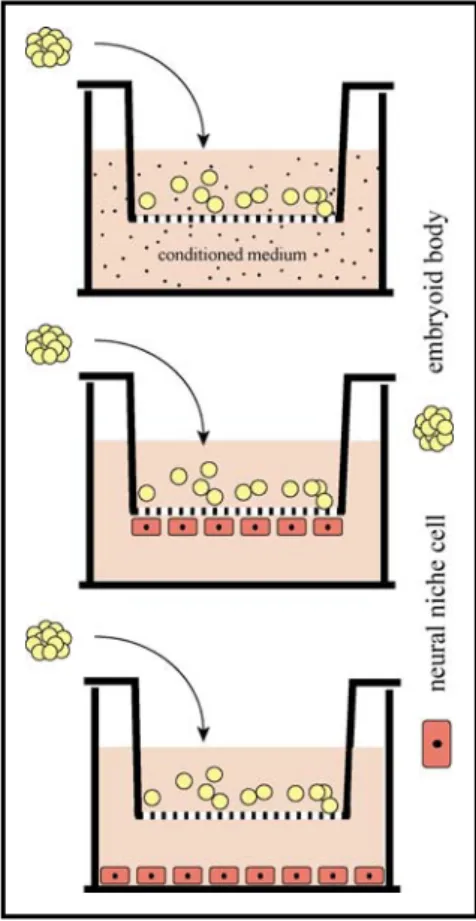
Figure 9 Co-culture settings for endothelial/ glial cell - stem cell interactions ... 39
Figure 10 Isolation and identification of primary rat brain endothelial cells... 51
Figure 11 Pre-differentiation of BAC7 embryonic stem cells to neuronal precursor cells... 53
Figure 12 RT-PCR analysis of Oct-4 and nestin expression in co-cultured ES cells ... 54
Figure 13 RT-PCR analysis of early neuronal marker neurofilament in co-cultured ES cells ... 56
Figure 14 FACS analysis of A2B5 expression in co-cultured stem cells ... 58
Figure 15 Phase contrast microscopy of ES cells in endothelial cell-conditioned
medium
... 59Figure 16 Fluorescence microscopy of GFP-expressing ES cells in conditioned medium... 60
Figure 17 Rotarod and Composite Neuroscore results following ES cell transplantation... 61
Figure 18 Coronal sections of injured rat brain following ES cell transplantation... 62
Figure 19 Phagocytosis of ES cells transplanted into traumatically injured rat brain ... 63
Figure 20 Brain extract-induced modulation of ES cell differentiation... 65
Figure 21 Expression of neural progenitor marker nestin by brain extract-treated ES cells... 66
Figure 22 Percentage of proliferating brain extract-treated ES cells ... 68
Figure 23 Changes in cell morphology following brain tissue extract treatment ... 70
Figure 24 Immunostaining of neuronal-like structures after brain extract-treatment ... 71
Figure 25 BDNF production by BAC7 embryonic stem cells ... 73
Figure 26 BDNF production by CGR8 embryonic stem cells ... 74
Figure 27 NGF release by BAC7 embryonic stem cells... 75
Figure 28 NT-3 release by BAC7 and CGR8 embryonic stem cells ... 77
Table 1 Primary cells and cell lines used... 32
Table 2 Primer sequences used for semi-quantitative (RT-)PCR and cDNA synthesis... 45
Table 3 Primary and secondary antibodies used for immunocytochemistry and FACS ... 49
1 I
NTRODUCTIONOne of today’s biggest challenges in molecular biological and biomedical research is the dissection of early developmental and regenerative processes that are responsible for the functional complexity of the mammalian central nervous system (CNS). The CNS assembly, consisting of a network of distinct cellular populations interwoven to a multifaceted functional entity contributes to our limited understanding. The elucidation of mechanisms and signalling pathways guiding neurogenesis and regeneration are the key to developing effective therapies for the treatment of various human neurodegenerative diseases and injuries (e.g. Morbus Parkinson, Morbus Alzheimer, Spinal Cord and Traumatic Brain Injury). The aetiology of neurodegenerative disease is associated with the progressive loss or damage of neuronal tissue leading to neuronal and behavioural deficits in the patient and is to date generally regarded as incurable. Moreover, the regenerative potential of the CNS is regarded as rather limited, and generally, neuronal populations cannot be replenished following cell death. Although many attempts to inhibit damaging pathological processes or to stimulate pathophysiological pathways e.g. by the administration of neuroprotective agents have been developed, these approaches could only attenuate post-traumatic pathophysiological processes, but have generally failed to restore functional deficits.
Putative treatment to overcome the pathophysiological symptoms of neurodegenerative diseases or injury lies in the rapidly developing field of cellular replacement strategies. The fundamental idea behind is to replace diseased or lost neuronal tissue by intracerebral cell or tissue transplantation resulting in restoration of form and function.
In the past, transplantation strategies mainly involved the transplantation of fetal tissues and considerable successes were achieved in the treatment of Parkinson’s and Huntington’s disease.
It was effectively demonstrated that fetal transplants could replenish destroyed and lost nervous
tissue by partial reconstruction and, to some extent recovery of cognitive and motor function was
achieved (Bachoud-Levi et al., 2000; Bjorklund and Lindvall, 2000; Lindvall, 1999). The lack of
availability of donor tissue, as well as concerns regarding the purity and viability of the
transplant population has led to the search for alternative sources of donor cells (Barinaga,
2000). Only recently, the discovery of pluripotent cell populations and consecutive advances in
stem cell biology has offered a promising future for cell replacement therapy.
Introduction
10
of stem cells. They allow detailed in vitro investigations into the course of cell lineage specification and the molecular mechanisms involved. Pluripotent embryonic stem cells represent an ideal model system to examine basic differentiation processes of neurogenesis by correlating the temporal regulation of genes and tissue morphogenesis during in vitro cell differentiation with normal development in vivo. Furthermore, stem cell-derived neural precursor cells have been shown to differentiate to neurons and glia during normal embryonic development of the brain (Brustle, 1999; Brustle et al., 1999). The isolation of pluripotent embryonic stem cells thus, not only provides an inevitable source for the study of neural differentiation processes on a cellular level but embryonic stem cells also represent ideal candidates for cellular replacement strategies.
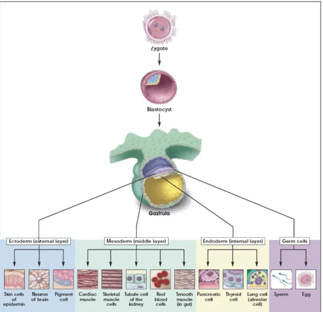
Pluripotent embryonic stem cell are derived from the inner cell mass of the blastocyst stage of embryonic development (E3.5) (Figure 1) (Evans and Kaufman, 1981; Martin, 1981). They are defined by their unlimited potential to self-renew and their ability to develop into cell types of all three primary germ layers including germ cells (Hubner et al., 2003). In contrast to terminally differentiated cells of the organism, embryonic stem cells display the absence of active senescence processes, which is attributed to their high telomerase activity (Tang et al., 2001;
Thomson et al., 1998). They can be maintained in vitro for prolonged periods of time in an undifferentiated state, but rely on the presence of leukæmia inhibitory factor (LIF) in culture medium and/ or the growth on mitotically inactivated mouse embryonic fibroblasts (MEF) (Smith et al., 1988; Williams et al., 1988). The developmental potential of embryonic stem cells has been demonstrated by the development of normal chimeras after incorporation into mouse blastocysts and in vitro differentiation of ES cells into a variety of endodermal, mesodermal and ectodermal derivatives (Figure 1) (Bradley et al., 1984; Nagy et al., 1990; Nichols et al., 1990).
To date, ES cell lines are available from a variety of species including rodents, rabbit, pigs,
primates and human embryos (Evans and Kaufman, 1981; Graves and Moreadith, 1993; Li et al.,
2003; Martin, 1981; Thomson et al., 1998; Thomson et al., 1995; Thomson et al., 1996). Since
their discovery, continuous research in the field of ES cell in vitro expansion and differentiation
shortly yielded a series of culture manipulations that promote the production of enriched
populations of various cellular lineages.
Figure 1 Lineage potential of pluripotent embryonic stem cells
Pluripotent embryonic stem cells of the blastocyst stage of embryonic development provide the basis for multilineage differentiation during gastrulation. In vitro ES cells can be expanded indefinitely and can be directed towards multiple cellular phenotypes including germ cells. Figure taken from Stem Cells: Scientific Progress and Future Research Directions, 2001
Introduction
12
1.2 In Vitro Differentiation of Embryonic Stem Cells Yields Adult Cellular Lineages The presence of LIF and/ or MEF in ES cell culture maintains ES cell’s self-renewal capacity and pluripotency, possibly acting via the STAT3 pathway (Niwa et al., 1998). Oct-4, a transcription factor of the POU domain, is expressed in stem cells of the early embryo maintaining their pluripotency and inhibiting lineage commitment. After gastrulation, Oct-4 gene expression is constrained to cells of the germ lineage (Pesce and Scholer, 2000, 2001;
Schoorlemmer et al., 1995; Yeom et al., 1996). In depth analysis of Oct-4 gene expression patterns in differentiating stem cells revealed that, rather than acting via a simple binary on-off control system, the precise level of Oct-4 gene expression governs distinct stem cell fates.
Accordingly, a critical amount of Oct-4 is required to sustain stem cell self-renewal, and up- or downregulation induces divergent developmental programmes. Hence, the transcription factor Oct-4 has been ascribed the role of a master regulator in pluripotent ES cells (Niwa et al., 2000).
Additionally, variant homeodomain containing protein Nanog, the SRY family member Sox2, Foxd3, a member of the forkhead winged-helix family and possibly Wnt signalling are also thought to play an important role in the inhibition of differentiation events (Avilion et al., 2003;
Hanna et al., 2002; Hart et al., 2004; Sato et al., 2004).
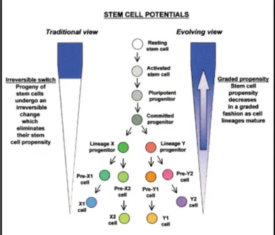
Upon removal of LIF and/ or MEF from ES cell culture, ES cells in vitro spontaneously differentiate into a broad spectrum of cellular lineages, progressively losing their pluripotent properties (Figure 2) (Blau et al., 2001; Keller, 1995). The most common method used for in vitro differentiation is the aggregation of ES cells into 3-dimensional ‘embryoid bodies’ (EBs) via ‘hanging drops’ (Boheler et al., 2002; Doetschman et al., 1985; Wobus et al., 1991).
Figure 2 Two conceptual views of decreasing stem cell potential during development.
Traditionally, lineage commitment has been viewed as one-way direction; pluripotent stem cells gradually lose their stem cell potential during progressive differentiation. The evolving view on the other hand assumes that cells in general have a recruitable propensity to act as stem cells, but proposes that this tendency is declining as ES cells differentiate.
Figure taken from Blau et al., 2001.
During embryoid body differentiation a number of cellular lineages coexist within these spherical structures, concomitantly with the regulated expression of marker genes associated with the development of primitive endoderm (hepatocyte nuclear factor), ectoderm (Pax6, Mash-1) and mesoderm (T brachyury, BMP-4) (Wiles and Johansson, 1997, 1999). Because of the structural resemblance of embryoid bodies with the organised development of the post- implantation embryo, these cellular aggregates are often exploited to investigate the complex interactions of cellular differentiation and gene expression during early embryogenesis in vitro (Abe et al., 1996; Itskovitz-Eldor et al., 2000; Leahy et al., 1999). The exposure of ES cells to various chemical agents, such as retinoic acid, dimethyl sulfoxide and 4-hydroxymethylbenzoic acid, as well as addition of growth factors and cytokines to culture medium (e.g. tumor necrosis factor-α, activin A, bone morphogenic protein-2 and-4, insulin, nerve growth factor, basic fibroblast growth factor, ciliary neurotrophic factor, triiodothyronine) has further led to the directed differentiation into tissue-specific cell types. One of the most extensively analysed differentiation paradigms comprises the spontaneous cardiac development of ES cells, firstly reported by Wobus and colleagues in 1991. Following the detection of spontaneously beating cardiomyocytes in ES cell culture, it was shown that the observed differentiation of ES cells towards terminally differentiated cardiomyocytes followed similar patterns as in vivo organ development (Boheler et al., 2002; Hescheler et al., 1997; Wobus et al., 1991).
Apart from cardiac differentiation, a range of other mesodermal cells types have been obtained from ES cells including chondrocytes, osteoblasts, adipocytes as well as endothelial cells (Buttery et al., 2001; Dani et al., 1997; Feraud and Vittet, 2003; Kramer et al., 2000).
Representatives of the endodermal lineages including pancreatic islets, hepatocytes, thyrocytes, lung and intestinal cells could also be derived by in vitro differentiation (Ali et al., 2002; Jones et al., 2002; Lin et al., 2003; Stoffel et al., 2004; Yamada et al., 2002). In view of later clinical application in neurodegenerative disease or injury paradigms, the in vitro differentiation of embryonic stem cells towards functional phenotypes of the neuronal lineage is of particular interest.
1.3 In Vitro Neuronal Differentiation Recapitulates In Vivo Neural Crest Development
Numerous publications describe the in vitro differentiation of ES cells to cells of the
ectodermal lineage including neural progenitor populations as well as terminally differentiated
Introduction
14
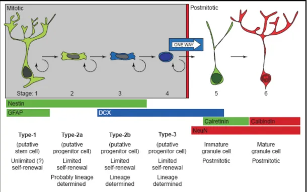
1997). Molecular mechanisms of RA action during embryogenesis and in the adult brain in vivo are proposed to function via a complex signalling pathway, but the mechanisms whereby RA induces neural differentiation of ES cells in vitro are yet not fully understood. Resembling in vivo neurogenesis, induction of neuronal differentiation in vitro follows a progressive restriction from bipotential radial glial-like stem cells via lineage-determined progenitor cells to early post- mitotic and mature neurons (Figure 3) (Alvarez-Buylla et al., 2001; Kempermann et al., 2004).
Limited mitotic activity and the developmentally regulated expression of various neural genes, transcription factors and proteins, as well as the display of differential cellular morphologies mark progressive lineage restriction (Guan et al., 2001; Lang et al., 2004; Rathjen and Rathjen, 2002; Stavridis and Smith, 2003). Following the aggregation of embryoid bodies and RA induction, the cellular spheres are dissociated and replated in monolayer culture. The subsequent withdrawal of serum from culture medium results in an inhibition of mesodermal lineage commitment, observable by the downregulation of mesodermal genes, such as T brachyury, cardiac actin and cardiac globin and the detection of a temporally controlled expression pattern of neural-specific genes (Bain et al., 1996). The characteristic developmental sequence of progressing neuronal differentiation events starts with upregulation of neural transcription factors Mash-1, Wnt-1, Sonic hedgehog (Shh), Pax6 and Engrailed-1 (En-1) (Joyner, 1996;
Mehler, 2002). Addition of basic fibroblast growth factor (bFGF) and epidermal growth factor
(EGF) yields high amounts of proliferating neural precursor cells marked by the elevated
expression (> 85%) of the intermediate filament protein nestin (Lee et al., 2000; Okabe et al.,
1996). Nestin mRNA expression is closely correlated with a mitotically active bipotential neural
progenitor cell state in the developing central nervous system in vivo from which glial and
neuronal cell emanate. Furthermore, in neural stem cells nestin gene expression has been shown
to diminish upon commitment to either the oligodendroglial or neuronal lineage. (Dahlstrand et
al., 1995; Mellodew et al., 2004). Thus, nestin gene expression marks a developmentally
decisive position during neuronal development. Accordingly, withdrawal of bFGF and EGF from
culture medium is followed by downregulation of nestin and Mash-1 gene expression. Neuron-
specific genes like the synaptic vesicle protein synaptophysin, Nurr1 and tyrosin hydroxlase are
in turn upregulated (Guan et al., 2001; Rathjen and Rathjen, 2002).
At an early stage of neural development genes encoding low (68 kDa) and middle (160 kDa) molecular weight neurofilament isoforms are expressed following neuronal lineage commitment and prior to cell cycle exit (Guan et al., 2001). Mature neurons are eventually characterised by the expression of high molecular weight (200 kDa) neurofilament proteins, neurocan, syntaxin, synaptobrevin, neuronal cell adhesion molecules (NCAM), class III β-tubulin and microtubule- associated proteins MAP2 and MAP5 (Bain et al., 1995; Fraichard et al., 1995; Guan et al., 2001; Strubing et al., 1995). Functional classification of post-mitotic neurons comprises the production of neurotransmitters dopamine, serotonin, gamma-aminobutyric acid (GABA) and glutamate, the expression of voltage-gated Ca
2+, Na
+, and K
+ion channels and expression of receptor-operated GABA
A, glycin, kainite, and N-methyl-D-aspartate (NMDA) ion channels
Figure 3 Six stages of progressive neuronal lineage restriction in the adult CNS
Although different hypotheses exist concerning the various stages of lineage commitment in the developing as well as in the adult nervous system, it is generally agreed on that the point of origin is a putative stem cells with radial glia and astrocytic properties. From there on lineage commitment is supposed to progress over (three) putative transiently amplifying progenitor cell stages. Different stages of development can be identified by morphological appearance, proliferative capacity and expression of stage-specific markers (e.g. Nestin, GFAP, DCX, NeuN, Calretinin and Calbindin). Hypothesis proposed by and figure taken from Kempermann et al., 2004.
Introduction
16
(Guan et al., 2001; Lang et al., 2004; Rathjen and Rathjen, 2002; Stavridis and Smith, 2003).
Glial differentiation has been monitored by the expression of glial fibrillary acidic protein (GFAP), OP4 (for astrocytes), and GalC and O4 (for oligodendrocytes). The addition of survival promoting factors (SPF) such as interleukin-1β, dibutyryl-cAMP, transforming growth factor-β
3, glial cell-derived neurotrophic factor and neurturin to ES cell differentiation culture can further promote lineage commitment and survival of functional neurons by preventing apoptosis (Rolletschek et al., 2001).
Successful in vitro differentiation of ES cells into functional phenotypes has consequently fostered development of cell replacement strategies. Subsequently, animal models were employed to validate stem cell differentiation in vivo.
1.4 Upon CNS-Transplantation Stem Cells Differentiate into Neural Phenotypes
The availability of basically unlimited numbers of ES cells and lineage-committed cell populations derived thereof has subsequently led to transplantation studies for the therapy of a variety of degenerative diseases. In animal models for cardiovascular disease, liver injury and diabetes integration of ES cell grafts with improved functional outcome could be demonstrated (Doss et al., 2004). However, compared to those promising results transplantation into the CNS still imposes a major challenge.
In first transplantation efforts into naïve embryonic mouse brain, Brüstle and colleagues demonstrated the differentiation of engrafted neural precursor cells into neurons, astrocytes and oligodendrocytes, which readily integrated into telencephalic, diencephalic, and mesencephalic regions of the brain (Brustle et al., 1997). A similar study by Zhang and co-workers confirmed these observations after transplantation of in vitro differentiated human ES cell-derived neural progenitor cells into the neonatal mouse brain (Zhang et al., 2001). In adult animal models, engraftment of nestin-positive ES cell-derived precursor cells into the striatum of adult rats similarly resulted in the differentiation into various neural phenotypes, detected by expression of neuronal (Thy-1) and astroglial (GFAP) proteins (Andressen et al., 2001; Arnhold et al., 2000b).
Long-term evaluation of stem cell-based transplantation into the brain of adult mice revealed a controlled differentiation pattern and the formation of mature neural grafts consisting of cells of all three major neural subtypes (Benninger et al., 2003). These observations demonstrate that in general replacement of nervous tissue by stem cell transplantation is possible, provided the host tissue is able to supply the necessary cues to direct grafted cells to a specific fate. This is particularly true for transplantation paradigms where stem cells are grafted into neonatal CNS.
Stimulated by these successful reports, implications for disease and injury were proposed.
Early experimental approaches utilised different models of myelin-deficiency. Learish and
colleagues injected ES-derived glial progenitor cells into the cerebral ventricle of myelin- deficient rats and Brüstle et al. transplanted ES-derived precursor into a rat model of human myelin disease. Subsequently, graft-derived production of myelin and efficient myelination of axons in brain and spinal cord were reported (Brustle et al., 1999; Learish et al., 1999). In 2002, the transplantation of undifferentiated ES cells into either chemically demyelinated rat spinal cord or likewise, into the spinal cord of myelin-deficient shiverer (shi/shi) mutant mice, led to differentiation of engrafted cells into oligodendrocytes accompanied by myelin production and remyelination of host axons (Liu et al., 2000). However, the described experimental settings of transplant-mediated repair have invariably been simplistic, with transplant paradigms characterised by a clear cellular deficit, a largely non-reactive CNS environment and a lack of inflammation. Although experiments such as these are fundamental to our understanding of the behaviour and differentiation potential of stem cell populations in vivo, they do not account for the multifaceted pathophysiology of a neurodegenerative disease state or injury, such as traumatic brain injury, which is accompanied by a complex and reactive CNS environment.
1.5 Reactive Microenvironment of Traumatic Brain Injury Limits Stem Cell Survival
In contrast to previously mentioned disease models, traumatic brain injury represents a rather
heterogeneous disease which is accompanied by a wide range of pathologies (Adams et al.,
2000; Davis, 2000). Traumatic brain injury is associated with a massive loss of multiple cell
types due to primary mechanical tissue disruption and bleeding, and the ensuing secondary
insults are the major cause of delayed brain damage. Activation of genomic, cellular and/or
biochemical cascades, the development of mass lesions and superimposed systemic insults in
response to traumatic brain injury additionally contribute to sustained cell necrosis, apoptosis
and neurological dysfunction (Davis, 2000; Sato et al., 2001). In particular, influx of
immunocompetent cells facilitated by break-down of the blood brain barrier and secretion of
inflammatory mediators and cytokines during the acute post-traumatic phase establish an
inhospitable microenvironment detrimental for cell survival (Lenzlinger et al., 2001; Morganti-
Kossmann et al., 1992). Over the years a variety of studies were conducted aiming at the cellular
replacement of lost or damaged nervous tissue in this specific disease environment. Stem cells of
different origin were transplanted into animal models of traumatic brain injury and analysed for
phenotypic and functional outcome. Transplantation paradigms involving immortalised
Introduction
18
expressing cells for engraftment, and actual contribution of grafted cells to functional recovery was not investigated (Boockvar et al., 2005; Philips et al., 2001; Riess et al., 2002). Similarly, the administration of bone marrow stromal cells (BMSCs) in Controlled Cortical Impact (CCI) models of traumatic brain injury led to improvement of neurological outcome, and moreover, expression of neuronal and glial markers could be observed (Lu et al., 2001; Mahmood et al., 2001a; Mahmood et al., 2001b). Nevertheless, reports on terminal differentiation of transplanted cells were rather inconsistent, and the mechanisms of graft function were not elucidated.
Following ES cell-derived neuronal and glial precursor transplantation into a Controlled Cortical Impact model of traumatic brain injury, Hoane and colleagues also reported improved functional outcome in addition to cell migration and reduction in lesion size (Hoane et al., 2004). Likewise, transplantation of ES cells into lateral fluid percussion (LFP)-injured rat brain led to significant recovery of motor function (Riess, unpublished data). Compared to engraftment into the naïve CNS, the survival rate of grafted cells in a trauma environment, if determined at all, was rather low. The small number of surviving cells and, in the majority of cases, lacking evidence of terminal differentiation suggest that functional improvements were not achieved on the basis of cellular replacement (Schouten et al., 2004). Alternative mechanisms of graft function involving trophic support provided by implanted cells or even stimulation of endogenous regenerative processes must thus be reconsidered. Furthermore, if differentiation of engrafted stem cells was in fact assessed in in vivo studies, migrating cells predominantly exhibited glial phenotypes in response to injury (Lacza et al., 2003). On the one hand, these studies provide encouraging reports on a functional level, and speculations on the molecular mechanisms involved. On the other hand it was demonstrated that pathological environmental conditions critically affect intrinsic stem cell properties and thus, emphasise our lack of understanding the control of stem cell differentiation in vivo (Keirstead, 2001). Successful differentiation of multipotent and progenitor cells into the three major CNS cell types in standardised in vitro culture cannot necessarily be transferred to a complex and multifaceted in vivo environment. Here, post- transplantation stem cell fate largely depends on both, intrinsic features of the particular cell type and the host environment (Cao et al., 2002a). Proposed stem cell-environment interactions were evidenced in two reports from 1996. Suhonen and colleagues transplanted adult hippocampal progenitors (AHPs) into neurogenic and non-neurogenic sites of the cerebellum of adult rats. In parallel, Svendsen and co-workers grafted stem cells derived from embryonic rat mesencephalon or striatum into the striatum of rats with either ibotenic acid or nigrostriatal dopamine lesions.
The first group demonstrated that grafted cells would only differentiate when grafted into
neurogenic, but not when transplanted into non-neurogenic regions of the brain. In contrast, the
second group showed a restricted proliferation and migration potential of grafted cells with only minor signs of differentiation when transplanted into lesioned CNS (Suhonen et al., 1996;
Svendsen et al., 1996). Both results soundly indicate a strong impact of the encountered milieu at the site of implantation on the proliferation and differentiation capacity of engrafted cells.
Moreover, Cao and co-workers showed that differentiation of neuronal-restricted precursors was inhibited when engrafted into the contused adult rat spinal cord, possibly due to the inhospitable environmental condition encountered (Cao et al., 2002b). These findings support the theory that the fate of stem cells is altered in response to pathological environmental conditions. Likewise, Okano and colleagues described that the post-traumatic microenvironment of the spinal cord was in an acute inflammatory stage following injury, unfavourable for the survival and differentiation of NSC transplants (Okano et al., 2003). Indeed, in a fluid percussion traumatic brain injury model, Molcanyi and co-workers demonstrated the loss of implanted stem cells due to extensive phagocytosis by activated microglia and macrophages (Molcanyi, unpublished data).
Consequently, graft - environment interactions play an important role in graft survival,
differentiation and integration, and finally, function (Tate et al., 2002). Although lineage
restriction and migration ability of a particular progenitor/ stem cell type have already been
correlated with stem cell origin, type and age, considerations must be extended to include the
graft-receiving host tissue. A familiar concept postulated in context with early embryonic
development and known as the microenvironmental “niche” has so only recently been envisaged.
Introduction
20
1.6 Molecular and Cellular Signals of the Environmental “Niche” Guide Cell Fate
From in vivo embryonic development it is known that the developmental fate of differentiating cells is largely depending on an intricate, site-specific interplay of specific regulatory genes, numerous signalling molecules and their receptory counterparts regulating the developmentally specific expression of proteins, thereby indicating different stages of development (Czyz and Wobus, 2001; Watt and Hogan, 2000). In particular, early neuronal development in vivo involves a sequential chain of fundamental processes that guide the way to the complex organisation of the mammalian CNS. Key molecular processes control crucial events in the life of a neuron, such as neural induction, regional and neuronal specification, migration of neurons, axonal growth and guidance and synapse formation. A precisely timed orchestration of signalling cascades and gene expression leads from neural tube segmentation through cell migration to interacting neuronal networks. Integrated regulatory mechanisms, responsible for the spatial organisation and ultimately, functional efficacy organise and manage the expression of tissue specific genes, proteins, receptors and ion channels in a developmentally controlled manner. The developmental “niche” takes part in directing programmed differentiation and is constituted of cells that, via secretion of humoral and signalling factors as well as ECM proteins, influence the developmental pathway of cells by directing their differentiation in an autocrine or paracrine manner.
In the course of in vivo embryonic development, transcriptional regulation during early spatial patterning of the neural tube is strongly influenced by cell-cell communications. Members of the Drosophila homologue segment polarity gene families engrailed (en), wingless (wnt) and pax functionally interact resulting in the morphogenesis and differentiation of specific brain regions, namely mesencephalon and anterior metencephalon (Joyner, 1996). The Wnt gene family consists of more than 10 secreted proteins that act locally via a yet unknown pathway but it is suggested that Wnt-1 represents the signal for En induction. Pax action involves cell-cell signalling and the expression of all Pax genes (except Pax1 and Pax9) is spatially and temporally restricted during CNS development (Gruss and Walther, 1992). The secreted protein of Sonic hedgehog (Shh) that was also first identified in Drosophila (Nusslein-Volhard and Wieschaus, 1980) represents another extracellular signalling mechanism. Initially expressed in the axial mesoderm, it can be found in the medial neural plate and the ventral neural tube, playing an important role in the patterning of face and ventral forebrain.
Throughout development, neuronal plasticity is restricted via progressive neuronal
specification; primary neurogenesis is followed by neuronal migration and the formation of
synaptic interactions. Again, studies in invertebrates paved the way for elucidating mammalian
cell specification processes. In Drosophila, neuronal fate largely depends on intercellular signalling processes. The Notch family of genes encode large proteins that contain segments in the membranes of cells. These proteins act as receptors for extracellular ligands that specify cell fate, leading to tissue organisation during development. The inhibition of Notch signalling promotes neuronal differentiation, whereas activation of the transmembrane receptor Notch results in a non-neural fate of cells (Marnellos et al., 2000). An interaction of cytoplasmatic proteins Notch-1 and Numb, located at opposite sites of ventricular epithelial cells has been proposed to decide on symmetric or asymmetric cellular division thereby directing cell fate (Figure 4) (Jan and Jan, 1998; Yoon and Gaiano, 2005). These perceptions fortify that instructive and restrictive cues are provided by the cellular and humoral environment and are able via intracellular signalling cascades to either potentiate maintenance and/ or proliferation of cell populations or alternatively, promote the commitment and maturation towards a specific lineage (Mehler, 2002; Mehler et al., 2000).
An extraordinary example for the influence of site-specific extracellular cues is the guidance of axons by chemoattractants that are secreted by the target under the influence of boundary regions that separate specific domains of the developing CNS (Wilson et al., 1993). These cues can either be repulsive or attractive, acting over long or short distances. It is assumed that gradients of different molecules are used for the establishment of neuron specification, neuron guidance and interactions between neurons. Diffusible factors secreted by axonal targets such as Netrin-1 protein and semaphorins also exert an important influence (Placzek et al., 1990). While Netrin-1 is a long-range chemoattractant, semaphorins are involved in the repellence of axon growth cones (Keynes and Cook, 1995; Raper, 2000; Serafini et al., 1996). Recently detected Slit proteins do also play a role in axon guidance and cell migration (Brose and Tessier-Lavigne, 2000).
Another example of secreted molecules that play a major role in neuronal development and
post-neurotraumatic injury are the neurotrophins. The neurotrophin family consists of a
functionally and structurally related class of neurotrophic factors that provide trophic support for
neurons during development and adult life. The neurotrophin family includes nerve growth
factor (NGF), brain-derived neurotrophic factor (BDNF) and neurotrophin-3 (NT-3) (Levi-
Montalcini, 1987). Neurotrophic factors mediate their cellular effects through the activation of
protein tyrosine kinase (trk) via binding of the trk receptor family, namely trkA for NGF, trkB
Introduction
22
1991; Vantini, 1992). In animal models of brain injury, the administration of neurotrophic factors via injection or via transplantation of cells genetically engineered to secrete neurotrophins has been shown to promote neuronal survival and repair by re-establishing functional connections and promoting the sprouting of neurits and the guidance of axons. In addition, neurotrophins delay apoptosis, prevent atrophy of axotomised neurons and enhance the expression of growth-associated genes. The neuroprotective and regenerative capacities of neurotrophic factors make them ideal candidates for injury-related applications and only lately, this protein family has been proposed a major role in stem cell-based functional recovery (Chen et al., 2005; Chen et al., 2002b; Lu et al., 2003; Mocchetti and Wrathall, 1995).
Recapitulating, a complex regulatory network that utilises cooperative, complementary, sequential and antagonistic extrinsic and intrinsic regulatory mechanisms is required to
Figure 4 Intercellular communication via Notch signalling generates lineage diversity
Within the developing and adult CNS Delta/
Notch interactions keep the balance between different modes of cell division, symmetric vs. asymmetric. Adjacent cells communicate via Delta/ Notch interaction determining their cell fate. Binding of Notch transmembrane receptor to its soluble ligand Delta leads to a cascade of intracellular events that antagonise neuronal gene expression and consequently, inhibit differentiation.
Differentiating neurons activate Notch signalling in adjacent cells to guarantee preservation of a pool of multipotent CNS progenitor cells. Notch signalling is generally involved in the maintenance of a multipotent state and later on in development promotes neuronal maturation and function. However, downregulation of Notch signalling is essential for the initiation of neurogenesis (Notch: Notch signalling is possibly involved; Off: downregulation of Notch is necessary). Both figures were taken from Yoon and Gaiano, 2005.
orchestrate the evolving profiles of lineage species during embryonic development. A minor shift in the character of a growth factor, signalling molecule concentration or ECM protein distribution constituting the developmental niche may therefore regulate and change depending cell fates. Factors and mechanisms responsible for developmental plasticity are still active in neonatal organs and especially in germinal centers of the adult organism where the composition of different cell types and their functional interaction has been termed “stem cell niche”.
1.7 Stem Cell Niche Controls Self-Renewal and Lineage Commitment
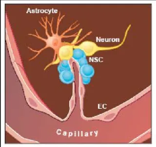
With regard to embryonic development the concept of the “niche” has been postulated and was re-introduced regarding the plasticity of adult stem cells (Schofield, 1983; Watt and Hogan, 2000). Adult stem cells can be found in a variety of adult tissues, some of them are known to undergo very limited regeneration or turnover, including the epidermis, skeletal muscle, bone marrow and the nervous system (Prockop et al., 2003; Watt and Hogan, 2000). Areas of neurogenetic activity were also found throughout the adult mammalian brain, especially in the hippocampus, the striatum and septum, as well as in the olfactory bulb and the subventricular zone (SVZ) (Bottai et al., 2003; Gage, 2000; Galli et al., 2003). Here, neural stem cells reside in close community with astroglial and microvascular endothelial cells, as well as with mature neurons of the CNS (Figure 5) (Doetsch, 2003;
Palmer et al., 2000; Wurmser et al., 2004). The residing neural stem cells (NSC) are generally defined as mitotically active cells that migrate along restricted pathways and have the ability to differentiate into mature neurons (Bottai et al., 2003; Gage, 2000; Galli et al., 2003). In vitro these cells retain their ability to self-renew, but in contrast to embryonic stem cells they can only generate lineage-restricted progeny (Czyz et al., 2003; Gritti et al., 2002). Maintenance of NSC plasticity in vivo is the pivotal role of the so- called stem cell niche and involves a variety of cell intrinsic as well as extrinsic mechanisms.
Figure 5 Stem cell niche of the adult brain In the adult CNS, neural stem cells (NSC) reside within distinct cellular compartments close to blood vessels. Astrocytes, microvascular
Introduction
24
In subsequent years, many in vitro studies into the nature of the stem cell niche and its influence on NSCs were conducted. So the site-specific migration, functional integration and differentiation of grafted NSCs was reported, proclaiming that NSCs responsiveness to environmental cues was restricted to their site of origin (Herrera et al., 2001). Moreover, it has been suggested that cues for migration and differentiation are dependent on recipient age, so that adult in contrast to fetal tissue is no longer able to provide the appropriate cues necessary for migration and differentiation of transplanted NSCs (Lim et al., 1997). In contrast, data by Shihabuddin and co-workers indicated that clonally expanded, multipotent adult progenitor cells from a non-neurogenic region were not lineage-restricted to their developmental origin but were able to generate region-specific neurons in vivo when exposed to the appropriate environmental cues (Shihabuddin et al., 2000). In a pilot in vitro investigation into the regional specification of neural stem cell, Hitoshi and colleagues confirmed that stem cells derived from different neurogenic sites of the brain, namely embryonic cortex, ganglionic eminence and midbrain/hindbrain, express separate molecular markers of regional identity in vitro. But, in the same study it was also shown that NSC’s regional identity could be altered by local inductive cues as observed in organotypic slice cultures (Hitoshi et al., 2002). The important role of extrinsic signals regulating intrinsic properties of neural progenitor cells was supported by Lillien and Raphael 2000 who related temporal changes during development to cell-cell signalling events within the developmental niche (Lillien and Raphael, 2000). Closer investigation into cell-cell communication within specific stem cell niches revealed that adjacent non-stem cells exerted significant influence on residing stem cell fate. It could be shown that ES cell-derived motoneurons extended long axons, formed neuromuscular junctions and induced
Figure 6 Neurogenesis in the adult CNS Within the neurogenic niches of the adult CNS, molecular and cellular signals collectively regulate, coordinate and control neurogenesis from neural stem cells.
Overlapping signalling pathways involve e.g. growth factors, neurotransmitters, guidance factors, metalloproteases and hormones. (a) Neurogenic niches of the dentate gyrus (DG) and forebrain. (b) In the dentate gyrus, new neurons are born in the subgranular zone just inside the granule cell layer (GCL). (c) Newly born neurons of the subventricular zone (SVZ) migrate through the rostral migratory stream (RMS) to the olfactory bulb (OB). (G: glomeruli of the olfactory bulb; LV: lateral ventricle).
Figure taken from Hagg, 2005.