© The Author(s) 2020 159
S. Jungblut et al. (eds.), YOUMARES 9 - The Oceans: Our Research, Our Future, https://doi.org/10.1007/978-3-030-20389-4_8
L.-E. Petersen (*) · M. Y. Kellermann
Institute of Chemistry and Biology of the Marine Environment, University of Oldenburg, Oldenburg, Germany
e-mail: lars-erik.petersen1@uni-oldenburg.de
8
P. J. Schupp (*)
Institute of Chemistry and Biology of the Marine Environment, University of Oldenburg, Oldenburg, Germany
Helmholtz Institute for Functional Marine Biodiversity at the University of Oldenburg (HIFMB), Oldenburg, Germany e-mail: peter.schupp@uni- oldenburg.de
Secondary Metabolites of Marine Microbes: From Natural Products Chemistry to Chemical Ecology
Lars-Erik Petersen, Matthias Y. Kellermann, and Peter J. Schupp
Abstract
Marine natural products (MNPs) exhibit a wide range of pharmaceutically relevant bioactivities, including antibi- otic, antiviral, anticancer, or anti-inflammatory proper- ties. Besides marine macroorganisms such as sponges, algae, or corals, specifically marine bacteria and fungi have shown to produce novel secondary metabolites (SMs) with unique and diverse chemical structures that may hold the key for the development of novel drugs or drug leads. Apart from highlighting their potential bene- fit to humankind, this review is focusing on the manifold functions of SMs in the marine ecosystem. For example, potent MNPs have the ability to exile predators and com- peting organisms, act as attractants for mating purposes, or serve as dye for the expulsion or attraction of other organisms. A large compilation of literature on the role of MNPs in marine ecology is available, and several reviews evaluated the function of MNPs for the afore- mentioned topics. Therefore, we focused the second part of this review on the importance of bioactive compounds from crustose coralline algae (CCA) and their role during coral settlement, a topic that has received less attention.
It has been shown that certain SMs derived from CCA and their associated bacteria are able to induce attach- ment and/or metamorphosis of many benthic invertebrate larvae, including globally threatened reef-building scler- actinian corals. This review provides an overview on bio- activities of MNPs from marine microbes and their potential use in medicine as well as on the latest findings of the chemical ecology and settlement process of scler- actinian corals and other invertebrate larvae.
Keywords
Marine natural products · Secondary metabolites · Marine bacteria · Marine fungi · Crustose coralline algae
· Settlement · Coral larvae
8.1 Introduction: Definition of Secondary Metabolism
Over millions of years, evolution has created a multitude of diverse organisms and biocoenosis. Besides individual dif- ferences within their appearance and way of life, the ability of absorbing, processing, and secreting substances from and into the environment can be found in all living organisms (Madigan et al. 2003). The biosynthesis and breakdown of these substances, including proteins, fats, or nucleic acids, is commonly known as primary metabolism with the com- pounds involved known as “primary metabolites” (Dewick 2002; Dias et al. 2012). The primary metabolism of plants, animals, humans, and prokaryotic microorganisms shows great similarity and displays the essential uniformity of all living matters; it thus serves as a driving force for the sur- vival and reproduction of all life (Kreis 2007). In contrast, the mechanism by which an organism synthesizes “second- ary metabolites” (SMs), frequently associated with the term
“natural products” (NPs), is known as secondary metabolism (Dias et al. 2012). SMs are defined as molecules with a molecular weight ranging between 100 and 1000 Da (Breinbauer et al. 2002) and, unlike primary metabolites, are
often found to be unique to an organism or a specific taxo- nomic group. They do not directly contribute to the basal metabolism of its producing organism but rather act as cru- cial factors to either attract, deter, or kill other organisms and thus increase their likelihood of survival (Kreysa and Grabley 2007). For example, SMs have been found in both prokary- otic and eukaryotic microorganisms, with unicellular bacte- ria (e.g., Bacillus spp., Pseudomonas spp.), eukaryotic fungi (e.g., Penicillium spp., Aspergillus spp.), filamentous actino- myces (e.g., Streptomyces spp.), and terrestrial plants being the most frequently studied and versatile producers (Bérdy 2005). Many SMs are only produced under specific circum- stances to serve different purposes: they can exile predators or competing organisms because of their toxic nature (Dewick 2002), act as attractants toward the same species for mating purposes (Gurnani et al. 2014), or serve as dyes for the expulsion and attraction of other creatures (Pichersky and Gang 2000). A possible explanation why organisms pro- duce a high variety of bioactive SMs is that these molecules provide producers with a selective advantage against com- peting organisms and, furthermore, act as an adaptation to environmental conditions (Jensen et al. 2005; O’Brien and Wright 2011; Letzel et al. 2013; Macheleidt et al. 2016).
Moreover, several natural products (NPs) have the ability to protect against nonbiological impacts, such as high light intensities or elevated temperatures, and to obtain reproduc- tion advantages for their producers (Ludwig-Müller and Gutzeit 2014). In the marine environment, SMs fulfill mani- fold tasks for their producers as they, for instance, act as a chemical defense against predators (Rohde et al. 2015;
Helber et al. 2017; Rohde and Schupp 2018) or have antimi- crobial effects against pathogenic microbes (Goecke et al.
2010; Rohde et al. 2015; Helber et al. 2018). Furthermore, MNPs are important for inducing larval settlement of benthic invertebrates (Yvin et al. 1985; Morse et al. 1988; Tebben et al. 2011, 2015; Harder et al. 2018), thereby maintaining and controlling community functioning and population dynamics. Besides their ecological impact, many NPs have been reported to exhibit a wide range of medically relevant bioactivities (Keller et al. 2005; Blunt et al. 2018), thus serv- ing as promising molecules for the development of new drugs or drug leads (Heilmann 2007).
8.2 Marine Natural Products Chemistry:
The Ocean as a Rich and Versatile Habitat
The ocean covers more than 70% of our planet’s surface and likely represents the origin of Earth’s life. In terms of species diversity, certain marine ecosystems, such as coral reefs, are thought to outnumber even tropical rain forests (Haefner 2003). Until today, the number of marine species that inhabit
the world’s oceans is not truly known; however, experts esti- mated a number approaching 1–2 million species (Simmons et al. 2005; Das et al. 2006). In the past, marine sponges were an interesting source for novel NPs; these sessile organisms can produce bioactive substances for chemical defense against natural predators, such as fishes (Rohde et al. 2015), as well as prevent overgrowth by competing organisms (Proksch 1994; Ortlepp et al. 2006). Furthermore, sponges serve as incubators for particular associated microorganisms like bacteria and fungi that also can contribute to the produc- tion of bioactive compounds (Radjasa et al. 2011; Wiese et al.
2011). Sponges being sessile, soft-bodied organisms, which mostly lack morphological defenses like biological armature or spines, depend to a large extend on bioactive metabolites for their survival and the survival of their associated micro- bial symbionts (Proksch et al. 2006). Accordingly, marine NP research has its origin in the discovery of the two nucleosides spongothymidine and spongouridine by Bergmann and coworkers in the 1950s, who isolated both active compounds from the Caribbean sponge Cryptotethya crypta (Bergmann and Feeneyz 1951; Bergmann and Burke 1955). These two SMs served as lead structures for the development of the syn- thetic antivirals cytarabine (Fig. 8.1a) and vidarabine (Fig. 8.1b) (Mayer et al. 2010) and, therefore, display exem- plarily the tremendous potential of MNPs for the develop- ment of new drugs (Gulder and Moore 2009). Although promising and still relevant, sponges and their associated microorganisms are not the only marine source producing bioactive compounds. Marine NP research has expanded its efforts in exploring worldwide oceans and their inhabitants from macro- to microorganisms as rich sources for novel SMs. Until today, this resulted in new MNPs being continu- ously described (Table 8.1) (Martins et al. 2014). For instance, 1163 novel compounds derived from marine organisms were described only in 2013 (Blunt et al. 2015).
Over the past decades, it has been obvious that unknown NPs are more likely found when high quality materials from novel sources are examined (Goodfellow and Fiedler 2010). Unfortunately, the acquisition of marine organisms, compared to that of terrestrial organisms, is often more dif- ficult and thus making the exploration and collection of marine samples (i.e., deep-sea organisms) very expensive (Molinski et al. 2009). However, past progress in marine technologies, such as easy accessible scuba diving equip- ment as well as remotely operated vehicles (ROVs), facili- tated the investigations beyond the intertidal areas and led to the exploration of new marine organisms that can poten- tially produce a huge range of novel chemical compounds with unique bioactivities (Gerwick and Moore 2012). Since many MNPs are released into the water, the concentration of bioactive compounds is rapidly diluted via diffusion pro- cesses, and thus MNPs must be highly potent to have a long-reaching effect (Haefner 2003). Past studies have
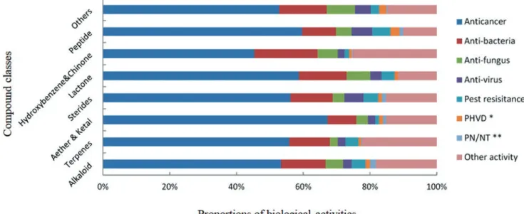
shown that MNPs cover a wide variety of biological activi- ties (Fig. 8.3), such as anticancer (Nastrucci et al. 2012), antibacterial (Hughes and Fenical 2010), antifungal, and antiviral effects (Mayer et al. 2013), making them a prom- ising source for novel drugs. Figure 8.3 shows that different chemical classes of MNPs are showing equal proportions among a vast set of bioactivities, leading to the assumption that most chemical structures could either be developed or serve as scaffolds for the development of new drugs against various diseases (Hu et al. 2015).
Besides the investigation on marine invertebrates or algae, modern marine biotechnology expanded its interests onto the exploration of marine bacteria and fungi, since the latter have been recognized as renewable producers of SMs (i.e., under controlled laboratory conditions) in the drug discovery process (Waters et al. 2010). Both, bacteria and fungi associated with marine macroorganisms have shown to be potent producers of bioactive substances, in some cases with prominent activities against several pathogenic germs, viruses, and tumor cells (Imhoff et al. 2011 and ref- erences therein).
8.2.1 Marine Bacteria: Widely Distributed Producers of Promising Natural Products
Marine microorganisms managed to conquer every marine habitat ranging from shallow and deep marine waters, polar regions, and deep-sea hydrothermal vents to diverse coral reef ecosystems. Particularly, the surface of macroorgan- isms, such as algae, sponges, and corals, is a favorable eco- logical niche for marine microorganisms. In many cases, bacteria live in close association with higher organisms and form symbiotic or mutualistic relationships (Lee et al. 2009;
Kazamia et al. 2012; Cooper and Smith 2015). There is growing evidence that the microbial community composi- tion on marine macroorganisms is habitat and even species specific. Examples include differences in communities found on the surface of different algae (Lachnit et al. 2009), between different parts of the rhizoid and phylloid of the brown alga Saccharina latissima (Staufenberger et al.
2008), between different sponge species (Thomas et al.
2016; Moitinho- Silva et al. 2017), as well as between outer
Table 8.1 Pipeline of marine pharmaceuticals until 2018 (according to http://marinepharmacology.midwestern.edu/clinical_pipeline.html, accessed 28 January 2018)
Compound Chemical class Source org. Therapeutic area Status 2018
Cytarabine Nucleoside Sponge Cancer FDA-approved
Vidarabine Nucleoside Sponge Antiviral FDA-approved
Ziconotide Peptide Cone snail Chronic pain FDA-approved
Trabectedin Alkaloid Tunicate Cancer FDA-approved
Brentuximab vedotin Antibody drug conjugate Mollusk Cancer FDA-approved
Eribulin mesylate Macrolide Sponge Cancer FDA-approved
Omega-3-acid ethyl ester Omega-3 fatty acid Fish Hypertriglyceridemia FDA-approved
Plinabulin Diketopiperazine Fungus Cancer Phase III
Plitidepsin Depsipeptide Tunicate Cancer Phase III
Bryostatin Macrolide lactone Bryozoan Alzheimer’s Phase II
Plocabulin Polyketide Sponge Cancer Phase II
Marizomib Beta-lactone-gamma-lactam Bacterium Cancer Phase I
Chemical structures of all compounds listed in this table can be found in Figs. 8.1 and 8.2 Fig. 8.1 (a) Cytarabine and
(b) vidarabine (Mayer et al.
2010) (created with ChemDraw, v. 16.0.1.4)
Fig. 8.2 Selected marine pharmaceuticals. (a) Ziconotide, (b) trabect- edin, (c) monomethyl auristatin E, (d) eribulin, (e) omega-3 fatty acid, (f) plinabulin, (g) plitidepsin, (h) bryostatin, (i) plocabulin, and (j) mar-
izomib (Mayer et al. 2010; modified from Lee et al. 2015; Pantazopoulou et al. 2018) (created with ChemDraw, v. 16.0.1.4)
Fig. 8.2 (continued)
and inner parts of the sponge Tethya aurantium (Thiel et al.
2007). However, the great microbial diversity of marine environments remains nearly untapped. Simon and Daniel (2011) estimated that less than 0.1%, probably solely 0.01%, of all microbes in the oceans have been characterized.
Molecular analysis of marine metagenomes revealed a great number of phylogenetic lines of so far uncultured groups of bacteria and archaea (DeLong et al. 2006; Simon and Daniel 2009; Hug et al. 2016). Besides their important roles in shaping community structures and in mediating microbe- microbe as well as microbe-host interactions, marine bacte- ria are suggested to represent a treasure box of new compounds for biotechnology. This assumption is due to their high biodiversity and the gap of knowledge regarding their potential of NP biosynthesis (Imhoff et al. 2011). Yet, much evidence is given that marine bacteria produce new compounds useful for the discovery of novel pharmaceuti- cals (Rahman et al. 2010; Waters et al. 2010; Blunt et al.
2018). From 1997 to 2008, about 660 new marine bacterial NPs were identified. Most of them originated from the classes Actinobacteria (40%) and Cyanobacteria (33%), followed by Proteobacteria (12%) and members of the Bacteroidetes and Firmicutes (5%) (Williams 2009). In comparison, 179 novel NPs have been isolated from marine bacteria in 2016. This is only a moderate increase compared to the average number of new marine bacterial compounds in the last 3 years, but a significant increase from the aver- age for the period of 2010 to 2012 (Blunt et al. 2018).
Members of the Actinobacteria are a rich source of NPs and hold an unmatched capacity for the generation of new drugs (Bull et al. 2005; Bull and Stach 2007; Fenical and Jensen
2006). The first bioactive compound extracted from a marine actinomycete was the antibiotic SS-228Y, showing antibac- terial activity to gram-positive bacteria. This biomolecule was proposed to be a peri- hydroxyquinone derivative pro- duced by Streptomyces purpurogeneiscleroticus (Chainia purpurogena) collected from sea mud (Okazaki et al. 1975).
Until today, the genus Streptomyces continues to be a pro- lific source of new and interesting chemistry; numerous compounds showed exciting bioactivities. For example, S.
spinoverrucosus, isolated from a sand sample from the Bahamian tidal flats, produced the dibohemamines A–C, three new dimeric bohemamines. These compounds were shown to be formed via a nonenzymatic process with form- aldehyde, which was also detectable in the growth media.
Both metabolites dibohemamines B (Fig. 8.4a) and C exhib- ited potent activity against lung cancer cells, with IC50 val- ues of 140 nM and 145 nM, respectively (Fu et al. 2016).
Another Streptomyces sp. isolated from a marine sediment sample collected off Oceanside, California, USA, produced the ansalactams A–D. The novel ansalactam derivatives (B–D) represent three new carbon skeletons and, therefore, display the plasticity within the ansamycin biosynthetic pathway. The latter three novel metabolites showed moder- ate antibacterial activity against MRSA (methicillin-resis- tant Staphylococcus aureus) (Wilson et al. 2011; Le et al.
2016). Apart from Streptomyces, species of the genera Salinispora and Marinispora were found to produce struc- turally novel bioactive compounds. A Salinispora tropica strain was isolated from a sediment sample, collected from a mangrove environment in Chub Cay, Bahamas. This strain produced several β-lactone-gamma- lactams, the salinospo-
Fig. 8.3 Analysis of new marine-derived compounds from 1985 to 2012 according to chemical classes and biological activities (∗PHVD, preven- tion of heart and vascular disease; ∗∗PN/NT, protection of neurons/neurotoxicity) (modified from Hu et al. 2015)
ramides, which represent a new family of SMs (Feling et al.
2003; Williams et al. 2005). Specifically, salinosporamide A (marizomib; see Fig. 8.2j) displayed potent in vitro cytotox- icity against HCT-116 human colon carcinoma with an IC50
value of only 11 ng mL−1. Furthermore, this compound showed great potency against NCI-H226 non-small cell lung cancer, SK-MEL-28 melanoma, MDA-MB-435 breast cancer, and SF-539 CNS cancer, all with LC50 values less than 10 nm (Feling et al. 2003). As displayed in Table 8.1, marizomib has entered Phase I human clinical trials for the
treatment of multiple myeloma (Martins et al. 2014; http://
marinepharmacology.midwestern.edu/clinical_pipeline.
html, accessed 28 January 2018). Asolkar et al. (2009) found three new cyclohexadepsipeptides, namely, aren- amides A–C, produced in the fermentation broth of a marine Salinispora arenicola, isolated from a sediment sample.
Arenamide A and B (Fig. 8.4b) blocked TNF (tumor necro- sis factor) induced activation in transfected 293/NFκB-Luc human embryonic kidney cells in a time- and dose-depen- dent manner with IC50 values of 3.7 μM and 1.7 μM, respec-
Fig. 8.4 (a) Dibohemamine B (Fu et al. 2016), (b) arenamide B (Asolkar et al. 2009), (c) marinomycin A (Kwon et al. 2006), and (d) dolastatin 10 (Bai et al. 1990) (created with ChemDraw, v. 16.0.1.4)
tively. The compounds also inhibited nitric oxide (NO) and prostaglandin E2 (PGE2) production with lipopolysaccha- ride (LPS)-induced RAW 264.7 macrophages. The authors suggest that the anti-inflammatory and chemoprevention characteristics of arenamides A and B are worth further investigation (Asolkar et al. 2009). Other examples for anti- biotics with antitumor activity from marine Actinobacteria are the marinomycins. A Marinispora strain, isolated from an offshore sediment sample, produced the marinomycins A–D. The most promising compound within this novel class of polyketides is marinomycin A (Fig. 8.4c). It shows selec- tivity against several human melanoma cell lines with an IC50 value of 5 nM for SK-MEL5 melanoma cells (Kwon et al. 2006). Besides Actinobacteria, members of marine Cyanobacteria are known to produce bioactive SMs too.
For example, the peptide dolastatin 10 (Fig. 8.4d) was origi- nally isolated from the sea hare Dolabella auricularia (Bai et al. 1990) but was then shown to be produced by the cya- nobacterium Symploca sp. (Luesch et al. 2001). This natural product (NP) was used as a model for the synthetic develop- ment of soblidotin, which has entered Phase III clinical tri- als (Mayer et al. 2010). The cyclic depsipeptide largazole is produced by another marine Symploca sp. and inhibited the growth of highly invasive transformed human mammary epithelial cells in a dose-dependent manner (GI50 7.7 nM). It induced cytotoxicity at higher concentrations (117 nM) (Taori et al. 2008). All these examples show the potential of marine bacteria, specifically Actinobacteria and Cyanobacteria, to produce chemicals that cover a broad range of bioactivities and might be used for the generation of novel drug candidates.
8.2.2 Marine Fungi: Bioprospecting the Future
Compared to bacteria, the basic knowledge on marine fungi, hereinafter referring to obligate and facultative marine fungi, is still deficient in matters of diversity and ecological impor- tance (Imhoff et al. 2011). The term “marine fungi” applies rather to an ecological background than to a distinct taxon- omy or a physiological approach (Kohlmeyer and Kohlmeyer 1979). Within biology, marine fungi are mainly separated into two groups, namely, obligate marine fungi, which grow and sporulate exclusively in marine habitats, and facultative marine fungi, which originate from freshwater or terrestrial milieus and are capable to grow also in the marine environ- ment (Kohlmeyer and Kohlmeyer 1979). By 1996, mycolo- gists estimated the number of marine fungi to be approximately 1500 species, and by 2011, biodiversity esti- mations of marine fungi were placed to be more than 10,000 species (Jones 2011).
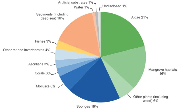
According to Overy et al. (2014), the examination of new substrata and geological locations will greatly increase the number of total species through the rapid discovery of new fungal species. However, marine fungal strains have been isolated from nearly every possible marine habitat until today, including soil and sediment (Wang et al. 2013; Simões et al. 2015), marine invertebrates (e.g., sponges and corals) (Wiese et al. 2011; Amend et al. 2012), marine plants (e.g., algae) (Loque et al. 2010), and marine vertebrates (fishes) (Rateb and Ebel 2011). Algae have been used primarily as a source for bioprospecting fungal diversity, closely followed by sponges and mangrove habitats (Fig. 8.5). Efforts to iso- late these symbionts within new and sometimes extreme habitats are still being made. A study on the fungal commu- nity by a culture-dependent approach revealed that several Antarctic sponges of the phylum Ascomycota were a rich source of associated fungi and novel SMs, with some of them showing antimicrobial, antitumoral, and antioxidant potential (Henríquez et al. 2014). Furthermore, due to the development of deep-sea instrumentation and new tech- niques used for sampling, the deep-sea habitat emerged as a new and highly promising source for marine fungal biodiver- sity, and thus an excessive number of novel fungal specimen have been retrieved (Wang et al. 2015). For example, Burgaud et al. (2009) investigated the biodiversity of cultur- able filamentous fungi and uncovered the presence of both Ascomycota and Basidiomycota associated with different deep-sea samples, including sediment, mussels, shrimps, and smoker rock scrapings. However, as an outcome of the recent bioprospecting efforts, biotechnological interests have mostly turned to marine microorganisms and notably fungi as a likely source for MNPs (Fig. 8.6) (Bhadury et al. 2006).
By 1992, only 15 marine fungal metabolites had been described (Fenical and Jensen 1993), and this number rose to 270 until 2002 (Bugni and Ireland 2004). Within the period from 2006 until mid-2010, Rateb and Ebel (2011) summa- rized 690 NPs from fungi isolated from marine habitats. With Penicillium spp. and Aspergillus spp. being the most potent producers, their study revealed that nearly 50% of the com- pounds are polyketides and their prenylated forms, whereas alkaloids, terpenoids, and peptides contributed 15%–20%
(Rateb and Ebel 2011). A famous example of a NP from a marine fungus is the diketopiperazine halimide (Fig. 8.6a), an aromatic alkaloid of a marine Aspergillus sp. isolated from the green alga Halimeda copiosa (Fenical 1999). Its synthetic analog, plinabulin (Fig. 8.2f), is showing antitumor activity by causing tubulin depolymerization, thereby leading to the dis- ruption of tumor cells followed by necrosis of the tumor itself (Gullo et al. 2006). Up to today, plinabulin is the only marine fungal synthetic analog that has entered clinical trials and suc- cessfully passed the first and second phase (Gomes et al. 2015;
www.beyondspringpharma.com/en/pipeline/, accessed 28
January 2018). A further example was given by Pontius et al.
(2008), who isolated chaetoxanthone B (Fig. 8.6b) from a marine Chaetomium sp., showing selective antimalarial activ- ity against Plasmodium falciparum (IC50 = 0.5 μg mL−1) and
moderate activity against Trypanosoma cruzi (IC50 = 1.5 μg mL−1) without or with only minimal cytotoxic- ity toward cultured eukaryotic cells. Another promising marine NP is the chlorinated benzophenone pestalone (Fig. 8.6c),
Algae 21%
Mangrove habitats 16%
Other plants (including wood) 6%
Sponges 19%
Molluscs 6%
Corals 3%
Ascidians 3%
Other marine invertebrates 4%
Fishes 3%
Sediments (including deep sea) 16%
Water 1%
Artificial substrates 1%
Undisclosed 1%
Fig. 8.5 Sources of marine fungi-producing MNPs until 2010 (reproduced from Rateb and Ebel 2011)
Fig. 8.6 (a) Halimide (Fenical 1999), (b)
chaetoxanthone B (Gademann and Kobylinska 2009), and (c) pestalone (Cueto et al. 2001) (created with ChemDraw, v.
16.0.1.4)
which has been isolated from the fungus Pestalotia sp., which is associated with the brown alga Rosenvingea sp. and was collected near the Bahamas Islands. Although pestalone was only produced when Pestalotia sp. was cocultured with a marine bacterium, this compound showed potent activities against methicillin- resistant Staphylococcus aureus and van- comycin-resistant Enterococcus faecium strains, indicated by minimum inhibitory concentrations (MIC) of 37 ng mL−1 and 78 ng mL−1, respectively (Cueto et al. 2001). These latter examples encourage the ongoing research activities on novel marine fungal species for the future development of new drugs. Considering that 38% of the approximately 22,000 bio- active microbial metabolites are of fungal origin, and that only about 5% of the world’s fungal taxa have been described, fungi exhibit a tremendous potential for the discovery of novel bioactive SMs (Schulz et al. 2008). To avoid the rediscovery of already known compounds, specialized and effective derepli- cation strategies need to be constantly employed (Martins et al. 2014). For this purpose, the most common techniques are a combination of chemical compound separation hyphenated to various spectroscopic or mass-selective detection methods such as high-performance liquid chromatography (HPLC) coupled to either a diode array detector (HPLC-DAD) or a mass spectrometer (HPLC-MS) (Wolfender et al. 2010).
Besides nuclear magnetic resonance (NMR) spectroscopy, HPLC-MS is another predominant analytical technique for the fast detection and identification of SMs and other small mol- ecules. A major advantage of MS over NMR is that MS-based methods are far more sensitive, making it the method of choice when it comes to first-pass compound detection and identifica- tion in high-throughput screening applications (Carrano and Marinelli 2015). Moreover, it provides accurate mass even within the nanogram range, which can be used as a search criterion or query in nearly all NP databases (Nielsen et al.
2011). On the contrary, NMR is by far the most efficient method to unambiguously elucidate complex structures of small molecules (Hubert et al. 2017). One of its advantages compared to MS strategies is that it serves as a quantitative analysis without the need of a suitable reference material (Kurita and Linington 2015). The 1H-NMR is also useful for evaluating the purity of a given sample. For example, impuri- ties such as lipids are somewhat invisible in HPLC-DAD-MS techniques due to their low UV absorption, hydrophobicity, and contumaciousness to ionization, but they can easily be seen in 1H- NMR (Carrano and Marinelli 2015). After the col- lection of UV/VIS absorption spectra, molecular mass, and further structure data, the gained information needs to be com- pared with database entries. Over the decades, many different databases covering a wide range of compounds have been established (Mohamed et al. 2016; Guijas et al. 2018), includ- ing general compound libraries like SciFinder (www. sci- finder.cas.org, accessed 28 January 2018), NP libraries such as
AntiBase (www.wiley-vch.de/stmdata/antibase.php, accessed 28 January 2018) or Dictionary of Natural Products (dnp.
chemnetbase.com, accessed 28 January 2018), and even some free-to-use databases like ChemSpider (www.chemspider.
com, accessed 28 January 2018), PubChem (pubchem.ncbi.
nlm.nih.gov, accessed 28 January 2018), or Metlin (metlin.
scripps.edu, accessed 28 January 2018). In addition to this widespread dereplication approach, fragmentation- based MS methods, also referred to as MS/MS or tandem mass spec- trometry, in combination with molecular networking are receiving increasing attention for the identification of unknown compounds. For example, the Global Natural Products Social (GNPS) molecular networking website (http://gnps.ucsd.edu) is an open-access knowledge base that aims to let NP chemists work together and share their raw, processed, or identified MS/MS spectrometry data. We believe that crowdsourced curation of freely available reference MS libraries as well as a fast-growing database of MS/MS spectra will rapidly acceler- ate the annotation and thus the search of prior unknown com- pounds (Wang et al. 2016; Kind et al. 2017; Quinn et al. 2017).
8.3 Marine Chemical Ecology: Predator- Prey Interactions and Competition During the last decades, marine chemical ecology has evolved from a young science with mostly NP chemists finding new SMs with potentially obscure ecological functions into a matured research field that simultaneously combines chemical and biological aspects. Besides their side effect of exhibiting utilizable bioactivities for humankind, chemical cues possess major influences on every organizational level in the marine system. Several reviews highlight the importance of chemical communication between benthic and pelagic organisms for a better understanding of marine ecosystem functioning (i.e., Hay 1996, 2009; Sieg et al. 2011; Paul et al. 2011; Puglisi et al. 2014). However, most marine organisms are rather orga- nized in highly biodiverse and productive communities occur- ring in ocean fringes, such as coral reefs or offshore zones, than being distributed all over the ocean (Simmons et al. 2005;
Das et al. 2006). Many of these biological communities are characterized by the presence of extremely harsh conditions in matters of UV radiation (light stress at water surface), tem- perature, pressure, and salinity. In addition to these environ- mental stressors, sessile benthic organisms are often in strong competition for available resources such as space (to settle and grow) and nutrients. As a result, survival and reproduction between the competing organisms can strongly depend on their ability to produce bioactive SMs (de Carvalho and Fernandes 2010). These bioactive substances can perform various tasks for their producers and associated organisms; for instance, SMs work as a chemical defense against predators
(Pohnert 2004; Kubicek et al. 2011; Rasher et al. 2013; Rohde et al. 2015; Helber et al. 2017), function as attractants toward consumers (Sakata 1989), have antimicrobial effects against pathogenic microbes (Goecke et al. 2010; Puglisi et al. 2014;
Helber et al. 2018), guide the opposing sex by letting individu- als find and evaluate potential mating partners through chemi- cal cues (Lonsdale et al. 1998; Li et al. 2002), or act as settlement cues for invertebrate larvae to initiate the transformation into a sessile, juvenile form (Morse et al. 1988;
Heyward and Negri 1999; Negri et al. 2001; Kitamura et al.
2009; Tebben et al. 2011, 2015; Sneed et al. 2014). For exam- ple, different classes of macroalgae defend themselves chemi- cally against herbivores and produce SMs with antimicrobial and antifouling activity (Schupp and Paul 1994; Paul et al.
2014; Schwartz et al. 2016). Specifically, brown algae of the family Dictyotaceae produce several classes of diterpenes that defend their producers against herbivores but have also shown activity against other competitors. It has been reported that natural concentrations of a diterpene of the dolastane class (Fig. 8.7a), originally isolated from the brown alga Canistrocarpus cervicornis, reduce feeding activity by the sea urchin Lytechinus variegatus (Bianco et al. 2010). In a study of Craft et al. (2013), lipophilic extracts of nine subtropical algae were offered to four subtropical and three cold-temper- ate sea urchins at two concentrations. While the extracts of the subtropical marine algae Caulerpa sertularioides, Dictyota pulchella, and D. ciliolate deterred all urchins, the other mac- roalgae extracts from the cold-adapted areas led to different feeding resistance patterns. Apart from anti-herbivore activity, many macroalgae are known for their antimicrobial and anti- fouling activity (Goecke et al. 2010, 2012; Pérez et al. 2016;
Schwartz et al. 2017). A review by Harder et al. (2012) high- lights the crucial role of halogenated furanones (Fig. 8.7b, c) within the red alga Delisea pulchra and how these compounds interact with the associated bacteria. The halogenated fura- nones deter fouling by bacterial pathogens and epiphytic bac- teria through interference with bacterial quorum sensing. By imitating quorum sensing-mediating acyl homoserine lactones to block the same receptor sites, halogenated furanones can manipulate bacterial colonization and biofilm formation as well as bleaching and diseases caused by pathogenic bacteria.
Besides macroalgae, sponges and their associated microbes are another prolific source of potentially novel NPs with prom- ising bioactivities. Although the ecological role of sponge crude extracts has been evaluated for numerous sponge spe- cies, assignment of activities to specific NPs is lacking behind.
Investigated bioactivities included antipredatory, antifouling, antimicrobial, and allelopathic functions (Rohde et al. 2015;
Helber et al. 2017, 2018). Several studies are providing evi- dence that sponges are chemically defended from predation and pathogens by compounds that either the host or other associated microorganisms had produced (Pawlik 2011;
Hentschel et al. 2012). The Mediterranean sponge Axinella verrucosa, collected from the Gulf of Naples, Italy, produces hymenidin (Fig. 8.7d) and debromo- carteramine A (Fig. 8.7e), two bromopyrroles that are also known from other sponges.
The n-butanol part of the A. verrucosa extract, containing the two bromopyrroles as well as the pure hymenidin, showed activity against microbial fouling and deterred feeding by the generalist shrimp Palaemon elegans at naturally occurring concentrations (Haber et al. 2011).
Several studies have shown that sponges of the same genus and even of the same species can produce different SMs. This circumstance raises the question to what extend SMs have evolutionary advantages for the survival of sponges. A study of Noyer et al. (2011) showed that several populations of Spongia lamella, collected in the Mediterranean Sea, spanning a region of 1200 km, had an extremely high intraspecific chemical diversity. While nit- enin (Fig. 8.7f) and ergosteryl myristate (Fig. 8.7g) were the major metabolites, the number of compounds as well as their concentrations changed among populations collected from different geographic locations. The authors suggested that these variations may have been due to both genetic and envi- ronmental factors. A further study on S. lamella revealed that the populations from the five regions (Portugal, Gibraltar, Baleares, Catalonia, and South France) significantly differed within their genetic and chemical diversity as well as their associated bacteria (Noyer and Becerro 2012). Similarly, Rohde et al. (2012) found different metabolites and com- pound concentrations in the tropical sponge Stylissa massa across different ocean basins and within sites. Compound concentration varied among individuals, and no correlation between compound concentrations and factors such as depth, UV, predation, and microbial growth could be identified. The authors concluded that concentrations could be affected by other selective pressures such as water temperature, water quality, light conditions, and food availability or that the observed variations reflected population-specific constitutive defenses. Another activity that has received increased atten- tion recently are allelopathic actions of sponges by which they can outcompete scleractinian corals. Sponges have become an increasingly dominant species in the Caribbean reefs (Maliao et al. 2008; Colvard and Edmunds 2011; Perry et al. 2013; Villamizar et al. 2013; Loh and Pawlik 2014) and to a lesser extend in the Indo-Pacific (Bell and Smith 2004;
Bell et al. 2013; Helber et al. 2017) as coral reef systems are permanently threatened by multiple decades of loss of reef- building corals due to climate change, disease, and pollution.
In contrast to the calcium carbonate skeleton of corals, sponge skeletons are made of silica or protein, making them less sensitive to ocean acidification and temperature shifts (Pawlik 2011; Bell et al. 2013). Apart from being environ- mentally more robust, sponges can also outcompete corals
Fig. 8.7 (a) A diterpene (Bianco et al. 2010), (b, c) two halogenated furanones (Harder et al. 2012), (d) hymenidin, (e) debromo-carteramine A (Haber et al. 2011), (f) nintenin, and (g) ergosteryl myristate (Noyer et al. 2011) (created with ChemDraw, v. 16.0.1.4)
by chemically affecting the coral symbionts through alle- lopathy. Crude extracts of several sponges collected from the Caribbean reefs were embedded in stable gels at natural con- centrations and caused a decrease in the photosynthetic potential of the symbiotic zooxanthellae from the brain coral Diploria labyrinthiformis. Interestingly, sponge extracts influenced the symbiotic zooxanthellae in two ways: impair- ing photosynthesis with bleaching and with only little or no bleaching at all (Pawlik and Steindler 2007). Similarly, organic extracts of three sponges collected from Zanzibar reduced the photosynthetic performance of symbionts in the scleractinian coral Porites sp. (Helber et al. 2018). In addi- tion to allelopathy on adults, it has been reported that sponge- derived SMs can negatively affect invertebrate larvae settlement too (Thompson 1985; Thompson et al. 1985;
Bingham and Young 1991; Hellio et al. 2005). Since there have been several reviews in recent years on the role of SMs in chemical ecology and specifically chemical defense (Paul et al. 2011; Pawlik 2011; Rohde and Schupp 2018), we decided to focus in the remaining part of this review on the role of SMs during the settlement process of invertebrates (a role which has to this point received less attention).
8.3.1 Marine Invertebrate Larvae Settlement:
Role of Secondary Metabolites
Many benthic marine invertebrates such as corals, sponges, mussels, or worms have a planktonic phase followed by a metamorphic event that transforms them into a less mobile or immobile, sessile benthic form. Since the process of attach- ment and metamorphosis for most organisms is generally irreversible (Thorson 1950), the choice of a suitable location for settlement is crucial for invertebrate larvae regarding sur- vival, population dynamics, and community functioning. In the past, two models have been developed to explain the settlement of marine invertebrate larvae: (1) the stochastic model postulated that the settlement process happens ran- domly as soon as suitable substrate becomes available and that postmetamorphic events arrange the final distribution of juveniles, and (2) the deterministic model suggested that specific environmental factors determine the attachment and metamorphosis of larvae as well as their final distribution.
Nowadays, there is great evidence that the settlement process of invertebrate larvae is mainly biologically and chemically driven, although environmental parameters may also influ- ence settlement behavior (Sebens 1983; Morse et al. 1988;
Mundy and Babcock 1998; Lau and Qian 2001; Lau et al.
2005; Tebben et al. 2015; Da-Anoy et al. 2017). Chemical settlement cues are produced by a variety of marine organ- isms. Some invertebrate larvae like to settle among individu- als of their own species, while others preferably settle upon other species, resulting in gregarious or associative settle-
ment, respectively. Gregarious settlement has been reported for many phyla including polychaete worms and barnacles (Hadfield and Paul 2001). Live adults of the polychaete Hydroides dianthus were capable of eliciting gregarious set- tlement responses in conspecific larvae. Interestingly, settle- ment in response to live adults with or without their tubes as well as to their amputated tentacular crowns was signifi- cantly greater compared to dead worms, empty tubes, or bio- film covered slides. Moreover, after extraction of aggregations of adult worms with organic solvents, the inductive capacity of the remaining tissue was lost, and the activity went into both the nonpolar and polar fractions of the crude extract (Toonen and Pawlik 1996). Gregarious settlement of inverte- brate larvae has long been assumed to be induced by contact with adult conspecifics (Crisp and Meadows 1962, 1963;
Clare and Matsumura 2000). It has been shown that a glyco- protein with high molecular weight isolated from the adult barnacle Amphibalanus amphitrite, termed the settlement- inducing protein complex (SIPC), induced settlement of cypris larvae (Matsumura et al. 1998; Dreanno et al. 2006).
Nevertheless, there are reports showing that waterborne cues are able to induce gregarious settlement as well. Endo et al.
(2009) isolated a previously undescribed ~32-kDa water- soluble protein from extracts of A. amphitrite adults that is distinct from SIPC and induced settlement of cyprids. This protein quickly induced searching behavior of conspecific larvae and was therefore proposed to act as a waterborne settlement pheromone. Elbourne and Clare (2010) provided evidence that settlement of A. amphitrite larvae can be induced by an unknown waterborne cue produced by con- specific adults both in the field and in the laboratory. These authors suggest that the ecological role of water-soluble set- tlement cues might be to facilitate the transition of inverte- brate larvae out of the plankton by stimulating searching behavior, rather than attachment and metamorphosis caused by surface-bound settlement cues. Besides gregarious settle- ment, associative settlement is another form and can be divided into several subcategories, including herbivorous/
predatory relationships, parasitic relationships, and nonpara- sitic or symbiotic relationships. There are already some fully and partially characterized chemical compounds described;
however, their ecological relevance often remains obscure (Pawlik 1992; Hadfield and Paul 2001). The quinol jacara- none (Fig. 8.8a), isolated from the red alga Delesseria san- guinea, induces larval settlement in Pecten maximus (Yvin et al. 1985), although this scallop has previously not been described to settle on this kind of red alga with any specific- ity (Chevolot et al. 1991; Nicolas et al. 1998). Another exam- ple is given by Williamson et al. (2000), who at first isolated a water-soluble complex of the sugar floridoside and isethi- onic acid in a 1:1 ratio from Delesseria pulchra. This floridoside- isethionic acid complex induced metamorphosis and reversible settlement in the sea urchin Holopneustes pur-
purascens. In a following study, Swanson et al. (2004) were unable to reproduce these results. Instead, they found that histamine (Fig. 8.8b), isolated from the polar extract of D.
pulchra, induced rapid settlement in 80–100% of the larvae
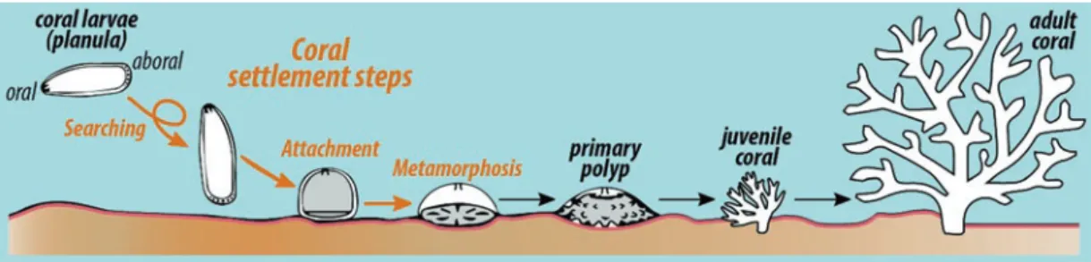
of H. purpurascens. As larval settlement can be distinguished between searching, attachment, and metamorphosis (see Fig. 8.9; exemplarily shown for coral larvae), it is question- able if a single cue can induce the settlement chain or if sev-
Fig. 8.8 (a) Jacaranone (Yvin et al. 1985), (b) histamine (Williamson et al. 2000), (c) tetrabromopyrrole (Tebben et al. 2011), (d) 11- deoxyfistularin-3 (Kitamura et al. 2007), and (e) luminaolide (Maru et al. 2013) (created with ChemDraw, v. 16.0.1.4)
Fig. 8.9 Settlement and early life stages of scleractinian corals. This figure highlights on the first steps of a coral larvae (searching, attachment, and metamorphosis) toward an adult coral
eral different cues are sequentially involved. Several studies on the settlement of different marine invertebrate larvae have indeed proven that single molecules can induce the whole chain of settlement, although only a few catalysts have been fully chemically characterized (Yvin et al. 1985; Pawlik 1986; Pawlik et al. 1991; Tsukamoto et al. 1999; Swanson et al. 2004; Dreanno et al. 2006). Only three of the latter studies have supported the ecological role of their investi- gated signaling molecules by also applying ecologically realistic concentrations (Pawlik 1986; Swanson et al. 2004;
Dreanno et al. 2006). Meanwhile, tetrabromopyrrole (Fig. 8.8c), a tetrabrominated pyrrole, has been isolated from a marine Pseudoalteromonas sp. associated to the crustose coralline algae (CCA) Neogoniolithon fosliei, showing set- tlement activity in larvae of the branching stony coral Acropora millepora. Interestingly, coral larvae directly underwent metamorphosis by developing into primary pol- yps within a few hours, but only a small amount of them conducted attachment to the substratum, a process nor- mally administered before metamorphosis is initiated (Tebben et al. 2011). Somewhat the same applies to 11- deoxyfistularin-3 (Fig. 8.8d), a bromotyrosine deriva- tive that has been isolated from an unnamed CCA overgrow- ing coral rubble collected in Okinawa, Japan. This secondary metabolite induced solely metamorphosis in larvae of the scleractinian coral Pseudosiderastrea tayamai.
Metamorphosis activity was further enhanced by the addi- tion of fucoxanthinol and fucoxanthin, which are two carot- enoids that had been isolated from the same CCA as well (Kitamura et al. 2007). Given the high number of studies on marine invertebrate settlement, it is very likely that addi- tional examples of larvae relying on waterborne or surface- bound cues for gregarious and associative settlement will be described in the future. Furthermore, we are convinced that future studies will not only focus on the discovery of novel chemical settlement cues but also provide more information on their role in the mechanism of the settlement cascade and on their broader ecological functions.
8.3.2 Coral Larvae Settlement: Search for Novel Settlement Cues
Coral reefs are among the world’s most diverse ecosystems and serve as nursery grounds and feeding areas for many reef-dependent animal species. Due to their relative complex physical structure, coral reefs shape the otherwise flat sea floor into a three-dimensional structure that provides a com- bination of food and shelter for a high biomass of commer- cially important fish species and other associated fauna (Moberg and Folke 1999). Besides their manifold ecosystem services, coral reefs affect humankind by having a major
impact on economy and politics. Coral reefs provide food via commercial fisheries (Pauly et al. 2002), protect coastlines from destruction by waves (Barbier et al. 2011), and generate income from food and tourism (Bellwood et al. 2004).
Unfortunately, corals reefs are also highly threatened ecosys- tems with some local and global factors being responsible.
Local factors include declining water quality, destructive fishing, and increased pollution from urban areas. Global factors are global warming and ocean acidification, due to a dramatic rise of carbon dioxide levels in the atmosphere over the past century (Hoegh-Guldberg et al. 2007). Stony “reef- building” corals (Scleractinia) live in symbiosis with micro- algae named zooxanthellae, which provide their coral hosts with up to 90% of their energy through photosynthesis (Stanley 2006). This relationship can be disrupted by envi- ronmental stressors such as long-lasting temperature increases together with intense periods of high sun irradi- ance. As a response to the latter stressor, corals expel their algae and thus lose their photosynthetic pigments at the same time, leading to the phenomenon of skeletal-looking bright white corals, a process better known as “coral bleaching”
(Ainsworth et al. 2016; Heron et al. 2016). Such bleaching events have been increasing in the last two decades, thereby affecting reefs on a global scale. The severity of events caused coral mortalities of over 60% in some locations (Eakin et al. 2010; Hughes et al. 2017). Predicted impacts of persistent bleaching events include a reduction of reef biodi- versity and coral cover, up to the total extinction of local coral species (Brainard et al. 2011, 2013). Furthermore, ocean acidification decelerates the calcification of corals by reducing the concentration of carbonate (CO32−), and thus making it even less available for marine calcifiers (Hoegh- Guldberg et al. 2007). To counteract the fast and massive coral decline, a better understanding of the recovery and population dynamics of stony corals needs to be developed.
because threatened coral reef systems depend on the recruit- ment of new individuals (Mumby and Steneck 2008). The recruitment process can be divided into (1) the development of competent larvae in the water column (spawners) or within the corals itself (brooders), (2) the settlement (searching, attachment, and larval metamorphosis) onto suitable sub- strata (Fig. 8.9), and (3) the survival of juvenile corals (Ritson-Williams et al. 2009). Since the survival rate of juve- nile corals is likely influenced by the type of substratum cho- sen for settlement (Harrington et al. 2004; Ritson-Williams et al. 2010), finding a suitable settlement ground may be a critical step within the recruitment process.
Although chemical cues are believed to serve as the pri- mary determinants of coral settlement, to some extent, physi- cal properties have shown to influence coral larvae settlement as well. In a field study, larvae of five different species of scler- actinian corals, including Goniastrea favulus, G. aspera,
Acropora tenuis, Oxypora lacera, and Montipora peltiformis, have shown to favor locations with lower light intensity (Mundy and Babcock 1998). Also, a study by Mason et al.
(2011) demonstrated that both larvae of Porites astreoides and Acropora palmata consistently settled on different red or red- orange plastic materials while, at the same time, disdaining other colors such as green, blue, or white. It was suggested that this consistent response to red or reddish surfaces is related to long-wavelength photosensitivity and thus might be a poten- tial strategy to artificially promote coral larvae settlement.
Over the past decades, many studies have shown that coral larvae settle in response to either live CCA or organic extracts of CCA. For instance, larvae of the agariciid corals Agaricia tenuifolia, A. humilis, and A. agaricites have reported to settle to different degrees of stringency and specificity on Caribbean CCA, specifically Hydrolithon boergesenii. The responsible morphogenic inducer was fractionated by ultrafiltration and shown to be a water-insoluble, ether- insoluble, and acetone- insoluble unstable biochemical, which is apparently associ- ated with the cell walls of the inducing CCA (Morse et al.
1988). Further studies by Morse and Morse (1991) have shown that the inductive molecule is a sulfated glycosaminoglycan. A field study of Price (2010) showed that larvae of several scler- actinian corals, including Pocillopora spp., Acropora spp., and Porites spp., do indeed prefer specific CCA species for in situ settlement, as they recruited more frequently on Titanoderma prototypum than on other CCA. A similar settlement specific- ity has been demonstrated for larvae of the scleractinian corals A. palmata and Montastraea faveolata. The latter two species have been tested for their rates of settlement on ten different species of red algae, including eight different CCA species, resulting in strong settlement preferences of larvae from both corals to different CCA. A. palmata settled on surfaces of H.
boergesenii, Lithoporella atlantica, Neogoniolithon affine, and Titanoderma prototypum, but showed no settlement on N.
mamillare. Larvae of M. faveolata settled on surfaces of Amphiroa tribulus, H. boergesenii, N. affine, N. munitum, and T. prototypum, but no settlement occurred on N. mamillare, Porolithon pachydermum, and a noncoralline Peyssonnelia sp.
The authors of this study suggested that patterns of coral dis-
tribution might be dependent on the red algae distribution (Ritson-Williams et al. 2014). However, in many cases of coral larvae settlement, the chemical identity of the pre- sumed settlement-inducing molecule is just poorly described or remains largely unknown. In the past, the identity of these CCA-associated chemical cues was presumed to be cell wall-bound and thought to be some kind of high molecular mass polysaccharides (Morse and Morse 1991; Morse et al.
1994, 1996). Other studies chemically fully described the chemical signaling molecules; however, the detected cue often did not initiate the entire settlement cascade (e.g., 11-deoxyfistularin-3, Fig. 8.8d) (Kitamura et al. 2007) or just function as a settlement enhancer, such as luminaolide (Fig. 8.8e). The macrodiolide luminaolide was originally isolated from the CCA H. reinboldii and greatly enhanced the metamorphosis activity in Leptastrea purpurea when combined with another fraction that eluted at 80% aqueous methanol by octadecyl silica gel column chromatography (Kitamura et al. 2009; Maru et al. 2013). Interestingly, chem- ical inducers for larval settlement were also discovered in coral rubble and the skeleton of the massive coral Goniastrea sp. (Heyward and Negri 1999), indicating that coral larvae settlement can be either induced by a variety of chemical cues or by specific cues from multiple sources. In the past, bacterial biofilms have received notable attention as suitable settlement ground for many marine invertebrate larvae (Johnson et al. 1991; Pawlik 1992; Huang and Hadfield 2003; Huggett et al. 2006; Hadfield 2011). It was shown that coral reef biofilms, which were more than 2 weeks old, are able to induce settlement in the scleractinian A. microph- thalma. FISH (fluorescence in situ hybridization) analysis revealed that the overall community composition of these biofilms was dominated by classes of Alphaproteobacteria, Betaproteobacteria, Gammaproteobacteria, and Cytophagia- Flavobacteria of Bacteroidetes (Webster et al. 2004). Apart from bacterial multispecies biofilms, a specific strain belong- ing to the genus Pseudoalteromonas (Pseudoalteromonas A3) isolated from the CCA H. onkodes was able to induce full settlement, including attachment and metamorphosis, in the reef- building corals A. willisae and A. millepora
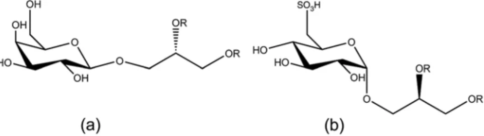
Fig. 8.10 Chemical structures of (a) (2S)-1-O-(7Z,10Z,13Z- hexadecatrienoyl)-3-O-β-D-galactopyranosyl-sn-glycerol and (b) (2R)-1
-O-(palmitoyl)-3-O-α-D-(6′-sulfoquinovosyl)-sn-glycerol (Tebben et al.
2015) (created with ChemDraw, v. 16.0.1.4)
(Negri et al. 2001). Another metamorphosis-inducing cue, named tetrabromopyrrole, was also isolated from a Pseudoalteromonas sp. (Tebben et al. 2011). This compound might have widespread importance among Caribbean corals as it induced settlement in the brooder Porites astreoides as well as in the spawning species Orbicella franksi and A. pal- mata (Sneed et al. 2014). Further studies on the ecological relevance of Pseudoalteromonads and tetrabromopyrrole in the coral settlement process revealed that the respective bacte- ria and its compound did not elicit the same rates of coral lar- vae settlement as CCA and instead introduced morphogenic processes that are often fatal to the larvae. Instead it was found that CCA-derived molecules, belonging to the chemical classes of glycoglycerolipids (Fig. 8.10a, b) and high molecu- lar weight polysaccharides, are the major contributors of the mixed fractions and caused larval settlement at equivalent concentrations present in live CCA (Tebben et al. 2015).
8.4 Conclusions
Secondary metabolites are investigated for their outstanding pharmaceutical applications as well as for their ecological relevance. Many MNPs have been found to elicit a broad range of bioactivities and, therefore, continue to be a prolific source for the generation of new drugs or drug leads. We believe that the exploration of new and extreme habitats will advance the discovery of novel macro- and microorganisms and, thus, might lead to the detection and isolation of novel NPs. Specifically, marine fungi still represent an underesti- mated but rich source for new SMs, although their distribu- tion and ecological role often remains scarce. The hyphenation of state-of-the-art techniques such as chromato- graphic separation, mass spectrometry, and nuclear magnetic resonance spectroscopy is a suitable way to facilitate NP screening effort. Particularly, the use of multiple secondary metabolite databases as well as MS/MS approaches in com- bination with molecular networking makes the search for novel NPs more efficient and, at the same time, lowers the risk of rediscovery. In terms of chemical ecology, SMs fulfill manifold roles for their producers. Besides predator-prey and algae-herbivore interactions, marine chemical ecology has also shifted its focus on marine invertebrate settlement behavior. Specifically, the role of SMs as signaling mole- cules for coral larvae settlement has gained interest during the last decades. CCA and their associated microorganisms are the best-known sources for coral larvae settlement cues, but until today, only few settlement compounds have been chemically fully described. Furthermore, the knowledge of the interplay between coral larva, settlement cue, settlement cue-producing organism (may it be the CCA or its associated
microbes), and other environmental factors such as light intensity is still limited and needs to be improved for a deeper comprehension of coral reef functioning. We are only at the beginning of understanding the role of SMs in the marine environment and many fascinating discoveries are yet to come.
Acknowledgments This study was carried out in the framework of the PhD research training group “The Ecology of Molecules” (EcoMol) supported by the Lower Saxony Ministry for Science and Culture. We also thank the reviewers for valuable comments which helped to improve the manuscript.
Appendix
This article is related to the YOUMARES 9 conference ses- sion no. 9: “Biodiversity of Benthic Holobionts: Chemical Ecology and Natural Products Chemistry in the Spotlight.”
The original Call for Abstracts and the abstracts of the pre- sentations within this session can be found in the Appendix
“Conference Sessions and Abstracts”, Chapter “7 Biodiversity of Benthic Holobionts: Chemical Ecology and Natural Products Chemistry in the Spotlight”, of this book.
References
Ainsworth TD, Heron SF, Ortiz JC et al (2016) Climate change dis- ables coral bleaching protection on the Great Barrier Reef. Science 352:338–342
Amend AS, Barshis DJ, Oliver TA (2012) Coral-associated marine fungi form novel lineages and heterogeneous assemblages. ISME J 6:1291–1301
Asolkar RN, Freel KC, Jensen PR et al (2009) Arenamides A–C, cyto- toxic NFκB inhibitors from the marine actinomycete Salinispora Arenicola. J Nat Prod 72:396–402
Bai R, Pettit GR, Hamel E (1990) Dolastatin 10, a powerful cytostatic peptide derived from a marine animal. Inhibition of tubulin polym- erization mediated through the vinca alkaloid binding domain.
Biochem Pharmacol 39:1941–1949
Barbier EB, Hacker SD, Kennedy C et al (2011) The value of estuarine and coastal ecosystem services. Ecol Monogr 81:169–193
Bell JJ, Smith D (2004) Ecology of sponge assemblages (Porifera) in the Wakatobi region, south-east Sulawesi, Indonesia: richness and abundance. J Mar Biol Assoc UK 84:581–591
Bell JJ, Davy SK, Jones T et al (2013) Could some coral reefs become sponge reefs as our climate changes? Glob Change Biol 19:2613–2624
Bellwood DR, Hughes TP, Folke C et al (2004) Confronting the coral reed crisis. Nature 429:827–833
Bérdy J (2005) Bioactive microbial metabolites. A personal view.
J Antibiot 58:1–26
Bergmann W, Burke DC (1955) Contributions to the study of marine products. XXXIX. The nucleosides of sponges. III. Spongothymidine and spongouridine. J Org Chem 20:1501–1507