AIMS Microbiology, 2(2): 205-221.
DOI: 10.3934/microbiol.2016.2.205 Received: 15 February 2016
Accepted: 06 June 2016 Published: 14 June 2016 http://www.aimspress.com/journal/microbiology
Research article
Diversity of Micromonospora strains from the deep Mediterranean Sea and their potential to produce bioactive compounds
Andrea Gärtner 1, Jutta Wiese 1, and Johannes F. Imhoff 1,2,*
1 GEOMAR Helmholtz Center for Ocean Research Kiel, RD3 Marine Microbiology, 24105 Kiel, Germany
2 Christian-Albrechts University of Kiel, 24118 Kiel Germany
* Correspondence: Email: jimhoff@geomar.de; Tel: +49-431-600-4450; Fax: +49-431-600-4482.
Abstract: During studies on bacteria from the Eastern Mediterranean deep-sea, incubation under in situ conditions (salinity, temperature and pressure) and heat treatment were used to selectively enrich representatives of Micromonospora. From sediments of the Ierapetra Basin (4400 m depth) and the Herodotos Plain (2800 m depth), 21 isolates were identified as members of the genus Micromonospora. According to phylogenetic analysis of 16S rRNA gene sequences, the Micromonospora isolates could be assigned to 14 different phylotypes with an exclusion limit of ≥ 99.5% sequence similarity. They formed 7 phylogenetic clusters. Two of these clusters, which contain isolates obtained after enrichment under pressure incubation and phylogenetically are distinct from representative reference organism, could represent bacteria specifically adapted to the conditions in situ and to life in these deep-sea sediments. The majority of the Micromonospora isolates (90%) contained at least one gene cluster for biosynthesis of secondary metabolites for non-ribosomal polypeptides and polyketides (polyketide synthases type I and type II). The determination of biological activities of culture extracts revealed that almost half of the strains produced substances inhibitory to the growth of Gram-positive bacteria. Chemical analyses of culture extracts demonstrated the presence of different metabolite profiles also in closely related strains.
Therefore, deep-sea Micromonospora isolates are considered to have a large potential for the production of new antibiotic compounds.
Keywords: Micromonospora; deep sea; natural products; PKS; NRPS
206
AIMS Microbiology Volume 2, Issue 2, 205-221.
Abbreviations:
PKS: polyketide synthase
NRPS: nonribosomal polypeptide-synthetase PT: phylotype
HPLC: high pressure liquid chromatography MS: molecular mass
MeCN: acetonitrile nt: nucleotide
1. Introduction
Members of the Actinomycetales are widely distributed in terrestrial as well as in aquatic and also marine ecosystems, where they are supposed to play an important role in the decomposition and recycling of biomaterials [1]. Maldonado et al. described first obligate marine species of the new genus Salinispora: Salinispora arenicola and Salinispora tropica [10]. Furthermore, two new genera, Salinibacterium and Serinicoccus were described containing solely marine isolates that tolerate high concentrations of sodium chloride [11,12]. They even have been recovered from the world’s deepest ocean trench, the Mariana Trench [2–4]. A controversial discussion exists about the question whether they are metabolically active in marine sediments or have been washed into the marine environment where they are just prevailing as spores. Culture experiments using marine salinity showed that many strains grow very well under these conditions and therefore appear well adapted to the marine environment [5,6]. In fact, evidence accumulates that they are active members of the microbial community in marine sediments [6–9].
A specific feature of the actinomycetes is their enormous contribution to the production of chemically diverse secondary metabolites and clinically relevant antibiotics. Indeed, the majority of bioactive compounds of microbial origin originates from actinomycetes and in particular from representatives of the genera Streptomyces and Micromonospora [13,14]. Novel biological active compounds have also been found in actinomycetes isolated from the marine realm [1,15–17]. These findings are a clear indication that actinomycetes from the marine environment may represent a valuable source for the discovery of novel natural products which has so far not been intensively studied. The genus Micromonospora was described by Orskov in 1923 and is represented by a total of 61 species and 7 subspecies in 2016 [18,19]. The first antibiotic isolated from a Micromonospora strain was micromonosporin [20]. After the discovery of the aminoglycoside antibiotic gentamicin in 1963 an intensive screening for new natural products in Micromonospora strains started [21]. Today more than 400 compounds are known that have been isolated from members of the genus Micromonospora [22]. The great potential of marine Micromonospora isolates to produce bioactive natural products was also highlighted by a study of marine actinomycetes isolated from Pacific sediments [23] which demonstrated strong anti-tumor activity of several culture extracts of Micromonospora strains. Cytotoxic activity against several human tumor cell lines was also observed for the levantilides, 20-membered cytotoxic macrolides produced by a deep-sea Micromonospora strain isolated from a Mediterranean deep-sea sediment sample in a previous study [24].
We have studied the bacterial diversity in the eastern basin of the Mediterranean Sea, which is
207
one of the most oligotrophic regions of the world’s oceans [28]. The extreme depletion of nutrients, in particular phosphorus, results in low primary production in waters distant from the coast [25].
Based on the poor primary production, minor amounts of organic matter reach the deep-sea floor of the Eastern Mediterranean Sea, in particular in one of the deep basins, the Ierapetra Basin, which is located approximately 30 nautic miles southeast of Crete. Studies on the macrofauna community showed that sediments in the Eastern Mediterranean Sea can be considered as event-driven ecosystems and the biota need to be adapted to few and pulsed nutrient input events [26]. In addition, the Mediterranean deep sea is characterized by a comparably high temperature, which may exceeds 13 °C in deep waters [27]. The unique and extreme conditions of the Eastern Mediterranean deep sea require specific adaptation of its inhabitants and therefore these sediments are an attractive source for the study of actinomycetes. In a previous study on the cultured biodiversity from Mediterranean deep sea sediments, representatives of Bacillus and Actinobacteria were found as major groups, the Actinobacteria representing almost a third of all isolated bacteria [28]. This result animated to perform the present study in which a highly diverse group of Actinobacteria were selectively enriched from Mediterranean sediments (4400 m and 2800 m deep). In particular, members of the genus Micromonospora, which are rarely described from deep-sea habitats, were identified and their potential to produce antibiotic compounds was studied.
2. Materials and Method 2.1. Sample collection
All samples were obtained during the Meteor research cruise 71 leg 2 southwards of Crete in January 2007. Sediment samples from two different sites at 4400 m (Ierapetra Basin; 34°30.296N, 26°11.507E) and 2800 m depth (Herodotos Plain; 33°42.989N, 26°20.329E) were collected using a multiple corer. The uppermost 5 cm of each sediment core were aseptically sub-sampled (from the Herodotos Plain also a sub-sample at 25 cm depth) and stored at 4°C until further treatment.
2.2. Isolation of Actinobacteria
15 Micromonospora strains as well as one representative each of the genera Knoellia (A207) and Marmoricola (A214), respectively, were isolated after pre-treatments with pressure incubation and/or heat treatment of the sediment samples (Table 1).
Pressure incubation. In order to selectively enrich deep-sea bacteria (not solely Actinomycetes) that are adapted to elevated hydrostatic pressure and spontaneous nutrient input, sediment samples were pre-incubated at in situ pressure and in situ temperature in sea water supplemented with N-acetyl-D-glucosamine prior to plating on agar media. For this purpose 10 ml of sediment were transferred into a plastic bag with addition of 9 ml of sterile-filtered Mediterranean seawater (taken from the multicorer) and 1 ml N-acetyl-D-glucosamine solution with a concentration of 100 µM.
N-acetyl-D-glucosamine (the monomer of chitin) is supposed to be an important carbon and nitrogen source for the microbial deep-sea community. The samples were incubated in compression-proof steel tubes at 280 bar (samples from the Herodotos Plain) or 440 bar (samples from the Ierapetra Basin) hydrostatic pressure and at 13.5 °C for 6 d.
208
AIMS Microbiology Volume 2, Issue 2, 205-X Page.
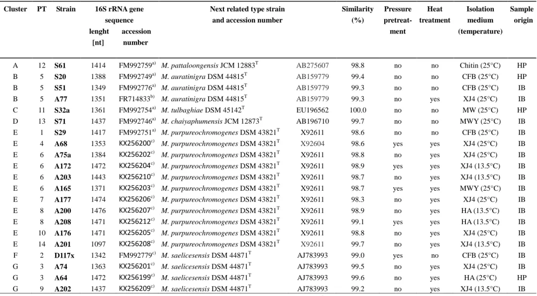
Table 1. Cultivation conditions and phylogenetic analyses of Micromonospora strains from the Eastern Mediterranean deep sea sediment.
Cluster PT Strain 16S rRNA gene sequence lenght accession [nt] number
Next related type strain and accession number
Similarity (%)
Pressure pretreat-
ment
Heat treatment
Isolation medium (temperature)
Sample origin
A 12 S61 1414 FM992759a) M. pattaloongensis JCM 12883T AB275607 98.8 no no Chitin (25°C) HP
B 5 S20 1388 FM992749a) M. auratinigra DSM 44815T AB159779 99.4 no no CFB (25°C) HP
B 5 S51 1349 FM992776a) M. auratinigra DSM 44815T AB159779 99.3 no no CFB (25°C) IB
B 5 A77 1351 FR714833b) M. auratinigra DSM 44815T AB159779 99.3 no yes XJ4 (25°C) IB
C 11 S32a 1361 FM992754a) M. tulbaghiae DSM 45142T EU196562 100.0 no no MW (25°C) HP
D 13 S71 1437 FM992746a) M. chaiyaphumensis JCM 12873T AB196710 99.7 no no MWY (25°C) IB E 1 S29 1417 FM992751a) M. purpureochromogenes DSM 43821T X92611 98.6 no no CFB (25°C) IB E 4 A68 1353 KX256200c) M. purpureochromogenes DSM 43821T X92604 98.6 yes yes XJ4 (25°C) IB E 6 A75a 1384 KX256202c) M. purpureochromogenes DSM 43821T X92611 98.8 no yes XJ4 (25°C) IB E 6 A172 1472 KX256204c) M. purpureochromogenes DSM 43821T X92611 98.9 yes yes XJ4 (13.5°C) IB E 6 A203 1443 KX256210c) M. purpureochromogenes DSM 43821T X92611 98.7 no yes XJ4 (13.5°C) IB E 6 A165 1371 KX256203c) M. purpureochromogenes DSM 43821T X92611 98.7 yes yes MWY (25°C) IB E 7 A177 1474 KX256206c) M. purpureochromogenes DSM 43821T X92611 98.3 no yes XJ4 (25°C) IB E 8 A200 1476 KX256207c) M. purpureochromogenes DSM 43821T X92611 98.9 no yes HA (13.5°C) IB E 8 A208 1471 KX256212c) M. purpureochromogenes DSM 43821T X92611 99.1 yes yes HA (13.5°C) IB E 10 A176 1471 KX256205c) M. purpureochromogenes DSM 43821T X92611 98.8 no yes XJ4 (25°C) IB E 14 A201 1097 KX256208c) M. purpureochromogenes DSM 43821T X92611 99.7 no yes XJ4 (13.5°C) IB
F 2 D117x 1342 FM992779c) M. saelicesensis DSM 44871T AJ783993 99.0 yes no CFB (25°C) IB
G 3 A74 1363 KX256201c) M. saelicesensis DSM 44871T AJ783993 99.5 no yes XJ4 (25°C) IB
G 3 A64 1472 KX256199c) M. saelicesensis DSM 44871T AJ783993 99.6 no yes HA (25°C) HP
G 9 A202 1437 KX256209c) M. saelicesensis DSM 44871T AJ783993 99.2 no yes XJ4 (13.5°C) IB
IB: Ierapetra Basin (4400 m); HP; Herodotos Plain (2800 m); PT: phylotype; a) from [28]; b) from [24]; c) this study;
Heat treatment. For selective isolation of Actinobacteria and other spore-forming bacteria, 2 g of the sediment samples were dried in a sterile Petri dish at room temperature for 3 months. Dry sediment was then heated to 120 °C for 1 hour and re-suspended in 19 ml deionized water. 200 µl of this suspension were plated on the isolation media.
Combination of both pre-treatments. A few bags were dried after the pressure incubation and in addition exposed to the heat treatment.
The following isolation media were used: a) CFB-medium: 15 g agar, 1 g tryptone, 0.5 g yeast extract, 0.5 g CaCl2·2H2O, 0.5 g MgCl2·7H2O in 1 L Mediterranean seawater; b) XJ4-medium: 15 g agar, 0.1 g histidine, 1 g raffinose, 0.5 g NaHPO4, 1.7 g KCl, 0.05 g MgSO4·4H2O, 0.01 g FeSO4·7H2O, 0.02 g CaCO3, 0.5 mg each of thiamine, riboflavine, niacine, pyridoxin, Ca-panthotenate, inositol and p-aminobenzoic acid, 0.25 mg biotin and 50 ppm K2Cr2O7 in 1 L of aquadest with addition of cycloheximide (50µg/ml); c) HA-medium: 1 g humic acid, 0.01 g FeSO4, 0.5 g Na2HPO4, 1.7 g KCl, 0.02 g CaCO3, 0.5 g asparagine and 50 ppm K2Cr2O7 in 1 L of deionized water; and d) MWY- medium: 15 g agar and 0.001 g yeast extract in 1 L Mediterranean seawater added with 50 µg/mL cycloheximide and penicillin G. Agar plates were incubated at 13.5 °C (in situ temperature) and 25 °C and checked regularly for growth. Using a binocular microscope the morphology of all colonies was checked and those appearing morphologically different were transferred to fresh agar medium until pure cultures were obtained. Isolates were stored at −80 °C using the Cryobank system (Mast Diagnostica GmbH, Reinfeld, Germany).
With the aim to test the capability of all Micromonospora sp. strains to cope with the availability of low nutrient concentrations, they were cultivated on a medium consistent of 1L Mediterranean Sea water supplemented with 15 g agar at 13.5 °C.
2.3. Phylogenetic analyses
DNA extraction, 16S rRNA gene amplification and sequencing were performed as described previously [29]. 16S rRNA gene sequences obtained during this study, were submitted to the GenBank database and were assigned accession numbers KX256199 - KX256213. Sequences were compared to NCBI database sequences using BLAST search [30]. Sequence alignment of Micromonospora sp. isolates, next related type strains and further representatives of the genus was performed using the ARB database [31]. Phylogenetic trees were calculated applying maximum likelihood analysis with PhyML software [32]. This analysis was performed assuming the GTR evolution model with an optimized gamma distribution parameter alpha and 100 bootstrap replicates [33]. For systematic consideration, sequences with similarities higher than 99.5 % were grouped as a phylotype together using Mothur [34].
2.4. Amplification of PKS-I, PKS-II and NRPS genes
DNA extracts of Micromonospora strains were analyzed for the presence genes for representative natural product biosynthesis pathways such as the polyketide synthase I and II (PKS-I and PKS-II) and non-ribosomal polypeptide synthetase (NRPS). The sequences of the respective primers are listed in Table 2. Detection of PKS-II genes was performed using the primers and PCR conditions as described by Metsä-Ketelä et al. [35]. For gene amplification, 50 pMol primer, 3.5 U TaqPolymerase, 10% DMSO and 5 µl template were applied. PKS-I and NRPS PCR assays were
210
AIMS Microbiology Volume 2, Issue 2, 205-221.
modified according to Ayuso-Sacido and Genilloud using PuReTaq® Ready-To-Go PCR Beads (GE Healthcare, Solingen, Germany) and 50 pMol of the primers K1 and M6R for the PKS-I gene [36].
The same system was used for amplification of NRPS genes with primers A3 and A7R and in addition primers A2f and A3r were used for amplification of NRPS according to the protocol of Martens et al., resulting in fragment length of 700 nt (NRPSlarge) and 300 nt, (NRPSshort), respectively [37]. PCR products were purified and sequenced using the BigDye Terminator v1.1 Sequencing Kit (Applied Biosystems, Life Technologies GmbH, Darmstadt, Germany) in a 3730-DNA-Analyzer (Applied Biosystems) as specified by the manufacturer. Sequences were edited by ChromasPro v.1.33, translated to amino acid sequences using BioEdit and compared to the NCBI database using BLAST search [30,38]. Matches of the blastx search were checked for sequence similarities and coverage regarding preferably genes of Micromonospora sp.
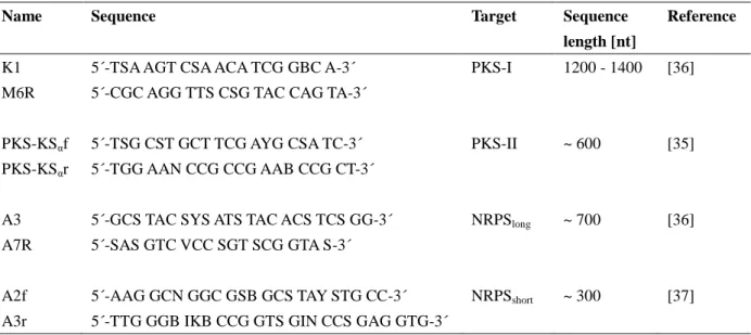
Table 2. Primers used in this study for the detection of PKS and NRPS gene fragments.
Name Sequence Target Sequence
length [nt]
Reference
K1 5´-TSA AGT CSA ACA TCG GBC A-3´ PKS-I 1200 - 1400 [36]
M6R 5´-CGC AGG TTS CSG TAC CAG TA-3´
PKS-KSαf 5´-TSG CST GCT TCG AYG CSA TC-3´ PKS-II ~ 600 [35]
PKS-KSαr 5´-TGG AAN CCG CCG AAB CCG CT-3´
A3 5´-GCS TAC SYS ATS TAC ACS TCS GG-3´ NRPSlong ~ 700 [36]
A7R 5´-SAS GTC VCC SGT SCG GTA S-3´
A2f 5´-AAG GCN GGC GSB GCS TAY STG CC-3´ NRPSshort ~ 300 [37]
A3r 5´-TTG GGB IKB CCG GTS GIN CCS GAG GTG-3´
2.5. Production of culture extracts
For HPLC-UV/MS analysis of the metabolite profiles and for antimicrobial test assays, culture extracts of all Micromonospora strains were prepared. Strains were cultivated in 100 ml liquid starch-peptone medium (1 g starch, 0.5 g soy-peptone and 1.5 g Tropic Marine® sea salt in 100 ml deionized water) using 300 ml Erlenmeyer-flasks with one baffle at 120 rpm. After incubation for 8 days at 28 °C 100 ml ethyl acetate (Picograde®, Promochem, Wesel, Germany) were added and the culture broth was homogenized using an Ultra-Turrax T25 basic disperser at 17500 rpm for 30 sec.
The supernatants were separated, dried and re-suspended in 1 ml methanol.
With the aim to study the effect of salt on the production of the compounds of interest, i.e.
S20-4, S20-5. S20-6, and S20-7, culture extracts of strain S20 were prepared after growth in starch-peptone medium with Mediterranean Sea water (3.86%) and 30 g/L Tropic Marine® sea salt, respectively, for 8 days at 28 °C.
2.6. HPLC analysis of culture extracts
Metabolite profiles of culture extracts were screened by HPLC-UV/MS. For reversed-phase
211
HPLC analysis, dried extracts were resolved in methanol. The measurements were carried out using a Phenomenex Onyx Monolithic C18 column (100 by 3.00 mm) on a VWR Hitachi Elite LaChrom system coupled to an electrospray ionization ion trap detector (Esquire 4000, Bruker Daltonics, Bremen). The solvents, water and acetonitrile (MeCN), were supplemented with 0.1% formic acid.
The gradient of MeCN was as follows: 0 min 5 %; 4 min 60 %; 6 min 100 %; flow rate: 2ml/min).
2.7. Fermentation of strain S20 and isolation of its metabolites
Micromonospora strain S20 was cultivated in 10 L of liquid starch-peptone medium (10 g starch, 5 g soy-peptone, 15 g Tropic Marine® sea salt and 1 g CaCO3 in 1L deionized water) using 2 L shaken Erlenmeyer-flasks with one baffle and filled with 1 L medium each. After 6 days of incubation at 28 °C and 120 rpm, the culture supernatant was separated from the cells by centrifugation at 8.000 rpm for 5 min (Beckmann_J2-MC). Afterwards the supernatant (9.5 L) was acidified by adding 20 ml HCl (15%) and transferred to an Amberlite XAD16-column. After washing the column with 500 ml 40% ethanol bound metabolites were eluted with 2 L 80% (v/v) ethanol and subsequently with 2 L 100% (v/v) ethanol. Both eluates were collected and freeze-dried. The dried extracts were dissolved in 5 ml methanol each and analyzed by HPLC-UV/MS for the presence of the compounds S20-4, S20-5, S20-6, and S20-7. Since the desired substances were found in both extracts, they were combined for further analyses. The compounds were isolated and purified by preparative reversed phase HPLC using a Phenomenex Gemini-NX column (C18, 5 μ, 110A, 100 × 21.20 mm) and a VWR Hitachi Elite LaChrom system consisting of an L-7150 pump, an L-2450 diode array detector, and an L-2200 autosampler. A water (A) / methanol (B) gradient (50% B to 100% B in 17 min; flow 15 ml/min) with 0.1% formic acid added to both solvents was applied.
2.8. Antimicrobial tests of culture extracts and metabolites
Antimicrobial activity of the culture extracts of the Micromonospora strains as well as of the metabolites produced by the strain S20 (S20-4, S20-5, S20-6, S20-7) was tested against Bacillus subtilis (DSM 347), Staphylococcus lentus (DSM 6672), Xanthomonas campestris (DSM 2405), and Candida glabrata (DSM 6425) using a resazurin assay according to Nagel at al. [38]. The concentration of culture extracts in the bioassay was 0.01 mg/200 µl, the compounds were tested at concentrations of 100 µM. A negative (no compound) and a positive control (100 µM of chloramphenicol for bacterial test strains and 100 µM of cycloheximid for the yeast) were measured on the same microtiter plate. All tests were run in triplicates.
3. Results
3.1. Isolation of Actinobacteria
Actinobacteria from Mediterranean deep-sea sediments were isolated after different pre-treatment procedures of the sediment samples, mostly from the uppermost 5 cm (Table 1).
Among 23 actinobacterial strains 21 belonged to the genus Micromonospora and one strain each affiliated to Knoellia and Marmoricola.
Ten Micromonospora strains as well as Marmoricola isolate A214 were obtained after treatment
212
AIMS Microbiology Volume 2, Issue 2, 205-221.
with dry heat on selective agar media. Marmoricola sp. strain A214 was isolated from the Herodotos Plain sediment after cultivation on XJ4-medium at 13.5 °C. It was the only isolate obtained from the 25 cm depth section of the sediment core. Four Micromonospora strains as well as Knoellia strain A207 were isolated after the combined pre-treatment approach, after incubation at in situ pressure with N-acetyl-D-glucosamine and subsequent heat treatment. The Knoellia sp. strain A207 was isolated originated from the Ierapetra Basin on HA-medium at 25 °C. Micromonospora sp. strain D117x was isolated after incubation at in situ pressure with enrichment on N-acetyl-D-glucosamine, without heat treatment. Six Micromonospora strains (S20, S29, S32a, S51, S61, and S71) were previously isolated from untreated sediment samples [28].
3.2. Identification and phylogenetic relations of the isolates
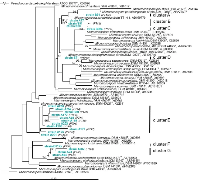
All 21 strains affiliating to the genus Micromonospora could be assigned to 14 different phylotypes (PTs) with an exclusion limit of ≥ 99.5% 16S gene sequence similarity (Table 1, Figure 1). The phylogenetic analysis of 16S rDNA gene sequences of the Micromonospora isolates revealed that most deep-sea strains were phylogenetically distinct from known reference organisms (Figure 1).
These phylotypes formed at least seven distinct clusters, in part with a single isolate and phylotype representing the cluster (clusters A, C, D, F). Cluster B (strains S20, S51 and A77) affiliates with Micromonospora auratinigra as next relative, cluster G (strains A64, A74, A202) affiliates with M.
saelicesensis and cluster E with M. purpureochromogenes (Table 1, Figure 1). Interestingly all strains of cluster G and cluster E (except strain S29) were isolated after heat treatment of the sediments and 4 out of 11 strains of cluster E in addition survived the pressure incubation. Despite their similarity to known reference species of Micromonospora, all isolates of clusters E, G and F (a single strain obtained after pressure treatment) formed separate branches in the phylogenetic tree (Figure 1). In addition to Micromonospora, members of rare actinomycetes were observed in the pre-treated sediment samples. Strain A207 affiliated to Knoellia subterranea DSM 12372T (AJ294413) with a sequence similarity of 97.9% (1476 nt) and strain A214 belonged to Marmoricola aurantiacus DSM 12652T (Y18629) with a sequence similarity of 99.2% (1439 nt).
213
Figure 1. Maximum likelihood phylogenetic subtree of 16S rRNA gene sequences from type strains affiliating to the genus Micromonospora as references and the Micromonospora strains derived from Eastern Mediterranean Sea deep-sea sediments.
The tree was constructed with 100 bootstrap replicates using phyML software. Despite the low bootstrap values, multiple calculations of the tree always resulted in identical clusters of the sequences. PT = number of phylotypes
3.3. Potential of Micromonospora strains to produce natural products
Culture extracts of almost half of the Micromonospora strains showed antimicrobial activity against Gram-positive bacteria but none inhibited growth of Gram-negative and eukaryotic test strain.
Ten strains showed antimicrobial activity against B. subtilis and most of these in addition exhibited antimicrobial activity against S. lentus (Table 3). In addition, 10 strains without antibiotic activity contained at least one of the tested biosynthetic genes for natural products of the PKS and/or NRPS type.
214
AIMS Microbiology Volume 2, Issue 2, 205-221.
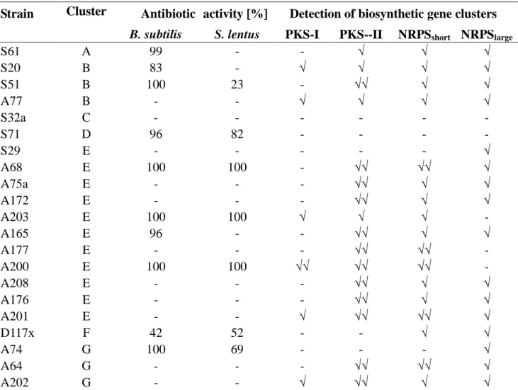
Table 3. Antibiotic activity of deep-sea Micromonospora strains and detection of gene clusters for PKS-I, PKS-II and NRPS.
Strain Cluster Antibiotic activity [%] Detection of biosynthetic gene clusters
B. subtilis S. lentus PKS-I PKS--II NRPSshort NRPSlarge
S61 A 99 - - √ √ √
S20 B 83 - √ √ √ √
S51 B 100 23 - √√ √ √
A77 B - - √ √ √ √
S32a C - - - -
S71 D 96 82 - - - -
S29 E - - - √
A68 E 100 100 - √√ √√ √
A75a E - - - √√ √ √
A172 E - - - √√ √ √
A203 E 100 100 √ √ √ -
A165 E 96 - - √√ √ √
A177 E - - - √√ √√ -
A200 E 100 100 √√ √√ √√ -
A208 E - - - √√ √ √
A176 E - - - √√ √ √
A201 E - - √ √√ √√ √
D117x F 42 52 - - √ √
A74 G 100 69 - - - √
A64 G - - - √√ √√ √
A202 G - - √ √√ √ √
√: positive PCR amplification; √√: positive PCR amplification and confirmation by sequence information; -: negative test result.
In order to substantiate the potential of producing natural products of known biosynthetic pathways, the presence of gene clusters for polyketide synthases of type I and type II (PKS-I, PKS-II) and for non-ribosomal peptide synthetase (NRPS) was analysed in all Micromonospora strains (Table 3, Figure 2). The amplification of non-ribosomal peptide synthetase genes by two primer sets and PCR protocols resulted in gene fragments of different length (NRPSshort and NRPSlarge). With a few exception all strains contained NRPS genes, most exhibited both gene fragments and a few strains showed amplicons only in one or the other protocol. While genes for PKS-II were present in all but 5 isolates, PKS-I was detected only in a few strains of clusters B, E and G. Sequence similarity to PKS-I and PKS-II genes was in the range of 72% and 91% and that of NRPS genes to reference sequences was >50% and (Table 4).
For those strains, which showed no inhibitory effects on the growth of the test strains, but were positive for PKS and/or NRPS it is expected that the cultivation conditions used in this study were not appropriate to stimulate the biosynthesis of antibiotic compounds.
215
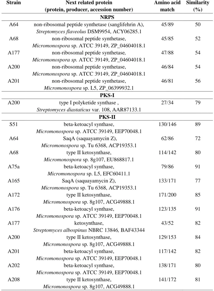
Table 4. NRPS, PKS-I and PKS-II gene products related to amino acid sequences derived from the respective PCR amplificates of Micromonospora strains.
Strain Next related protein
(protein, producer, accession number)
Amino acid match
Similarity (%) NRPS
A64 non-ribosomal peptide synthetase (sanglifehrin A), Streptomyces flaveolus DSM9954, ACY06285.1
45/89 50
A68 non-ribosomal peptide synthetase,
Micromonospora sp. ATCC 39149, ZP_04604018.1
45/85 52
A177 non-ribosomal peptide synthetase,
Micromonospora sp. ATCC 39149, ZP_04604018.1
47/88 54
A200 non-ribosomal peptide synthetase,
Micromonospora sp. ATCC 39149, ZP_04604018.1
46/84 54
A201 non-ribosomal peptide synthetase, Micromonospora sp. L5, ZP_06399932.1
46/81 56
PKS-I A200 type I polyketide synthase ,
Streptomyces diastaticus var. 108, AAR87133.1
27/34 79
PKS-II
S51 beta-ketoacyl synthase,
Micromonospora sp. ATCC 39149, EEP70048.1
130/146 89
A64 SaqA (saquayamycin Z),
Micromonospora sp. Tu 6368, ACP19353.1
62/86 72
A68 type II ketosynthase,
Micromonospora sp. 8g107, EU868817.1
114/142 80
A75a beta-ketoacyl synthase,
Micromonospora sp. L5, EFC60411.1
79/86 91
A165 SaqA (saquayamycin Z),
Micromonospora sp. Tu 6368, ACP19353.1
133/171 77
A172 type II ketosynthase,
Micromonospora sp. 8g107, ACG49888.1
171/200 85
A176 beta-ketoacyl synthase,
Micromonospora sp. ATCC 39149, EEP70048.1
123/135 91
A177 ketosynthase,
Streptomyces albospinus NBRC 13846, BAF43344
43/52 82
A200 type II ketosynthase,
Micromonospora sp. 8g107, ACG49888.1
129/153 84
A201 beta-ketoacyl synthase,
Micromonospora sp. ATCC 39149, EEP70048.1
117/142 82
A202 beta-ketoacyl synthase,
Micromonospora sp. ATCC 39149, EEP70048.1
138/171 80
A208 type II ketosynthase,
Micromonospora sp. 8g107, ACG49888.1
141/172 81
216
AIMS Microbiology Volume 2, Issue 2, 205-221.
Figure 2. Presence of PKS-I, PKS-II and NRPS biosynthesis gene clusters in deep-sea Micromonospora isolates.
3.4. Chemical metabolite profiles of selected strains
Culture extracts of Micromonospora strains showed large variation in the secondary metabolite profiles as analyzed by HPLC-MS/UV analysis. Similar metabolite profiles were found in representatives of cluster E (strains A177, A200, A203 and A208). While strains A64 and A74 were phylogenetically closely related by affiliating to cluster G, they showed different metabolite profiles.
In contrast, the already known compounds levantilide A and its derivative levantilide B were produced by members of different clusters, i.e. the original producer strain A77 [24] and strain S51, which affiliates both to cluster B, but also strain A74 (cluster G).
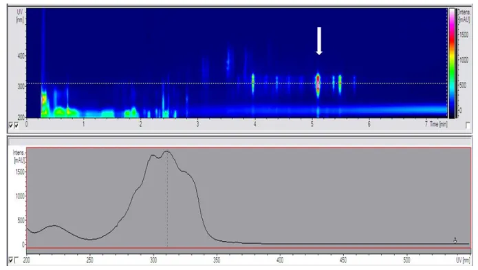
The HPLC-MS/UV profile of strain S20 revealed four unknown metabolites, which were not produced by any of the other strains tested. The main compound S20-4 exhibited a molecular weight of 940 (Figure 3). The production of this substance was initiated at the end of the log-phase and lasts throughout the stationary phase, but was independent from the salt concentration. It was found at salinities up to 3.0% of Tropic Marine® salt and also when seawater from the habitat (salinity of 3.9%) was used for cultivation. Three derivatives of this major compound (S20-5, S20-6, and S20-7) showing the molecular weights of 954, 982, and 940, respectively, were observed. Interestingly, these compounds showed variations in the bioactivity towards B. subtilis. While S20-7 exhibited no antibiotic activity, the compounds S20-4, S20-5 and S20-6 inhibited growth of B. subtilis with 99 %, 100 % and 32 % at 100 µM, respectively.
217
Figure 3. HPLC-UV profile of Micromonospora strain S20. Top: The DAD spectra of the chromatogram of the crude extract showed several compounds. The strongest UV signal was exhibited by the unknown compound (m/z = 940) which is marked with an arrow. Bottom: The UV spectrum of this compound is characterized by two maxima at 298 nm and 310 nm as well as a shoulder at 330nm.
4. Discussion
Even though reports on Actinobacteria isolated from marine habitats are accumulating, so far only few reports deal with Actinobacteria from deep-sea sediments. In the present study, Actinobacteria were isolated from an extreme oligotrophic marine deep-sea habitat, the majority of which was classified as belonging to the genus Micromonospora. This is the first detailed study on the phylogenetic diversity of deep-sea Micromonospora strains from the oligotrophic deep Mediterranean Sea and on their potential to produce bioactive natural products. Diversity, bioactivity and the potential for secondary metabolite biosynthesis of these bacteria were analyzed in this study.
4.1. Isolation and phylogenetic analyses of the Actinobacteria
A number of treatments have been applied to improve the isolation success of Actinobacteria.
Dry-heat has been found to be useful for the selective enrichment of non-streptomycete actinobacteria [40] and we have used this pre-treatment with dry heat to selectively enrich actinobacteria and to isolate members of Micromonospora, Knoellia and Marmoricola. The selectivity of this pre-treatment is demonstrated by the observation, that members of Streptomycetes were completely missing after the heat-treatment approach applied in this study. This finding demonstrates that in addition to the media and cultivation conditions the sample pre-treatment is important for the isolation of a diverse array of Actinobacteria. The increase in isolation efficiency of these Actinobacteria is of particular interest,
218
AIMS Microbiology Volume 2, Issue 2, 205-221.
because the diversity of non-streptomycete Actinobacteria is hardly covered in eubacterial environmental gene libraries.
The high diversity and number of phylotypes of the genus Micromonospora (7 clusters, 14 PTs) recovered from the deep-sea sediments in this study is remarkable. In a previous study on these sediments only two phylotypes of Streptomyces (a total of 8 strains) were recovered [28]. This is in accordance with a study at the Pacific coast off San Francisco in which recovery of actinomycetes from shallow versus deep sediments was compared [5]. These authors discovered an increasing diversity of Micromonospora relative to Streptomyces with increasing water depth. In the Trondheim Fjord (Norway), a higher abundance of Micromonospora strains was observed among isolates from deeper sediments (450 m) compared to shallow water sediments and Micromonospora isolates were the major group of actinobacteria in the deeper sediment samples [41].
Noteworthy, most Micromonospora isolates of the present study phylogenetically clustered apart from already described type strains of this genus and distinct clusters could be detected by phylogenetic analysis of the 16S rDNA gene sequences. Clusters E and F (D117x, A165, A68, A208 and A172) contain isolates obtained after pretreatment at in situ pressure and may be considered as (Table 1). As these strains obviously are adapted to changes in hydrostatic pressure (during sampling and incubation procedures) and were selected after pressure incubation, they might be considered to be well adapted to the conditions prevailing in the Mediterranean deep sea sediments. The survival of long lasting treatments of high hydrostatic pressure at low temperature apparently is common to Micromonospora strains and was already noticed earlier [40]. All Micromonospora isolates grew well under characteristic features of the deep Eastern Mediterranean Sea, at sea water from the habitat, at minor nutrient concentrations (i.e. Mediterranean Sea water supplemented with 15 g agar) and at low temperature (13.5°C). In conclusion, these strains cope well the conditions prevailing in situ, appear well adapted to the conditions of the Mediterranean deep sea and therefore may be regarded as indigenous inhabitants of the investigated sediments.
4.2. Natural products from deep-sea Micromonospora strains
As the deep sea generally is characterized by oligotrophy with spontaneous and punctual nutrient input, inhabitants must be adapted to these features and compete with others when nutrients become available. Therefore, the use of antimicrobial compounds in order to outcompete competitors when nutrient become available appears to be one reasonable strategy and might explain why secondary metabolite production is an important feature of deep-sea bacteria.
Antimicrobial activity of the Micromonospora strains was specifically targeted towards Gram-positive bacteria and approx. half of the tested strains inhibited growth of B. subtilis and half of these strains inhibited in addition S. lentus. If one considers, that Gram-positive bacteria and in particular members of the genus Bacillus are important players in the marine sediments, they can be expected being primary competitors of Actinobacteria. According to a former study, isolates from the Mediterranean deep-sea sediments were dominated by a highly diverse group of Gram-positive bacteria, in particular by members of the genus Bacillus, all of which cope quite well with minor nutrient concentrations [28]. Since the Bacillus strains and the Micromonospora strains have similar nutrient requirements, the production of antibiotics might be a useful tool of Micromonospora to compete against Bacillus in marine environments. Inhibition of Bacillus might be of advantage for the occurrence, persistence and growth of the antibiotic producing Micromonospora community. The
219
importance of bioactive secondary metabolites for Micromonospora is supported by the detection of multiple biosynthesis genes for secondary metabolite production (PKS-I, PKS-II and NRPS) in the great majority of these isolates (86%). As far as tested, the Micromonospora strains provide a rich source of compounds, which will be identified in further studies.
5. Conclusion
Growth characteristics of Micromonospora strains isolated from Mediterranean deep sea sediments and their affiliation to phylogenetic clusters apart from known type strains indicate their potential to adapt to the conditions prevailing in situ. At least some of them can cope with pressure conditions prevailing in these sediments. Therefore, they may be regarded as indigenous bacteria of these deep-sea sediments. They have a broad genetic potential to produce secondary metabolites, which quite likely is significantly exceeding those detected in the culture extracts. As we know, that cultivation conditions have strong impact on the production of secondary metabolites [42], the choice of media and cultivation conditions are of major importance for the production of secondary metabolites. Further experiments are expected to significantly enlarge the number of metabolites produced by the Micromonospora strains isolated from deep sea sediments.
Acknowledgments
The authors are grateful to Arlette Wenzel-Storjohann for taking care about the antibiotic assays, to Dr. Markus Schilhabel and his team at the Institute for Clinical Molecular Biology (University Hospital Schleswig-Holstein, Kiel, Germany) for providing Sanger sequencing and to the DFG for support through the Clusters of Excellence “Inflammation at interfaces” and “Future Ocean”.
Conflict of Interest
Authors declare no conflict of interest in this paper.
References
1. Fenical W, Jensen PR (2006) Developing a new resource for drug discovery: marine actinomycete bacteria. Nat Chem Biol 2: 666–673.
2. Pathom-aree W, Stach J, Ward A, et al. (2006a) Diversity of actinomycetes isolated from Challenger Deep sediment (10.898 m) from the Mariana Trench. Extremophiles 10: 181–189.
3. Pathom-aree W, Nogi Y, Sutcliffe IC, et al. (2006b) Dermacoccus abyssi sp. nov., a piezotolerant actinomycete isolated from the Mariana Trench. Int J Syst Evol Microbiol 56: 1233–1237.
4. Colquhoun JA, Heald SC, Li L, et al. (1998) Taxonomy and biotransformation activities of some deep-sea actinomycetes. Extremophiles 2: 269–277.
5. Prieto-Davó A, Fenical W, Jensen PR (2008) Comparative actinomycete diversity in marine sediments. Aquat Microb Ecol 52: 1–11.
6. Jensen PR, Dwight R, Fenical W (1991) Distribution of actinomycetes in near-shore tropical marine sediments. Appl Environ Microbiol 57: 1102–1108.
7. Jensen PR, Gontang E, Mafnas C, et al. (2005a) Culturable marine actinomycete diversity from
220
AIMS Microbiology Volume 2, Issue 2, 205-221.
tropical Pacific Ocean sediments. Environ Microbiol 7: 1039–1048.
8. Grossart HP, Schlingloff A, Bernhard M, et al. (2004) Antagonistic activity of bacteria isolated from organic aggregates of the German Wadden Sea. FEMS Microbiol Ecol 47: 387–396.
9. Moran MA, Rutherford LT, Hodson RE (1995) Evidence for indigenous Streptomyces populations in a marine environment determined with a 16S rRNA probe. Appl Environ Microbiol 61: 3695–3700.
10. Maldonado L, Stach J, Pathom-aree W, et al. (2005) Diversity of cultivable actinobacteria in geographically widespread marine sediments. Antonie van Leeuwenhoek 87: 11–18.
11. Han SK, Nedashkovskaya OI, Mikhailov VV, et al. (2003) Salinibacterium amurskyense gen.
nov., sp. nov., a novel genus of the family Microbacteriaceae from the marine environment. Int J Syst Evol Microbiol 53: 2061–2066.
12. Yi H, Schumann P, Sohn K, et al. (2004) Serinicoccus marinus gen. nov., sp. nov., a novel actinomycete with L-ornithine and L-serine in the peptidoglycan. Int J Syst Evol Microbiol 54:
1585–1589.
13. Berdy J (2005) Bioactive microbial metabolites. J Antibiot 58: 1–26.
14. Bernan VS, Greenstein M, Carter GT (2004) Mining marine microorganisms as a source of new antimicrobials and antifungals. Curr Med Chem: Anti-Infect Agents 3: 181–195.
15. Fiedler HP, Bruntner C, Bull AT, et al. (2005) Marine actinomycetes as a source of novel secondary metabolites. Antonie van Leeuwenhoek 87: 37–42.
16. Jensen PR, Mincer TJ, Williams PG, et al. (2005b) Marine actinomycete diversity and natural product discovery. Antonie van Leeuwenhoek 87: 43–48.
17. Magarvey NA, Keller JM, Bernan V, et al. (2004) Isolation and characterization of novel marine-derived actinomycete taxa rich in bioactive metabolites. Appl Environ Microbiol 70:
7520–7529.
18. Kawamoto I (1989) Genus Micromonospora. Bergey's Manual of Systematic Bacteriology 4:
2442–2450.
19. Euzéby, JP. List of prokaryotic names with standing in nomenclature. Available online:
http://www.bacterio.cict.fr/ (accessed 06. February 2016).
20. Waksman SA, Geiger WB, Bugie E (1947) Micromonosporin, an antibiotic substance from a little-known group of microorganisms. J Bacteriol 53: 355–357.
21. Weinstein MJ, Luedemann GM, Oden EM, et al. (1963) Gentamicin, a new antibiotic complex from Micromonospora. J Med Chem 6: 463–464.
22. The Combined Chemical Dictionary on DVD (Version 18:2; December 2014) CRC Press, Taylor
& Francis Group, Boca Raton, Florida.
23. Zheng Z, Zeng W, Huang Y, et al. (2000) Detection of antitumor and antimicrobial activities in marine organism associated actinomycetes isolated from the Taiwan Strait, China. FEMS Microbiol Let 188: 87–91.
24. Gärtner A, Ohlendorf B, Schulz D, et al. (2011) Levantilides A and B, 20-membered macrolides from a Micromonospora strain isolated from the Mediterranean deep-sea sediment. Mar Drugs 9:
98–108.
25. Krom MD, Kress N, Brenner S, et al. (1991) Phosphorus limitation of primary productivity in the Eastern Mediterranean Sea. Limnol Oceanogr 36: 424–432.
26. Kroencke I, Tuerkay M (2003) Structural and functional aspects of the benthic communities in the deep Angola Basin. Mar Ecol Prog Ser 260: 43–53.
221
27. Zenetos A, Kurz T, Briand F (2003) CIESM Atlas of exotic species in the Mediterranean.
CIESM Publ., Monaco.
28. Gärtner A, Blümel M, Wiese J, Imhoff, JF (2011) Isolation and characterisation of bacteria from the Eastern Mediterranean deep sea. Antonie van Leeuwenhoek 100: 421–435.
29. Gärtner A, Wiese J, Imhoff JF (2008) Amphritea atlantica gen. nov., sp. nov., a gammaproteobacterium from the Logatchev hydrothermal vent field. Int J Syst Evol Microbiol 58: 34–39.
30. Altschul SF, Gish W, Miller W et al. (1990) Basic local alignment search tool. J Mol Biol 215:
403–410.
31. Ludwig W (2004) ARB: a software environment for sequence data. Nucl Acids Res 32: 1363–
1371.
32. Guindon S, Gascuel O (2003) A simple, fast, and accurate algorithm to estimate large phylogenies by maximum likelihood. Syst Biol 52: 696–704.
33. Keane TM, Creevey CJ, Pentony MM, et al. (2006) Assessment of methods for amino acid matrix selection and their use on empirical data shows that ad hoc assumptions for choice of matrix are not justified. BMC Evol Biol 6: 29.
34. Homepage Mothur. Available online: http://www.mothur.org/wiki/Download_mothur.
35. Metsä-Ketelä M, Salo V, Halo L, et al. (1999) An efficient approach for screening minimal PKS genes from Streptomyces. FEMS Microbiol Let 180: 1–6.
36. Ayuso-Sacido A, Genilloud O (2005) New PCR primers for the screening of NRPS and PKS-I systems in actinomycetes: detection and distribution of these biosynthetic gene sequences in major taxonomic groups. Microbiol Ecol 49: 10 – 24.
37. Martens T, Gram L, Grossart HP, et al. (2007) Bacteria of the Roseobacter clade show potential for secondary metabolite production. Microbiol Ecol 54: 31–42.
38. Hall TA (1999) BioEdit: A user-friendly biological sequence alignment editor and analysis program for Windows 95/98/NT. Nucl Acids Symp Ser 41: 95–98.
39. Nagel K, Schneemann I, Kajahn I, et al. (2012). Proposed beneficial effects of 2,4-Diacetylphloroglucinol-producing pseudomonads on the marine alga Saccharina latissima.
Aquatic Microbiol Ecol 67: 239-249.
40. Weyland H (1984) Actinomycetes of the bottom sediments of various seas. 2. Colloque International de Bacteriologie Marine, Brest (France), 1-5.
41. Bredholt H, Fjaervik, E, Johnsen G, Zotchev SB (2008). Actinomycetes from sediments in the Trondheim Fjord, Norway: diversity and biological activity. Mar Drugs 6: 12–24.
42. Schulz D, Beese P, Ohlendorf B, Erhard E, Zinecker H, Dorador C, Imhoff JF (2011) Abenquines A–D: aminoquinone derivatives produced by Streptomyces sp. strain DB634. J Antibiot 64: 763–768.
© 2016 Johannes F. Imhoff, et al., licensee AIMS Press. This is an open access article distributed under the terms of the Creative Commons Attribution License (http://creativecommons.org/licenses/by/4.0)