T HE EFFECTS OF OXYTOCIN :
REGULATION OF STRESS AND ANXIETY BY MEANS OF OXYTOCIN RECEPTOR - COUPLED
SIGNALING CASCADES
DISSERTATION ZUR ERLANGUNG DES
DOKTORGRADES DER NATURWISSENSCHAFTEN (DR. RER. NAT.) DER FAKULTÄT FÜR BIOLOGIE UND VORKLINISCHE MEDIZIN
DER UNIVERSITÄT REGENSBURG
vorgelegt von Benjamin Jurek aus Altötting
im Jahr 2013
2
3
Das Promotionsgesuch wurde eingereicht am: 18.12.2013
Die Arbeit wurde angeleitet von: Prof. Dr. rer. nat. Inga. D. Neumann
Unterschrift:
4
Content overview
General Introduction ... 8
Materials & Methods ... 52
Results ... 67
General Discussion ... 116
Summary – concluding remarks ... 129
Perspectives ... 131
Abbreviations ... 133
German summary ... 136
References ... 140
Acknowledgments ... 152
CV and Publications ... 154
Author´s declaration ... 140
5
Content
General Introduction ... 8
1. Anxiety ... 8
1.1 Anxiety disorders ... 8
1.2 Anxiety-related genes... 9
1.3 Anxiety-related behavior in animal models ... 10
2. The neuropeptide oxytocin (OT) ... 11
2.1 Central and peripheral OT release ... 12
2.2 The OTR and OTR-coupled signaling cascades ... 14
2.3 OT and the regulation of stress responses and anxiety ... 17
2.4 Cross-talk between the OTR and CRFR coupled signaling in the periphery ... 19
3. Anxiety, stress, and its key regulator CRF ... 20
3.1 Definition of the term “stress” ... 20
3.2 Differentiation between physical, psychological, and cellular stress ... 21
3.3 Regulation of the CRF neuron ... 22
3.4 Transcriptional regulation of the CRF gene ... 23
3.5 Peripheral effects of CRF ... 24
3.6 Central effects of CRF ... 26
4. OTR-coupled signaling cascades and the regulation of CRF gene transcription ... 28
4.1 The ERK cascade ... 30
4.2 P38 and JNK ... 39
4.3 The transcription factor CREB ... 41
4.4 The CRTC family of CREB co-activators ... 44
4.5 Salt-inducible kinase (SIK)... 47
4.6 Calcineurin ... 48
4.7 Pin1 and its interactions with ERK1/2 and CRTC2 ... 49
5. Aim of the present thesis ... 49
Materials & Methods ... 52
1. Animals ... 52
1.1 Surgical procedures ... 52
1.2 Light-dark box ... 54
1.3 Restraint Stress ... 54
1.4 Verification of cannula placements ... 55
2. Cell culture and stimulation ... 56
2.1 Primary hypothalamic neurons ... 56
2.2 H32 cells ... 57
6
2.3 Be(2)-M17 cells ... 57
3. Western blot analysis of protein phosphorylation and nuclear translocation ... 58
4. RNA isolation and qPCR ... 61
5. siRNA transfection ... 63
6. Chromatin immunoprecipitation (ChIP) ... 64
7. Statistical analyses ... 65
Results ... 67
Part I: Differential effects of oxytocin on MAPK activity ... 67
Introduction to Part I ... 68
Outline of experimental protocols part I... 70
Results Part I ... 74
Part II: Stress and oxytocin; molecular mechanisms that regulate CRF gene expression ... 90
Introduction to Part II ... 91
Outline of experiments part II ... 93
Results Part II ... 96
General Discussion ... 116
1. OT and MAPK signaling in the PVN ... 116
1.1 Downstream targets of OT-induced MEK1/2 ... 118
1.2 Effects of OT on ERK phosphorylation ... 119
1.3 Effects of the reproductive status on MAPK signaling and anxiety ... 121
1.4 Differences in transcriptional regulation in males and females ... 122
2. OT release and its influence on the stress response ... 123
2.1 Regulatory mechanisms of CRF gene transcription ... 125
2.2 Other potential targets of CREB/CRTC signaling ... 128
Summary - concluding remarks ... 129
Perspectives ... 131
Abbreviations ... 133
German summary - Deutsche Zusammenfassung ... 136
References ... 140
Acknowledgments - Danksagung ... 152
CV and Publications ... 154
1. Curriculum vitae ... 154
2. Publications ... 155
Author´s declaration - Eidesstattliche Erklärung ... 156
7
General Introduction
8
General Introduction
1. Anxiety
1.1 Anxiety disorders
Anxiety is an adaptive, evolutionary conserved response to frightening stimuli. These stimuli often initiate a stress response, characterized by the activation of the hypothalamo- pituitary-adrenal (HPA) axis and subsequent secretion of adrenal steroids (Aguilera 1998).
This leads to increased heart rate, vigilance, a decrease in feeding, and exploration of the environment (Korte 2001). However, if anxiety becomes excessive, it poses a threat to a person’s or animal’s mental well-being and then falls under the classification of anxiety disorders. About 18 % of the American adults and 14 % of EU citizens were affected by anxiety disorders in 2010 (Wittchen et al. 2011), which include panic disorders, obsessive- compulsive disorder, post-traumatic stress disorder, social anxiety disorder, and generalized anxiety disorder. Such high numbers imply the need for efficient medication. However, current treatments, such as the use of benzodiazepines and selective serotonin re-uptake inhibitors (SSRIs), are not fully satisfactory because of addictive effects, development of tolerance, and poor efficacy in a subset of patients. Therefore, new anxiolytic drugs are needed and unraveling the molecular mechanisms of anxiety regulation in the brain could contribute to the quest for proper medication. Current research in this field is devoted to finding genes that are important for anxiety and stress.
9 1.2 Anxiety-related genes
In recent years, some candidate genes have been found to be involved in the regulation of anxiety and, if dys-regulated, may cause anxiety disorders (Hovatta and Barlow 2008). These genes are involved, for example, in the regulation of G-protein signaling (Rgs2), GABA A receptor regulation (Gabra2), neurotrophic factor signaling (TrkB), peroxisome proliferator- activated receptor gamma coactivator 1 alpha (Ppargc1a) signaling, opioid µ-receptor signaling (Opmr1) (Sokolowska and Hovatta 2013), or in HPA axis regulation and dendritic spine formation (corticotrophin releasing factor; CRF) (Maras and Baram 2012). Also, several other anxiety-relevant genes that code for neuropeptides are involved in the regulation of anxiety and stress, such as the neuropeptide Y (Giesbrecht et al. 2010), neuropeptide S (Ionescu et al. 2012), substance P (Ebner and Singewald 2006), nociceptin/orphanin FQ (Martin-Fardon et al. 2010), arginine-vasopressin (AVP), and oxytocin (OT) (Neumann and Landgraf 2012) to name a few. The plethora of neuropeptides regulating an organism’s stress and anxiety response is necessary to fine-tune the response to the apparent stressor.
It lies beyond the scope of this work to address all anxiety and stress-related neuropeptides;
the focus will be therefore on CRF and OT. The transcriptional regulation of CRF was intensely studied in this work, because it has anxiogenic properties when it is released in the amygdala (Mountney et al. 2011). Furthermore, it is the first factor in the HPA axis and therefore an important stress-related neuropeptide.
Another factor that relates to this work is Rgs2, whose transcription is regulated by the neuropeptide OT (Okimoto et al. 2012), and, in a different context, via the CREB transcriptional coactivator 3 (CRTC3) (see below) (Song et al. 2010). It is thus possible that OT controls the expression of genes related to anxiety and stress by modulating a CRTC-
10
dependent pathway. This is one of the hypotheses that were tested in the work presented here.
1.3 Anxiety-related behavior in animal models
Although identification of genes involved in anxiety is useful, it has become more and more accepted over the years that one single gene is unlikely to be the regulator of complex emotions like anxiety. It is more likely the combination of psychological/environmental factors (stress), and the genetic predisposition that underlies the individual’s susceptibility for developing anxiety disorders (Bakshi and Kalin 2000; Razafsha et al. 2013). Nevertheless, the genetic background is one important factor in the etiology of anxiety disorders and can be studied using a variety of methods like genome-wide expression profiling, proteomics, or quantitative trait locus mapping (Sokolowska and Hovatta 2013). To investigate the involvement of these genes in anxiety-like behavior several behavioral test paradigms, such as the elevated plus maze, open field test, light dark box (LDB), and novelty suppressed feeding test can be applied (Sokolowska and Hovatta 2013).
To assess the effects of OT on non-social anxiety-related behavior in my rat studies I monitored rats in the LDB. This test makes use of a conflict the rats will experience, i.e. the rats’ exploratory drive versus the fear of open and bright spaces. Therefore, the more time a rat spends in the bright compartment of the LDB, the less anxious it is.
11 2. The neuropeptide oxytocin (OT)
In the search for endogenous anxiolytic factors, the neuropeptide OT was discovered to exert anxiolytic activity in the central amygdala (CeA) of rats in 2001 (Bale et al. 2001). It is produced only in two hypothalamic nuclei, the supraoptic and the paraventricular nuclei (PVN), from where it is distributed by axonal projections (Knobloch et al. 2011) and, more locally, dendritic release and diffusion (Landgraf and Neumann 2004; Ludwig and Leng 2006). OT, known as the “love, trust or cuddle hormone”, is also available as a nasal spray, which has been reported to increase trust and to reduce cortisol levels during couple conflicts in humans (Kosfeld et al. 2005; Ditzen et al. 2009). When released in the brain, it has been well acknowledged for its regulation of reproductive behavior, such as maternal care and aggression, pair bonding, sexual behavior and anxiety-like behavior (Bosch et al.
2005; Bales et al. 2007; Waldherr and Neumann 2007). Further studies in humans revealed a role for OT in enhancing the individuals’ social skills and to antagonize depression, post- traumatic stress disorder, autism, and other psychiatric illnesses. However, OT also stimulates aggression against group-outsiders, selfish behavior depending on the social context, personality, and gender (Bakermans-Kranenburg 2013) and can even be fear- enhancing (Guzman et al. 2013). The molecular underpinnings of this wide range of behavioral effects are rather unknown and under intense scientific investigation. To reveal some of these molecular mechanisms of OT actions was, therefore, in the focus of this thesis.
12 2.1 Central and peripheral OT release
Neuropeptides are amino acid assemblies that are synthesized on ribosomes in the cytoplasm, packed into Golgi vesicles and post-translationally modified in their transport vesicles. Neuronal dendrites are capable of synthesizing neuropeptides, such as OT, to allow fast dendritic release, while axonal synthesis could not be observed (Tiedge et al. 1999).
Although peripheral release of OT from the neurohypophysis into blood is under the control of a variety of stimuli and neurotransmitters, noradrenaline (norepinephrine), histamine, and excitatory amino acids have been directly associated with peripheral release, especially in response to suckling. More recent studies revealed the involvement of these neurotransmitters in the regulation of central release of OT as well (for review see (Bealer et al. 2010)). Following an appropriate stimulus, OT is centrally released from either axon terminals as a rapid synaptic neurotransmitter (Dabrowska et al. 2011), or from dendrites, soma, or non-terminal axonal regions as a non-synaptic neuromodulator (Tobin et al. 2012).
A neuromodulator is recognized by slow substance diffusion via the extracellular fluid, and binding to nearby or distant receptors in brain regions that are not necessarily connected via axonal projections. Interestingly, central release and peripheral release from neurohypophysial terminals into the blood stream can be either independent or coordinated, depending on the type of stimulus (Neumann and Landgraf 2012). Stimuli like birth, suckling, sexual activity, and a variety of stressors trigger coordinated central and peripheral release of OT, whereas other stressors, such as social defeat, exclusively trigger central release without altering peripheral levels (Engelmann et al. 1999; Landgraf and Neumann 2004). This differential release of a neuropeptide from separate compartments of a single neuron implies the need for fine-tuned regulatory mechanisms. Somato-dendritic, but not axon terminal release, is triggered by depolarization-induced release of calcium
13
(Ca2+) from intracellular stores via voltage operated Ca2+ channels (VOCCs), which leads to the depolymerization of filamentous (F)-actin to monomer G-actin. F-actin is a barrier for Large Dense Core Vesicles (LDCV), which prevents exocytosis at the cell membrane, whereas depolymerized G-actin allows fusion of LDCV with the membrane and subsequent exocytosis of incorporated neuropeptides. Binding of OT to the OT receptor (OTR) leads to a rise in intracellular Ca2+ levels, which induces “priming” of the OT neuron for potentiated dendritic OT release (Ludwig et al. 2002). However, the physiological meaning of this self-potentiated dendritic release is not yet fully understood.
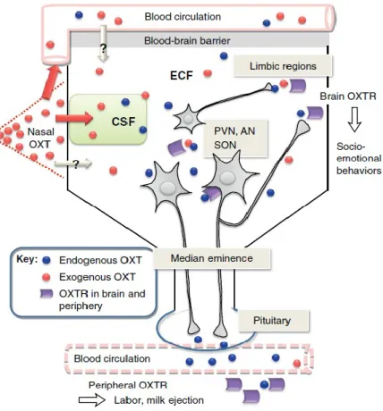
The classical action potential-driven axonal release from neurohypophysial terminals into the blood stream leads to the well-known peripheral effects of OT, like induction of labour or milk ejection.
Figure 1 The brain OT system: neuronal projections, release, receptor-mediated effects, and external application. In brief, central release of OT as a neuromodulator occurs from dendrites and perikarya, in
14
addition to axon terminal release as a neurotransmitter. Central release can occur independently and simultaneously of peripheral release. Taken and adapted from (Neumann and Landgraf 2012)
2.2 The OTR and OTR-coupled signaling cascades
All of the effects that OT exerts on animal behavior and physiology, including its anxiolytic activity, are the consequence of its binding to the OTR and the subsequent induction of intracellular physiological responses. The OTR is a 7-trans-membrane domain class I G- protein coupled receptor (GPCR). The human OTR gene consists of 4 exons and 3 introns, and the promoter consists of a variety of species-dependent transcription factor binding sites. The OTR gene is differentially expressed in various tissues and under different physiologic conditions (virgin vs. post-partum), regulated by DNA methylation of a specific CpG site in the promoter (Mamrut et al. 2013). The sequence homology between the mammalian OTR and vasopressin receptor 1 (V1) is nearly 50%; between the OTR and V2R it is even lower (40%) (Gimpl and Fahrenholz 2001).
It should be noted that the expression of the OTRs at the cell membrane and their affinity for OT are both regulated. For example, OTRs can be desensitized upon permanent agonist stimulation. This process involves multiple mechanisms, and can occur at transcriptional, translational or protein levels. The fastest desensitizing mechanism includes phosphorylation of the receptor and subsequent ß-arrestin binding. This leads to uncoupling of the GPCR from its G-proteins and endocytosis of the receptor. It was found that 60 % of the OTR expressed in human kidney fibroblasts (HEK293 cells) is desensitized already after 5-10 min of agonist stimulation (Gimpl and Fahrenholz unpublished data). However, due to the artificial nature of this system (the OTR has been transfected into the cell), the
15
interpretation of the data has to be taken with care. Moreover, exposure of naturally OTR- expressing myometrial cells to OT for 20 hours lead only to an almost 10-fold reduction in OT binding, with no changes in the OTR protein level.
Although only one type of OTR exists, it can occur in a high or in a low affinity state. The OTR requires at least two essential components for high-affinity binding of OT: divalent cations, such as Mg2+ or Mn2+, and cholesterol. These two components determine if the OTR displays the high-affinity state with a Kd > 1-50 nM, or the low affinity state with Kd > 100 nM. The conversion of the two affinity states is reversible. Cholesterol seems to stabilize the OTR for agonists in a high affinity state and acts, like the divalent cations, as an allosteric modulator (Gimpl and Fahrenholz 2001).
OTRs are functionally coupled mostly to Gq/11α class GTP binding proteins that stimulate together with Gβγ the activity of phospholipase C (PLC). This leads to the generation of inositol-3-phosphate (IP3) and 1,2-diacylglycerate (DAG). IP3 triggers Ca2+-release from intracellular stores, whereas DAG stimulates protein kinase C (PKC), which phosphorylates downstream targets. The rise in intracellular Ca2+ stimulates the Ca2+/calmodulin system that activates the myosin light chain kinase, which in turn leads to smooth muscle contraction, e.g. in myometrial cells during labour. However, the OTR does not exclusively bind the Gq- protein, but also Gi-protein subunits. This implies a differential downstream signaling of the activated OTR, since Gi-proteins inhibit adenylyl-cyclase, which reduces cAMP levels (Gimpl and Fahrenholz 2001; Busnelli et al. 2012).
Zhong and co-workers revealed the involvement of the trans-activated epidermal growth factor receptor (EGFR) in extracellular signal regulated kinase 1/2 (ERK1/2) activation by the myometrial OTR (Zhong et al. 2003). In the PVN, it is this pathway that mediates the
16
anxiolytic activity of OT (Blume et al. 2008). Blockade of the mitogen-activated protein (MAP) kinase “MAP ERK kinase 1/2” (MEK1/2) by the inhibitor U0126 attenuated the anxiolytic effect of OT in male rats, indicating a central role of MEK1/2 in the mediation of the anxiolytic effect of OT.
In the hippocampus, activation of OTRs lead to Gq/11 – IP3 activation, as well as EGFR- ERK1/2-mediated translational induction of the PKC subform PKMζ via the mammalian target of Rapamycin (mTOR) complex (Lin et al. 2012). The interplay between these cascades triggers long term potentiation (LTP) in the hippocampus, a mechanism also involved in the establishment of long-lasting spatial memory (Tomizawa et al. 2003). Blocking the MAPK cascade with the MEK inhibitor U0126 inhibited cyclic AMP responsive element binding protein (CREB) phosphorylation and subsequent spatial memory formation during motherhood (Tomizawa et al. 2003).
A global knockout of the OTR in male mice results in profound behavioral and physiological alterations such as impaired sociability and impaired preference for social novelty, impaired cognitive flexibility, increased aggression (Chini et al. 2008; Sala et al. 2011; Sala et al. 2013), but also obesity and dysfunction in body temperature when exposed to cold (Nishimori et al.
2008). In the ventral and dorsal striatum, it also has been reported that OTRs can occur as heterodimers coupled to a dopamine D2 receptor, which is thought to produce the rewarding effect of a social stimulus during pair bonding (Romero-Fernandez et al. 2013).
This would increase the rich repertoire of intracellular responses to OTR activation, and makes very specific responses to particular stimuli possible.
In addition, our group found that OTR activation in hypothalamic neurons lead to the incorporation of transient receptor potential V2 (TRPV2) channels into the cell membrane
17
and a subsequent influx of Ca2+ from the extracellular space (unpublished data). This influx may increase cellular excitability, protein synthesis and neurotransmitter release via several Ca2+-dependent signaling cascades. Activation of the TRPV2 channel is upstream of MEK1/2 phosphorylation, and it has therefore been postulated that blocking TRPV2 channels pharmacologically prevents the anxiolytic activity of OT.
2.3 OT and the regulation of stress responses and anxiety
It has long been established that physical as well as psychological stressors induce OT release into the peripheral circulation (Lang et al. 1983; Gibbs 1984). Later studies revealed the central release of OT in the hypothalamus and lateral septum upon an emotional stressor, which appeared to be independent from peripheral release (Engelmann et al. 1999).
Furthermore, intra-cerebro-ventricular (icv) administration of a selective OTR antagonist (OTA) disinhibited basal and stress-induced (forced swim) HPA axis activity in male and female rats (Neumann et al. 2000), as indicated by increased adrenocorticotropic hormone (ACTH) and corticosterone release into the blood. Retrodialysis of the OTA in the PVN resulted in similar effects in unstressed males and females, locating OTs’ effects on the HPA axis to this specific brain region. Interestingly, the expression of the main HPA axis activator, CRF, was more enhanced following a physical stressor (4 h of restraint) in the PVN of OT gene deficient male mice, when compared to wild type mice suggesting a central inhibitory role of OT in the regulation of CRF gene transcription (Nomura et al. 2003)
In 2004, Windle and co-workers infused OT icv at 1 or 10 ng over a 7-day period via an osmotic minipump in female, ovariectomized, estradiol-treated rats and subsequently subjected the rats to 30 min restraint stress. This treatment significantly reduced both basal
18
and restraint-induced plasma ACTH and corticosterone levels. CRF mRNA, which was measured by in situ hybridization in brain slices collected 210 min after the end of the stressor, increased in saline-treated restraint animals, and this increase was absent in OT- treated stressed rats (Windle et al. 2004). One year later, Ebner et al (2005) found that OT is released in the CeA and directs the stress-coping behavior toward a more passive coping style, as indicated by an increased floating time in the forced swim test. Blocking the OTR by an OTA likewise increased the release of excitatory amino acids (glutamate and aspartate) indicating an inhibitory effect of OT in the amygdala (Ebner et al. 2005). This was confirmed by a study that revealed that OT excites a population of GABAergic interneurons in the CeA, thereby reducing the activity of output neurons in the CeA that control the physiological expression of fear (Huber et al. 2005). The relation of axonal OT release in the CeA to fear responses has been investigated with the help of a sophisticated optogenetic approach, revealing decreased freezing responses in fear-conditioned rats upon blue-light-induced release of endogenous OT (Knobloch et al. 2011).
Besides regulating stress-coping and anxiety in the CeA, OT regulates sexual behavior and anxiety in the PVN of males and females. Endogenous, mating-induced release of OT in the PVN of male rats was found to be causally involved in mating-induced anxiolysis for up to 4 h (Waldherr and Neumann 2007). In addition, exogenous OT infused into the PVN of male rats also induces anxiolysis (Blume et al. 2008).
In contrast to the studies cited above, which suggest that central OT inhibits HPA axis activity, peripheral OT might be stimulatory at the level of the pituitary gland. Peripheral OT acts as ACTH and corticosterone secretagogue, increasing both basal and stress-induced plasma ACTH levels (Gibbs 1984; Petersson et al. 1999) after chronic (Ondrejcakova et al.
19
2010) or acute (Stachowiak et al. 1995) subcutaneous (s.c.) injection. Similar effects have been found in vitro in pituitary explants (Antoni et al. 1983).
2.4 Cross-talk between the OTR and CRFR coupled signaling in the periphery
What could be the physiological consequences of activation of the OTR and its downstream intracellular signaling pathways? One of the targets of the OT system appears to be the myometrial CRF system. An interesting study by Grammatopoulos et al (1999) investigated the crosstalk between the OTR and the CRF receptor (CRFR) in human myometrium at term (Grammatopoulos and Hillhouse 1999). They found that OT reduces CRF binding to the CRFR in human myometrial membranes in a time-dependent manner by reducing the affinity of the CRF receptor without affecting the number of receptors. The inhibitory effect of OT on CRF binding first appeared 15 – 20 min after the onset of OT stimulation. The maximum of CRF-binding inhibition (-50%) by OT was reached at 30 min and remained constant for at least 2 h. The effect was observed at OT concentrations between 1 – 100 nM OT. Further investigations in human myometrial cells suggested a role of PKC in CRF receptor desensitization. OTR binding activates the PLC pathway via Gq- proteins, which leads to rapid release of Ca2+- ions from intracellular Ca2+ stores. Intracellular Ca2+ activates PKC and induces its translocation from the cytosol to the cell-membrane, where it phosphorylates the CRF receptor. Phosphorylation of the receptor leads to reduced receptor affinity for its agonist. It is interesting to note that this mechanism is only apparent in term, but not pre- term myometrium membranes. The authors speculate that CRF might influence myometrial contractility, and that OT antagonizes the relaxing effect of the activated CRFR. Additionally, studies in the pituitary gland describe opposite (i.e. potentiating) effects of PKC on CRF
20
receptor signaling (Cronin et al. 1986). It is not clarified to date how this highly dynamic interplay is regulated at the molecular level. It is tempting to speculate, however, that different sub-forms of PKC, like the PKMζ (Lin et al. 2012) are responsible for tissue and state (pre-term / term) specific effects.
3. Anxiety, stress, and its key regulator CRF
A physiological state that is intimately connected with anxiety is stress. A fearful stimulus will generally not only induce anxiety behavior, but also leads to neural and peripheral adaptations that enable an individual to cope with the stimulus; this is known as the stress response. Both OT and CRF are involved, and interact in anxiety behavior as well as in the stress response.
3.1 Definition of the term “stress”
It is essential for a living organism to maintain a constant inner environment. This ability to create an inner environment, independent from the external environment, was termed
“homeostasis” by the American physiologist Walter Bradford Cannon (Cannon 1929, 1939).
Disturbing homeostasis can be perceived as threat, provoking a response from the organism to regain a constant inner environment. It was the endocrinologist Hans Selye who termed the threat as “stressor” and defined stress as the “nonspecific response of the body to any demand placed upon it” (Selye 1936). Later studies revealed that Selye’s definition of stress as “nonspecific” was not accurate (Pacak et al. 1998). The stress response is specific to the kind of stressor and depends on the organism’s perception of the stressor, and the ability to cope with it (Goldstein and Kopin 2007).
21
3.2 Differentiation between physical, psychological, and cellular stress
For a better understanding of the work presented in this thesis, it is of importance to differentiate between physical, psychological and cellular stress, and the processing of each at the cellular level. The term “stress” is not always clearly defined in the literature, especially at the cellular level, which imposes the need to clarify and define terms that are used in this work. Severe physical stressors like cold, blood loss or injuries recruit the brainstem and hypothalamic regions, whereas the kind of stress we perceive every day and influences the organism’s homeostasis mostly, is of psychological nature, such as social embarrassment, examinations, or deadlines (Joels and Baram 2009). This physical/psychological stress is processed at a high level of different interacting networks in the brain and does normally not include damage to a single neuron. Cellular stress, to the contrary, imposes damage to single neurons and, as a consequence of an increasing imbalance in central networks, to the whole organism. This kind of stress can be caused by UV radiation, hypoxia, or chemical compounds (Chu et al. 2004). Cells respond differentially to these different kinds of stressors, however, one universal stress responsive protein is the MAPK family member “stress-activated protein kinases” (SAPK) and its most common member JNK has been reported to be activated in response to both cellular (e.g. glutamate- induced excitotoxicity) (Bogoyevitch et al. 2004) and psychological/physical stressors, such as forced swim stress in mice (Liu et al. 2004).
22 3.3 Regulation of the CRF neuron
Under basal conditions the CRF neuron is under inhibitory control by GABAergic interneurons, which act via GABA A receptors expressed on hypophyseotropic CRF neurons.
If a stressor is perceived, depending on the type of stressor (blood loss, immune challenge, hypoglycemia), it is directly transmitted to the PVN via the brain stem and spinal cord projections to the PVN, or, in case of psychological stressors, integrated via limbic structures, such as the CeA, hippocampus, medial prefrontal cortex, or the bed nucleus of the stria terminalis (BNST). Additionally, CRF neurons in the BNST are innervated by oxytocinergic projections from magnocellular neurons of the PVN, thereby integrating stressors (CRF) and stress-protecting actions of OT to fine tune the organism’s stress response. The BNST is a central relay station to integrate limbic inputs toward the PVN. The reciprocal innervation of CRF and OT neurons between PVN and BNST as described above is thought to be involved in balancing stress and affect (Dabrowska et al. 2011). Additionally, CRF neurons are under control of somatostatinergic / dopaminergic interneurons in the hypothalamus. Long-day photoperiods induce a depressive and anxiety-related behavior in nocturnal rats, via the induction of CRF release by somatostatinergic interneurons. These interneurons can switch their expression profile and start to synthesize dopamine, which down-regulates CRF release and plasma corticosteroids in response to a short-day photoperiod (prolonged active phase) (Dulcis et al. 2013).
Other neurotransmitters, such as dopamine, histamine, and acetylcholine are likely to modulate CRF neurons indirectly by modulating the activity of afferent neurons (Lee et al.
2008), for review see (Aguilera and Liu 2011)). Influences from the periphery are mediated by hormones such as angiotensin II, prolactin and leptin. Since these peptides cannot cross the blood-brain-barrier, specialized organs (circumventricular organs) receive these
23
hormonal signals and transmit them to central nuclei. Circumventricular organs, such as the subfornical organ, express the angiotensin II receptor to recognize peripherally released angiotensin II, which is the end product of the renin-angiotensin system, and transmit this stress-induced signal directly to the PVN. Another circumventricular organ, the choroid plexus, is involved in the uptake of prolactin, which results in hypo-responsiveness of the HPA axis during lactation (Blume et al. 2009).
3.4 Transcriptional regulation of the CRF gene
There is one gene encoding for CRF, which is located on the long arm of chromosome 8, with one promoter region, one intron (800 bp) and two exons (582 bp). The gene codes for the inactive pro-hormone. The pro-hormone is transcribed, translated, and processed into the active CRF peptide. There is a 97% homology of the promoter region between humans, mice, rats, and sheep, which suggests that signals leading to CRF gene transcription are highly conserved among mammals (King and Nicholson 2007). This allows for comparisons between animal and human hypothalamic response to stress. The human CRF gene promoter contains several response elements (RE) that, upon binding of their cognate factors, either activate or repress transcription of the CRF gene. There are three elements (metal-responsive transcription factor 1 RE, hybrid steroid RE, ecdysone RE) which activate gene transcription by inhibiting two repressive elements (Ying Yang RE, negative glucocorticoid RE (nGRE)). If these repressive elements are inhibited, the cyclic AMP RE (CRE) is free to bind CREB and its co-activators. There are also two canonical TATA-Box elements present in the CRF gene, only one of which seems to be involved in transcriptional regulation, the second one is thought to be silent (King and Nicholson 2007). The best studied inhibitory mechanism of CRF
24
transcription is the negative feedback regulation by glucocorticoids (GC). The activated HPA axis leads to the production and release of GCs, which cross the blood brain barrier to bind the GC receptor (GR) in the brain (hippocampus, PVN). This complex translocates to the nucleus and binds the nGRE in the CRF promoter. This inhibits the binding of the transcriptional machinery to the CRE region, thereby reducing CRF gene transcription (Jeanneteau et al. 2012).
Recent studies showed that the transcription of the CRF gene is also under the control of the CREB co-activator CRTC2. The molecular machinery employed is described in more detail in the CRTC section below. In brief, in vitro and in vivo studies revealed that CREB phosphorylation is essential but not sufficient for forskolin (FSK) or restraint stress-induced CRF gene transcription. The activation, i.e. dephosphorylation of the co-activator CRTC2 and its subsequent translocation to the nucleus is essential for a full transcriptional response (Liu et al. 2008; Liu et al. 2011).
3.5 Peripheral effects of CRF
CRF is the central regulator of the HPA axis, which mediates the organism´s response to a perceived stressor. The main regulatory brain region of the HPA axis is the hypothalamic PVN. The PVN contains the highest concentration of CRF neurons in the brain. It lies adjacent to the 3rd ventricle and consists of large magnocellular and smaller parvocellular neurons.
The medial-dorsal parvocellular CRF neurons release CRF via the external zone of the median eminence into the hypophyseal portal blood capillaries to the adenohypophysis. These CRF- neurons co-express and co-release AVP together with CRF in response to a variety of psychological and physiological stressors into the portal circulation (Aguilera 1994, 1998;
25
Aguilera and Liu 2011). This increase serves as a trigger for the release of effector-hormones, such as ACTH, from the adenohypophysis into the blood stream. Interestingly, the circadian activation of the CRF neuron is delayed to the daily changes in circulating ACTH and corticosterone in rats (Watts et al. 2004), suggesting that HPA axis activity is driven by the release of CRF pool contents, followed by the transcriptional replenishment of the pools.
During acute stress, CRF and AVP are rapidly released into the portal blood system to induce the full ACTH response. This release is followed by an increase of CRF gene transcription, and later on, AVP gene transcription (Herman et al. 1992; Ma et al. 1997). However, CRF is considered to be largely responsible for HPA axis activation, while AVP was reported not to be essential, but potentiating the organism’s response to a stressor (Aguilera et al. 2008).
Interestingly, other researchers favor a model of synergistic actions of AVP and CRF, instead of additive effects, to fully induce the stress response (Chen et al. 2008; Spiga et al. 2009).
The binding of CRF to the CRF receptor 1 (CRFR1) expressed by the corticotrophe cells in the pituitary gland, activates adenylate cyclase, thus increasing cAMP levels. cAMP, in turn, activates protein kinase A (PKA), which translocates to the nucleus and up-regulates the expression of pro-opiomelanocortin (POMC). POMC can be cleaved in several active peptides, one of which is ACTH. The release of ACTH into the circulation will induce the synthesis and secretion of glucocorticoids (GC) upon binding to the melanocortin-2-receptor (Mc2r) expressed in the zona fasciculata of the adrenal cortex. The synthesis of GCs is rate- limited by the availability of the steroidogenic acute regulatory protein (StAR). The StAR enzyme stimulates the initial mitochondrial metabolism of cholesterol to pregnenolone by enhancing cholesterol transfer from the outer membrane to cytochrome P450 11A1 (P450scc) in the inner membrane (Stocco et al. 2001; Stocco et al. 2001; Jefcoate 2002). To initiate transcription of the StAR enzyme, the cooperative actions of multiple transcription
26
factors, including CREB (Clem et al. 2005) and its co-activator CRTC2 (Takemori et al. 2007;
Liu et al. 2012), CCAAT enhancer- binding protein (C/EBP β), steroidogenic factor 1 (SF-1) (Manna et al. 2003; Manna et al. 2003) and GATA4 (Silverman et al. 2006) are required. The proximal region of the mouse StAR promoter contains three canonical 5’ CRE half-sites (TGAC) that are bound by the activated CREB/ATF-1/CBP-p300/CRTC2 complex (Takemori et al. 2007; Liu et al. 2012).
3.6 Central effects of CRF
CRF plays an important role in the regulation of the HPA axis, but recent findings have shown that production and central release of CRF during stress influences neuronal structure and the functionality of the hippocampus and other brain regions. In fact, whereas mild or short- lasting stress enhances hippocampal function by augmenting synaptic plasticity, chronic stress impairs learning and memory by retraction of pyramidal cell dendrites, likely to be caused by the chronic presence of stress-induced CRF. The integrity of dendrites is only guaranteed if excitatory synapses are present on specialized structures called dendritic spines. The picture below represents an organotypic slice taken from a murine hippocampus and incubated with 100 nM CRF for 2 weeks. This treatment induces a significant loss of dendritic branching (Maras and Baram 2012).
27
Figure 2 100 nM of CRF applied onto hippocampal organotypic slice cultures reduced dendritic complexity. Picture taken from (Maras and Baram 2012)
The number and functionality of dendritic spines is highly dynamic and can be influenced by neurotransmitters, hormones, and growth factors, which are in turn governed by environmental signals like stress (Maras and Baram 2012). Chronic stress, for instance, tends not only to cause dendritic retraction, but also inhibits neurogenesis and reduces survival of hippocampal neurons in a sex-dependent manner (Hillerer et al. 2013). In addition, acute (i.e. 30 min) restraint stress with subsequent intermittent tail stimulations increase spine densities in the hippocampal CA1 area of male rats (Shors 2009), while the same stimulus causes a decrease in hippocampal spine density in females. These sex-dependent stress- induced effects are also observed in the medial prefrontal cortex (mPFC), where stress decreases dendritic branching and length of pyramidal neurons in males, but increases these parameters in females (Garrett and Wellman 2009; Shansky and Morrison 2009). Stress- induced remodeling of layer V neurons in the mPFC has an impact on projections to subcortical regions like the BNST and amygdala, both being related to regulation of adaptive and maladaptive responses to stress (Gabbott et al. 2005; Conrad et al. 2011; Radley and Sawchenko 2011). The amygdala is most often linked to anxiety. However, recent human
28
and rodent studies revealed that the amygdala is more relevant to fear or short term anxiety responses, whereas the BNST is more involved in sustained anxiety (Davis et al. 2010; Leuner and Shors 2013). Intriguingly, the BNST is reciprocally connected with the PVN, with oxytocinergic projections toward the BNST, and CRF fibers originating in the BNST projecting toward the PVN (Dabrowska et al. 2013). This anatomical arrangement represents a direct morphological link of the CRF system with the oxytocinergic system. Considering the sex dependency of the stress response, reproduction-induced neuroplasticity (likely to be related to the oxytocinergic system), and the common brain regions these systems share, it is possible that OT is a sex-specific regulator of the CRF-driven stress response with major impact on the morphology of neurons in stress-related brain regions. It is tempting to speculate that the release of OT during stress serves as a modulator of CRF expression in a time-dependent manner, to prevent neuronal damage by large amounts of stress-induced CRF (see Results).
4. OTR-coupled signaling cascades and the regulation of CRF gene transcription
CREB and CRTC signaling is one example of how intracellular effectors of receptor activation can modulate gene expression. Today, literally hundreds if not thousands of such factors have been characterized, and some of them will be highlighted here as they might be involved in OT-mediated signaling and social behavior, or the control of CRF gene expression and stress responses.
Between 1994 and 2001, substantial progress has been made in the understanding of the mammalian MAP kinases. The first MAP kinase to be discovered was the extracellular signal regulated kinase 1 (ERK1), which is encoded by the MAPK3 gene. The closely related ERK2
29
(encoded by the MAPK1 gene) was identified in the same year. In the following years a plethora of mammalian MAPKs were identified, revealing an evolutionary highly conserved, and ubiquitously expressed enzymatic machinery in all eukaryotic cells. Mitogens, the first identified stimulators of MAPK signaling gave the protein family its name, although a diverse set of extracellular stimuli can activate MAPKs. Stimuli like hormones, growth factors, cytokines, GPCR regulated agents, transforming growth factor (TGF)-ß-related agents, and environmental stresses. As diverse as the stimuli are the effects on the cell physiology.
MAPKs are involved in the regulation of gene transcription, protein biosynthesis, cell cycle control, apoptosis, and cell differentiation (Kyriakis and Avruch 2012).
The substantial core of MAPK signaling is a three-tiered module, in which activating signals are passed on as phosphorylation of specific motifs (Thr-X-Tyr) in the kinase subdomain VIII activation loop of the MAPK. The upstream kinases of MAPK are named dual specificity MAPK Kinase, MAP2K, MEK, or MKKs. These kinases are activated by Serine/Threonine (Ser/Thr) phosphorylation by a wide variety of MAPK kinase kinases (MAP3K) (Kyriakis and Avruch 2012).
MAPKs have remarkable substrate specificity. They phosphorylate Ser/Thr residues only if they are followed by a proline, and the presence of a MAPK docking site. Despite the high selectivity of the individual elements of the core signaling module, MAPK show promiscuous functions in related signaling cascades and are often regulated by a variety of extracellular signals. The question arose, how MAPKs maintain their signal/substrate specificity if the components are directed by proline and MAPK docking sites on the one hand, but can couple to multiple substrates on the other hand. The answer to this question lies in the function of scaffolding proteins, which sequester the elements of the cascade, bring them in
30
close physical contact, and thereby maintain signal specificity, and subcellular localization (Kyriakis and Avruch 2012; Roskoski 2012).
4.1 The ERK cascade
The highest upstream activators of the ERK cascade are receptor tyrosine kinases (RTKs) like the EGFR, which recruit the membrane associated proto-oncoprotein GTPase Rat sarcoma (Ras) via several possible intermediate signaling proteins (see Figure 3), depending on the subcellular localization of the cascade. Ras in turn phosphorylates the family of Raf MAP3Ks, such as Raf-1 (also known as c-Raf), A-Raf, and B-Raf. Raf-1 was first identified 1983 in murine retrovirus infected fibroblast cells, and named Virus-induced Rapidly Accelerated Fibrosarcoma (V-Raf). Later studies revealed the ubiquitous expression in all eukaryotic cells, so the gene’s name was changed to cellular-Raf (c-Raf) (Osborne et al. 2011). C-Raf is the gatekeeper of the entire ERK cascade and therefore regulated at multiple steps. It contains an auto-inhibitory region, which physically blocks the kinase domain of the protein. Only GTP-bound Ras can compete with the auto-inhibitory block, leading to a conformational change and the relief of the kinase domain. Scaffolding proteins such as kinase suppressor of Ras (KSR) or 14-3-3 are either inhibitors or activators of Ras signaling, depending on their phosphorylation status. Non-phosphorylated 14-3-3 does not bind Ras, and this in turn is regulated by the TGF beta activated kinase 1 (TAK1) or the Protein phosphatase 1 (PP1) / Protein Phosphatase 2A (PP2A). Once c-Raf is in an active state, with its auto-inhibitory site deactivated, 14-3-3 can act as an activating scaffolding protein, by bringing Ras and its substrates (e.g. MEK1/2, BAD, adenylate cyclase and others) in close proximity (Osborne et al. 2011; Kyriakis and Avruch 2012). Interestingly 14-3-3 is also a scaffolding protein for
31
CRTCs, representing a direct spatial link between the MAPK cascade and the CRTCs (see CRTC section of the Introduction). MEK1 (also known as MAP2K1, MAPKK1, MKK1) and MEK2 are the direct downstream substrates of c-Raf. The protein MEK contains a phospho-domain in its C-terminus, and a trifunctional N-terminal sequence with an inhibitory segment, a nuclear export sequence, and an ERK binding site (Fischmann et al. 2009). Contrary to the complexity observed in c-Raf activation, MEK and ERK are simply activated by dual phosphorylation of their respective activating residues (Ser218 and Ser222 for MEK1; Ser222 and Ser226 for MEK2; Thr185 and Tyr 187 for ERK2 or Thr203, Tyr205 for ERK1).
Interestingly, phosphorylation of Ser212 in MEK1 decreases the kinase activity of MEK in vivo, without interfering with the ERK2 binding or other activating phosphorylated residues (Gopalbhai et al. 2003). Other residues have been shown to be inhibitory as well, such as T286, which is phosphorylated by cyclin dependent kinase 5 (Cdk5). This kinase has several functions in cell proliferation, brain development and synaptic vesicle exocytosis.
Furthermore, T292 in MEK1 is a substrate of ERK, which implies a negative feedback loop to reduce ERK activation by direct inhibition of MEK by ERK.
For many years, ERK was the only known substrate of MEK, only recently (2006-2010) new MEK targets were identified, such as PPARγ, MyoD, PI3K, or LIMKinase 1 in a variety of tissues (Brandt et al.; Jo et al.; Klemke et al.; Burgermeister et al. 2007). All these recent findings imply the possibility of MEK activation without subsequent ERK phosphorylation.
The distribution of the MAPK cascade in the rat brain has been studied intensively. MEK1 has been shown to be expressed in many brain regions, including the cortex, hippocampus, amygdala, bed nucleus of the stria terminals (BNST), lateral septum, hypothalamus (most notably in the PVN), cerebellum, and the brainstem (Flood et al. 1998).
32
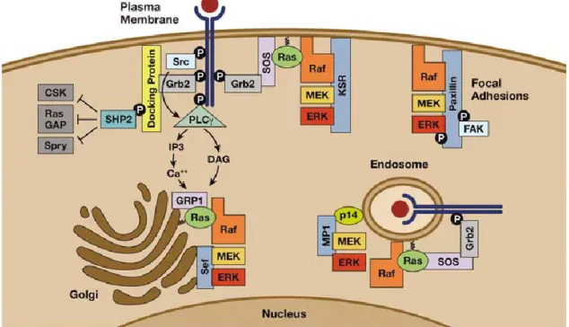
Figure 3 Receptor tyrosine kinase (RTK) to ERK signaling: Ras and ERK activation at various intracellular compartments. At the plasma membrane, activated RTKs promote Ras activation through the recruitment of growth factor receptor bound protein 2 / Son of sevenless (Grb2/Sos) complexes. Kinase suppressor of Ras (KSR) is an ERK scaffold that facilitates Ras dependent ERK cascade activation at the plasma membrane, whereas Paxillin directs ERK activation at focal adhesions. Active signaling complexes containing internalized receptors, Grb2/Sos and Ras are also found on endosomes. MEK partner-1 (MP1) is an MEK1/ERK1- specific scaffold that localizes to late endosomes through an interaction with the adaptor protein p14. Activated RTKs can also direct the activation of Golgi-associated Ras through a signaling route involving sarcoma (Src), Phospho-lipase-C (PLC)-γ and Ras-general receptor of phosphoinositides-1 (GRP1). Similar expression to fgf genes (Sef) is a Golgi-localized scaffold that recruits activated MEK and promotes ERK activation. Active ERK is retained on the Sef/MEK complex and confined to cytosolic substrates. The tyrosine phosphatase Src homology phosphatase-2 (Shp-2) is another effector of RTKs that positively regulates Ras signaling by antagonizing the ability of negative regulators, such as cellular-Src kinase (CSK), RasGAP and Sprouty, to access and downregulate critical enzymes involved in Ras activation. Picture taken from (McKay and Morrison 2007)
ERK1 and ERK2, two of the direct downstream targets of MEK1/2, transduce extracellular signals to the nucleus to regulate processes as diverse as proliferation, differentiation, apoptosis, and synaptic plasticity (Zhuang and Schnellmann 2006). When dephosphorylated, ERKs are basically incapable of catalyzing the transfer of phospho-groups (20 to 30 pmol min−1 mg−1), mainly because interactions with cognate substrates are hindered by the
33
structural configuration of the activation loop of the kinase. MEK-mediated activation induces conformational changes that enhance the catalytic activities of the ERKs by about 5 orders of magnitude (5 μmol min−1 mg−1) (Chang and Karin 2001; Kyriakis and Avruch 2012).
After being dually-phosphorylated, ERK1 dimerizes, which enables it to generate peak kinase activity, while a dual-phosphorylated monomer ERK1 is thought to maintain basal activity (Philipova and Whitaker 2005). However the nuclear translocation seems to be dimerization- independent, but is controlled by the rate of phosphorylation (Lidke et al.). These facts lead to the hypothesis that dual-phosphorylated ERK dimers, with the help of cytosolic scaffolding proteins that act as assembling platforms, target cytosolic proteins, and dual- phosphorylated monomers, that are released from these scaffolds, have nuclear targets. The release of ERK from its scaffolding proteins and the redistribution over cellular compartments, including the nucleus, cytosol, plasma membrane, intracellular vesicles, mitochondria and cytoskeletal components, enables contact with and subsequent phosphorylation of nearly 200 target proteins (Whitehurst et al. 2002; Yoon and Seger 2006). While there is evidence for a carrier-independent mechanism of nuclear entry of ERK2 (Whitehurst et al. 2002) by direct binding of ERK2 to a nuclear pore complex (Matsubayashi et al. 2001), the nuclear import of ERK1 is poorly studied. However, ERK1 seems to shuttle between cytosol and nucleus at a much slower rate than ERK2, and this difference is caused by differences in an N-terminal domain of ERK1. This N-terminal domain forms the main difference between ERK1 and ERK2; moreover, they share 85% of amino-acid sequence identity, and all other known functional domains (Marchi et al. 2008).
This shuttling of ERK is independent of its phosphorylation status, which might point toward a kinase-independent function of ERK in the nucleus. Indeed, recent studies discovered new
34
targets of ERK that are not necessarily modified by phosphorylation. ERK is involved in important cellular processes, such as chromatin remodeling, DNA transcription, and cell cycle regulation, acting independently of their kinase activities. In more detail, one example concerns the posttranslational modification of dual specificity phosphatase-6 (DUSP6) also known as MAPK phosphatase-3 (MKP-3), whose catalytic activity is increased 30 fold upon binding to ERK2. DUSPs specifically target MAPKs to fine tune their activity by dephosphorylation. Most DUSPs are encoded by inducible genes, the transcription of which is mainly regulated by MAPK themselves, or glucocorticoids. A second level of regulation is posttranslational modification of the phosphatase by its target. The binding of ERK2 to DUSP6 is sufficient to induce an allosteric conformational change that leads to the reorganization of the consensus acidic residue in the phosphatase, which results in enhanced catalytic activity. The specific recognition of ERK2 by MKP-3 is mediated by a conserved motif within the N terminal region of DUSP6 (Camps et al. 1998). Another non- canonical target that does not require kinase activity of ERK is poly (ADP-ribose) polymerase 1 (PARP-1), which is bound by phosphorylated ERK2, and activated without phospho-transfer activity. PARP-1 subsequently binds the transcription factor Elk-1, thus enhancing its transcriptional activity of genes such as c-fos. Acetylation of the core histones H3 and H4 is also enhanced in response to ERK2-mediated activation of PARP-1 (Cohen-Armon et al.
2007).
ERK2 is not only a signaling mediator between proteins, but also a DNA binding factor. High- throughput screening of the human protein-DNA interactome in HeLa cells identified ERK2 as a transcriptional repressor of interferon-γ(IFN-γ)–induced genes (Hu et al. 2009). Analysis of 82 genes that were increased in expression in response to knockdown of ERK2 revealed
35
G/CAAAG/C as the consensus sequence for the binding of ERK2. Within promoters, these ERK2-binding sites appear at around −90 base pairs, a typical distribution for many transcriptional regulators (Roy et al. 2000).
Besides these recently discovered non-canonical targets, ERK has its “normal” downstream signaling cascade substrates. The first identified substrate, which is exclusively phosphorylated by ERK1/2 and not by JNK or p38 (see p38 and JNK paragraph in the Introduction) was the ribosomal S6 Kinase (Rsk). This kinase derives its name from its kinase activity on the small ribosomal protein S6. However it does not represent the major kinase of the S6 protein, which is the S6K. Rsk is activated by ERK1/2, which phosphorylates the C- terminal catalytic domain. The activated catalytic domain auto-phosphorylates Rsk at Ser380, which provides a docking site for 3-phosphoinositide-dependent-kinase 1 (PDK1), which finally leads to full activation by phosphorylating Ser222 in the activation loop of the N-terminal domain (for review (Kyriakis and Avruch 2012). One factor that leads to MEK, ERK and Rsk activation in a hypothalamic neuronal cell line is AVP (Chen et al. 2009).
The shuttling of ERK and RSK is, among others, regulated by phosphoprotein enriched in Astrocytes-15 (PEA-15- or PED), a small protein (15 kDa) with an N-terminal death effector domain and an irregular C-terminal tail. The name suggests that it is expressed solely in astrocytes, but later studies revealed its ubiquitous expression in all neural cell types with a highly conserved structure among mammals (Araujo et al. 1993). The activation of PEA-15 and its control over ERK is tightly regulated. Under resting conditions, ERK is bound to PEA- 15 in the cytosol. Upon EGFR-induced Ca2+ influx, activated Ca2+-calmodulin dependent kinase II (CaMKII) phosphorylates the Ser116 residue of PEA-15, which allows the PKC mediated phosphorylation of Ser104. Dual-phosphorylation of PEA-15 releases ERK from
36
PEA-15 and allows its shuttling to the nucleus. The phosphorylation status of ERK is not altered by PEA-15 binding, so that PEA-15 restricts ERK activity to cytosolic targets, such as RSK or stathmins (Renault et al. 2003) (for the role of stathmins in behavior see Introduction JNK paragraph). The effects of PEA-15 on RSK are bi-functional. First, PEA-15 blocks nuclear accumulation of RSK after epidermal growth factor stimulation. Second, PEA-15 inhibits RSK2 kinase activity by 50 %. RSK1 is not bound by PEA-15 (Vaidyanathan and Ramos 2003;
Vaidyanathan et al. 2007). The central importance of PEA-15 activity on MAPK cascades has been demonstrated by a PEA-15 knockout mouse line, which displayed altered CREB and c- fos mediated gene transcription activity. This results in a heightened stress reactivity and anxiety and impairment of spatial memory (Ramos et al. 2009).
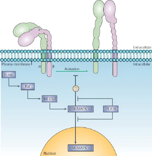
Figure 4 Depiction of the MAPK pathway and its inhibitory scaffolding protein PEA-15. Taken from (Kinbara et al. 2003)
37
Furthermore, the MAPK-interacting kinases (MNKs) are downstream ERK 1/2 targets. MNKs are involved in translational regulation, which occurs at the 5’ methylguanosine cap structure. The eukaryotic initiation factor (eIF)-4E tethers mRNA onto a scaffolding protein, and with the help of other co-factors, unwinds the secondary structure of the mRNA, which allows the ribosomal complex to scan the mRNA. The exact role of eIF-4E phosphorylation by MNKs in transcriptional regulation is still under debate. However, directly related to eIF-4E phosphorylation is another interesting translational repressor binding protein (4E-BP1), which interrupts the eIF-4E interaction with its co-factors, but dissociates from the translational complex upon phosphorylation induced by the mammalian target of rapamycin (mTOR) (Sonenberg and Gingras 1998). Studies have shown a co-localization of activated (i.e.
phosphorylated) mTOR and oxytocinergic neurons of the PVN (Lembke et al.) in the context of energy balance in fasted rats. mTOR is a protein central to the regulation of protein translation, and has shown to be involved in mediating the fast antidepressant effects of NMDA antagonists by synapse formation (Li et al.). The central role of mTOR in the regulation of mRNA translation, its regulation by MAP Kinases (see Figure 5) and its co- localization with OT in the PVN, identifies the possibility for a role in mediating the effects of OT on protein synthesis (Devost et al. 2008), and hence sustained anxiolytic effects.
38
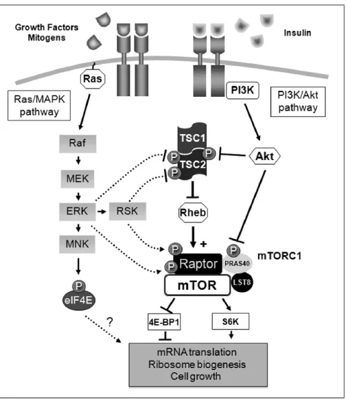
Figure 5 Schematic representation of the molecular mechanisms employed by the Ras/MAPK pathway to regulate mTORC1 signaling. Activation of the Ras/MAPK pathway stimulates mTORC1 activity through the coordinated action of the kinases ERK1/2 and Rsk on the tuberous sclerosis (TSC) protein complex upstream mTORC1, and on Raptor, an important partner of mTOR within mTORC1. Taken from (Carriere et al. 2011)
Another family of ERK substrates are the Mitogen- and Stress-activated Protein Kinases (MSKs). MSKs can be phosphorylated by ERK1/2 and p38 (for p38 see respective chapter in Introduction), and are therefore mediators of many physiological and pathological stimuli.
Their most acknowledged activity lies in the phosphorylation of CREB and nuclear factor κB (NF- κB).
Recent studies revealed an additional role for MSKs in histone phosphorylation, which leads, just like histone acetylation, to chromatin relaxation and facilitated binding of proteins
39
involved in transcription. It has been reported that MSKs were necessary for EGF-induced Histone 3 (H3) Ser10 phosphorylation of the cellular-Finkel-Biskis-Jinkins murine osteosarcoma virus homolog (c-Fos) - promoter (Duncan et al. 2006). MSKs are predominantly found in the nucleus and can partly be relocated to the cytosol upon GC signaling. It is therefore suggested that MSK1 contributes to the anti-inflammatory actions of GCs, by impairing transcription of genes related to inflammatory processes by nuclear export of MSKs. Gene transcription is regulated by transcription factors, of which CREB is the most studied one. The influence of MSKs on CREB and its result on gene transcription will be described in the CREB paragraph of the Introduction and in the Results section.
4.2 P38 and JNK
P38α was the first isolated isoform of the stress-, inflammatory cytokines, and endotoxin- induced MAPK p38. There are four p38 genes (p38α to p38δ), however, p38 is a MAPK often overseen and poorly studied. There is evidence, that p38 disrupts BDNF-induced TrkB signaling, thereby inhibiting long term potentiation (LTP) (Tong et al.), but the effects of p38 are often studied in relation with other members of the MAPK family, because exclusive p38 activation is rarely seen (Chang 2012). The downstream targets of p38 are mainly MNK1/2, which regulates the activity of eIF-4E, and MSK1/2, which is the main kinase of the transcription factor CREB (see CREB paragraph in the Introduction).
The dominant stress-induced MAPK is the stress activated protein kinase (SAPK) or c-Jun N- terminal kinase (JNK). It is named after its substrate, the transcription factor c-Jun, short for cellular–ju nana, which is Japanese for 17 and refers to the avian sarcoma virus 17, from which the protein has been originally isolated. C-Jun recognizes and binds to the enhancer
40
heptamer motif 5'-TGA(C/G)TCA-3'. It activates gene expression through direct interaction with enhancers, and has structural similarities with the activating protein 1 (AP1) (Bohmann et al. 1987). There are three main isoforms, JNK1, JNK2, and JNK3, encoded by the MAPK 8, 9, and 10 genes, respectively. Each JNK isoform contains the typical MAPK Thr-X-Tyr phospho-acceptor loop in its kinase subdomain VIII. The JNK specific phosphorylation sites are Thr183 and Tyr185 (for review (Kyriakis and Avruch 2012). The JNK1 and JNK2 isoforms are ubiquitously expressed, while JNK3 is almost exclusively found in the brain and testis.
Upstream of JNK are the MAP2Ks MKK4 and MKK7. MKK4 is localized in the cortex, hippocampus, amygdala, BNST, thalamus, hypothalamus, cerebellum, and brainstem of the adult rat central nervous system (Flood et al. 1998), and was shown to co-activate p38, while MKK7 is a specific upstream kinase of JNK, expressed at a much lower level, especially in the liver (Nishina et al. 1999). However, whereas MKK4 is present in cell body, dendrites and axons of neurons, MKK7 is exclusively detected in the nucleus. JNK is phosphorylated by MKK4/7 upon stressful stimuli in an isoform-dependent manner. It has been shown that JNK2/3 are strongly activated by trophic withdrawal, while JNK1 phosphorylation remained constant (Coffey et al. 2002). Constant JNK1 activity is related to the stability of microtubule associated protein, which influences dendritic and axonal outgrowth. It is therefore highly possible that MKK4 plays a prominent role in regulating the establishment of functional neural circuits in the brain via JNK1 (for review see (Wang et al. 2007). The interplay between MKK7 and JNK can be disrupted by activated (hormone-bound) GR. It was shown before that GC can repress the activation of the transcription factor activator protein 1 (AP- 1), and this repression is likely mediated via physical interaction with JNK through a hormone-regulated docking site located in the ligand-binding domain of the GR. Thus, stress- induced GCs can interrupt JNK-mediated stabilization of microtubules, which is a necessary
41
prerequisite for axonal branching and therefore neuronal plasticity and memory formation.
In fact, GCs are not the only regulators to induce dendritic and axonal outgrowth via JNK.
Jeanneteau et al could show that BDNF-induced MAPK phosphatase-1 (MKP-1; also known as dual specificity phosphatase-1 (DUSP-1)) activation (via TrkB-ERK signaling) leads to JNK dephosphorylation, microtubule destabilization (via stathmin dephosphorylation), and subsequent axonal branching (Jeanneteau et al.). Stathmins, the downstream effectors of JNK, have been shown to be involved in the establishment of social and parental behavior in the basolateral amygdala of female rats (Martel et al. 2008). This pathway shows one possibility of how intracellular cascades can, by influencing cytosolic target proteins, change neuronal circuits and thereby the behavior of an animal.
4.3 The transcription factor CREB
Cyclic AMP responsive element binding protein (CREB) is a transcription factor that promotes genome-wide changes in gene expression if activated by hormonal (GPCR) stimuli, Ca2+-influx, growth factors, osmotic stress and ultraviolet irradiation. CREB-regulated gene expression, such as that of c-fos, BDNF, neuronal nitric oxide synthase (nNOS), somatostatin, peroxisome proliferator-activated receptor γ (PPARγ) co-activator 1α (PGC1α), B cell lymphoma 2 (Bcl-2), the StAR enzyme and CRF (Yamamori et al. 2004; Sasaki et al. 2011), is the molecular basis of adaptive behavioral changes that occur in response to a changing environment, learning and memory in mature organisms, as well as cell proliferation and differentiation in a range of cell types of developing vertebrates (Bonni et al. 1999; Riccio et al. 1999; Walton et al. 1999).