Oxytocin Receptor-Mediated Signaling in Astrocytes
DISSERTATION ZUR ERLANGUNG DES
DOKTORGRADES DER NATURWISSENSCHAFTEN (DR. RER. NAT.) DER FAKULTÄT FÜR BIOLOGIE UND VORKLINISCHE MEDIZIN
DER UNIVERSITÄT REGENSBURG
Vorgelegt von Carl-Philipp Meinung
aus Meiningen
Im Jahr
2020
[1]
Das Promotionsgesuch wurde eingereicht am: 30.04.2020
Die Arbeit wurde angeleitet von: Prof. Dr. Inga Neumann und PD Dr. Barbara Di Benedetto
...
Carl-Philipp Meinung
[2]
Oxytocin Receptor-Mediated Signaling in Astrocytes
DISSERTATION ZUR ERLANGUNG DES
DOKTORGRADES DER NATURWISSENSCHAFTEN (DR. RER. NAT.) DER FAKULTÄT FÜR BIOLOGIE UND VORKLINISCHE MEDIZIN
DER UNIVERSITÄT REGENSBURG
Vorgelegt von Carl-Philipp Meinung
aus Meiningen
Im Jahr
2020
[3]
DECLARATION
Herewith, I declare that this thesis is my own work and I did not make use of any other sources and auxiliary means besides those listed in the bibliography.
Regensburg, 30.04.2020 ...
Carl-Philipp Meinung
[4]
Table of contents
ABSTRACT ... 6
1 INTRODUCTION ... 7
1.1 The neuropeptide oxytocin ... 7
1.2 The OXTR and its downstream effectors ... 9
1.3 Astrocytes – Maintenance of CNS homeostasis and active participation in neuronal communication ...13
1.4 Astrocytic networks and their regulation ...17
1.5 The tripartite synapse ...18
1.7 The small GTPase Gem as a potential mediator of OXT actions on astrocytes 22 1.8 Aims and objectives ...24
2 MATERIALS AND METHODS ...26
2.1 Animals...26
2.2 Cannula Implantations ...26
2.3 Microinfusions ...26
2.4 Preparation of acute hippocampal ex vivo slices ...28
2.5 Cells ...28
2.5.1 Transfection of Astrocytes by Electroporation ...29
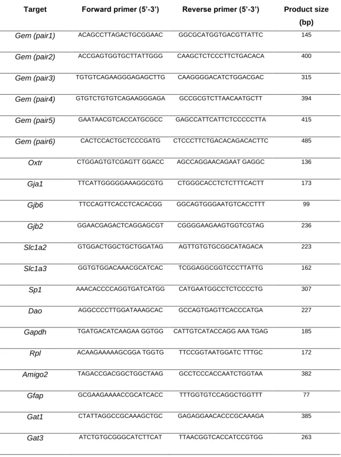
2.6 RNA-Isolation ...33
2.6.1 Reverse Transcriptase PCR (RT PCR), Endpoint PCR and quantitative PCR (qPCR) ...33
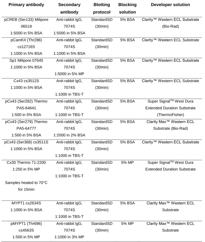
2.7 Protein Extraction ...36
2.8 SDS-PAGE and Western Blot Analysis ...36
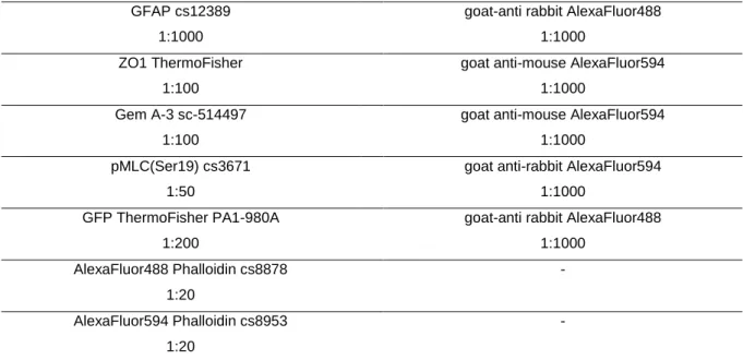
2.9 Immunocytochemistry ...39
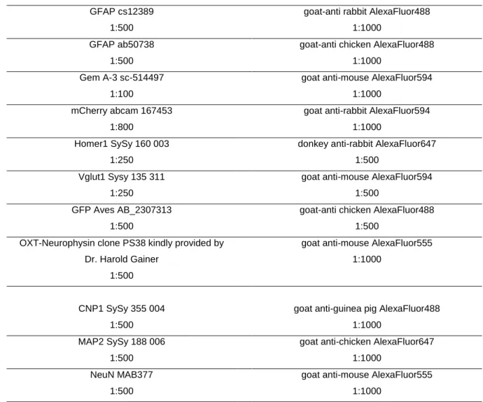
2.10 Immunohistochemistry ...40
2.11 Gap-junctional intercellular communication (GJIC) ...41
2.12 Bioimaging and Image Analysis ...41
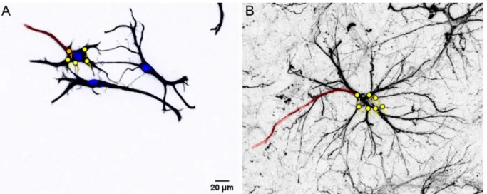
3D-reconstruction of GFP-expressing astrocytes ...42
Determination of astrocyte-synapse spatial relationship by STED nanoscopy ...44
Colocalization studies ...44
Intensity measurements and determination of above threshold cells in vitro and in vivo ...44
2.13 Statistical Analysis ...44
[5]
3 RESULTS ...46
3.1 Establishment of primary rat cortical astrocyte cultures ...46
3.2 Characterization of the effects of OXT on astrocytes ...47
Effects of OXT on astrocytic signaling cascades and proteins in vitro and in vivo ...47
Effects of OXT on the expression of selected astrocytic genes ...48
Effect of OXT on distribution of astrocytic gap-junctions and its impact on intercellular connectivity ...50
OXT-induced changes in astrocytic cytoskeletal dynamics and the impact on astrocyte-neuron spatial relationships ...53
3.3 Involvement of the Sp1 – Gem signaling axis ...57
The involvement of the small GTPase Gem in the effects of OXT on astrocytes ....57
Potential involvement of astrocytic gap-junctions in OXT-induced cytoskeletal remodeling ...62
Regulation of Gem by OXT on a genomic level ...64
Differential regulation of the Sp1-Gem-ROCK axis in neuronal cells ...66
3.4 Establishment of astrocyte-specific Oxtr/Gem-knockdown vectors ...67
4 DISCUSSION ...70
4.1 Effects of OXT on astrocytic signaling cascades and proteins ...71
4.2 Effect of OXT on expression and distribution of astrocytic gap-junctions and its impact on intercellular connectivity ...73
4.3 OXT-induced changes in astrocytic cytoskeletal dynamics and the impact on astrocyte-neuron spatial relationships ...75
4.4 The involvement of the small GTPase Gem in the effects of OXT on astrocytes ...77
4.5 Involvement of astrocytic gap-junctions in OXT-induced cytoskeletal remodeling ...79
4.6 Transcriptional regulation of Gem by OXT ...80
4.7 Differential regulation of the Sp1-Gem axis in neuronal cells ...82
4.8 Establishment of astrocyte-specific Oxtr/Gem-knockdown vectors ...83
4.9 Conclusion and future perspectives ...84
References ...88
Appendix 1 ... 106
Appendix 2 ... 115
Appendix 3 ... 129
Danksagung ... 130
[6]
ABSTRACT
Especially in higher vertebrates, astrocytes are an indispensible part of signal processing
within the brain. Thus, the mode of action of a neuroactive peptide such as OXT cannot be
fully understood without this integral part of the CNS. The effects of OXT on neuronal cells
have been well characterized, while its effects on astrocytic cells, specifically on OXTR-
coupled signaling and its resulting cellular consequences, are poorly understood and might
very well differ. To characterize the effect of OXT on astrocytic gene expression, intracellular
signaling, as well as astrocyte-specific proteins, synthetic OXT was either administered icv in
male Wistar rats or applied to cultured rat primary cortical astrocytes. Due to the results of this
analysis implying an acute OXT-induced cytoskeletal remodeling and alterations to gap-
junction coupling, I next examined the underlying molecular mechanisms and cellbiological
consequences of these alterations. Here I found that OXT led to rapid elongation and formation
of astrocytic processes in vitro and in vivo, while simultaneously impairing astrocytic
intercellular connectivity. Mechanistically, both of these effects were OXTR-specific, conveyed
via PKC and, to a lesser extent, MEK1/2 signaling. Notably, OXT-induced cytoskeletal
remodeling and impairment of gap-junctions were characteristic for OXT, since its closely
related sister-peptide AVP did not affect the examined parameters. CLSM and STED-
microscopy following icv or ex vivo administration of OXT furthermore revealed changes to
astrocyte-neuron spatial relationships in two brain regions associated with high
responsiveness of astrocytic markers to OXT, i.e. PVN and hippocampus. In depth in vitro
studies identified the previously undescribed Sp1-Gem signaling axis to be at the base of these
effects. A combination of knockdown, knockout and overexpression experiments revealed that
OXT drives Gem expression via the transcription factor Sp1 and that Gem is required and
sufficient for the effects of OXT on astrocytes. The Sp1-Gem axis was differentially regulated
by OXT in neuronal cells, identifying it as key driver in the cell type-specific response of
astroglial cells to OXT. Based on these findings, astrocyte-specific AAV-mediated Gem or Oxtr
shRNA knockdown vectors were established as tools for a targeted manipulation of astrocytic
OXTR signaling and future assessment of astrocytic contribution to the physiological and
behavioral effects of OXT. To this end, shRNA oligonucleotides were screened for knockdown
efficiency in vitro and subsequently packaged into viral vectors providing astrocyte-specific
expression via transcriptional control of shRNA expression under the hGFAP promoter.
[7]
1 INTRODUCTION
1.1 The neuropeptide oxytocin
Due to its various physiological and behavioral functions, there has been a growing scientific interest in the nonapeptide oxytocin (OXT) and its cognate receptor over the last decade.
However, the research on OXT and its closely related sister-peptide arginine vasopressin (AVP) dates back more than a century. Besides a multitude of physiological functions, this research unraveled a remarkable degree of evolutionary conservedness of the OXT/AVP systems (Acher et al., 1995; Donaldson and Young, 2008; Hoyle, 1999), indicating a high degree of selective pressure acting on both, the genes coding for OXT/AVP homologs, as well as their receptors. As a result of a gene duplication of their common ancestor gene vasotocin, which homologs can be traced back to invertebrate phylae like annelida or mollusca, OXT/AVP-like neuropeptides are found in all vertebrate species. Their evolutionary conservation extends beyond the chemical structures of the peptides and their receptors, as it can also be observed in a similar anatomical distribution of the synthesizing neurons and receptor expression patterns (Grinevich et al., 2016; Vargas-Pinilla et al., 2015). Moreover, striking functional similarities exist, with OXT/AVP homologs regulating osmotic homeostasis and social/sexual behaviors throughout large parts of the animal kingdom (Lema et al., 2015;
Soares et al., 2012; Van Kesteren et al., 1995).
In mammals, OXT and AVP are mainly synthesized in magnocellular neurons of the paraventricular nucleus (PVN) and the supraoptic nucleus (SON) of the hypothalamus in a mutually exclusive manner (Mohr et al., 1988; Sofroniew, 1983). A detailed immunohistochemical analysis in rats revealed an additional expression in magnocellular accessory nuclei of the hypothalamus constituting around one-third of OXT/AVP-positive cells (Rhodes et al., 1981). An additional site of OXT synthesis is posed by parvocellular neurons of the PVN, but also scattered extra-hypothalamic neurons (De Vries and Buijs, 1983;
Knobloch and Grinevich, 2014). Contrary to magnocellular neurons, these cells do not project to the neurohypophysis, but instead form connections with a) areas in the brain stem and spinal cord, where they are involved in the regulation of autonomic processes and pain perception (Swanson et al., 1980) and b) magnocellular neurons of the SON/PVN to regulate OXT release (Eliava et al., 2016).
OXT synthesizing cells express the Oxt gene, which encodes a signal peptide (SP), the
nonapeptide and its attached neurophysin (NP). The 4850bp gene contains three exons, two
introns (Ivell and Richter, 1984), as well as a promoter region for which binding of various
hormone receptors (Adan et al., 1993; Richard and Zingg, 1990; Sladek and Somponpun,
2004) and the transcription factor CREB (Sharma et al., 2012) was identified. The newly
[8]
synthesized SP-OXT-NP pre-peptide is packaged into neurosecretory large dense-core vesicles (Tooze, 1998) where it undergoes extensive posttranslational modifications (Altstein and Gainer, 1988; Gainer et al., 1977). The magnocellular cells of the PVN and SON send axonal projections to the neurohypophysis, a neuro-hemal organ of neuronal (i.e. ectodermal) origin. OXT-containing vesicles are stored in and released from neuronal terminals into neurohypophysial capillaries, permitting entry into the peripheral blood stream (Hatton, 1990).
Within the brain, OXT neurons project to various mesolimbic and forebrain structures like the bed nucleus of the stria terminalis (BNST), septal nuclei, nucleus accumbens, prefrontal cortex, medial and central amygdala, hippocampus and the anterior olfactory nucleus (Dolen et al., 2013; Grinevich et al., 2016; Sofroniew, 1980). In contrast to peripheral OXT release, intracerebral OXT release seems to occur non-synaptically, as neither presynaptic localization of OXT containing vesicles nor postsynaptic oxytocin receptors (OXTR) could be observed yet (Knobloch et al., 2012; Theodosis, 1985). Oxt mRNA has been detected in dendrites (Mohr and Richter, 2003), suggesting local synthesis of OXT and consequent dendritic release (Pow and Morris, 1989). Indeed, Pow and Morris were the first to describe such dendritic release of nonapeptides using electronmicroscopic tools (Morris and Pow, 1991). Instead of being transmitted synaptically, OXT is released axo-dendritically at axonal projection sites, or somato-dendritically within the PVN/SON. These diffusion-like neuropeptide actions led to the view of OXT as neuromodulator, rather than neurotransmitter (Landgraf and Neumann, 2004;
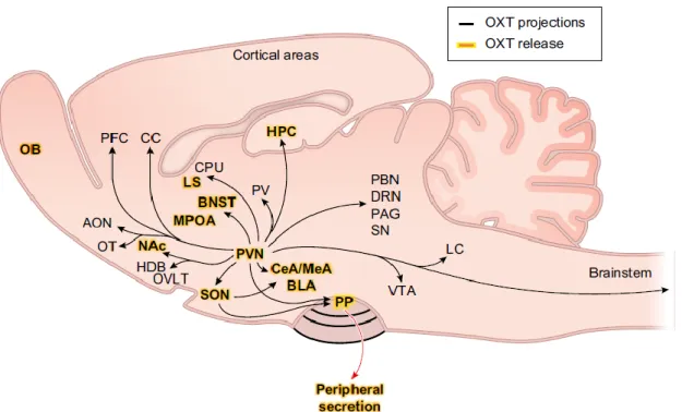
Leng and Ludwig, 2008). The anatomical distribution of OXT projections and release sites is summarized in Fig. 1.
Figure 1. Projections and release sites of the OXT system in an anatomical scheme of the rat brain (sagittal slice).
[9]
OXTergic projections originating from the PVN/SON are depicted as black lines, connecting OXTR expressing brain areas. Brain regions where OXT release has directly been shown are highlighted in yellow. AON, anterior olfactory nucleus; OB, olfactory bulb; OT, olfactory tubercle; Nac, nucleus accumbens; OVLT, organum vasculosum laminae terminalis; SON, supraoptic nucleus; PVN, paraventricular nucleus of the hypothalamus; PP, posterior pituitary;
PFC, prefrontal cortex; CC, cingulate cortex; MPOA, medial preoptic area; BNST, bed nucleus of the stria terminalis;
LS, lateral septum; CPu, caudate putamen;PV, paraventricular nucleus of the thalamus; CeA, central amygdala;
MeA, medial amygdala; BLA, basolateral amygdala; VTA, ventral tegmental area; LC, locus coeruleus; PBN, parabrachial nucleus; DRN, dorsal raphe nucleus; PAG, periaqueductal gray; SN, substantia nigra; HPC, hippocampus; HDB, nucleus of the horizontal limb of the diagonal band. Scheme adapted from (Jurek and Neumann, 2018).
Interestingly, peripheral and central release can occur coordinated or independently, increasing the response specificity of the OXT system to external stimuli. Stimuli triggering simultaneous release include parturition, lactation (suckling), physical and emotional stress, as well as osmotic challenge, mating and social interaction (reviewed in(Jurek and Neumann, 2018). It should, however, be noted that differences in temporal dynamics of central and peripheral release do still exist for these stimuli. An examples for a stimulus triggering independent central OXT is the anorexic neuropeptide α-melanocyte stimulating hormone (α- MSH). While α-MSH induced OXT release within the SON, it inhibited release into the bloodstream (Sabatier et al., 2003).
Dependent on the stimulus and site of release, OXT drives an adequate physiological or behavioral response, including the modulation of physiological parameters such as pain perception, appetite and HPA axis activity, but also the regulation of complex social behaviors and emotionality. A large body of literature has demonstrated that the endogenous OXT system promotes learning and memory functions, maternal behavior, sexual aggression, social preference and bonding (reviewed in Jurek and Neumann, 2018). Moreover, its robust anxiolytic, anti-stress, and pro-social effects have brought the brain OXT system up for discussion as a potential therapeutic target for psychopathologies such as anxiety disorders (Labuschagne et al., 2010; Landgraf and Neumann, 2004; MacDonald and Feifel, 2014), major depressive disorder or autism spectrum disorder (Bakermans-Kranenburg and van Ijzendoorn, 2014). In this context, a detailed understanding of the mode of action of OXT is of particular relevance and might contribute to the identification of new treatment options.
1.2 The OXTR and its downstream effectors
On a cellular level, OXT mediates its functions mainly via the OXTR. In the periphery, the Oxtr
gene displays a widespread expression pattern, including the renal cortex, adrenal medulla,
heart, retina, skin, fat tissue, the enteric nervous system, bones and taste buds (Colaianni et
al., 2014; Deing et al., 2013; Eckertova et al., 2011; Gutkowska and Jankowski, 2012; Halbach
et al., 2015; Ostrowski et al., 1995; Taylor et al., 1989). In the brain, Oxtr expression is found
in all above mentioned brain areas (for review, see(Jurek and Neumann, 2018) and various
[10]
non-neuronal cell types (Di Scala-Guenot and Strosser, 1992a; Yuan et al., 2016). OXTR- positive cells display a wide range of properties that in some cases are even co-characteristic.
For example, Oxtr expressing neurons within the PVN are exclusively glutamatergic, whereas within the BNST OXTR-positive neurons are of GABAergic nature (Dabrowska et al., 2013).
The highly conserved 17kb Oxtr gene consists of four exons and three introns and codes for a 389 amino acid 7-transmembrane domain (TM) G protein-coupled receptor (GPCR) (Kimura et al., 1992; Rozen et al., 1995). The first three extracellular loops of the OXTR are most critical for OXT binding by interacting with the tertiary structure of the peptide (Postina et al., 1996).
OXT does not exclusively bind to OXTRs (
Ki =0.79nM), but also to the vasopressin receptors V1a (
Ki =120nM), V1b (
Ki=1.782nM)and V2 (
Ki =1.544nM), although with lower affinity (Akerlund et al., 1999). This fact makes it both difficult and important to validate OXTR specificity in any OXT-dependent effect to be studied.
Oxtr expression is under tight transcriptional and post-transcriptional control, strongly enhancing the regulatory capacity of the OXT-OXTR system. This is reflected by binding sites for various transcription factors/repressors like Sp1, AP1/2, c-Myb, NF-κB, estrogen receptors, C/EBP and Peg3 in the Oxtr sequence (Frey et al., 2018; Terzidou et al., 2006)and reviewed in(Blanks et al., 2007), as well as additional epigenetic and miRNA-based mechanisms (Beery et al., 2016; Choi et al., 2013). Moreover, the availability of the ligand itself seems to be able to affect OXTR quantities, since both chronic intracerebroventricular (icv), as well as repeated intranasal administration reduced Oxtr mRNA expression in various brain regions (Huang et al., 2014; Peters et al., 2014). Extracellular signals regulating Oxtr expression include labor- induced mechanical stretch and interleukin-β release (Terzidou et al., 2011; Terzidou et al., 2005), as well as estrogen and progesterone (Quinones-Jenab et al., 1997; Schumacher et al., 1990).
Once the receptor is expressed and trafficked to its subcellular localization, the biochemical environment of the plasma membrane and interaction partners within the membrane are able to alter both signal perception, i.e. affinity, and activation patterns of downstream effectors (Busnelli et al., 2016; Gimpl and Fahrenholz, 2001; Reversi et al., 2006; Romero-Fernandez et al., 2013; Wiegand and Gimpl, 2012; Wrzal et al., 2012). Additionally, the coupled signaling cascades will determine the cellular and, later on, network output. Due to its major role in uterine contractions during birth, these cascades were initially mainly studied in myometrial cells.
The first level of signal processing following ligand binding is characterized by the respective subforms of G
α/G
β/G
γproteins immediately coupled to the receptor, which can vary dependent on the cell type and physiological state. For example, the OXTR is coupled to the inhibitory G
αprotein subforms G
αi1-3, G
αoAand G
αoBin myometrial cells (Busnelli et al., 2012), whereas
[11]
coupling to the activating G
q/11subform and subsequent phospholipase C (PLC) activation was described in myometrial membranes (Ku et al., 1995). Another example is the G
q/11–mediated increased contractility of myometrial cells of non-pregnant rats vs. the G
βγ-dependent decrease of contractility in myometrial cells of pregnant rats (Zhou et al., 2007). As exemplified here, such differential coupling can in consequence lead to a contrary response to the same signal, reflecting the highly context-dependent nature of OXTR signaling.
As a general characteristic of GPCRs, the second level of signal processing is an activation of second messengers by either the activated G proteins or direct interactions. This mechanism provides an enormous amount of amplification, enabling small quantities of a ligand to trigger a significant response. The main second messenger recruited by G proteins is Ca
2+, which can be released from internal stores and/or enter cells from the extracellular space. G
q/11coupled OXTR signaling activates phospholipase C, which in turn leads to the cleavage of phosphatidylinositol 4,5-bisphosphate to inositol-3-phosphate (IP3) and diacylglycerol (DAG).
IP3 binding to IP3 receptors located in the endoplasmatic reticulum subsequently triggers Ca
2+release from intracellular stores (for review see(Mikoshiba, 2007). In cells of the central nervous system, this has been described for both neuronal (Ayar et al., 2014) and astrocytic cells (Di Scala-Guenot et al., 1994) in the context of OXT. In contrast, OXT led to a decrease of intracellular calcium in lipopolysaccharide-challenged microglial BV-2 cells (Yuan et al., 2016). The full extent of signal amplification additionally requires Ca
2+influx from the extracellular space via calcium channels. For OXT, the involvement of various transient receptor potential cation channels (TrpC; specifically TrpC1/TrpC3-6;(Chung et al., 2010;
Murtazina et al., 2011; Shlykov et al., 2003; Ulloa et al., 2009))and transient receptor potential
vannilloid channels (TrpV; specifically TrpV2/TrpV4;(van den Burg et al., 2015; Ying et al.,
2015), as well as voltage operated channels (Sanborn, 2007) has been reported. That this
aspect of OXTR signaling is of critical relevance at the behavioral level was demonstrated in
2015 by van den Burg et al., as pharmacological blockade of TrpV2 within the PVN prevented
the acute anxiolytic effect of OXT by inhibiting downstream activation of the mitogen activated
protein kinase (MAPK) pathway that had been previously shown to mediate this effect (Blume
et al., 2008; Jurek et al., 2012). Other Ca
2+-dependent downstream effectors of the OXTR are
the calmodulin dependent kinases II/IV (Jurek et al., 2015), calcineurin (Pont et al., 2012) and
protein kinase C (PKC; (Devost et al., 2008a). We recently found that OXT induces de novo
protein synthesis in neuronal cells in a PKC-dependent manner by stimulating the translational
activator eukaryotic elongation factor 2 (Martinetz et al., 2019). Notably, one of the newly
synthesized proteins, neuropeptide Y receptor Y5, was sufficient and necessary for the
anxiolytic effect of acute OXT within the PVN. In a broader picture, this demonstrates an
intracellular feed-forward mechanism of OXT, supporting its own effect on gene expression
(see below) by facilitating the translation of the newly transcribed mRNAs.
[12]
The third level of signal transduction is additionally shaped by downstream effectors not dependent on increased cellular Ca
2+levels. The transactivation of the epidermal growth factor receptor (EGFR) and subsequent activation of MAPK pathways studied both in myometrial cells and neurons, is a link of OXTR signaling to such effectors (Blume et al., 2008; Lin et al., 2012; Zhong et al., 2003). Although the direct signal transducer is yet unknown, OXTR-coupled signaling triggers the auto-phosphorylation activity of the EGFR tyrosine kinase domains, which subsequently recruits the membrane-bound GTPase Rat sarcoma (Ras). Full MAPK activation is then accomplished by a phosphocascade of [c-Raf-1(Map3k)/Map2k/ERK1/2], although ERK1/2 independent signaling was observed for the Map2k family member MEK1/2 in some cases (Fischmann et al., 2009; Jurek et al., 2015; Jurek et al., 2012; Kim et al., 2015).
OXT-induced phosphorylation peaks for c-Raf and ERK1/2 have been described as early as
5min or 10min, respectively (Blume et al., 2008), however the timecourse of ERK1/2
phosphorylation in particular seems to be cell type-dependent (Terzidou et al., 2011). Other
members of the MAPK family that have been linked to OXTR signaling in myometrial cells are
p38 and ERK5 (Brighton et al., 2011; Devost et al., 2008b; Kim et al., 2017). In general, MAP
kinases are involved in the regulation of a wide variety of cellular processes ranging from cell
differentiation and migration, apoptosis to regulation of gene expression in response to
external stimuli. In the context of OXT, exertion of such transcriptional control has been mainly
studied for two distinct transcriptional regulators. First, the transcription factor CREB was found
to mediate OXTR/MAPK-induced spatial memory formation during motherhood (Tomizawa et
al., 2003), hippocampal long-term potentiation (Lin et al., 2012), as well as Crf expression
(Jurek et al., 2015). Second, the myocyte enhancer factor 2 (MEF-2) was activated by OXT in
myometrial (Devost et al., 2008b) and neuronal cells (Meyer et al., 2018), leading to neurite
outgrowth in the latter. In general, OXT seems to have profound effects on the formation and
elongation/retraction of cellular processes. While OXT caused less ramification in hippocampal
glutamatergic neurons ex vivo (Ripamonti et al., 2017), it induced process elongation in human
neuroblastoma and glioblastoma cells (Lestanova et al., 2016; Lestanova et al., 2017). The
latter effect was accompanied by a variety of changes in the expression of genes associated
with cytoskeletal dynamics. In line, OXT was found to increase myometrial contractility via
activation of the RhoA/ROCK signaling pathway, most pronounced during late pregnancy
(Gogarten et al., 2001; Tahara et al., 2002). The GTPase RhoA and its effector ROCK belong
to the major regulators of the cellular cytoskeleton and as such are involved in cellular
migration, morphology, adhesion, motility and smooth muscle contraction. (Van Aelst and
D'Souza-Schorey, 1997). ROCK targets Ser19 of the myosin light chain (MLC;(Totsukawa et
al., 2000), while simultaneously inhibiting myosin light chain phosphatase (MYPT) via
phosphorylations at threonines 696 and/or 853 (Feng et al., 1999; Kawano et al., 1999) and,
in consequence, increases F-actin contractility.
[13]
Finally, the desensitization of the OXT-OXTR signaling axis is initiated via OXTR phosphorylation by the G protein-coupled receptor kinase 2 and is already initiated 4s following ligand binding (Hasbi et al., 2004). This phosphorylation enables subsequent binding of β- arrestin2, which in turn uncouples the receptor from its G proteins and simultaneously acts as an adapter for clathrin-mediated endocytosis (Goodman et al., 1996; Smith et al., 2006). The endocytotic vesicles are stored intracellulary, and the receptor is reinserted into the membrane around 4h after the initial internalization (Conti et al., 2009). In addition, β-arrestin provides a negative feedback mechanism by inhibiting insertion of TrpV channels into the plasma membrane (Ying et al., 2015). Interestingly, β-arrestin additionally seems to play a role in the nuclear translocation of the OXTR, a process so far exclusively described in osteoblasts (Di Benedetto et al., 2014). A potential secondary negative feedback mechanism is the prevention of prolonged calcium influx via TrpC3/5 channels by inhibitory phosphorylation of these channels by PKC (Venkatachalam et al., 2003).
Taken together, the high degree of regulatory capacity, from ligand over receptor to the cell type-specific identity of coupled downstream effectors, enable the OXT-OXTR system to bring about physiological and behavioral responses, which are diverse and highly specific at the same time. In the brain, OXT actions are not restricted to neurons, which makes an understanding of these actions on other cell types of the CNS imperative.
1.3 Astrocytes – Maintenance of CNS homeostasis and active participation in neuronal communication
The first description of a neural cell that would later be classified as glia cell dates back to 1851
(Müller, 1851). Heinrich Müller had described cells of the retina, which were later named Müller
cells, while six years later Karl Bergmann described radial like cells of the cerebellum (later
named Bergmann glia;(Bergmann, 1857). Carl Frommann coined the term glia cell (from
greek: γλία, glue), when describing ‘Leim erfüllte Interstitien’ (glue-filled
interstitiae;(Frommann, 1867). Based on their star-shaped appearance with processes
pointing in all directions, Michael von Lenhossék was the first to use the term ‘astrocyte’ in
1895 (Lenhossék, 1895). This term was later on popularized by Santiago Ramón y Cajal, who
developed the first astroglia-specific staining technique based on gold and mercury chloride-
sublimate staining (Garcia-Marin et al., 2007). These anatomical studies fostered speculations
on the physiological functions of these cells, some of them turning out to be surprisingly correct
when examined experimentally later on. Such examinations were enabled by
electrophysiological experiments in the late 1950s providing the first evidence of neuron-glia
interactions (Hertz, 1965; Hild et al., 1958; Orkand et al., 1966). The establishment of purified
[14]
cultures by Jean de Villis allowed research to provide insights into astroglial biology on a single- cell level (Morrison and de Vellis, 1981). The findings of the following decades (see below) even led some researchers to call for a shift from a neurocentric to a gliocentric view of the brain. The glia cells that populate the CNS (neuroglia) are characterized by form, function and developmental origin. Neuroglia are subdivided into macro and microglia, with the first including cells of neuroectodermal origin (astrocytes, oligodendrocytes, ependymal cells), while the latter are of mesodermal origin and originate from macrophages invading the brain during early development (Sierra et al., 2014).
Evolutionary, neuro-supportive glial cells could be traced back to higher Platyhelminthes, with support of neuronal cells by ‘proto-astrocytes’ first observed in Nemathelmintes (Golubev, 1988; Oikonomou and Shaham, 2011). Neuroglia were then found in all higher taxa including Arthropoda, Mollusca and Annelida (Hartline, 2011), as well as vertebrates with a general trend for increased glia to neuron-ratios throughout the course of evolution (Friede, 1954;
Reichenbach, 1989). This increase follows the increase in brain thickness, as well as neuronal energy expenditure and reflects the resulting elevated demand for metabolic support and homeostatic maintenance. Moreover, the complex astrocyte-neuron interplay on the synaptic level (for a more detailed description see 1.4) allowed for a progressive increase in the computational power of the CNS, which is also reflected by large increases in astrocytic size, complexity and signal procession speed particularly seen in humans. This has been remarkably demonstrated by engraftment of human glial progenitor cells (hGPCs) into neonatal mice. Chimeric mice devoloped mature hominid astrocytes, which caused sharp enhancements of LTP, as well as improved learning capabilities in a variety of behavioral tests (Han et al., 2013). Despite significant advances in the identification of astrocytes (see below), their exact abundance, especially in relation to other cell types, is still under debate. In rodents, the glia to neuron ratio is around 0.3 – 0.4, with 10-20% of CNS cells being astrocytes (Sun et al., 2017), whereas the glia-neuron ratio in higher mammals increases to around 1.5-2.0 (Pelvig et al., 2008; Sherwood et al., 2006). However, of these glia cells only ~20-40% where found to be astrocytes, while oligodendrocytes make up ~50% and microglia ~5-10%
(Mittelbronn et al., 2001). Verkhratsky and Nedergaard (2018) describe astrocytes throughout
the course of evolution as ‘highly opportunistic supportive cells that tailor their form and
function to match the demands of progressively changing nervous tissue. In this context, the
CNS evolved through division of functions between cell types: the neurons become mostly
responsible for rapid propagation of signals associated with action potential and chemical
synapses, whereas neuroglia assumed the responsibility for homeostasis and defense’. In
case of astrocytes, these homeostatic functions are manifold and include ionostasis, pH
buffering, H
2O homeostasis and thereby regulation of extracellular space volume, reactive
oxygen species homeostasis, neurotransmitter uptake and recycling, neurovascular coupling,
[15]
clearance of waste products, systemic energy homeostasis, regulation of food intake and nutrient shuttling to neurons. This homeostatic focus is reflected by a broad variety of membrane transporters, ion channels and metabolic enzymes being the most highly expressed astrocytic genes (Cahoy et al., 2008). Hence, astrocytes are indispensable to maintain a stable molecular environment within the CNS and thereby support vital neuronal functions.
Developmentally, astrocytes, in contrast to microglia, originate from neuroepithelium-derived neuronal progenitors (Kriegstein and Alvarez-Buylla, 2009) and differentiate to astrocytes after the neurogenic period of the CNS, in which early neurons populate neuronal layers. In rodents, the subsequent gliogenic switch, characterized by the expression of gliogenic transcription factors like NFIA or Sox9 (Deneen et al., 2006; Freeman, 2010), occurs on embryonic day (E) 12 in the spinal cord and around E16-18 in the cortex. Neurons and astrocytes born in the same region will generally develop together, and by that give rise to regional specificity (Gao et al., 2014; Magavi et al., 2012). However, the described embryonic astrogliogenesis accounts only for a part of adult CNS astrocytes. In rodents, the number of non-neuronal cells increases from 4 million to over 140 million during the second and third postnatal weeks (Bandeira et al., 2009), whereas in cats the astrocyte-to-neuron ratio almost doubles from ~0.8 in young kittens to ~1.48 in adult animals (Brizzee and Jacobs, 1959). An important factor in this postnatal increase is most likely the retained (low) proliferative capacity of astrocytes, which distinguishes them from the majority of neurons (Ge and Jia, 2016).
The identification of astrocyte-specific markers facilitated the understanding of astroglial biology to a great extent. However, even today, specific identification and targeting of astrocytes is not trivial and topic of ongoing debates, since the high degree of morphological and transcriptomic heterogeneity rendered identification of an universal astrocytic marker impossible to the date. Therefore, only a combination of techniques and markers led to a more concise picture. Early on, classical histological techniques based on Cajal’s sublimated gold- chloride staining accomplished labeling of astroglial filaments and endfeet. Later, glial fibrillary acidic protein (GFAP) within the CNS was identified to be exclusively expressed in astrocytes and has been the most commonly used astrocytic marker since. GFAP is an intermediate filament of the astrocytic cytoskeleton (Bignami et al., 1972; Hol and Pekny, 2015; Ludwin et al., 1976), which displays a subpopulation and region-specific heterogenic expression in vivo.
For example, 60% of astrocytes in the adult hippocampus are GFAP positive, while this holds
true for only 12% of astrocytes in the mouse entorhinal cortex. GFAP seems to be generally
upregulated in reactive astrocytes in vivo (Bushong et al., 2002; Nolte et al., 2001; Ogata and
Kosaka, 2002; Walz and Lang, 1998), while almost all astrocytes are GFAP positive in vitro
(Walz, 2000; Yeh et al., 2013). Even though GFAP stains the astrocytic cytoskeleton, its use
for this purpose is somewhat limited by its lack of localization to finer and distal processes
[16]
(Connor and Berkowitz, 1985). Other proposed markers are the calcium binding protein S100B (Savchenko et al., 2000), the glutamate transporters EAAT1 and EAAT2 (Jungblut et al., 2012), the enzyme glutamine synthetase (Anlauf and Derouiche, 2013), the intermediate filament vimentin (Pekny et al., 1999), the water channel aquaporin 4 (Nielsen et al., 1997), the transcription factor Sox9 (Sun et al., 2017), the foliate metabolism enzyme aldehyde dehydrogenase 1 family member L1 (ALDH1L1;(Cahoy et al., 2008) and the gap-junction proteins Cx30/Cx43 (Dermietzel et al., 1991; Nagy et al., 1999). All of these markers provide distinct advantages and disadvantages in terms specificity, inclusiveness and subcellular distribution depending on the specific aim of the study and therefore have to be chosen carefully beforehand (for a concisive review see(Verkhratsky and Nedergaard, 2018).
Additionally, astrocytes can be visualized by either dye-loading with a patch pipette or expression of a fluorescent protein/calcium indicator, e.g. EGFP, under the promoter of an astrocytic marker gene like Gfap. Due to the subcellular distribution of fluorescent dyes or proteins to even fine astrocytic processes, these techniques allow for a more detailed analysis of the morphology of astrocytes and their spatial relationship to neighbouring cells. The application of the above mentioned markers and approaches led to the identification of a variety of astrocytic subpopulations. First, protoplasmic astroglia represent the major population of astrocytes in the grey matter of the brain and spinal cord. These cells possess a small soma (~10µm in diameter) with 5-10 primary processes (~50µm in length) that branch to a dense peripheral arborization underlying their spongiform appearance. A single protoplasmic astrocyte in the rodent cortex may contact 4-8 neurons, surround ~300-600 neuronal dendrites, and interact with 20,000-120,000 synapses residing within its domain (Bushong et al., 2002;
Halassa et al., 2007b). Second, fibrous astrocytes populate the white matter of the CNS and are organized in rows between the axonal bundles. Their arborization is less complex than that of protoplasmic astrocytes, and their overlapping processes reflect the absence of domain organization characteristic for protoplasmic cells. The processes of fibrous astrocytes establish several perivascular endfeet and send numerous long (up to 100µm) extensions that contact axons at nodes of Ranvier (Lundgaard et al., 2014).
The morphological heterogeneity of astrocytes seems to be mirrored by a remarkable degree
of molecular heterogeneity (Chai et al., 2017), which is believed to play a role in their ability to
specifically accompany distinct neuronal circuits despite their high spatial overlap (Martin et
al., 2015). Fluorescence activated cell sorting with subsequent RNA sequencing revealed that
astrocytes are especially enriched in transcripts of genes involved in cellular metabolism
compared to neurons (Lovatt et al., 2007). Later studies showed a strong increase in
expression of phagocytotic genes in mature (17-30d) vs. immature (7-8d;(Cahoy et al., 2008)
astrocytes, further supporting the involvement of astroglia in synaptic pruning (Chung et al.,
[17]
2013). In conditions of CNS injury and disease, astrocytes switch to a so-called reactive state, which is characterized by alterations in the astrocytic gene expression profile (Zamanian et al., 2012). Specifically, this state is more directed towards interactions with the immune system and cytoskeletal motility. Notably, astrocytes cultured in vitro display a similar transcriptome to such a reactive state, stressing the importance to validate in vitro findings under non-reactive, more physiological conditions, i.e. in vivo. In contrast, astrocytes show very similar electrophysiological properties in all brain regions, a feature for which the above mentioned heterogeneity of astrocytes is not observed (Du et al., 2016). In general, astroglia possess a hyperpolarized resting potential (~80mV) and low input resistance, which is reflected by an almost linear current to voltage relationship (Chvatal et al., 1995; Dallerac et al., 2013; Mishima and Hirase, 2010).
1.4 Astrocytic networks and their regulation
A characteristic feature of astrocytes is their high degree of intercellular connectivity via gap- junctions. In a variety of tissues these specialized subcellular areas allow a tightening of the intercellular cleft to ~2-3nm (Evans and Martin, 2002), and the connexons residing within these areas permit intercellular transport of ions, second messengers, nucleotides, siRNA and metabolites smaller than 1kDa (Harris, 2007; Tabernero et al., 2006; Valiunas et al., 2005). In the grey matter, two neighbouring astrocytes are connected with about 230 gap-junctions on average. Injection of Lucifer yellow or biocytin into a single astrocyte results in staining of ~50–
100 adjacent astroglial cells. The concept of a panglial syncytium connecting all macroglia into a single functional network, which has been described in invertebrates (Mugnaini, 1986), does not fully apply to the mammalian CNS. In many brain regions anatomically segregated astroglial networks follow anatomical structures (Giaume et al., 2010; Roux et al., 2011) and even coupling between adjacent astrocytes is not always present, as 15-20% of neighbouring astrocytes were found to be uncoupled (Houades et al., 2006; Meme et al., 2009). Thus, astroglial coupling is not only defined by spatial proximity, and astroglial networks may represent a non-binary second level of information processing parallel to that formed by neurons.
A single gap-junction is composed of two adjacent (homo-or heteromeric) connexons that are assembled from six connexin (Cx) subunits. The Cx gene family has 21 members in humans coding for 4-TM proteins with differing molecular mass which also underlies their nomenclature (e.g. Cx26, Cx43) (Dermietzel et al., 1990; Saez et al., 2003). Several hundred connexons form so-called gap-junctional plaques between two coupled cells in a homo-or heterocellular manner (e.g. astrocyte-oligodendrocyte or astrocyte-neuron;(Altevogt and Paul, 2004;
Alvarez-Maubecin et al., 2000). Homocellular astrocytic gap-junctions are formed by
[18]
Cx26,Cx30 and Cx43, with Cx43 being the most abundant, ubiquitously expressed isoform (Giaume et al., 1991; Kunzelmann et al., 1999; Nagy et al., 2004). Cx30 is most prominently found within the thalamus and leptominges, but not in the white matter (Sohl et al., 2004), while Cx26 expression is restricted to subcortical areas like the hypothalamus and subthalamic nuclei (Nagy et al., 2011). The expression of connexins seems to be regulated by neuronal factors, since co-culturing neurons with astrocytes upregulates Cx43 and triggers Cx30 expression. Without exposure to neuronal factors, cultured astrocytes show detectable levels of Cx30 only in a mature state (21d onwards;(Koulakoff et al., 2008). The biophysical properties of connexons are regulated by multiple factors including pH, transjunctional or membrane voltage (Herve and Derangeon, 2013), subunit composition, intracellular calcium levels and phosphorylation state, which is controlled by protein kinases A, C, and G, as well as MAPK signaling (Ek-Vitorin et al., 2006). Phosporylation of Cx43 at Ser368 by PKC or at Ser279/Ser282 by MAPK signaling can additionally lead to internalization and possible subsequent degradation of the gap-junction which involves internalization into a specific doublemembrane vacuole termed annular junction or connexosome (Kjenseth et al., 2010).
These regulatory mechanisms contribute to the high turnover rate of connexins with a half-life of several hours. In addition, connexons can act as gated pores, known as hemichannels (Esseltine and Laird, 2016), which have been identified in astrocytes in vitro and in vivo and can be formed by all three types of astrocytic connexons (Giaume et al., 2013). Generally, hemichannels are in a closed state, but their opening can be triggered by low external calcium concentration, substantial depolarization, specific intracellular Ca2
+signals or exposure to proinflammatory agents (Orellana et al., 2012; Orellana et al., 2009). Hemichannels are discussed to be one of the major ways for astroglial secretion of neuroactive substances (see the concept of gliotransmission and the tripartite synapse below).
Notably, recent studies have demonstrated a variety of non-channel functions for astrocytic connexins, including synapse invasion (Pannasch et al., 2014), synaptic glutamate clearance (Pannasch et al., 2019), cellular migration and adhesion (Ghezali et al., 2018). These functions seem to be accomplished by close interactions with other membrane proteins (e.g. glutamate transporters) or adapter proteins like ezrin (Dukic et al., 2017; Pidoux et al., 2014), which connect connexins to the cytoskeleton of its harboring cell and, thereby, form a membrane bound signaling hub capable of integrating signals from different cellular compartments.
1.5 The tripartite synapse
With their perisynaptic astrocytic processes (PAPs), astrocytes are in contact with at least half
of all neuronal synapses. PAPs express high levels of glutamate transporters, as well as ezrin
and radixin, which anchor them to the astrocytic cytoskeleton and may be at the base of the
[19]
rapid morphological plasticity that has been described for astrocytic processes (Derouiche and Frotscher, 2001; Hirrlinger et al., 2004; Lavialle et al., 2011). Furthermore, PAPs have an extremely high surface to volume ratio and express a barrage of receptors, ion channels and transporters that couple astrocytic homeostatic functions to neuronal activity (Grosche et al., 2002). Together with the concept of ‘gliotransmission’ describing that astroglia release neuroactive substances (Araque et al., 2014), these observations led to the model of the tripartite synapse (Araque et al., 1999; Halassa et al., 2007a), in which astrocytes are not merely seen as passive housekeeping cells, but are acknowledged as active participants of signal transduction in the brain (Fig.2). At its core this concept is based on a bidirectional communication between synaptic elements and PAPs, by which neuronal activity is sensed by astrocytes and triggers rapid alterations in synaptic coverage, as well as release of neuroactive substances. The first will in turn affect the efficiency of neurotransmitter reuptake and, thus, availability of neurotranmsitters in the synaptic cleft, while the latter directly shapes synaptic communication (Dityatev and Rusakov, 2011). In addition, findings that astrocytes are critical for synapse formation, maturation, maintenance, as well as elimination further stress their important role in shaping neuronal communication. Exemplary, this was demonstrated by a reduction of synaptic density and dendritic spines following disruption of direct astrocyte- neuron contacts (Lippman Bell et al., 2010; Nishida and Okabe, 2007). In consequence, altered spatial relationships between astrocytes and neurons may affect higher cognitive processes, as suggested by Ostroff et al. (2014). Here, rapid retraction of astroglial processes from synapses in the lateral amygdala was found to be a prerequisite for synaptic remodeling associated with memory consolidation during Pavlovian fear conditioning (Ostroff et al., 2014).
In general, synaptic coverage by astrocytes is highly dynamic and dependent on the brain
status. During sleep, for example, synaptic coverage is decreased, while in wakefulness the
opposite is observable (Bellesi et al., 2015).
[20]
Figure 2. Neuron-astrocyte interactions at the tripartite synapse. At the synaptic cleft, thin terminal structures of highly arborized astrocytic processes form perisynaptic processes (PAPs) and are in close contact with synaptic boutons. PAPs modulate the synaptic environment by uptake of ions and neuroactive substances, whereas gliotransmitters released from PAPs actively alter synaptic communication. In parallel, astrocytes maturate and maintain synapses via contact-dependent mechanisms. The highly plastic spatial relationship between neurons and astrocytes determines the efficiency of all of these functions and is dependent on physiological states and neuronal activity. Illustration created on BioRender.com.
1.6 Neuron-glia interactions in the OXT system
The above described ability of astrocytes to rapidly respond to an altered environment of neuroactive substances is provided by the expression of numerous ionotropic and metabotropic receptors. Astrocytes monitor synaptic transmission by brain regionspecific expression of receptors for almost all neurotransmitters and neuromodulators (Cahoy et al., 2008; Neary et al., 2004; Verkhratsky and Nedergaard, 2018). The hypothalamic SON has emerged as an important model system to study the plasticity of such neuron-glia interactions.
Pioneering studies observed a (reversible) reduction in glial coverage of SON OXT neurons
during pregnancy and lactation, a physiological condition associated with hyperactivity of the
OXT system (Theodosis et al., 1986a). In consequence, the surfaces of ~50% of all OXTergic,
but not AVPergic, neurons become directly juxtaposed and, in some cases, form ‘shared-
synapses’ in which two presynaptic boutons were observed to target a single postsynaptic
element. A direct involvement of OXT was further suggested by identical observations
following administration of chronic OXT icv via osmotic minipumps for 6d (Theodosis et al.,
1986b). Interestingly, AVP administration had no effect on SON remodeling. Follow-up studies
[21]
demonstrated that these changes occur only in rats undergoing a prolonged diestrus and are dependent on the concomitant actions of progesterone and estradiol (Montagnese et al., 1990). Mechanistical work in acute SON slices of pregnant rats (PD19) further characterized the effect of OXT (100nM) as OXTR-specific, Ca
2+- and GABA-dependent, as well as requiring de novo protein synthesis (Langle et al., 2003). The resulting consequences of this neuron- glia remodeling for neuronal communication are increased glutamate availability and release probability (Oliet et al., 2001), as well as an elevated glutamate spillover from uncovered to neighboring synapses (Piet et al., 2004), which in turn leads to a stronger depression of GABAergic transmission via activation of presynaptic mGluRs. Moreover, the gliotransmitter D-serine, an endogenous co-agonist at NMDA receptors and, therefore, critical for the induction of long-term potentiation is less available at synapses lacking glial coverage (Panatier et al., 2006).
On an intracellular level, ERK1/2 has been implicated in OXT-induced retraction of astrocytic processes in acute SON slices of lactating rats, as pre-incubation with the MEK1/2 inhibitor U0126 decreased miniature EPSC frequency and prevented OXT-evoked (10pM) neuronal bursts, as well as neuronal F-actin dynamics. Notably, differential ERK1/2 activation patterns were observed as early as 5min post-application, as OXT increased cytosolic pERK1/2 levels in neurons, whereas it triggered an elevation of nuclear pERK1/2 in astrocytes (Wang and Hatton, 2007). Furthermore, bath application of 10pM, but not 1nM, OXT reduced levels of GFAP in acute SON slices of both lactating female rats and virgin male rats independently from neuronal activity (Wang et al., 2017; Wang and Hatton, 2009).
To this date, it remains unclear, whether the above described findings are due to direct action
of OXT on astrocytes. In support of this hypothesis are the findings of Di-Scala Guenot and
Strosser (1992a, 1992b, 1994), who demonstrated reversible binding of the radio-iodinated
OXTR antagonist [125I]OTA to cultured hypothalamic and hippocampal astrocytes (Di Scala-
Guenot and Strosser, 1992a). In contrast to neurons, astrocytes displayed both low and high
affinity binding sites. Follow-up studies with synthetic OXT revealed that Mg
2+-dependent
binding of OXT dose-dependently (starting at 10nM) triggers Ca
2+release from astrocytic
intracellular stores, with some cells showing Ca
2+oscillations (Di Scala-Guenot et al., 1994; Di
Scala-Guenot and Strosser, 1992b). Astrocytic Oxtr expression seems to be regulated by
intercellular interactions, as TGF-β1/2 released from neuronal cells increased Oxtr mRNA in
cultured astrocytes, whereas direct contact decreased OXTR binding and simultaneously
increased Oxtr mRNA (Mittaud et al., 2002). However, the exact type of regulation might be
exerted by a combination of released and contact-dependent factors, since contact to neuronal
membranes alone decreased both [125I]OTA binding, as well as Oxtr mRNA (Mittaud et al.,
2002). Notably, the Oxtr expressed by cultured astrocytes is in fact identical to the transcripts
[22]
expressed in neuronal and uterine cells (Strosser et al., 2001). In the context of development, prolonged exposure of rat neural progenitor cells to OXT drove them more into a neuronal lineage than into the astrocytic/oligodendrocytic fate (Palanisamy et al., 2018). However, the molecular and physiological consequences of astrocytic OXTR signaling remain largely unknown and might be, at least partially, different from neuronal OXTR signaling due to the cell type-specific gene expression profile and physiological roles of astrocytes.
In addition to effects of OXT on astrocytes, direct actions of astrocytes on OXT neurons have been reported. This was suggested by findings that icv administration of the gliotoxin L- aminoadipic acid (L-AAA) suppressed OXT neuronal activity in SON slice preparations and blocked the occurrence of the milk ejection reflex, which essentially depends on OXT secretion into blood (Wang and Hatton, 2009). It should, however, be mentioned that gliotoxins such as L-AAA have recently been criticized for lacking astrocyte specificity and inducing non- physiological effects.
1.7 The small GTPase Gem as a potential mediator of OXT actions on astrocytes Due to its preferential expression in astrocytes (Piddini et al., 2001; Zhang et al., 2014) and its significant upregulation within the PVN following icv administration of OXT for 30min in a RNA microarray study (Martinetz et al., 2019), one such cell type-specific molecular link may be the protein Gem (GTP binding protein overexpressed in skeletal muscle). As a member of the RGK (Rad/Rem/Rem2/Gem/Kir) monomeric GTPases, Gem belongs to the Ras-superfamily and hence displays a Ras-like core domain, in which GTPase activity is located (Correll et al., 2008;
Splingard et al., 2007). However, unlike most GTPases, RGKs are not predominantly regulated
as nucleotide-dependent molecular switches. In most cases, their GTPase activity is below
detection level, and GTP binding does not induce conformational changes characteristic for
Ras-like GTPases (Cohen et al., 1994; Opatowsky et al., 2006; Sasson et al., 2011). Instead,
atypical extensions of both N- and C-terminus provide additional binding and phosphorylation
sites for regulatory proteins and downstream effectors. In the periphery, RGKs are widely and
differentially found in a variety of tissues, with Gem being predominantly expressed in the gall
bladder, urinary bladder, heart, kidney, lung, testes, uterus and adrenal glands (Maguire et al.,
1994). The expression of Gem is specifically induced by mitogenic and cytokine stimuli. For
example, the PKC activator PMA and, to a greater extent, the acetylcholine analog carbachol
both triggered Gem expression in neuroblastoma cells (Leone et al., 2001). In blood T cells,
increased quantities of Gem were detected following exposure to either fetal bovine serum
(FBS) or PMA (Maguire et al., 1994). Interleukin-1α, TNFα and LPS stimulation of porcine
aortic endothelial cells, but not thymus, spleen or lymph cells, yielded similar results (Vanhove
[23]
et al., 1997). Transcriptional control of the Gem gene was so far only studied in blood T cells, in which Gem expression is driven by the transcription factors Tax and CREB (Chevalier et al., 2014).
Functionally, two main roles have been described for Gem. First, the majority of studies demonstrate profound effects on cytoskeleton-dependent processes, like cellular migration, cell division, adhesion and elongation/ramification of cellular extensions. In a variety of cell types, overexpression of Gem induces cellular elongation, cell flattening, loss of stress fibres and focal adhesions, as well as increased migration (Chevalier et al., 2014; Leone et al., 2001;
Piddini et al., 2001; Ward et al., 2002). Gem is exerting its effects through direct and indirect interactions with the RhoA/ROCK pathway and actin filaments/microtubules. Binding to its effector Gem interacting protein (Gmip) triggers the Rho GTPase activating protein (RhoGAP) activity of Gmip, which in turn distinctly inhibits RhoA, but not other members of the Rho- GTPase family (Hatzoglou et al., 2007). The Gem-Gmip complex is recruited to the plasma membrane by the active (i.e. phosphorylated) form of the membrane-cytoskeletal linker ezrin.
Additionally, inhibition of Gem-binding to its interaction site in ROCK1 alters the substrate specificity of ROCK1 and specifically prevents downstream phosphorylation of the ROCK substrates MLC/MYPT (Ward et al., 2002) independent of RhoA. This not only plays a role in cellular morphology/migration, but was shown to be critical for vesicular transport and exocytosis. JFC1 vesicles are able to recruit Gmip to locally inhibit RhoA and by that transverse cortical actin structures that otherwise inhibit exocytosis (Johnson et al., 2012). Second, Gem inhibits the Ca
v1.2subunit of the voltage gated L-type calcium channel (L-VGCC) by sequestering its pore-forming β-subunit in the cytoplasm and immobilizing its voltage sensor (Yang et al., 2012). This inhibition was shown to be critical for Ca
2+-dependent growth homone release from neurosecretory cells (Beguin et al., 2001) and activity dependent arborization of mouse neurons (Krey et al., 2013).
A finely balanced posttranslational control involving various phosphorylation sites in the N- and
C-terminal domains of Gem, as well as binding of the regulatory proteins CaM and 14-3-3
governs the two main functions of Gem. In its unbound form, Gem is imported into the nucleus
via the importin α5, whereas either CaM or 14-3-3 binding localize Gem to the cytoplasm
(Mahalakshmi et al., 2007a; Mahalakshmi et al., 2007b) and stabilize the protein (Ward et al.,
2004). This cytoplasmatic localization is required for binding of Gem to the β-subunit of the L-
VGCC and the subsequent inhibition of the channel. Simultaneously, the cytoskeletal effects
of Gem are inhibited by conjunct CaM and 14-3-3 binding through inhibition of its interaction
with Gmip and possibly ezrin (Beguin et al., 2005; Hatzoglou et al., 2007). In response to
phosphorylations at S289/S261 in its C-terminal domain by PKC and/or cdc42, 14-3-3 binding
[24]
is prevented which shifts the balance in favor of cytoskeletal regulation through ezrin/Gmip and subsequent RhoA/ROCK inhibition (Ward et al., 2004).
Despite its well-described regulation, only a single study has examined the physiological role of Gem. Gem knockout mice are glucose intolerant and have an impaired glucose-stimulated release of insulin, as well as abnormal pancreatic β-cell Ca
2+signaling (Gunton et al., 2012).
Given the multitude of cellular effects exerted by Gem, it is likely that its physiological role extends far beyond that. Despite its high expression in astrocytes, the specific role of Gem in the brain is largely unclear.
1.8 Aims and objectives
The neuropeptide OXT exerts manifold regulations of physiological and emotional processes.
Its modes of action on neuronal cells have been well characterized. However, its effects on astrocytic cells, specifically on OXTR-coupled signaling cascades and the expression of astrocytic genes, are poorly understood and might very well differ from those on neurons.
Astrocytes are increasingly appreciated as indispensable components of the CNS that actively shape information processing. Thus, the biology of a neuroactive signaling peptide like OXT cannot be fully understood without a more holistic and integrative approach to the CNS.
Therefore, the first aim of my thesis was to characterize the effects of OXT on astrocytic signaling cascades and gene expression in vitro and to compare the resulting activation pattern to published data of neuronal cells. In this context, I furthermore aimed to examine the acute effect of centrally administered OXT on astrocyte-specific proteins in brain regions associated with actions of the OXT system. To this end, synthetic OXT was either administered icv in male Wistar rats or applied to cultured rat primary cortical astrocytes, and the effects on the above mentioned parameters were analyzed by either (phospho-specific) immunoblotting, qPCR or immunostainings.
Based on my findings of rapid OXT-induced alterations of astrocytic cytoskeletal dynamics and
gap-junction coupling, the second aim of my thesis was to examine the underlying mechanisms
and cellbiological consequences of the observed effects. In this context, I further aimed to
examine potential OXT-induced changes in astrocyte-to-neuron spatial relationships in two
brain regions, i.e., the PVN and hippocampus, which had shown the highest responsiveness
of astrocytic markers to OXT. To achieve this, I applied a combined approach of CLSM/STED-
microscopy following icv and ex vivo administration of OXT, as well as various genetic and
pharmacological manipulations in vitro.
[25]
Based on these findings, the third aim of my thesis was to establish astrocyte-specific AAV- mediated shRNA knockdown vectors as tools for a targeted manipulation of astrocytic OXTR signaling and future assessment of astrocytic contribution to the physiological and behavioral effects of OXT. For this purpose, shRNA nucleotides targeted against candidates identified in the second part of my thesis were screened for knockdown efficiency in vitro and subsequently packaged into viral vectors providing astrocyte-specific expression.
Overall, this thesis aims to provide a) a better understanding of the effects and underlying
mechanisms of OXT actions on astrocytes and b) a tool to study the involvement of astrocytes
in the physiological and behavioral effects of OXT.
[26]
2 MATERIALS AND METHODS 2.1 Animals
The examination of the effects of central OXT infusion and astrocyte-specific knockdown of Gem or OXTR was performed in adult male Wistar rats (250-300g; Charles River, Sulzfeld, Germany) housed under standard laboratory conditions. After surgery, rats were single- housed in polycarbonate observation cages five days before biological sample isolation.
Experiments were performed in the light phase between 0800 and 1200 hour, in accordance with the Guide for the Care and Use of Laboratory Animals of the Government of Oberpfalz and the guidelines of the NIH.
Due to the advantages of transgenic animals, connexin knockout studies were performed in acute slices derived from adult male C57BL/6 mice (Connexin30 knockout (Cx30KO), Connexin43 knockout (Cx43KO) and wild type C57BL/6;(Pannasch et al., 2014) housed under standard laboratory conditions in accordance with the regulations of the guidelines of the European Community Council Directives of January 1st 2013 (2010/63/EU) and of the local animal welfare committee (certificate A751901, Ministère de l’Agriculture et de la Pêche). All efforts were made to reduce the number of animals used, as well as their suffering.
2.2 Cannula Implantations
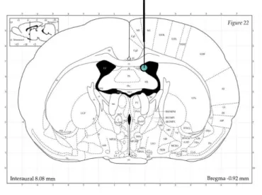
For implantation of guide cannulas, rats were anesthesized with isoflurane (Isofluran Baxter, Baxter Germany GmbH, Unterschleißheim, Germany) and fixed into a stereotactic frame. For icv infusions, unilateral, stainless steel cannulas (21G, 12mm long, Injecta GmbH, Klingenthal, Germany) were implanted 2mm above the right lateral ventricle (Fig.3; AP: -1.0mm bregma, ML: +1.6mm lateral, DV: +1.8mm below the surface of the skull;(G Paxinos, 2008). The guide cannula was fixed with two stainless steel screws using dental cement. After surgery, an antibiotic (100µl, 2.5% Baytril®, Bayer Vital GmbH, Klingenthal, Germany) was administered subcutaneously to avoid post-surgical infections. The guide cannula was kept feasible with a dummy cannula, which was cleaned every day during handling. Rats were handled daily for 5 days to reduce non-specific stress responses during experiments.
2.3 Microinfusions
For examination of the effects of central OXT infusion on astrocytes, rats received an icv
infusion of either vehicle (Veh, sterile Ringer solution, pH 7.4, 5µl) or synthetic OXT (1 nmol/5
µl;(Blume et al., 2008). For this, an infusion cannula (30G, 14 mm) connected to a Hamilton
syringe via polyethylene tubing was lowered into the guide cannula (Fig.3) and infusions were
[27]
slowly performed over 1min. Marks on the tubing allowed precise control of the volume administered. Following the infusion, the system was left in place for at least 10s to allow diffusion to occur. After withdrawal of the infusion cannula, the stylette was again inserted into the guide cannula. After termination of the experiment, rats were killed by CO
2-exposure followed by cervical translocation. In order to control for correct for cannula placement, 2µl ink were injected and brains were harvested. Next, coronal sections were prepared using a razorblade and only animals with ink distribution were included in the statistical analysis
Figure 3. Placement of guide cannula (thick black line) and infusion cannula (thin black line) for icv administration of OXT. Coordinates for stereotactic implantation of the guide cannula used were AP: -1.0mm bregma, ML: +1.6mm lateral, DV: +1.8mm. Torquise circle marks point of infusion. Illustration adopted from Paxinos and Watson (2006).