Citation: Prantl, L.; Brix, E.; Kempa, S.; Felthaus, O.; Eigenberger, A.;
Brébant, V.; Anker, A.; Strauss, C.
Facial Rejuvenation with
Concentrated Lipograft—A 12 Month Follow-Up Study.Cells2021,10, 594.
https://doi.org/10.3390/cells10030594
Academic Editor: Bruce A. Bunnell
Received: 4 February 2021 Accepted: 3 March 2021 Published: 8 March 2021
Publisher’s Note:MDPI stays neutral with regard to jurisdictional claims in published maps and institutional affil- iations.
Copyright: © 2021 by the authors.
Licensee MDPI, Basel, Switzerland.
This article is an open access article distributed under the terms and conditions of the Creative Commons Attribution (CC BY) license (https://
creativecommons.org/licenses/by/
4.0/).
sally.kempa@ukr.de (S.K.); oliver.felthaus@ukr.de (O.F.); andreas.eigenberger@ukr.de (A.E.);
vanessa.brebant@ukr.de (V.B.); Alexandra.anker@ukr.de (A.A.)
* Correspondence: catharina.strauss@ukr.de
† The authors Lukas Prantl and Eva Brix contributed equally to this work.
Abstract: Lipofilling is a popular technique to treat volume loss in aging patients. The isolated adipose tissue is composed of adipocytes and stromal vascular fraction cells, which include adipose- derived stem cells (ASC). We hypothesize that the patient’s wrinkle severity scale (WSS) and patient’s satisfaction on the global aesthetic improvement scale (GAIS) can be improved after using concen- trated lipoaspirate. Fourteen patients (54 years ± 11.09 years) with volume loss in the midface area underwent waterjet-assisted liposuction (Human Med AG, Schwerin, Germany). Fat was cen- trifuged in an ACP Double Syringe (Arthrex GmbH, Munich, Germany) using Rotofix 32A centrifuge (Andreas Hettich, GmbH & Co.KG, Tuttlingen, Germany). Homogenization was performed using the double syringe and a 1.4 mm female–female luerlock connector. After a second centrifugation, patients received periorbital (PO) and nasolabial (NL) lipografting. ASC count was performed after enzymatical digestion. Vitality of cells was assessed using a resazurin assay. During long-term follow up (12 months, n = 10), we found a high patient’s satisfaction (GAIS 1+/ − 0.52) and a good improvement of the WSS during short- and long-term follow-up. The ASC count of processed lipoaspirate was 2.1-fold higher than of unprocessed lipoaspirate (p < 0.001). The difference of ASC in sedimented and simply centrifuged lipoaspirate was also significant (p < 0.05). Facial rejuvenation with concentrated fat graft offers good results concerning objective aesthetic outcome and patient’s satisfaction.
Keywords: adipose-derived stem cells; facial rejuvenation; patient’s satisfaction; fat grafting; stromal vascular fraction
1. Introduction
Lipofilling, the autologous transplantation of adipose tissue, has been used since the end of the 19th century, especially for posttraumatic or congenital defects. Currently, lipofill- ing has become a popular technique to treat volume and contour abnormalities in aesthetic and reconstructive surgery. The processed fatty tissue can be seen as a natural filler instead of commercially available products [1,2]. Fat grafting is used widely in clinical practice for various indications, but there are many variations on fat harvesting, preparation, and reinjection [3–6]. Facial plastic surgery, for reconstructive and cosmetic reasons, aims to achieve harmony of facial features in case of volume loss, aging or just as an aesthetic tool.
Lipofilling promises to establish a custom fit long-term outcome with minor adverse effects as opposed to the more severe adverse effects that temporary fillers such as hyaluronic acid filler may cause [7,8]. Autologous fat grafts offer several benefits, including lack of immunogenicity, simplicity of surgical procedure, low cost, and easy accessibility. Further- more, it has become increasingly common to utilize fat that has been cryopreserved after
Cells2021,10, 594. https://doi.org/10.3390/cells10030594 https://www.mdpi.com/journal/cells
the initial fresh fat graft for potential future application [9]. Fat grafts are harvested from a region that is generally more abundant and injected into a secondary site.
The initial isolated adipose tissue is composed of adipocytes and stromal vascular fraction cells, which include adipose-derived stem cells (ASCs), preadipocytes, fibroblasts, vascular endothelial cells, and a variety of immune cells [10]. ASCs, which are cells of mesenchymal origins, are believed to be responsible for not only improving tissue contour of dermal and subdermal tissue but also weakening scar adhesions and paling the scars themselves. Preclinical studies suggested that ASC live longer than normal fat cells. ASCs offer a great regenerative potential for angioneogenesis, cell differentiation, and cell proliferation [11]. The beneficial effects to fat graft survival are believed to depend on secreted factors [12,13]. A higher stem cell percentage resulting in a higher growth factor secretion at the grafting site might be able to overcome the problem of volume losses of up to 70 percent [14,15]. The development of fine harvesting and injection cannulas made it possible to inject small adipose tissue particles in small volume areas, such as the face [16].
Liposuction should be performed gently with special cannulas under continuous negative pressure to guarantee maximal vitality and purity of adipocytes [17–19]. To achieve maximum viability in lipotransfer the optimal vacuum pressure for liposuction is between
− 0.5 and − 0.55 bar [20,21].
To properly address volume loss rates which vary up to 70% [14] volume deficits are often overcorrected. This may result in further surgeries and patient’s dissatisfaction with the results. The main surgical goal seems to be increasing of uptake rate while keeping adverse effects at a low rate and to achieve a stable long-term result immediately after the treatment.
Several studies have proven processing of lipoaspirate can increase healing rate [22,23], but there is still no universal agreement of how one should harvest, process, or graft fat [24].
Accurate photographic documentation has become essential in reconstructive and aes- thetic plastic surgery both for clinical and scientific purposes. Obtaining standardized, con- sistent, and relevant digital images is not easy outside a photographic studio [25,26]. Furthermore, the assessment of the outcome often seems subjective while digital photography remains a main tool for visualization of results [27].
Additionally, for objective evaluation of treatment outcome several tools were de- veloped as 3D photographs, which show the volume uptake in several parts of the face or the reduction of wrinkles. Satisfaction of patients with aesthetic procedures can easily be monitored by using the global aesthetic improvement scale (GAIS) [28]. The wrinkle severity scale (WSS) is a helpful tool for physicians to monitor the success of antiaging interventions [29]. It is quite popular for facial fat grafting not only among plastic surgeons but also dermatologists and craniofacial surgeons.
We hypothesize that using centrifugated and concentrated lipoaspirate improves patient’s wrinkle severity scale (WSS) and patient’s satisfaction (GAIS).
2. Materials and Methods
2.1. Patient Demographics Informed Consent
A total of twelve healthy, tumor-free women and two tumor-free man aged on average
54 years ± 11.09 years (n = 14; range from 30 to 72 years) underwent first water-assisted
liposuction at different regions of the body (abdominal, flanks, hips or legs) and after
lipoaspirate processing, fat grafting for volume loss in nasolabial fold (NLF) and periorbital
(PO) (Table 1).
8 w/47 abdomen 3/3 2/2
9 w/55 legs 5/5 2/2
10 m/30 Abdomen 2,5/2,5 /
11 w/72 legs 1/1 1.5/1.5
12 w/56 Flanks/axillary
region 2,5/2,5 3/3
13 w/69 abdomen 5/5 5/5
14 w/59 abdomen 3/3 /
PO = periorbital; NLF = nasolabial fold.
Interventions have been approved by the Ethics Committee of the University Hospital of Regensburg (No.17-520-101). All patients agreed to explained surgical intervention (liposuction, lipoaspirate processing and facial lipofilling for nasolabial or periorbital folds) and documentation by digital and 3D photography by signature. Inclusion criteria were any kind of volume loss in midface area.
Exclusion criteria were additive filler therapy or midface surgery performed. 4 patients missed follow up and were excluded in long-term follow-up analysis.
2.2. Liposuction Technique
All patients received a waterjet-assisted liposuction. In preparation for harvesting lipoaspirate, a 0.9% (weight/volume) solution of sodium chloride containing adrenaline at a concentration of 1:200.000 was infiltrated over approximately 15 min by means of a 2.5-mm injection cannula (Human Med AG, Schwerin, Germany). The volume was equal to the volume of fat tissue harvested. The lipoaspirate was harvested using 3.8-mm cannulas (Human Med AG, Schwerin, Germany) and the Body-Jet (Human Med AG), which allows a water jet–assisted liposuction. This medicinal product ensures an even negative pressure of less than 0.5 mbar.
2.3. Processing of Lipoaspirate
Lipoaspirate is extracted from solution in suction container (Human Med AG) after sedimentation and filling into 15 mL double syringe (Arthrex ACP Double Syringe System, Munich, Germany). Centrifugation is performed at 2500 rpm for 4 min (Rotofix 32 A, Andreas Hettich GmbH & Co.KG, Tuttlingen, Germany) to separate fat, oil, and tumescent solution, a three-layer can be seen in double syringe afterward.
After extracting the oil into smaller inner syringe, the aqueous phase at the bottom
of the syringe is discarded. The following emulsification and homogenization step is
done by using a Tulip-1.4-mm connector (Tulip Medical Products, San Diego, CA, USA)
attached to a second syringe. The lipoaspirate was forced manually in both directions with
high velocity through the double syringes. After a second centrifugation at 2500 rpm for
4 min, the released lipids were removed by transfer in the inner syringe by slowly pushing
down on the outer syringe while slowly pulling up the plunger of the small inner syringe
(see Figure 1)
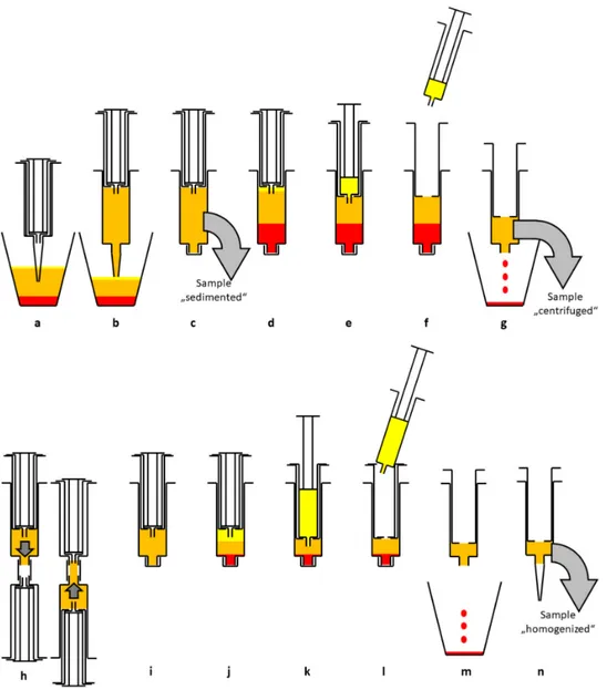
Figure 1. Schematic diagram of the enrichment process. After sedimentation in the suction container (a) lipoaspirate is transferred to a 15 mL double syringe (b). The sample “sedimented” is taken for analysis (c). After centrifugation (2500 rpm, 4 min) three layers can be seen (d). The upper oil phase is transferred to the small inner syringe (e) and discarded (f).
The blood and tumescent solution are discarded as well (g). The sample “centrifuged” is taken for analysis. The syringe is connected to a Tulip-1.4-mm connector and another syringe and the lipoaspirate is emulsified by forcing it through the connector at least 10 times (h). The now homogenized lipoaspirate (i) is centrifuged again at 2500 rpm for 4 min, resulting in three layers (j). The upper oil phase from disrupted adipocytes is transferred to the small inner syringe (k) and discarded (l). The aqueous phase is discarded, too (m). The remaining lipoaspirate contains a high percentage of ASCs and is ready for lipofilling (n). The sample “homogenized” is taken for analysis.
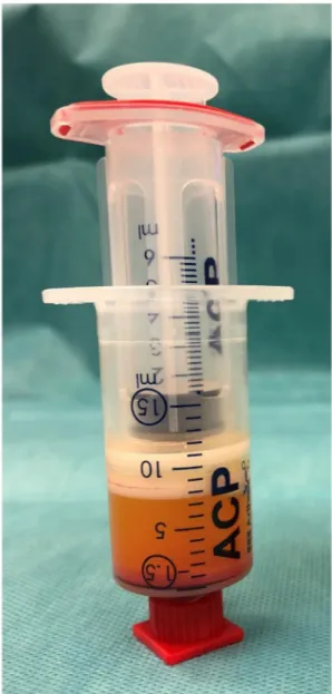
This emulsified, homogenized fat graft (about 1.5 mL) was transferred to 1 mL syringes
via the 2.4- or 1.4-mm transfer device (see Figure 2).
Figure 2. Emulsified, homogenized fat graft with oily phase and cell pellet at the bottom.
2.4. Stem Cell Isolation and Counting
Sedimented, centrifuged, and emulsified, homogenized lipoaspirate was enzymat- ically digested as described previously [10]. Briefly, 1 mL of α -MEM (Gibco (Thermo Scientific, Waltham, MA, USA) containing 0.2% (w/v) collagenase (from Clostridium histolyticum, Sigma Aldrich, St. Louis, MO, USA) was added to 1 mL of lipoaspirate.
Samples were incubated for 45 min at 37
◦C under constant agitation and centrifuged at 500 rpm for 5 min, subsequently. The supernatant was discarded, and the cells were resuspended in cell culture medium. After a small volume was set aside for the vitality assay, the cells were seeded into T25 cell culture flasks and grown for 3 days at 37
◦C and 5%
CO2 in a humidified atmosphere. Plates were washed every day to remove nonadherent cells. The plastic-adherent cells were counted at five randomly chosen sites (1 mm
2each).
The total cell number was calculated using the total cell culture area (25 cm
2).
2.5. Cell Vitality Assay
Vitality of cells isolated from sedimented, centrifuged, and homogenized lipoaspirate
was evaluated using a resazurin assay. The cells set aside after isolation were seeded
in 96-well plates in quintuplicates. For measurement, cell culture medium was supple-
mented with 0.07 µ M resazurin (Sigma Aldrich). Metabolic conversion of resazurin into
the fluorescent resorufin was detected using a multiwell plate reader (VarioScan, Thermo Scientific).
2.6. Flow Cytometry
Three days after seeding cells were washed with PBS (phosphate-buffered saline, PAAL- aboratories, Pasching, Austria) and detached by incubation with 500 µ L Trypsin/EDTA (Promo-Cell) for 5 min at 37
◦C. Cells were distributed to FACS tubes. After centrifuging (300 rpm, 5 min) the supernatant was removed and the cells were resuspended in 40 µ L staining buffer consisting of PBS containing 0.01% sodium azide, 0.5% BSA, and 2 nM EDTA.
Then 5 µL of CD44 antibody or isotype control antibody (Alexa-Fluor488 antimouse/human CD44 Clone IM7 or Alexa-Fluor488 mouse IgG1 isotype control (FC) Clone MOPC-21. BioLe- gend) were added and the cells were incubated on ice in the dark for 1 h. After addition of 1 mL staining buffer, the cells were centrifuged and the supernatant was removed. Cells were resuspended in 500 µ L staining buffer and measured using the FACS Canto II (BD Biosciences, Heidelberg, Germany). At least 50,000 events of each sample were recorded.
2.7. Lipofilling Technique
Fat grafts which resulted after homogenization and two rounds of centrifugation (see 2.3) (1–3 mL) were injected into nasolabial folds and/or periorbital folds (within sub- dermal layer similar a filler, subcutaneous tissue and under superficial musculoaponeurotic system) in multiple tissue planes, tunnels, areas with a syringe connecting with a blunt needle whose external diameter is 1 or 1.5 mm and 2 mm. For additional volume effect homogenized fat (after one centrifugation and homogenization with intersyringe shifting) was also used. For the subdermal/dermal injection, a 20 G needle (0.603 mm) was used.
Care was taken to inject fat grafts in only small quantities in one place each time (maximum:
3 mL), radially from distal to proximal. The syringe was drawn back before each injection to check blood return to avoid hematoma or to inject fat grafts into blood vessels.
In the periorbital area, extra care was taken to prevent intra-arterial injection. Gen- tle massage was done with finger or palm of the surgeon to ensure a smooth correction.
2.8. Digital Photography
Documentation of patient’s face was done preoperatively and at 3 months, 6 months and 12 months after lipofilling using a digital photograph Nikon Lumix NR/DMC-5272 (Nikon Corporation, Tokyo, Japan). Setting takes place while patient standing in upright position. Illumination was done by two lamps at 45
◦with respect to the patient on a plane parallel to frontal lamp. A minimum of five pictures needed to be taken: frontal view, oblique view (left/right) and lateral view (right/left).
2.9. 3D Photography and Wrinkle Severity Rating Scale
Documentation of the patient’s face was done preoperatively and at 3 months, 6 months and 12 months after lipofilling using a 3D skin camera (Antera 3D
®, Miravex Limited, Ireland) which enables the user to identify skin surface, wrinkles or hyperpigmentations.
The camera is used for each region (nasolabial fold or periorbital) in three different angles:
frontal view; oblique vies (45 degrees) and lateral view.
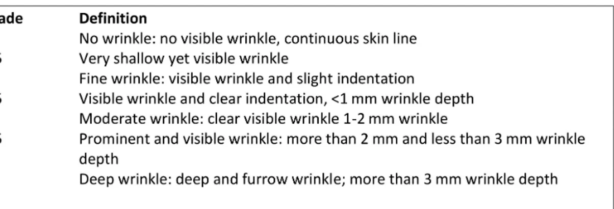
The depth of the facial folds was measured using the modified wrinkle severity scale
by Fitzpatrick [24,25] which has a wide use in clinical examination (Figure 3). Pre- and
postoperative fold measurements were contrasted and compared.
Figure 3. Wrinkle severity scale for nasolabial or periorbital folds (WSS).
2.10. Evaluation of Patient’s Satisfaction
To evaluate patient’s subjective appreciation of the surgical procedure the global aesthetic improvement scale (GAIS) was used (Figure 4) The documentation was primary 6 weeks after surgical intervention and at least at 9 to 12 months after surgical intervention (long term follow up).
Figure 4. Patient’s satisfaction measured by global aesthetic improvement scale (GAIS).
2.11. Statistical Analysis
The unpaired students t-test was used to compare the pre- and postoperative mea- surements and the patient’s satisfaction. Values of p < 0.05 were considered statistically significant (*: p value < 0.05; **: p value < 0.01; ***: p value < 0.001).
3. Results 3.1. Cell Isolation
The number of adherent cells isolated after homogenization and centrifugation from
1 mL lipoaspirate is 2.1-fold higher than the number of cells that were isolated from 1 mL
of unprocessed tissue (Figure 5). This increase in cell yield is verified by the data from the
cell vitality assay (Figure 6).
Figure 5. Number of cells isolated from varying tissue processings. Mean values and standard deviations are shown.
Student’s test was used to assess statistical significance (* p < 0.05; *** p < 0.001). (sed.: sedimented; centr.: centrifuged;
homog.: homogenized).
Figure 6. Vitality of cells isolated from varying tissue processings. Mean values and standard deviations are shown. Student’s t-test was used to assess statistical significance (*** p < 0.001).
(sed.: sedimented; centr.: centrifuged; homog.: homogenized).
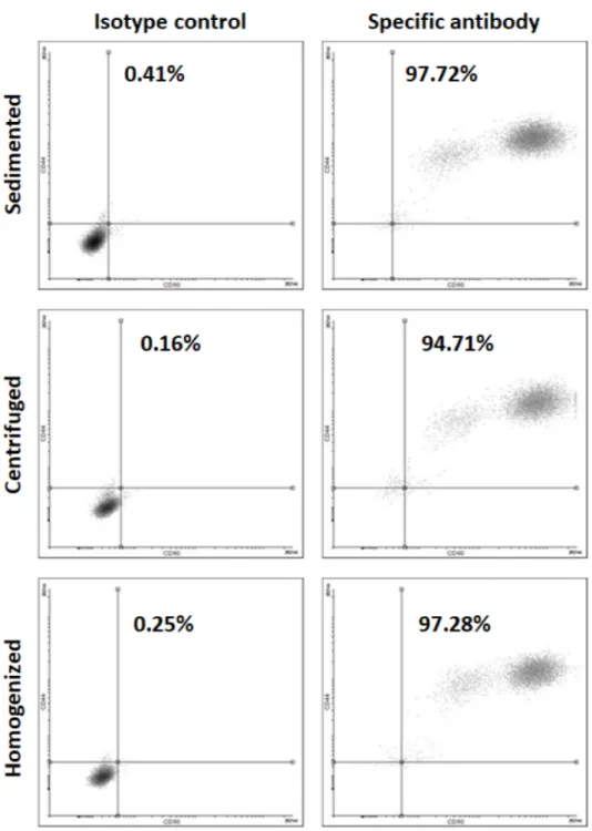
The cells isolated from the differently processed tissues are positive for MSC marker
CD44 (Figure 7).
Figure 7. Flow cytometry for MSC marker CD 44 for the cells isolated from varying tissue processings.
3.2. Lipofilling Volume
In 13 patients concentrated graft was used for NLF with mean volume (cc) of 2.5 cc
± 1. 38 cc (n = 13; range 1 to 5 cc). Lipofilling in periorbital (PO) region was done in 14 patients, the mean volume was 2.21 cc ± 1.01 cc (n = 14; range 1–5 cc) (Table 1).
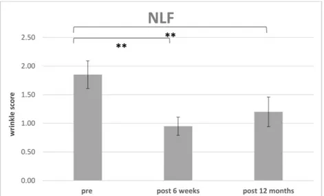
3.3. Improvement of Wrinkle Severity Scale (WSS)
The improvement of WSS preoperative for each region (PO or NLF) were compared
to postoperative at 6 weeks (n = 14) (Table 2) and the endpoint was defined after 12 months
(n = 10) (Table 3) (Figures 8 and 9). 4 patients missed the endpoint because of additional
surgical interventions and/or missing follow up.
Table 2. Follow up 6 weeks: Wrinkle severity scale (WSS) for different facial regions pre- and postoperative and global aesthetic improvement scale (GAIS).
Patient WSS NLF Pre OP
WSS NLF Post
WSS PO Pre OP
WSS PO Post OP
GAIS (1.5 Months)
1 1.5 0.5 1.5 1 1
2 2 1 1.5 1 1
3 2 1 1.5 0.5 1
4 2 1 1.5 0.5 1
5 1.5 1 1.5 0.5 1
6 2 1 1.5 0.5 2
7 2 1 1.5 0.5 1
8 2 1 1.5 0.5 1
9 2 1 1.5 0.5 1
10 / / 1.5 1 0
11 2 1 1.5 1 2
12 1.5 1 1.5 1 1
13 1.5 0.5 1.5 1 1
14 / / 2 1.5 1
The postoperative WSS for NLF after 12 months was 1.20±0.26 (n= 10. range 1 to 1.5). For PO the initial WSS was measured with 1.50±0.00 (n= 14;) and after 6 weeks with 0.79±0.32 (n= 14; range 0.5–1.5). After 12 months and loss of 4 patients, the score was measured with 1.05±0.15 (n= 10; range 1–1.5).
Table 3. Follow-up 12 months: Survey of patients undergoing lipofilling for rejuvenation for na- solabial and periorbital folds.
Patient WSS NLF Pre OP
WSS NLF Post OP
WSS PO Pre OP
WSS PO Post OP
GAIS (>6 Months)
1 1.5 1 1.5 1 1
2 2 1.5 1.5 1.5 1
3 2 1 1.5 1 1
4 2 1 1.5 1 1
5 1.5 1 1.5 1 1
6 2 1.5 1.5 1 2
7 2 1.5 1.5 1 1
8 2 1.5 1.5 1 1
9 2 1.5 1.5 1 1
10 / / 1.5 1 0
WSS = wrinkle severity scale; NLF = nasolabial fold; PO = periorbital region; GAIS = global aesthetic improve- ment scale.