Isolation and functional characterization of Arabidopsis powdery mildew effector proteins
Inaugural-Dissertation zur
Erlangung des Doktorgrades
der Mathematisch-Naturwissenschaftlichen Fakultät der Universität zu Köln
vorgelegt von Ralf Weßling
aus Rheine
Köln, Juni 2013
Die vorliegende Arbeit wurde am Max-Planck-Institut für Pflanzenzüchtungsforschung in Köln in der Abteilung für Pflanze-Mikroben Interaktionen (Direktor: Prof. Dr. Paul Schulze-Lefert) angefertigt.
Berichterstatter: Prof. Dr. Paul Schulze-Lefert Prof. Dr. Martin Hülskamp Dr. Frank Takken
Prüfungsvorsitzender: Prof. Dr. Ulf-Ingo Flügge
Tag der Disputation: 7.12.2012
Für Lisa und Oskar,
meine kleine Familie
I
Publications
Spanu P. D., Abbott J. C., Amselem J., Burgis T. A., Soanes D. M., Stüber K., Ver Loren van Themaat E., Brown J. K. M., Butcher S. A., Gurr S. J., Lebrun M.-H., Ridout C. J., Schulze-Lefert P., Talbot N. J., Ahmadinejad N., Ametz C., Barton G. R., Benjdia M., Bidzinski P., Bindschedler L. V., Both M., Brewer M. T., Cadle-Davidson L., Cadle-Davidson M. M., Collemare J., Cramer R., Lopez-Ruiz F., Frenkel O., Godfrey D., Harriman J., Hoede C., King B. C., Klages S., Kleemann J., Knoll D., Koti P. S., Kreplak J., Lu X., Maekawa T., Mahanil S., Micali C., Milgroom M. G., Montana G., Noir S., O’Connell R. J., Oberhaensli S., Parlange F., Pedersen C., Quesneville H., Reinhardt R., Rott M., Sacristán S., Schmidt S. M., Schön M., Skamnioti P., Sommer H., Stephens A., Takahara H., Thordal-Christensen H., Vigouroux M., Weßling R., Wicker T., and Panstruga R. 2010. Genome expansion and gene loss in powdery mildew fungi reveal functional tradeoffs in extreme parasitism. Science 330: 1543-1546
Weßling R., Schmidt S. M., Micali C. O., Knaust F. Reinhardt R., Neumann U., Ver Loren van Themaat E., and Panstruga, R. 2012. Transcriptome analysis of enriched Golovinomyces orontii haustoria by deep 454 pyrosequencing. Fungal Genetics and Biology 49: 470-482.
Weßling, R. and Panstruga, R. 2012. Rapid quantification of plant-powdery mildew interactions
by qPCR and conidiospore counts. Plant Methods 8: 35.
II
III
Table of Contents
Publications ... I
Table of Contents ... III
Abbreviations ... V
Summary ... VII
Zusammenfassung ... IX
1 Introduction ... 1
1.1 Plant immunity ... 1
1.1.1 Non-host resistance and MAMP-triggered immunity – two sides of the same coin ... 1
1.1.2 Plant hormones – integrators of multiple defense responses ... 3
1.1.3 Adapted pathogens have evolved to cause disease ... 5
1.2 Effectors of microbial pathogens ... 6
1.2.1 Effectors as avirulence determinants ... 6
1.2.2 Effectors as virulence factors ... 7
1.3 G. orontii and the powdery mildew infection of Arabidopsis ... 10
1.3.1 Quantification of powdery mildew infection ... 11
1.4 Thesis aims ... 12
2 Results ... 13
2.1 Rapid quantification of plant-powdery mildew interactions by qPCR and conidiospore counts... 13
2.1.1 The powdery mildew infection on seedlings ... 13
2.1.2 qPCR-based quantification of G. orontii infection ... 14
2.1.3 Spore counts of G. orontii ... 16
2.2 Transcriptome analysis of enriched G. orontii haustoria ... 18
2.2.1 Sequencing and EST analysis of a haustorial cDNA library ... 18
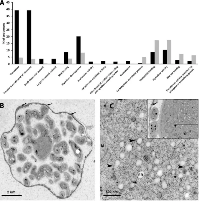
2.2.2 Functional classification indicates high protein turn-over in haustoria ... 19
2.2.3 ROS-detoxifying enzyme transcripts are abundant in G. orontii haustoria ... 21
2.2.4 Low abundance of nutrient transporter transcripts in haustoria ... 21
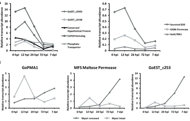
2.2.5 qRT-PCR analysis of haustorial transcripts ... 23
2.3 Functional characterization of G. orontii effector candidates ... 25
2.3.1 G. orontii effector prediction and cloning ... 25
2.3.2 Most effector candidates suppress induced cell death ... 27
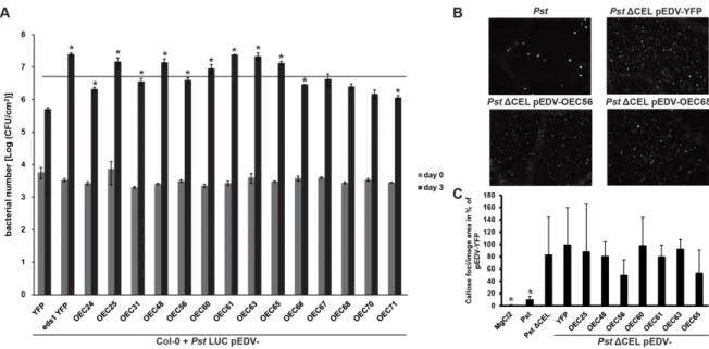
2.3.3 Bacterial delivery of selected effectors interferes with host immunity ... 29
2.3.4 Effectors show differential localization and expression patterns ... 31
2.3.5 Systematic Y2H reveals effector host targets and convergence onto hubs of the host cellular network. ... 33
2.3.6 Unrelated phytopathogens target overlapping sets of host proteins ... 36
2.3.7 Effector targets function in immunity towards G. orontii ... 38
2.3.8 Bimolecular fluorescence complementation (BiFC) confirms Y2H interactions ... 39
3 Discussion ... 42
3.1 Quantification of powdery mildew infections by qPCR and spore counts ... 42
3.1.1 qPCR-based quantification of G. orontii biomass ... 42
IV
3.1.2 Spore counts are a valid method to determine G. orontii reproductive success ... 43
3.1.3 Comparison of developed to existing methods of powdery mildew quantification ... 44
3.2 Transcriptome analysis of enriched G. orontii haustoria ... 46
3.2.1 Assembly and annotation of the haustorial library ... 46
3.2.2 Transcripts of primary metabolism dominate the haustorial transcriptome ... 47
3.2.3 Haustoria produce multiple ROS scavenging molecules ... 47
3.2.4 Transporter transcripts are not abundant in the haustorial cDNA library ... 48
3.2.5 Conserved pathogenicity genes are expressed in G. orontii haustoria ... 49
3.3 Functional characterization of G. orontii effector candidates. ... 51
3.3.1 OECs display common features of effector proteins ... 51
3.3.2 Cell death suppression by OECs ... 52
3.3.3 The virulence enhancing effect of OECs ... 53
3.3.4 Lessons from OEC localization ... 54
3.3.5 Expression of OECs – do effectors come in waves? ... 54
3.3.6 The OEC-Arabidopsis interactome reveals potential virulence targets ... 55
3.3.7 Unrelated phytopathogens converge onto hubs in the plant cellular network ... 56
3.3.8 Hubs are involved in the defense response ... 58
3.3.9 Y2H interactions can be confirmed by BiFC ... 65
3.4 Concluding remarks ... 66
4 Materials and Methods ... 67
4.1 Materials ... 67
4.1.1 Plant material ... 67
4.1.2 Strains and plasmids ... 67
4.1.3 Reagents, chemicals and antibiotics ... 68
4.1.4 Antibodies ... 68
4.1.5 Media ... 69
4.2 Methods ... 69
4.2.1 Standard molecular and biochemical methods ... 69
4.2.1 Powdery mildew inoculations ... 71
4.2.2 Quantification of powdery mildew infection ... 72
4.2.3 Generation and analysis of the haustorial library ... 73
4.2.4 qRT-PCR ... 74
4.2.5 Transmission electron microscopy ... 74
4.2.6 Prediction and bioinformatic analysis of effectors ... 74
4.2.7 Pseudomonas EDV assays ... 75
4.2.8 Transient expression assays ... 77
4.2.9 Yeast-two-hybrid ... 78
5 References ... 80
6 Supplemental Data ... 104
6.1 Supplemental Figures ... 104
6.2 Supplemental Tables ... 106
Acknowledgements ... 119
Erklärung ... 121
Lebenslauf ... 123
V
Abbreviations
ABA abscisic acid
AC average coverage
ACC 1-aminocyclopropane-1-carboxylic acid AI-1 Arabidopsis interactome version 1
APC Anaphase-promoting complex
Arabidopsis Arabidopsis thaliana
Avr Avirulence
Bgh Blumeria graminis f.sp. hordei
BiFC bimolecular fluorescence complementation BAK1 brassinosteroid receptor 1 (BRI1)–associated kinase Bgh Blumeria graminis f.sp. hordei
BIK1 Botrytis-induced kinase 1
BR Brassinosteroid
CC coiled-coil domain
Cf Cladosporium fulvum
cfu colony forming unit
Ch Colletotrichum higgansianum CLSM confocal laser scanning microscopy
CUL cullin
CRLs cullin RING ligases
CSEP candidate secreted protein
CSN COP9 signalosome
C-terminal carboxy terminal
Da Dalton
DMSO dimethylsulfoxide
DNA deoxyribonucleic acid
dpi days post inoculation
DRMIP-HESP developmentally regulated MAPK interacting protein-haustorially expressed protein domain
eds enhanced disease susceptibility edr enhanced disease resistance
EDTA Na
2-ethylenediamine tetraacetic acid EDV effector detector vector EF-Tu elongation factor Tu
EFR EF-Tu receptor
ER endoplasmic reticulum
EST expressed sequence tag
ET ethylene
ETI effector-triggered immunity
FDR false discovery rate
FLS2 flagellin sensing 2
f.sp. formae specialis
GA gibberellic acid
GABA γ-aminobutyric acid
GO gene ontology
Hpa Hyaloperonospora arabidopsidis hpi hours post inoculation
HR hypersensitive response
JA jasmonic acid
JAZ jasmonate-ZIM domain containing protein
LRR leucine-rich repeat
MAMP microbe-associated molecular pattern MAP mitogen-activated protein
MES 2-(N-morpholino)ethanesulfonic acid
VI
NLP necrosis and ethylene-inducing proteins (NEP1)-like proteins
mM millimolar
µM micromolar
MFS major facilitator superfamily
MTI MAMP-triggered immunity
MVB multi-vesicular body
Nb Nicotiana benthamiana
NB nucleotide binding site
N-terminal amino terminal
OEC G. orontii effector candidate
ORF open reading frame
o/n overnight
PCR polymerase chain reaction Pi Phytophthora infestans
PPIN-1 plant pathogen interactome version 1
PR pathogenesis-related protein
PRA prenylated rab acceptor
PRR pattern-recognition receptor
Pst Pseudomonas syringae pv tomato DC3000 Psy Pseudomonas syringae (several pathovars)
pv. pathovar
PVC prevacuolar compartment
qPCR quantitative real time PCR
qRT-PCR quantitative reverse transcription PCR
R resistance
RIN4 RPM1 interacting protein 4
RNA ribonucleic acid
rpm revolutions per minute
RT room temperature
RING really interesting new gene
RLK receptor-like kinase
RLP receptor-like protein
ROS reactive oxygen species
RT room temperature
SA salicylic acid
SAG101 senescence-associated gene 101
SCF S-phase kinase-associated protein (SKP), CUL1, really interesting new gene (RING) box 1 (Rbx1), F-box protein complex
SDS-PAGE sodium dodecyl sulfate (SDS) polyacrylamide gel electrophoresis SNAP synaptosomal-associated protein
SNARE soluble N-ethylmaleimide sensitive factor attachment protein receptor TAL transcription-activator like
TCP teosinte branched1, cycloidea (CYC), proliferating cell factor (PCF)
TF transcription factor
TIR Toll-interleukin1 receptor
TM transmembrane
T3SS Type III secretion system
T-DNA transfer DNA
VAMP vesicle-associated membrane protein YFP yellow fluorescent protein
Y2H yeast two-hybrid
°C degrees Celsius
VII
Summary
Plants are resistant to the majority of potential pathogenic microbes. Adapted pathogens can however overcome plant defense and induce susceptibility. The molecular processes underlying this adaptation are only partially understood. Obligate biotrophic pathogens, which require a living host for growth and reproduction, establish especially intimate relationships with their plant hosts.
A crucial aspect of this lifestyle is the formation of a specialized infection structure termed the haustorium. Haustoria are believed to represent pivotal sites of nutrient uptake and deliver effectors, proteins that manipulate the host cell during infection to promote susceptibility. While the effector arsenal of pathogenic bacteria has been investigated intensively, the repertoires and host targets of fungal effectors are currently underexplored. The work presented here thus aims at characterizing virulence mechanisms employed by the obligate biotrophic Ascomycete Golovinomyces orontii, the causal agent of the powdery mildew disease in Arabidopsis thaliana (hereafter Arabidopsis). To this end, the haustorial transcriptome of G. orontii was obtained by pyrosequencing of a cDNA library generated from isolated haustorial complexes. Transcripts coding for gene products with roles in protein turnover, detoxification of reactive oxygen species and fungal pathogenesis were abundant, while surprisingly transcripts encoding presumptive nutrient transporters were not highly represented in the haustorial cDNA library.
A substantial proportion (~38%) of transcripts encoding predicted secreted proteins comprised effector candidates. These candidates were cloned and found to frequently suppress induced plant cell death. A subset of effectors enhanced bacterial virulence and could suppress callose deposition, indicating a role in defense suppression. Transcript profiling of these effectors suggested their sequential delivery during pathogenesis. Furthermore, subcellular localization revealed diverse target compartments in the host. In a complementing approach, a large-scale yeast 2-hybrid (Y2H) assay was performed on the 84 cloned effector candidates and revealed convergence onto 61 potential host targets. These targets were enriched in transcription factors and components involved in development and cellular trafficking. Bimolecular fluorescence complementation assays confirmed the interaction of selected effectors with their host interactors.
Finally, the Y2H targets of effectors were used to construct an integrated protein-protein interaction network of Arabidopsis and the three adapted pathogens Pseudomonas syringae (Psy), Hyaloperonospora arabidopsidis (Hpa) and G. orontii. This network revealed pathogen- specific as well as nine common host targets. These common targets are highly connected in the Arabidopsis cellular network. After the development of suitable quantitative methods, the important role of these common targets in the Arabidopsis immune response was validated by screening respective T-DNA insertion lines. In sum, my work supports the hypothesis that pytopathogenic microbes target hubs in the host cellular network to promote susceptibility. The effector targets identified will therefore form the basis of subsequent effector research in G.
orontii.
VIII
IX
Zusammenfassung
Pflanzen sind immun gegen den Großteil potentieller Schädlinge. Angepasste Schädlinge können die pflanzlichen Verteidigungsmechanismen allerdings überwinden und so die Pflanze für eine Infektion empfänglich machen. Die molekularen Prozesse, die dieser Anpassung zugrundeliegen sind nur teilweise verstanden. Obligat biotrophe Schädlinge, welche auf einen lebenden Wirt für Wachstum und Vermehrung angewiesen sind, etablieren eine besonders enge Beziehung mit ihrem pflanzlichen Wirt. Ein kritischer Aspekt dieser Lebensform ist die Bildung einer spezialisierten Infektionsstruktur, des Haustoriums. Das Haustorium ist entscheidend für sowohl die Nahrungsaufnahme als auch die Sekretion von Effektoren, kleinen Proteinen die die Wirtszelle manipulieren und so empfänglich für eine Besiedlung machen. Während die Effektorarsenale von Bakterien bereits intensiv erforscht wurden, sind die Effektoren von pilzlichen Schädlingen sowie ihre Zielproteine in der Wirtszelle noch weitgehend unbekannt. Die hier präsentierte Arbeit zielt deshalb auf das Verständnis der Virulenzmechansimen des obligat biotrophen Schlauchpilzes (Ascomycet) Golovinomyces orontii, des Erregers des Mehltaus auf Arabidopsis thaliana (folgend Arabidosis). Dazu wurde das haustorielle Transkriptom von G.
orontii durch die Pyro-Sequenzierung einer cDNA-Bibliothek aus isolierten haustoriellen Komplexen charakterisiert. Viele Transkripte kodierten für Proteine mit Funktionen in der Proteinumsetzung, der Entgiftung von reaktiven Sauerstoffspezies und pilzlicher Pathogenese.
Überraschenderweise konnten nur wenige Transkripte für mögliche Nährstofftransporter identifiziert werden.
Ein substanzieller Anteil (38%) der Transkripte für vorhergesagte sekretierte Proteine kodiert für Effektorkandidaten. Diese Kandidaten wurden kloniert und konnten häufig induzierten pflanzlichen Zelltod unterdrücken. Eine Untergruppe der Effektorkandidaten erhöhte die Virulenz von Bakterien und verringerte teilweise die Ablagerung von Callose. Zusammengenommen weist dies auf eine Rolle in der Unterdrückung der pflanzlichen Verteidigungsmechanismen hin. Die Analyse der Transkriptprofile dieser Kandidaten deutete auf eine sequentielle Produktion während der Infektion hin. Außerdem zeigten Lokalisationstudien unterschiedliche subzelluläre Zielkompartimente der einzelnen Effektorkandidaten. In einer komplementären Herangehensweise wurde eine großmaßstäbliche Hefe-Zwei-Hybird (Y2H) Analyse der 84 klonierten Effektorkandidaten durchgeführt. Dieser Ansatz enthüllte die Konvergenz der Kandidaten auf 61 potentielle Zielproteine des Wirtes.
In diesen Zielproteinen sind Transkriptionsfaktoren sowie Komponenten aus der
Pflanzenentwicklung und dem zellulären Transport überrepräsentiert. Versuche über die
bimolekulare Fluoreseszens-komplementation bestätigten die Interaktion von ausgewählten
Effektoren mit den jeweiligen Wirtsproteinen. Schließlich wurden die Y2H-Interaktoren der
Effektorkandidaten zur Konstuktion eines integrierten Protein-Protein Interaktions-Netzwerks von
Arabidopsis und den drei adaptieren Schädlingen Pseudomonas syringae (Psy),
X
Hyaloperonospora arabidopsidis (Hpa) und G. orontii genutzt. Dieses Netzwerk enthüllte sowohl
Schädling-spezifische als auch gemeinsame Zielproteine im Wirt. Die gemeinsamen Zielproteine
sind hochvernetzt im zellulären Netzwerk von Arabidopsis. Nach der Entwicklung geeigneter
quantitativer Methoden konnte eine Rolle dieser Proteine in der Immunantwort von Arabidopsis
durch die Analyse von entsprechenden T-DNA-Insertionsmutanten bestätigt werden. Die hier
präsentierte Arbeit unterstüzt die Hypothese das mikrobielle Pflanzenschädlinge hochvernetzte
Proteine in der Pflanze angreifen um ihre Anfälligkeit zu erhöhen. Die identifizierten Effektoren
und Zielproteine werden daher die Grundlage für eine weitere Erforschung des Arabidopsis-
Mehltaus bilden.
1
1 Introduction
1.1 Plant immunity
Plants are sessile organism and have to continuously adapt to a changing environment. They are continuously exposed to pathogens with various infection strategies and feeding habits, including viruses, bacteria, fungi, nematodes and insects. The world is however still a green place as plants, similarly to invertebrates and vertebrates, possess an innate immune system that detects and wards off potential pathogens. Accordingly, plants are resistant against the vast majority of pathogenic microbes, a phenomenon termed non-host resistance. In comparison to vertebrates however, plants do not have an adaptive immune system with dedicated mobile immune cells but rather rely on the ability of each cell to defend against invaders (Ausubel, 2005). Plant innate immunity involves both preformed barriers and a multitude of responsive processes triggered by the recognition of potential pathogens, orchestrated by transcriptional reprogramming and executed by defense responses such as cell wall reinforcement and the delivery of anti-microbial compounds (Jones and Dangl, 2006; Thordal-Christensen, 2003). The immune response can be broadly divided into two distinct but overlapping layers: microbe-associated molecular pattern (MAMP)- and effector-triggered immunity (MTI and ETI, Jones and Dangl, 2006). Adapted pathogens are able to overcome plant innate immunity to cause disease.
1.1.1 Non-host resistance and MAMP-triggered immunity – two sides of the same coin
Plants are resistant to the majority of potential pathogens; they are non-hosts for these pathogens. This is due to the multilayered defense system of plants and specific requirements of the pathogens. In general, pathogens require certain cues to initiate pathogenesis, including surface topologies and cuticular waxes (Thordal-Christensen, 2003). Subsequently, successful pathogens need to overcome preformed plant defenses like secondary metabolites or the rigid cell wall (Mysore and Ryu, 2004). In the first responsive layer of defense, plants, analogous to animals, recognize MAMPs, conserved and indispensable microbial signatures such as bacterial flagellin, the elongation factor Tu (EF-Tu), fungal chitin and many more (Boller and Felix, 2009).
These cellular components create a “non-self” signal that is perceived by the plant. The active epitopes of MAMPs are recognized by membrane-resident pattern-recognition receptors (PRRs) of the receptor-like kinase (RLK) and receptor-like protein (RLP) type (Boller and Felix, 2009).
These PRRs are distinguished by their domain structure. RLKs such as flagellin sensing 2 (FLS2)
and the EF-Tu receptor (EFR) contain an extracellular leucine-rich repeat (LRR) domain, a short
transmembrane (TM) domain and an intracellular kinase domain (Gómez-Gómez and Boller,
2000; Zipfel et al., 2006). RLPs such as chitin elicitor receptor kinase 1 (CERK1), by contrast
harbor an extracellular LysM domain, a TM domain and a short cytoplasmic domain without
kinase function (Miya et al., 2007; Wan et al., 2008). The receptor proteins can be either
2
restricted to specific plant families or widely conserved, as shown for EFR, which is specific to the Brassicaceae and FLS2, which has also been found in tobacco, tomato and rice (Hann and Rathjen, 2007; Kunze et al., 2004; Robatzek et al., 2007; Takai et al., 2008). Transfer of EFR to tobacco and tomato generates responsiveness to elf18, the active epitope of EF-Tu, and enhanced resistance to bacteria, indicating that the downstream signaling components of the receptor are conserved (Lacombe et al., 2010). The transfer of EFR shifts the recipient plant towards a non-host state, further underlining the role of PRRs in non-host resistance.
The recognition of MAMPs by FLS2 and EFR, but not CERK1, triggers their association with brassinosteroid receptor 1–associated kinase (BAK) 1, phosphorylation of both partners and the subsequent activation of plant responses (Chinchilla et al., 2007). This is a key step in receptor activation, as bak1 mutants are strongly impaired in downstream signaling events (Chinchilla et al., 2007). CERK1 functions independent of BAK1 but also associates with Botrytis-induced kinase (BIK) 1, a cytoplasmic kinase with a key role in the relay of receptor signals (Lu et al., 2010; Zhang et al., 2010). Subsequent to receptor complex activation, a stereotypical signaling cascade is activated that is mostly independent of the MAMP applied. There is a marked Ca
2+- influx, an oxidative burst occurs and MAP (mitogen-activated protein) kinases are activated (Boller and Felix, 2009). Subsequently, the phytohormone ethylene (ET) is produced and transcriptional reprogramming occurs (Zipfel et al., 2006; Zipfel et al., 2004). Even later, callose deposits appear and seedling growth is inhibited, indicating the reallocation of energy to the defense response (Gómez-Gómez and Boller, 2000; Gómez-Gómez et al., 1999). MAMP- triggered responses limit development and growth of both adapted and non-adapted pathogens and thus significantly contribute to plant immunity (Miya et al., 2007; Wan et al., 2008; Zipfel et al., 2006; Zipfel et al., 2004). It has to be noted that MAMPs, by definition, are also contained in commensal and beneficial microorganisms. The plant thus has to discriminate between neutral or beneficial and pathogenic microbes, as defense induction against the former would be detrimental for the plant. Specificity is probably achieved through the integration of MAMP and danger-associated molecular pattern (DAMP) perception. DAMPs are plant-derived signals released by pathogen activities such as cell wall degradation, sucrose degradation and cell permeabilization and comprise oligogalacturonides, extracellular sugars, or endogenous elicitor peptides (Doares et al., 1995; Herbers et al., 1996; Huffaker et al., 2006). Perception of these DAMPs by membrane-resident RLKs initiates a signaling cascade that is integrated with MAMP- signaling to promote defense response induction (Brutus et al., 2010; Krol et al., 2010;
Yamaguchi et al., 2010; Yamaguchi et al., 2006).
But what are the executers of MTI? This question leads back to the genetic dissection of non-host
resistance, especially to the characterization of components involved in the interaction of
Arabidopsis thaliana with the non-adapted powdery mildew pathogen of barley, Blumeria
graminis f.sp. hordei (Bgh). In this patho-system, non-host resistance at the pre-penetration stage
is conferred by two parallel pathways. The first pathway leads to vesicle-mediated secretion of
3 unknown defense compounds and involves soluble N-ethylmaleimide sensitive factor attachment protein receptor (SNARE)-complex formation by the membrane-resident syntaxin PEN1/SYP121, the synaptosomal-associated protein (SNAP) 33 and the endomembrane-resident vesicle- associated membrane proteins (VAMPs) 721 and 722 (Collins et al., 2003; Kwon et al., 2008).
The second pathway generates active glucosinolates for antifungal defense. It involves the biosynthesis of 4-methoxyindol-3-ylmethylglucosinolate by CYP81F2, a P450 monooxygenase, and subsequent activation by the peroxisomal β-glycosyl hydrolase PEN2 (Bednarek et al., 2009;
Clay et al., 2009; Lipka et al., 2005). The active compound is then presumably secreted by PEN3, a pleiotropic drug resistance/ATP-binding cassette transporter (Stein et al., 2006). Both pathways limit the colonization by adapted as well as non-adapted pathogens and are thus also involved in MTI (Bednarek et al., 2009; Hiruma et al., 2010; Kwon et al., 2008; Stein et al., 2006). CYP81F2, PEN2 and PEN3 are required for MAMP-induced callose deposition and PEN1, VAMP722 and PEN3 accumulate in callosic haustorial encasements of the adapted powdery mildew fungus G.
orontii, clearly demonstrating their role in MTI (Clay et al., 2009; Meyer et al., 2009). Components of the two pathways are transcriptionally induced after challenge with G. orontii and part of a transcriptional regulon conserved in both Arabidopsis and barley, showing that an ancient transcriptional program controls their expression (Chandran et al., 2010; Humphry et al., 2010).
Additional inducible defense components comprise the phytoalexin camalexin, which is involved in resistance to both adapted and non-adapted necrotrophic pathogens and the pathogenesis- related (PR) proteins, which include chitinases and glucanases (Schlaeppi et al., 2010; Stotz et al., 2011; Thomma et al., 1999b; van Loon et al., 2006). The induction of defense responses is costly for the plant and therefore needs to be tailored towards invading microbes. One mechanism allowing a specific defense response is the production of different plant hormones and the cross-talk of their signaling modules.
1.1.2 Plant hormones – integrators of multiple defense responses
MAMP and DAMP perception induces the generation of several defense-related hormones, most prominently salicylic acid (SA), jasmonic acid (JA) and ET (Pieterse et al., 2009; Tsuda and Katagiri, 2010). These hormonal pathways act mostly antagonistically, with SA controlling defense against biotrophic and JA/ET synergistically controlling the defense response against necrotrophic pathogens (Glazebrook, 2005). However, all three pathways contribute positively to MTI and are probably needed to amplify the defense signal (Tsuda et al., 2009).
Induction of SA production requires the defense regulator enhanced disease susceptibility (EDS)
1 and its interaction partner phytoalexin deficient (PAD) 4, both of which also regulate SA-
independent responses (Falk et al., 1999; Feys et al., 2001; Zhou et al., 1998). EDS1 also
interacts with and signals through senescence-associated gene (SAG) 101, putatively forming a
ternary complex with PAD4 (Feys et al., 2005; Zhu et al., 2011). All three proteins are also
involved in basal immune responses, and define a third layer of post-invasive resistance against
4
Bgh (Lipka et al., 2005). The pen2 pad4 sag101 triple mutant renders Arabidopsis a host for Bgh, as the fungus can in rare cases complete its life cycle on these plants (Lipka et al., 2005).
SA is produced predominantly through a chloroplast-localized pathway from chorismate by the isochorismate synthase ICS1/SID2 (Wildermuth et al., 2001). The mode of SA perception has long remained elusive, but two recent publications argue for either NPR1 (nonexpressor of pathogenesis-related genes 1) or its paralogs NPR3 and NPR4 as SA receptors (Fu et al., 2012;
Wu et al., 2012). Previously, NPR1 has been characterized as the key signal transducer downstream of SA production, as loss of NPR1 impairs both local and systemic resistance to pathogens (Cao et al., 1994; Mou et al., 2003). In the non-induced state, NPR1 oligomers are localized in the cytoplasm. Upon SA-induced redox changes the monomers are liberated, enter the nucleus and initiate signaling through TGA transcription factors (TFs) (Mou et al., 2003; Tada et al., 2008). This leads to the activation of downstream responses such as PR gene expression, the establishment of systemic acquired resistance in non-challenged leaves and contributes to the hypersensitive response (HR), a localized plant cell death (Durrant and Dong, 2004).
Constitutive signaling is prevented by constant proteasome-dependent degradation of NPR1 in the nucleus (Spoel et al., 2009). The cytosolic pool of NPR1 is important for the repression of JA signaling (Spoel et al., 2003).
JA is a lipid-derived compound originating from α-linolenic acid that is produced by several enzymatic reactions in both the chloroplast and peroxisomes (Wasternack, 2007). It is perceived through a receptor complex containing the ubiquitin-conjugating E3 ligase SCF
COI1(S-phase kinase-associated protein, cullin (CUL) 1, really interesting new gene (RING) box 1 (Rbx1), F-box protein) (Pauwels and Goossens, 2011). COI1 is an F-box protein and thus functions as the eponymous substrate adaptor of the complex (Xie et al., 1998). It interacts with several Jasmonate-ZIM domain containing (JAZ) proteins that repress JA signaling by binding to the JA- responsive TF MYC2 and recruiting the corepressor TOPLESS (Pauwels et al., 2010). Binding of JA-isoleucine, the bioactive JA conjugate, to the SCF
COI1-JAZ coreceptor complex induces ubiquitination of JAZ proteins by SCF
COI1and subsequent degradation of the JAZs (Chini et al., 2007; Sheard et al., 2010; Thines et al., 2007; Yan et al., 2007). This allows the induction of JA- responsive genes by the TF MYC2. In addition to its role in the defense response, JA also influences several developmental processes, as exemplified by the male sterility phenotype of the coi1 mutant (Pauwels and Goossens, 2011; Wasternack, 2007; Xie et al., 1998).
Similar to JA, ET is a versatile hormone involved in defense responses as well as plant development. The production of ET from its precursor S-adenosylmethionine via 1- aminocyclopropane-1-carboxylic acid (ACC) synthase and ACC oxidase is rapidly induced by MAMP perception (Boller and Felix, 2009; Wang et al., 2002). Five membrane-bound ET receptors have been described which constitutively repress ET signaling (Wang et al., 2002).
Upon ET perception these receptors are inactivated, leading to derepression of the pathway and
activation of gene expression through EIN2 and the TFs EIN3 and EIL3 (Shan et al., 2012). JA
5 and ET predominantly act synergistically. This synergism is in large parts controlled by the two TFs ERF1 and ORA59 (Lorenzo et al., 2003; Pré et al., 2008). JA- and JA/ET-induced genes can thus be separated by the requirement of either MYC2 or ERF1 and ORA59 for their induction (Lorenzo et al., 2004).
The outcome of hormone signaling is governed by a large network of crosstalk. In addition to the archetypical JA-SA antagonism, many interactions of different hormone branches have been described. They include, but are not limited to, synergism of ET and SA signaling, antagonism between abscisic acid (ABA), JA/ET and SA responses, antagonism between SA and auxin signaling, a role of gibberellic acid (GA) signaling in SA-JA crosstalk and antagonism between cytokinins and SA and auxin (Anderson et al., 2004; Leon-Reyes et al., 2009; Naseem et al., 2012; Navarro et al., 2008; Wang et al., 2007; Yasuda et al., 2008). The complexity of hormonal crosstalk is essential for integrating both immune and developmental signals and generating an appropriate response. The use of proteasome mediated degradation is another common theme in these hormone signaling pathways. Constitutive repression of ET signaling is mediated through the constant degradation of EIN2 and EIN3 through SCF
ETP1/2and SCF
EBF1/2, respectively (Guo and Ecker, 2003; Potuschak et al., 2003; Qiao et al., 2009). Additionally, SCF
COI1, SCF
TIR1and SCF
GID2perceive the plant hormones JA, auxin and GA, respectively (Santner and Estelle, 2009).
Finally, the potential perception of SA by an E3 ligase complex containing CUL3, NPR3 and/or NPR4 also provides a link to SA signaling (Fu et al., 2012). Despite the complexity and redundancy of plant innate immunity, adapted pathogens have found ways to disturb or abuse the plant defense response and cause disease. One such mechanism is the production of the phytotoxin coronatine, a structural analog of JA-Isoleucine, by the hemibiotrophic bacterium Pseudomonas syringae pv. tomato DC3000 (Pst) (Weiler et al., 1994; Yan et al., 2009).
Coronatine activates JA-signaling and induces susceptibility owing to the suppression of SA- induced responses by hormonal crosstalk (Brooks et al., 2005; Uppalapati et al., 2007).
1.1.3 Adapted pathogens have evolved to cause disease
Arabidopsis can be colonized by many microbial pathogens, including bacteria and the
filamentous oomycetes and fungi. Their lifestyle ranges from necrotrophy, where nutrients are
obtained by lysis of the host, to obligate biotrophic pathogens that can only grow and reproduce
on the living host (O’Connell and Panstruga, 2006). Obligate biotrophy has evolved in unrelated
pathogens several times independently, indicating that this life-style provides a selective
advantage (Kemen and Jones, 2012; O’Connell and Panstruga, 2006). Accordingly, obligate
biotrophy is associated with specific genomic adaptations, including increased genome size
mediated by the expansion of transposable elements, reduced sets of lytic enzymes and
enzymes for the production of secondary metabolites and the loss of some biosynthetic pathways
(Kemen and Jones, 2012; Schmidt and Panstruga, 2011). Obligate biotrophic pathogens use a
characteristic feeding organ, the haustorium to establish an intimate relationship with their host
6
and facilitate the uptake of carbohydrates, amino acids and possibly water from the host (Gil and Gay, 1977; Hahn and Mendgen, 1997; Voegele and Mendgen, 2003). Nutrient uptake is thought to be driven by a proton gradient across the membrane that is generated by fungal H
+-ATPases (O’Connell and Panstruga, 2006). Haustoria remain separated from the host cell by the extrahaustorial matrix, the battleground of the host-pathogen interaction, and the extrahaustorial membrane (EHM), a derivative of the plant plasma membrane whose composition is modified remarkably (Koh et al., 2005; Meyer et al., 2009; Micali et al., 2011). Arabidopsis can be colonized by several obligate biotrophs, including the oomycetes downy mildew Hyaloperonospora arabidopsidis (Hpa) and white rust Albugo species as well as powdery mildew fungi (Kemen and Jones, 2012). The haustoria of obligate biotrophic pathogens have been shown to secrete effectors, small proteins that undermine the host immune system and reprogram the host cell for compatibility (Kemen et al., 2005; Sohn et al., 2007; Stergiopoulos and de Wit, 2009). These effectors can in turn be recognized by the plant, triggering ETI.
1.2 Effectors of microbial pathogens 1.2.1 Effectors as avirulence determinants
It has long been recognized that plants are resistant to specific isolates of adapted pathogens. A
conceptual framework for these observations was first provided by the gene-for-gene concept,
which stated that isolate-specific resistance requires complementary Avirulence (Avr) and
resistance (R) genes in host and pathogen, respectively (Flor, 1971). Interactions of plants and
adapted pathogens can thus be classified as compatible (R or Avr gene absent) or incompatible
(R and Avr gene present). The cloning and analysis of several Avr/R gene combinations has
elucidated the molecular mechanisms underlying the gene-for-gene concept and shown that Avr
genes encode effectors, thus coining the term ETI (Dangl and Jones, 2001; Jones and Dangl,
2006; Van Der Biezen and Jones, 1998). R genes predominantly encode cytoplasmic proteins
with a modular structure. They contain a central nucleotide-binding (NB) domain, a C-terminal
LRR domain and one of two possible N-terminal domains (Takken and Tameling, 2009). The N-
terminal coiled-coil (CC) or Toll/Interleukin-1 receptor (TIR) domains form the signaling hubs of
the proteins. The LRR domain generates the recognition specificity of R proteins by either binding
to Avr proteins (direct recognition) or by monitoring effector-induced modifications of host proteins
(guardees). This “guard” model has provided a valuable extension of the gene-for-gene concept
(Dangl and Jones, 2001; Van Der Biezen and Jones, 1998). One of the best studied guardees is
the plasma membrane-resident RPM1 interacting protein 4 (RIN4). It is targeted by at least four
independent effectors (AvrRpm1, AvrB, AvrRpt2 and HopF2) and guarded by two distinct CC-NB-
LRR proteins, RPM1 and RPS2, which recognize its phosphorylation or cleavage, respectively
(Kim et al., 2005a; Kim et al., 2005b; Mackey et al., 2002; Wilton et al., 2010). The guard model
has recently been modified to also encompass decoys, non-functional proteins that mimic effector
targets solely to trigger R protein mediated resistance (van der Hoorn and Kamoun, 2008). It is
7 important to note that in contrast to non-host resistance, which acts at the species level, R gene- mediated resistance is cultivar-specific.
The signaling cascades downstream of R protein activation are only poorly understood. TIR-NB- LRRs and CC-NB-LRRs activate at least partially distinct signaling modules, as illustrated by the differential requirement of EDS1 or NDR1 (non-race specific resistance 1), respectively, for the activation of downstream responses (Aarts et al., 1998). The most frequent executor of ETI is the HR, but resistance and HR can also be uncoupled (Bendahmane et al., 1999; Clough et al., 2000; Heidrich et al., 2011). Notably, ETI can also be characterized as an enhanced and prolonged MTI response (Tsuda and Katagiri, 2010). There is a significant overlap of transcriptional changes induced by MTI and ETI and both MTI and ETI trigger reactive oxygen species (ROS) production and MAP kinase activation (Navarro et al., 2004; Torres et al., 2006;
Underwood et al., 2007). In ETI, the kinase activation is however more extensive and ROS production is biphasic with a second prolonged ROS burst probably triggered by effector recognition (Torres et al., 2006; Underwood et al., 2007). Hormonal responses are also involved in MTI and ETI alike, playing synergistic and compensatory roles in the former and latter, respectively (Tsuda et al., 2009). In addition, the separation of MAMPs from effectors and PRRs from R proteins is sometimes difficult (Thomma et al., 2011). The rice Xa21 and tomato Cf-2 R proteins are transmembrane proteins with an extracellular LRR domain and thus resemble PRRs.
They do however specifically recognize the bacterial effector protein Ax21 and modifications of the tomato cysteine protease Rcr3, respectively (Lee et al., 2009; Rooney et al., 2005). Notably, the recognized epitope of Ax21 was mapped to a 17 amino acid sulfated peptide that is conserved in all Xanthomonas species, thus resembling a MAMP (Lee et al., 2009).
1.2.2 Effectors as virulence factors
Effectors can be defined as “all pathogen proteins and small molecules that alter host-cell structure and function” (Hogenhout et al., 2009). These compounds are predominantly proteins and can be divided into apoplastic and intracellular effectors, based on their localization. They have been extensively characterized in bacterial pathogens (Feng and Zhou, 2012). Pathogenic bacteria use a syringe-like structure, the Type III secretion system (T3SS), to insert effectors into the plant cell (Jin and He, 2001). The Pst genome contains 28 well expressed effectors and the function of many of these has been elucidated (Cunnac et al., 2009; Feng and Zhou, 2012).
Collectively, effectors interfere with many events of MTI but have varying contributions to susceptibility (Cunnac et al., 2011). Many bacterial effectors target PRR complexes at the plasma membrane and interfere with subsequent MAP kinase signaling cascades (Cheng et al., 2011;
Cui et al., 2010; Feng et al., 2012; Gimenez-Ibanez et al., 2009; Göhre et al., 2008; Wang et al.,
2010; Zhang et al., 2010; Zhang et al., 2007)
.Additional functions include the modification of RNA
metabolism and the interference with vesicle trafficking and secretion (Bartetzko et al., 2009; Fu
et al., 2007; Nomura et al., 2006). Effectors of Xanthomonas can also abuse the host
8
transcriptional machinery by acting as TFs that induce susceptibility genes (Kay et al., 2007;
Römer et al., 2007). These TAL (transcription activator-like) effectors recognize specific DNA elements through their central repeat domain (Boch et al., 2009; Moscou and Bogdanove, 2009).
The susceptibility genes induced include two SWEET-type sugar exporters, suggesting that effectors also affect pathogen nutrition (Chen et al., 2010). In response, plants have evolved atypical R genes which are transcriptionally induced by TAL effectors and induce a cell death response, thus complying with the gene-for-gene model (Gu et al., 2005; Römer et al., 2007).
In comparison to bacteria, the effector functions of filamentous pathogens are far less understood. Cloned Avr effector proteins have been found to be secreted via the classical vesicle trafficking pathway and bioinformatic predictions of unknown secreted proteins has thus been used to define candidate effector sets (Saunders et al., 2012; Schmidt and Panstruga, 2011).
These analyses revealed large (>200) effector candidate sets, complicating the selection of appropriate candidates (Schmidt and Panstruga, 2011). Most information is currently available on the function of apoplastic effectors. These can roughly be divided into cell wall degrading enzymes, toxins, protease inhibitors and effectors preventing chitin degradation or signaling (de Jonge et al., 2011). Cell-wall degrading enzymes are more prevalent in the genomes of necrotrophic and hemibiotrophic relative to biotrophic pathogens, as biotrophic pathogens do not lyse their host cells for nutrient acquisition (Schmidt and Panstruga, 2011). Toxins are also associated with hemibiotrophic and necrotrophic pathogens. A good example are the necrosis and ethylene-inducing proteins (NEP1)-like proteins (NLPs), that are present in genomes of pathogenic fungi, oomycetes and even bacteria and induce membrane permeabilization (Ottmann et al., 2009). NLPs are also present in the genomes of biotrophic pathogens, but these orthologs do not induce necrosis (Cabral et al., 2012). Protease inhibitors are effectors of many pathogens and have even evolved independently to target the same protease. This is evident for the cysteine protease Rcr3 of tomato, which is inhibited by effectors of Cladosporium fulvum (Cf), Phytophthora infestans (Pi) and the nematode Globodera rostochiensis (Lozano-Torres et al., 2012; Song et al., 2009). Interestingly, Rcr3 is guarded by the atypical LRR transmembrane R protein Cf-2 (Dixon et al., 2000; Rooney et al., 2005). Finally, Cf Avr4 protects the fungus from plant chitinases and Cf Ecp6 scavenges chitin in the apoplast, thereby preventing chitin-triggered responses (de Jonge et al., 2010; van den Burg et al., 2006). Orthologs of Ecp6 have been detected in many other fungal species, indicating the chitin scavenging is a common virulence mechanism (Bolton et al., 2008).
For cytoplasmic effectors, most progress has recently been made in oomycete pathogens.
Protein sequence comparison of several oomycete Avr effectors allowed the delineation of the
common RXLR-(D)EER motif (short RXLR), which was subsequently shown to be involved in
effector uptake (Dou et al., 2008; Rehmany et al., 2005; Whisson et al., 2007). Subsequently, the
LXLFLAK motif was shown to mediate uptake in another group of oomycete intracellular
effectors, the Crinkler proteins (CRNs) (Haas et al., 2009; Schornack et al., 2010). The RXLR and
9 LXLFLAK motives allow rapid bioinformatic searches for effector candidates in the genomes and/or transcriptomes of oomycete species, which can be subjected to subsequent biological analysis (Vleeshouwers et al., 2008; Wang et al., 2011). These screens have uncovered several previously uncloned Avr effectors and corresponding R genes, clearly demonstrating the power of the approach (Goritschnig et al., 2012; Vleeshouwers et al., 2008). Large-scale approaches have also revealed MTI suppression functions and subcellular localization of both Hpa and P. sojae effector candidates (Caillaud et al., 2012; Fabro et al., 2011; Wang et al., 2011). Recently, the functions of AvrBlb-2, which inhibits the secretion of an immune protease, and CRN8, a functional serine/threonine RD kinase have also been determined (Bozkurt et al., 2011; van Damme et al., 2012). Additionally, the first described intracellular host target of a filamentous pathogen effector, the E3 ubiquitin ligase CMPG1 was identified by analysis of the Pi effector Avr3a (Bos et al., 2010). A common theme of both oomycete and fungal effectors is the suppression of plant cell death, which has been frequently reported (Dou et al., 2008; Kleemann et al., 2012; Wang et al., 2011). Recently, a large-scale yeast two-hybrid (Y2H) screen of effectors from Hpa and several Pseudomonas syringae (Psy) pathovars has recently revealed many potential host targets (Mukhtar et al., 2011). The authors showed that these unrelated pathogens target overlapping sets of proteins, suggesting that effectors show convergent evolution for the inhibition of key defense targets.
The functional analysis of fungal cytoplasmic effectors is currently lacking behind. Although the uptake of effectors into host cells has been described, the uptake signal remains enigmatic (Kemen et al., 2005; Khang et al., 2010). Additionally, the frequent cloning of Avr proteins has not led to the definition of effector functions or their role in virulence (de Jonge et al., 2011;
Stergiopoulos and de Wit, 2009). Cloned effect have however been utilized to define the spatiotemporal organization of effector delivery (Khang et al., 2010; Kleemann et al., 2012).
Research on the maize smut fungus Ustilago maydis has revealed Pep1, an effector necessary for cell to cell movement of the pathogen and Cmu1, a secreted chorismate mutase that probably interferes with SA production and also moves to neighboring cells (Djamei et al., 2011;
Doehlemann et al., 2009). Recently, Pep1 was shown to inhibit a host peroxidase, thus preventing a localized oxidative burst (Hemetsberger et al., 2012). The xylem-colonizing fungus Fusarium oxysporum f.sp. lycopersici secretes Avr1, which is recognized by the intracellular R protein I-1 and interferes with resistance triggered by I-2 and I-3 (Houterman et al., 2008). The exact mechanism of this inference remains to be determined. The effector functions of obligate biotrophic fungal pathogens have so far not been elucidated. Several Avr proteins of rust fungi have been cloned, but remain to be characterized functionally (Catanzariti et al., 2006;
Stergiopoulos and de Wit, 2009). In powdery mildew fungi, attempts to clone Avr genes have
been mostly unsuccessful. These pathogens cause extensive yield losses worldwide, and effector
proteins might provide important insights into their virulence mechanisms.
10
1.3 G. orontii and the powdery mildew infection of Arabidopsis
Powdery mildew fungi are widespread pathogens infecting more than 10,000 plant species, including many agronomically relevant crops (Takamatsu, 2004). They are obligate biotrophic parasites and are thus dependent on a living host to complete their life cycle. In recent years Arabidopsis has been used to achieve great progress in the dissection of the interaction of powdery mildews with their host (Consonni et al., 2006; Shen et al., 2007). Arabidopsis can be colonized by four powdery mildew fungi: Erysiphe cruciferarum, Golovinomyces cichoracearum, Golovinomyces orontii and Oidium neolycopersici, (see Micali et al. (2008) for review). Until recently research in this field was primarily focussed on the plant side of the interaction.
Accordingly, several Arabidopsis mutants with both enhanced and reduced susceptibility to powdery mildew have been characterized (Consonni et al., 2006; Dewdney et al., 2000; Vogel and Somerville, 2000; Vogel et al., 2002; Xiao et al., 2001).
Most powdery mildew fungi grow epiphytically, completing their life cycle on the leaf surface. After landing of a conidiospore on the leaf a germ tube emerges, which subsequently differentiates an appressorium at the site where the fungal sporeling attempts to break through the host cuticle and cell wall. Following successful penetration, the haustorium invaginates the plant plasma membrane and matures into a lobe-shaped structure. Subsequently, secondary hyphae emerge from the spore, spread epiphytically and secondary haustoria are inserted into neighbouring host cells. Around seven days post inoculation (dpi) abundant epiphytic conidiation is apparent, generating the characteristic white powdery mildew pustules (Micali et al., 2008). While the interaction of Bgh with barley follows the gene-for-gene concept and up to 85 R genes with different specificities against powdery mildew isolates have been characterized in barley, no canonical powdery mildew R genes have been found in Arabidopsis yet (Joergensen, 1994;
Micali et al., 2008). G. orontii infections of Arabidopsis have only been reported in 1998 and the
fungus first coevolved with the plant family Asteraceae before experiencing frequent host jumps
(Matsuda and Takamatsu, 2003; Plotnikova et al., 1998). Arabidopsis might thus not have
coevolved with G. orontii long enough to select for canonical R genes. Broad-spectrum resistance
to powdery mildew pathogens has however been observed. Loss of function mutations in the
mildew resistance locus O (MLO) gene render barley plants resistant to all powdery mildew
isolates (Büschges et al., 1997). Similarly, the combined loss of the orthologous MLO2, MLO6
and MLO12 genes generates broad-spectrum powdery mildew resistance in Arabidopsis
(Consonni et al., 2006). Additionally, the RPW8 locus that encodes the non-canonical TM-CC R
proteins RPW8.1 and RPW8.2 confers resistance to several powdery mildew species in a wide
range of Arabidopsis accessions (Göllner et al., 2008; Xiao et al., 2001). Similar to canonical R
proteins of the TIR type, the RPW8 proteins require EDS1 and additional SA signaling
components to induce HR (Xiao et al., 2005). Strikingly, RPW8.2 localizes to the EHM and
increases the formation of callosic haustorial encasements as well as localized defense
responses (Micali et al., 2011; Wang et al., 2009).
11 Previously, transcriptomic studies of the barley pathogen, Bgh, have been used to reveal the timing and stage-specificity of fungal gene expression, indicating the co-regulation of large gene sets during infection (Both et al., 2005a; Both et al., 2005b). To understand the basis of biotrophy, it is crucial to also characterize the set of haustorially expressed genes. In rust fungi, for instance, these studies provided first indications for the potential importance of haustorially-expressed sugar and amino acid transporters during fungal pathogenesis (Hahn and Mendgen, 1997;
Jakupovic et al., 2006). In addition, many effector genes have been discovered by transcriptomic studies of haustoria (Catanzariti et al., 2006; Hahn and Mendgen, 1997). The initial genome analysis of Bgh revealed 248 candidate effectors, of which very few could also be identified in the genomes of the pea powdery mildew pathogen, Erysiphe pisi and G. orontii (Spanu et al., 2010).
The effector repertoire of G. orontii has not been explored. This pathogen and its effectors can serve as a model system for dicot-infecting powdery mildew fungi, which are the vast majority of powdery mildews species described to date (Glawe, 2008; Takamatsu, 2004). Recently, a protocol for the efficient preparation of powdery mildew haustoria from Arabidopsis has been developed, allowing the in depth transcriptomic analysis of these important infection structures (Micali et al., 2011).
1.3.1 Quantification of powdery mildew infection
Currently, the quantification of powdery mildew infection on plants is based on three major
methods that all have certain limitations: macroscopic categorization and microscopy-based
penetration and conidiophore counts (Consonni et al., 2006; Reuber et al., 1998; Vogel and
Somerville, 2000). For crude categorization, disease symptoms can be scored by eye at late
stages of pathogenesis (7-14 dpi) and ratings assigned based on the severity of disease
symptoms (Humphry et al., 2010; Reuber et al., 1998). While this method is quick and suitable for
high throughput, it is prone to subjectivity, relies on equal inoculation densities and can only
reveal strong differences in colonization that are readily visible to the naked eye. Assessment of
host cell entry by penetration counts is a quantitative way to measure powdery mildew infection,
but this method is limited to differences in susceptibility that are already manifested at early
stages of infection (Consonni et al., 2010; Consonni et al., 2006). In addition, it requires time-
consuming staining and mounting steps of multiple microscopic samples and the subsequent
assessment of hundreds of interaction sites. Finally, conidiophore counts have been used to
characterize small mutant sets in detail (Consonni et al., 2006; Reuber et al., 1998; Vogel and
Somerville, 2000). This method requires tight control of inoculation density to ensure the
presence of single fungal colonies, and, similar to penetration counts, necessitates tedious
staining and mounting of multiple microscopic samples. In addition, hyper-susceptibility of
genotypes can sometimes not be resolved by this technique (Reuber et al., 1998). Based on their
microscopic nature involving staining of specimens, the latter two methods are unsuitable for the
analysis of large sample contingents such as mutant collections or segregating populations. A
microscopy-based quantification method for the analysis of intermediate stages (fungal colonies)
12
has also been developed, but either requires tedious manual micro-photographic time series or expensive automated microscopy systems (Göllner et al., 2008; Seiffert and Schweizer, 2005;
Baum et al., 2011). Additional methods are thus needed to facilitate large-scale quantitative analysis of mutant populations.
1.4 Thesis aims
Effectors are important pathogenicity agents of plant pathogens. They suppress the plant innate immune system to induce susceptibility of the host. While increasing knowledge has been gained on the function of bacterial effectors, the contributions of effectors from filamentous pathogens to virulence are still mostly enigmatic. This is especially true for effectors of the powdery mildew fungi, important pathogens of both monocot and dicot plants. The effector repertoire of Bgh has only been defined by bioinformatic analysis, and information on the effector arsenal of dicot infecting powdery mildews is lacking completely.
The aim of this thesis was therefore to (I) characterize a transcriptome library of isolated G. orontii haustoria, (II) predict and clone effector candidates from this library and (III) analyze these effector candidates functionally.
The haustorial library was queried for gene ontology (GO) annotations and enrichment of GO
terms. Additionally, the highest expressed transcripts and those encoding secreted proteins or
transmembrane proteins were analyzed in detail. Finally, the library was used as a starting point
for the prediction of G. orontii effector proteins. To explore the functions of effector candidates,
these were analyzed for their ability to suppress induced cell death and enhance bacterial
virulence. Additionally, the subcellular localization and temporal expression patterns of selected
candidates were explored. In a complementary approach, all candidate effectors were subjected
to a large scale Y2H screen. The interactors of the effectors were used to construct an integrated
protein-protein interaction network of Arabidopsis and the three adapted pathogens Pst, Hpa and
G. orontii. This network revealed common host targets whose involvement in the Arabidopsis
immune response was examined by screens of respective T-DNA insertion lines. Finally, selected
Y2H interactions were to be confirmed by bimolecular fluorescence complementation. The
development of suitable assays for the medium to high-throughput quantitative analysis of
powdery mildew susceptibility completes this work.
13
2 Results
2.1 Rapid quantification of plant-powdery mildew interactions by qPCR and conidiospore counts
At the beginning of my thesis I realized that the current protocols available for the quantification of powdery mildew infections were not sufficient for the medium to large-scale screenings I had to conduct. In addition, the recent advances in the field of powdery mildew research clearly necessitate the development of such protocols for the validation of -omics approaches (Hückelhoven and Panstruga, 2011). At the time, the quantification of powdery mildew infection on plants was based on three major methods that all had certain limitations: macroscopic categorization and microscopy-based penetration and conidiophore counts (Consonni et al., 2006; Reuber et al., 1998; Vogel and Somerville, 2000). I thus aimed at developing two complementary methods with superior performance relative to present methods. I focused on a quantitative real time PCR (qPCR)-based and a spore count-based method as these procedures have been successfully applied to other plant-pathogenic microorganisms before (Brouwer et al., 2003; Feys et al., 2005; Gachon and Saindrenan, 2004; Silvar et al., 2005; Stuttmann et al., 2011). Yet, to the best of my knowledge, these methods have not been adapted to the quantification of powdery mildew pathogenesis.
2.1.1 The powdery mildew infection on seedlings
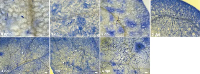
First, a cytological analysis of the G. orontii infection on wild type (ecotype Col-0) seedlings was conducted (Figure 1). The use of seedlings allowed a more rapid screening of phenotypes (2-3 week old instead of 4-5 week old plants) and produced an averaging effect due to the use of many individuals (up to a hundred in the case of spore counts) per genotype and biological replicate. The susceptibility of a genotype can vary based on environmental conditions like pot humidity and averaging across pots can help to control this effect. A settling tower was used as this method allows a uniform and controlled inoculation and can be used efficiently for larger amounts of genotypes (Adam et al., 1999; Humphry et al., 2010; Reuber et al., 1998). Inoculation densities were kept low (~750 spores/cm
2) to discern single powdery mildew colonies on the leaves.
The life cycle of G. orontii can be separated into several distinct stages, but so far its timing has not been characterized on Arabidopsis seedlings. At 1 day post inoculation (dpi) most primary haustoria had been formed and growth of secondary hyphae began (
Figure 1). Hyphal development continued slowly until 2 dpi and increased rapidly after the
formation of secondary haustoria at 3 dpi. The Col-0 ecotype is highly susceptible to G. orontii
and conidiophores therefore sometimes already formed at 4 dpi. Subsequently, the number of
14
conidiophores increased and numerous growing chains of conidiospores were observed at 5 and 6 dpi. The observations of the infection process on seedlings were in line with previous reports on G. orontii infections of 4-5 week old Arabidopsis plants (Micali et al., 2008; Plotnikova et al., 1998). The use of seedlings therefore faithfully reflected the timing of the natural infection process on mature plants.
Figure 1: Powdery mildew disease progression on Arabidopsis seedlings. Microscopic images of powdery mildew disease progression on Col-0 plants. Samples were harvested at the indicated time points and stained with Coomassie Brilliant Blue.
Arrows indicate conidiospore chains and arrowheads point to the initial spore. Images are representative of three independent experiments. Scale bar is 50 µm.
2.1.2 qPCR-based quantification of G. orontii infection
Methods on the basis of qPCR have been developed for biomass quantification of many plant- pathogenic microorganisms (Brouwer et al., 2003; Gachon and Saindrenan, 2004; Silvar et al., 2005). For this procedure, the quantitative extraction of pure genomic DNA as well as the efficient and specific amplification of target sequences is key. Therefore a phenolic extraction technique for genomic DNA isolation was used that was previously found to allow quantitative DNA isolation from both fungal and bacterial plant pathogens (Brouwer et al., 2003). The protocol was modified by introducing a disruption step of frozen material, as direct disruption of fresh seedlings was inefficient in my hands. Subsequently a series of qPCR primers from arbitrarily chosen genes was designed to amplify either G. orontii or Arabidopsis genomic sequences and tested for amplification efficiency. For efficient primer pairs the annealing temperatures and primer concentrations were optimized to obtain most specific PCR results. Primer dimers were not detected for any of the primer pairs by either melting curve analysis or agarose gel electrophoresis. Subsequently, a 5-fold dilution series of genomic DNA from heavily infected Col- 0 tissue (harvested at ~14 dpi) was performed to generate a standard curve for primer efficiency calculation across the dynamic range (Supplemental Figure 1A). All tested primer pairs were found to have high amplification efficiencies of 90 to 100% (Supplemental Table 1). For the G.
orontii primer pairs, marginal background amplification of unspecific products was detected on an
uninfected Col-0 control sample (Supplemental Figure 1 B). The unspecific amplification did not
affect the procedure since it was associated with Ct values (>35) that were outside the range
used in the subsequent experiments (ca. 25-35). The amplification of G. orontii-derived amplicons
15 relative to products obtained from Arabidopsis genomic DNA was used for the quantification of fungal biomass. This measure controls for variation in both sample harvesting and the efficiency of DNA isolation.
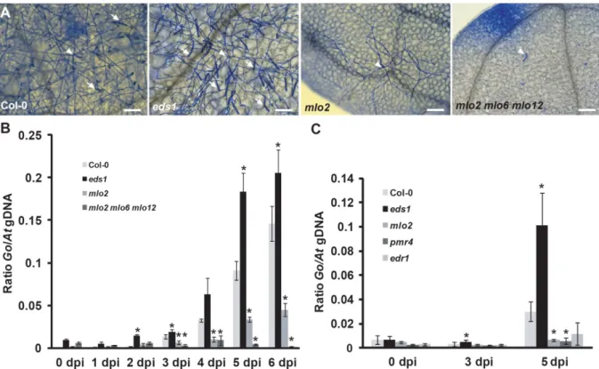
Figure 2: Time series analysis of powdery mildew infection by qPCR. (A) G. orontii-infected leaves were harvested at 5 dpi from Col-0 wild type and indicated mutant plants and stained with Coomassie Brilliant Blue. Arrows indicate conidiospore chains and arrowheads point to the initial spore. Images are representative of three independent experiments. (B) qPCR analysis of a time series of powdery mildew infection on Col-0 wild type, eds1, mlo2 and mlo2 mlo6 mlo2 plants. Ratios of G. orontii to Arabidopsis gDNA were determined by qPCR with primers R189/R192 and R193/R194, respectively. Bars represent the mean ± standard deviation of three technical replicates from a DNA sample of ten pooled seedlings grown in five different pots (two seedlings/pot used). (C) qPCR analysis of powdery mildew infection on Arabidopsis mutants that show powdery mildew-induced cell death. Representative time points of infection on Col-0 wild type, eds1, mlo2, pmr4 and edr1 plants were used. Ratios of G.
orontii to Arabidopsis gDNA were determined by qPCR with primers R189/R192 and R193/R194, respectively. Bars represent the mean ± standard deviation of three DNA samples (each derived from ten pooled seedlings grown in five different pots) with three technical replicates each. Asterisks indicate statistically significant differences to Col-0 in two-tailed Student’s t-test (p<0,05). Data shown are representative of three independent experiments. Scale bar is 50 µm.