S1 Leitlinie zur Diagnostik und Therapie der kutanen Larva migrans (Creeping Disease)
Zusammenfassung
Die kutane Larva migrans ist eine in ihrem klinischen Bild typische Hautinfektion, die durch aktives Eindringen und anschließende epider-
Cord Sunderkötter
1Esther von Stebut
2male Wanderung von Nematodenlarven hervorgerufen wird. Dieses ty-
Helmut Schöfer
3pische klinische Bild wird durch Larven von Hakenwürmern, meist An-
Martin Mempel
4cylostoma braziliense, selten andere bei Kaniden und Feliden vorkom- mende Hakenwurmarten, verursacht.
Dieter Reinel
5Ziele der Leitlinie sind die Verbesserung der Versorgung der Patienten
durch Optimierung von Diagnostik und Therapie bei Infektionen mit
Gerd Wolf
6Volker Meyer
1Larva migrans cutanea sowie die Verbesserung der Kenntnisse von
Ärztinnen und Ärzte über aktuelle Therapieoptionen.
Alexander Nast
7Schlüsselwörter:Leitlinie, Diagnostik, Therapie, kutane Larva migrans, Creeping Disease
Gerd-Dieter Burchard
8,91 Klinik für Hautkrankheiten – allgemeine Dermatologie und Venerologie,
Universitätsklinikum Münster (UKM), Münster, Deutschland 2 Hautklinik der
Universitätsmedizin, Johannes-Gutenberg- Universität, Mainz, Deutschland
3 Klinik für Dermatologie, Venerologie und Allergologie, Klinikum der Johann Wolfgang Goethe Universität, Frankfurt/M., Deutschland 4 Elmshorn, Deutschland 5 Hamburg, Deutschland 6 Grafschaft-Ringen,
Deutschland
7 Division of Evidence based Medicine (dEBM), Klinik für Dermatologie, Venerologie und Allergologie, Charité – Universitätsmedizin Berlin, Deutschland
8 Sektion Tropenmedizin und Infektiologie,
Unversitätsklinikum Hamburg-Eppendorf, Hamburg, Deutschland 9 ifi – Institut für
Interdisziplinäre Medizin, Hamburg, Deutschland
Ziele der Leitlinie
Verbesserung der Versorgung der Patienten durch Opti- mierung von Diagnostik und Therapie bei Infektionen mit Larva migrans cutanea. Verbesserung der Kenntnisse von Ärztinnen und Ärzten über aktuelle Therapieoptionen.
Methodik
S1 Leitlinie, nicht systematische Literaturrecherche, Konsensusprozess im Umlaufverfahren
Einleitung
Die kutane Larva migrans ist eine in ihrem klinischen Bild typische Hautinfektion, die durch aktives Eindringen und anschließende epidermale Wanderung von Nematoden- larven hervorgerufen wird. Dieses typische klinische Bild wird durch Larven von Hakenwürmern, (meistAncylosto- ma braziliense, selten andere bei Kaniden und Feliden vorkommende Hakenwurmarten, siehe unten) verursacht.
Die Weiterentwicklung dieser tierischen Hakenwurmlarven ist im Menschen nicht möglich.
Abzugrenzen ist die Larva migrans cutanea von der Larva currens. Hierunter versteht man eine Reaktion auf Larven vonStrongyloides stercoralis, vorwiegend am Rumpf oder Gesäß lokalisiert. Diese zeigt eine weniger irreguläre Wanderung, in weiten und weniger scharf begrenzten Gängen die Extremitäten entlang (bis zu 5 cm pro Stunde – daher Larva currens) und verschwindet meist rasch.
Auf die Larva currens wird in dieser Leitlinie nicht einge- gangen. Eine Larva migrans cutanea (als Syndrom) ist ebenfalls abzugrenzen von dem Symptom „Creeping eruption“ – hierunter versteht man wandernde subkutane Schwellungen mit Rötung und Urticaria der darüberliegen- den Haut, zum Beispiel bei Gnathostomiasis, kutaner Paragonimiasis (Lungenegel-Infektion) oder Fasziolose (Leberegel-Infektion), ähnliche wandernde Symptome sind auch möglich bei einer Myiasis („migratory myiasis“), also bei einem Befall mit Fliegenmaden.
Erreger
Bei der kutanen Larva migrans durch tierpathogene Ne- matodenlarven (meistAncylostoma braziliense, aber auch Ancylostoma caninum oder Uncinaria stenocephala) bleibt die Infektion auf die Haut beschränkt. Die Larven dringen in die intakte Epidermis (Füße, Gesäß oder ande- re exponierte Areale) ein, vermutlich über Haarfollikel oder Schweißdrüsen, und bohren sich unter Hinterlassung charakteristisch gewundener Gänge durch die Epidermis.
Die Basalmembran vermögen sie nicht zu durchdringen.
Sie bewegen sich je nach Art unterschiedlich schnell vorwärts, in der Regel bis zu 1 cm pro Tag. Die juckenden Effloreszenzen sind Ausdruck einer Immunreaktion auf die Wurmlarven und ihre Produkte. Für diese auf Hunde
und Katzen spezialisierten Parasiten stellt der Mensch einen Fehlwirt dar, und die Larven sterben nach einigen Wochen ab.
Epidemiologie
Die kutane Larva migrans ist verbreitet an warmen, san- digen Orten (vor allem Strände) in Südamerika, Afrika, in der Karibik, Südostasien, südöstliche Staaten der USA, teilweise auch am Mittelmeer [1].
Die Prävalenz ist dort hoch, wo in Gegenden mit warm- feuchtem Klima barfuß laufende Menschen mit Exkre- menten freilaufender Tiere in Kontakt kommen. In Deutschland tritt die Erkrankung vor allem auf bei Reisen- den, die Strände mit frei laufenden infestierten Tieren besucht haben. Auswertungen in internationalen Beob- achtungsnetzwerken zu Erkrankungen bei Reisenden und Migranten zeigen, dass die kutane Larva migrans eine der am häufigsten importierten Hautkrankheiten ist. So machte unter ihnen in einer weltweiten Untersuchung die Larva migrans cutanea 10% der dermatologischen Diagnosen aus [2]. Auch in Auswertungen einzelner Zentren ist die Larva migrans cutanea eine der häufigsten Diagnosen (in Frankreich [3], oder in Spanien [4]).
Klinik der Larva migrans
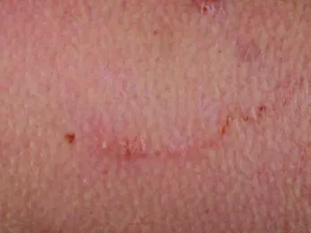
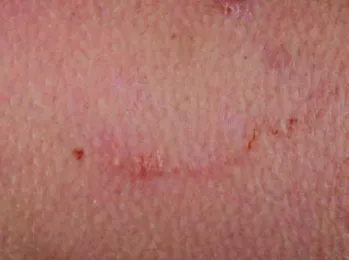
Einige Tage nach Kontakt mit verseuchten Böden entsteht zunächst eine Papel, von dieser breitet sich jeweils ein gewundener Gang mit lokal begrenzter Entzündungsreak- tion aus (Abbildung 1). Die Inkubationszeit kann Tage, aber auch Wochen oder Monate betragen [5]. Infestatio- nen mit mehreren Larvae migrantes gleichzeitig sind möglich (Abbildung 2). Die Gänge können mit einer serösen Flüssigkeit gefüllt sein und es können sich als Folge der Entzündungsreaktion Bläschen bilden. Juckreiz ist ein typisches Symptom. Er kann bereits vor Auftreten der ersten Effloreszenz beginnen und nimmt im Verlauf deutlich zu [6]. Die kutane Form ist selbstlimitierend, da die tierpathogenen Larven im Menschen absterben. In einigen Fällen können die Effloreszenzen über mehrere Monate persistieren, ohne dass histologisch Larven de- tektiert werden; die Ursache ist nicht ganz geklärt und kann u.a. dennoch in einer Persistenz einer Larve oder in einer anhaltenden Immunreaktion auf Larvenbestand- teile liegen [7]. Aufgrund der Eintrittspforte oder aufgrund von Kratzexkoriationen kann sich jedoch sekundär eine bakterielle Infektion (zum Beispiel ein Erysipel) entwi- ckeln, eine vor allem in den Endemiegebieten häufige Komplikation.
Sunderkötter et al.: S1 Leitlinie zur Diagnostik und Therapie der kutanen ...
Abbildung 1: Serpiginöse Spur der kutanen Larva migrans mit nur mäßiger Entzündungsreaktion
Abbildung 2: Befall mit mehreren Nematoden und Begleitentzündungsreaktion
Prophylaxe
Auf möglicherweise befallenen Stränden und Böden sollte Schuhwerk mit festen Sohlen (zum Beispiel Sanda- len) getragen werden. Badegäste sollten sich nicht direkt auf den Sand und auch nicht auf Handtücher legen, sondern Matratzen oder Liegen nutzen. Strandareale, die regelmäßig von den Gezeiten gespült werden, sollten bevorzugt werden. Wenn möglich, sollten Strände genutzt werden, an denen sich weder Katzen noch Hunde aufhal- ten.
Diagnostik/Differentialdiagnose
Die serpiginösen Exantheme sind pathognomonisch, die entsprechende Reise- und Expositionsanamnese unter- mauert den Verdacht.
Bei der Infestation mitStrongyloides stercoralisbeginnen die Effloreszenzen meist in der Perinealregion und breiten sich in weiten und weniger scharf begrenzten Gängen an den Extremitäten aus (bis zu 5 cm pro Stunde, daher Larva currens genannt).
Indikation zur Therapie
Eine Infektion mit einer kutanen Larva migrans ist selbstlimitierend und endet in der Regel nach 1–3 Mona- ten mit dem Tod und der Resorption der Larve. Eine Therapie sollte vor allem wegen des ausgeprägten und anhaltenden Juckreizes, der psychischen Belastung eines Parasitenbefalls und der Gefahr einer möglichen Super- infektion zum Beispiel nach Kratzexkoriationen erfolgen.
Therapie
Lange wurde in Deutschland und anderen Ländern die Lokaltherapie mit Thiabendazol in einer lipophilen Grundlage empfohlen [8]. Hierzu gab es zwar kaum Stu- dien, aber der Therapie wurde eine hohe therapeutische Breite zugeschrieben. Thiabendazol ist seit 1988 nicht mehr in Deutschland zugelassen und auch nicht mehr als Reinsubstanz in Pharmaqualität erhältlich.
Wir unterbreiten nach Durchsicht der Literatur und nach Erfahrungsberichten folgende Vorschläge zur Therapie:
Erwachsene und Jugendliche:
1. Ivermectin 1x 200 µg/kgKG (Stromectol 3 mg, Mecti- zan 3 mg, Ivermec 6 mg, Revectina 6 mg Tablette) Orale Gabe; 2 Stunden vor und nach Einnahme des Medikamentes sollte keine Nahrung aufgenommen werden. In der Regel reicht die einmalige Gabe aus, wenn nach 10 Tagen aber keine Besserung eingetre- ten ist, kann eine zweite Dosis gegeben werden [9].
Falls bei ausgeprägtem Befall Ivermectin nicht verfüg- bar, nicht wirksam oder nicht verträglich ist, stellt die systemische Therapie mit Albendazol eine Alternative dar.
2. Albendazol 800 mg/Tag oral für 3 Tage (zum Beispiel Eskazole 400 mg Tabletten); bei Jugendlichen unter 60 kg, aber über 6 Jahre: 15 mg/kg Körpergewicht bis maximal 800 mg/Tag), bei Unverträglichkeit (gastrointestinale Beschwerden) Dosisreduktion auf 400 mg/Tag für 5 Tage.
Einnahme in 2 Einzeldosen morgens und abends zu den Mahlzeiten.
3. Lokaltherapie mit Albendazol 10% in lipophiler Grundlage (zum Beispiel Albendazol 1200 mg (ent- sprechend 3 Tabletten zu 400 mg) in Vaselinum al- bum ad 12,0) drei mal täglich für 7–10 Tage auf ei- nem genügend großen Areal auftragen. Anwendung vor allem bei Kindern unter 15 kg Körpergewicht (kein Ivermectin) oder Kindern unter 6 Jahren (keine genü- gende Erfahrung mit Albendazol) sowie bei Patienten mit diskretem Befund (nur eine Läsion, kein quälender Juckreiz) und bei Patienten, die eine Systemtherapie ablehnen. Siehe auch Kommentar unten zum Bezug von Albendazol für die Lokaltherapie).
Begleitend empfehlen wir zur Milderung der Entzün- dung und des Juckreizes eine kurzzeitige (1–5 Tage)
Lokaltherapie mit einer glukokortikoidhaltigen Creme (Expertenmeinung) und bei Bedarf systemische Anthi- staminika zur Juckreizstillung.
Schwangerschaft und Stillzeit:
In der Schwangerschaft dürfen weder Ivermectin noch Albendazol eingenommen werden. Hier können wir bis- lang nur die oben erwähnte symptomatische Therapie empfehlen, das heißt zur Milderung der Entzündung und des Juckreizes eine Lokaltherapie mit einer glukokortiko- idhaltigen Creme und bei Bedarf die bei Schwangerschaft zugelassenen systemischen Anthistaminika zur Stillung des Juckreizes.
Unter einer wirksamen Behandlung sollte der Juckreiz nach 3, die Dermatitis nach 7 Tagen deutlich rückläufig sein.
Nicht empfohlen werden:
1. Topische Anwendung von Mebendazol
Begründungen: a) Die Wirkung scheint im Vergleich zu Albendazol bei Larva migrans und allgemein gegen Hakenwürmer schwächer zu sein [10], [11]; b) die intestinale Resorption ist nicht ausreichend, so dass es unter den gebräuchlichen Dosierungen zwar gegen intestinale Nematoden, nicht aber gegen Nematoden im Gewebe wirksam ist; c) entsprechend waren keine Publikationen zum systemischen Einsatz von Meben- dazol gegen Larva migrans auffindbar.
2. Kryotherapie (obwohl mitunter genannt), da der Auf- enthaltsort der Larve (meist 1–2 cm im Umkreis des sichtbaren Gangendes) nicht genau bekannt ist [12]
und ihre Empfindlichkeit auf Kälte in der Haut nicht hoch ist (>5 min Überleben bei –21°C).
Kommentar zu Ivermectin
Ivermectin ist in Deutschland nur als Tierarzneimittel unter dem Namen Eraquell Tabs 20 mg ad usum veteri- narium (zum tierärztlichen Gebrauch) zugelassen. In Frankreich, Belgien und den USA besteht eine Zulassung am Menschen für die Behandlung einer viszeralen Form der verwandten intestinalen Strongyloidiasis durch den ZwergfadenwurmStrongyloides stercoralis.
Die Tabletten lassen sich aber über jede Apotheke aus dem Ausland unter den Handelsnamen Stromectol®3 mg, Ivermec®6 mg oder Revectina®6 mg Tabl. (N1) beziehen.
Der Patient soll darüber aufgeklärt werden, dass Ivermec- tin in Deutschland nicht und im Ausland nicht direkt für Larva migrans Infektionen zugelassen ist. Die Aufklärung sollte zusammen mit einer Nutzen-Risiko-Abwägung des Off-Label Einsatzes in der Patientenakte dokumentiert werden.
Bei Kindern unter 15 kg Körpergewicht und bei Schwan- geren ist die Anwendung kontraindiziert. Beeinträchtigen- de unerwünschte Wirkungen sind transient und zudem unter der millionenfachen Anwendung gegen Onchozer- kose selten beschrieben worden [13].
Kommentar zu Albendazol systemisch
Albendazol ist für die systemische Gabe zur Zeit nur in einer Packungsgröße mit 60 Tabletten (N3) zu einem Verkaufspreis von 590,38 € [Importe: 549,09–556,09 €/
Preise: Stand: 05/2013] als Eskazole®[14]) erhältlich.
Zu wünschen wäre eine kleinere Packungsgröße, da die Inzidenz der Fälle einer Larva migrans nicht niedrig ist und somit häufiger nur kleine Mengen erforderlich sind.
Wenn Eskazole®verordnet wird, sind die Apotheken auch bei Verwendung von nur 6 Tabletten für die orale Therapie laut der rechtsverbindlichen „Hilfstaxe für Apotheken“
berechtigt, auf den Einkaufspreis der Packung 90% auf- zuschlagen und die ganze Packung zu berechnen. Klini- kapotheken können für den stationären Gebrauch in der internen Verrechnung nur die benutzte Menge in Rech- nung stellen. Das wäre theoretisch auch für Apotheken möglich, steht aber nicht im Einklang mit der „Hilfstaxe für Apotheken“. Albendazol ist in Deutschland als Antihel- minikum zugelassen, darunter unter anderem für den Behandlungsversuch bei Befall mitStrongyloides stercora- lis(siehe oben), aber nicht für die Behandlung von Larva migrans, so dass sein Gebrauch hierfür außerhalb der Zulassung liegt (so genannter „Off Label“ Einsatz). Folglich soll der Patient darüber informiert werden, und die Auf- klärung sollte zusammen mit einer Nutzen-Risiko-Abwä- gung des Off-Label Einsatzes in der Patientenakte doku- mentiert werden.
Kommentar Albendazol topisch
Die vorgeschlagene Rezeptur entstammt einem Fallbe- richt [15]: Albendazol 1,2 g, Vaselinum album ad 12,0 g.
Die Koautorin nutzt Albendazol 10,0, Paraff. subliq. 10,0, weißes Vaselin ad 100,0.
Wenn für die Rezeptur Eskazole®verordnet wird, sind die Apotheken auch bei Verwendung von nur 3 Tabletten berechtigt, den vollen Preis zu erheben (siehe oben).
Seit 2013 wird Albendazol aber auch in Form der Rein- substanz für Apotheken angeboten (von der Firma Fagron, Barsbüttel) und könnte dadurch kostengünstiger für eine lokale Anwendung verarbeitet werden. Unsere bisherige Erfahrung hat indes gezeigt, dass Lieferschwierigkeiten auftreten können (offenbar vor allem aufgrund mangeln- der Reinheit der von unterschiedlichen Zulieferern stammenden Substanzen). Außerdem müssen Apotheken nach Auslieferung einer reinen Substanz mit Analysenzer- tifikat vor einer pharmazeutischen Verarbeitung von Al- bendazol einen Identitätsnachweis mittels Infrarotspek- troskopie und Dünnschichtchromatographie durchführen.
Über Infrarotspektroskope verfügen aber nur wenige Apotheken.
Daher sollte sich der verordnende Arzt vorher erkundigen a) welche Apotheke den geforderten Identitätsnachweis durchzuführen vermag, und b) ob für diese Apotheke Al- bendazol lieferbar ist. Für die Apotheke ist beachtenswert, dass sie für die Rezeptur eventuell eine größere Mindest- menge abnehmen und bezahlen muss als sie für ein einzelnes Rezept berechnen darf.
Sunderkötter et al.: S1 Leitlinie zur Diagnostik und Therapie der kutanen ...
Sollten diese Erfordernisse aber erfüllt sein, ergäbe sich daraus für den Patienten eine deutlich kostengünstigere Lokalbehandlung als unter Verwendung von Eskazole®. Daher empfehlen wir, dass solche Einrichtungen, die häufiger Patienten mit Larva migrans behandeln, sich langfristig mit einer Apotheke über die oben genannten Bedingungen verständigen, damit sie nicht jedes Mal er- neut abgeklärt werden müssen.
Beurteilung der Wirksamkeit
Es gibt nur wenige vergleichende prospektive randomi- sierte Studien. Die einmalige Gabe von Ivermectin (n=10 Patienten) zeigte sich in einer vergleichenden randomisierten Studie wirksamer als 400 mg Albendazol (n=11 Patienten) [16]. Für Ivermectin im Vergleich zu Al- bendazol könnte auch sprechen, dass es bei Strongyloi- diasis (also einer Erkrankung durch einen verwandten Wurm) in einer prospektiven Studie in einer Einmaldosie- rung wirksamer war als 800 mg Albendazol für 7 Tage [17].
Neben vergleichenden Studien gibt es eine Reihe von teilweise retrospektiven Auswertungen mit einzelnen Medikamenten:
Ivermectin:In zwei kleineren Studien in den 90iger Jahren war eine Einmaldosis bei Patienten aus Kamerun und bei französischen Touristen gut wirksam [18], [19]. Dies ließ sich dann in größeren Studien bestätigen [9]. In einer Studie in Paris wurden 62 Europäer mit Ivermectin behan- delt, nach einer Dosis betrug die Heilungsrate 77%, mit ein bis zwei zusätzlichen Dosen 97% [20]. Schließlich gibt es noch eine weitere aktuelle Studie aus Frankreich, in welcher 59 von 62 Patienten mit Ivermectin geheilt werden konnten [21] und eine Studie aus einem brasilia- nischen Endemiegebiet der zufolge alle 92, zum Teil schwer befallenen Patienten 2–4 Wochen nach einmali- ger Gabe von 200 µg/kg signifikant angesprochen haben [22].
Albendazol:In einer retrospektiven Erhebung an 78 Pati- enten mit kutaner Larva migrans führten 1x400 mg/Tag Albendazol p.o. nach 7 Tagen bei allen zur Heilung. Zwei Patienten hatten unter der Medikation Übelkeit oder verstärkten Pruritus [23]. In weiteren publizierten Fallse- rien oder retrospektiven Erhebungen kamen verschiedene Dosen und Behandlungsdauern zum Einsatz, wobei unter 800 mg/Tag für 3 Tage 100%ige Heilungsraten, unter 400 mg/Tag hingegen einzelne Therapieversager berich- tet wurden [24], [25]. In der Dosis von 800 mg/Tag scheint es gegenüber Ivermectin eine höhere Rate an unerwünschten (meist gastrointestinalen) Nebenwirkun- gen zu haben [26].
Zur Lokaltherapie mit Albendazol wurden ein Fallbericht [15] und eine Empfehlung im Rahmen einer Übersicht [11] gefunden. Topisches Albendazol hat in dem Fallbe- richt bei 2 Kindern 1 Woche nach Behandlung zu einer rezidivfreien Abheilung geführt [15]. Außer diesem Fall- bericht sind keine genügenden Daten zu Hautpenetration, Wirksamkeit und Verträglichkeit bekannt. Die Autorinnen und Autoren dieser Leitlinien haben einzelne Fälle eben-
falls erfolgreich topisch mit 10% Albendazol behandelt.
Die Empfehlung, Albendazol 10% in lipophiler Grundlage, 3-mal täglich für 7–10 Tage auf einem genügend großen Areal aufzutragen, entstand in Analogie zur früher oft verwandten Thiabendazol-haltigen Rezeptur und gemäß des publizierten Fallberichtes. Die Größe des Behand- lungsareals ergibt sich aufgrund des Wissens, dass die Larve oft mehrere Zentimeter weiter gewandert ist, als die Entzündung anzeigt.
Glukokortikoide: Direkte Evidenz zur Wirksamkeit einer begleitenden Lokaltherapie mit Glukokortikoiden zur Milderung der Entzündung und des Juckreizes bei Larva migrans liegen nicht vor.
Anmerkungen
Gültigkeit
Die vorliegende Leitlinie hat eine Gültigkeit bis April 2017.
Die Autorengruppe entscheidet über die Notwendigkeit einer Aktualisierung. Erstellungsdatum: Mai 2013.
ICD-Code (ICD 10)
B76.9
AWMF-Register-Nummer
013/087
Interessenkonflikte
Siehe Anhang 1.
Finanzierung
Die Erstellung der Leitlinie erfolgte als Eigenleistung der Autoren. Es erfolgte keinerlei Finanzierung und es erfolgte keine äußere Einflussnahme auf die Inhalte der Leitlinie.
Genehmigungen
Autorisiert durch Deutsche Dermatologische Gesellschaft (DDG).
Autorisiert für die elektronische Publikation: AWMF online.
Autorisiert für Publikation in GMS ID durch John Wiley
& Sons Ltd Blackwell Verlag im Rahmen der interdiszipli- nären Erstellung dieser Leitlinie unter Mitwirkung der PEG (ursprünglich erschienen im Journal der Deutschen Der- matologischen Gesellschaft 2014 [27]).
Beteiligte Fachgesellschaften und deren Vertreter
• Deutsche Dermatologische Gesellschaft (DDG)//Ar- beitsgemeinschaft für dermatologische Infektiologie und Tropendermatologie (ADI-TD): E. von Stebut-Bor-
schitz, H. Schöfer, M. Mempel, D. Reinel, V. Meyer, A. Nast, C. Sunderkötter
• Deutsche Gesellschaft für Tropenmedizin und Interna- tionale Gesundheit e.V. (DTG): G.-D. Burchard, D. Reinel
• Society for Dermatology in the Tropics: D. Reinel
• Paul Ehrlich Gesellschaft: C. Sunderkötter
Anhänge
Verfügbar unter
http://www.egms.de/en/journals/id/2014-2/id000011.shtml 1. Anhang1_id000011.pdf (122 KB)
Interessenkonflikte
Literatur
1. Galanti B, Fusco FM, Nardiello S. Outbreak of cutaneous larva migrans in Naples, southern Italy. Trans R Soc Trop Med Hyg.
2002 Sep-Oct;96(5):491-2. DOI: 10.1016/S0035- 9203(02)90415-3
2. Lederman ER, Weld LH, Elyazar IR, von Sonnenburg F, Loutan L, Schwartz E, Keystone JS; GeoSentinel Surveillance Network.
Dermatologic conditions of the ill returned traveler: an analysis from the GeoSentinel Surveillance Network. Int J Infect Dis. 2008 Nov;12(6):593-602. DOI: 10.1016/j.ijid.2007.12.008 3. Caumes E, Carrière J, Guermonprez G, Bricaire F, Danis M,
Gentilini M. Dermatoses associated with travel to tropical countries: a prospective study of the diagnosis and management of 269 patients presenting to a tropical disease unit. Clin Infect Dis. 1995 Mar;20(3):542-8. DOI: 10.1093/clinids/20.3.542 4. Ramírez-Olivencia G, Bru Gorraiz FJ, Rivas González P, Lago
Núñez M, Herrero Mendoza MD, Puente Puente S. Patologia dermatologica y medicina tropical. Resultados de un estudio prospectivo (2004–2007)[Skin diseases and tropical medicine.
Results from a prospective study (2004–2007)]. Rev Clin Esp.
2009 Dec;209(11):527-35. DOI: 10.1016/S0014- 2565(09)73058-9
5. Siriez JY, Angoulvant F, Buffet P, Cleophax C, Bourrat E. Individual variability of the cutaneous larva migrans (CLM) incubation period. Pediatr Dermatol. 2010 Mar-Apr;27(2):211-2. DOI:
10.1111/j.1525-1470.2010.01107.x
6. Feldmeier H, Schuster A. Mini review: Hookworm-related cutaneous larva migrans. Eur J Clin Microbiol Infect Dis. 2012 Jun;31(6):915-8. DOI: 10.1007/s10096-011-1404-x
7. Veraldi S, Persico MC, Francia C, Schianchi R. Chronic hookworm- related cutaneous larva migrans. Int J Infect Dis. 2013 Apr;17(4):e277-9. DOI: 10.1016/j.ijid.2012.11.002 8. Hochedez P, Caumes E. Hookworm-related cutaneous larva
migrans. J Travel Med. 2007 Sep-Oct;14(5):326-33. DOI:
10.1111/j.1708-8305.2007.00148.x
9. Van den Enden E, Stevens A, Van Gompel A. Treatment of cutaneous larva migrans. N Engl J Med. 1998 Oct
22;339(17):1246-7. DOI: 10.1056/NEJM199810223391714 10. Steinmann P, Utzinger J, Du ZW, Jiang JY, Chen JX, Hattendorf J,
Zhou H, Zhou XN. Efficacy of single-dose and triple-dose albendazole and mebendazole against soil-transmitted helminths and Taenia spp.: a randomized controlled trial. PLoS ONE.
2011;6(9):e25003. DOI: 10.1371/journal.pone.0025003 11. Wolf P, Ochsendorf FR, Milbradt R. Aktuelle
Therapiemöglichkeiten bei Larva migrans cutanea [Current therapeutic possibilities in cutaneous larva migrans]. Hautarzt.
1993 Jul;44(7):462-5.
12. Albanese G, Venturi C, Galbiati G. Treatment of larva migrans cutanea (creeping eruption): a comparison between albendazole and traditional therapy. Int J Dermatol. 2001 Jan;40(1):67-71.
DOI: 10.1046/j.1365-4362.2001.01103.x
13. Meinking TL, Taplin D, Hermida JL, Pardo R, Kerdel FA. The treatment of scabies with ivermectin. N Engl J Med. 1995 Jul;333(1):26-30. DOI: 10.1056/NEJM199507063330105 14. Eskazole® Tabletten. In: Rote Liste 2012. Frankfurt/Main: Rote
Liste Service GmbH; 2012. p. 387.
15. Caumes E. Efficacy of albendazole ointment on cutaneous larva migrans in 2 young children. Clin Infect Dis. 2004
Jun;38(11):1647-8. DOI: 10.1086/421026
16. Caumes E, Carriere J, Datry A, Gaxotte P, Danis M, Gentilini M.
A randomized trial of ivermectin versus albendazole for the treatment of cutaneous larva migrans. Am J Trop Med Hyg. 1993 Nov;49(5):641-4.
17. Suputtamongkol Y, Kungpanichkul N, Silpasakorn S, Beeching NJ. Efficacy and safety of a single-dose veterinary preparation of ivermectin versus 7-day high-dose albendazole for chronic strongyloidiasis. Int J Antimicrob Agents. 2008 Jan;31(1):46-9.
DOI: 10.1016/j.ijantimicag.2007.08.014
18. Caumes E, Datry A, Paris L, Danis M, Gentilini M, Gaxotte P.
Efficacy of ivermectin in the therapy of cutaneous larva migrans.
Arch Dermatol. 1992 Jul;128(7):994-5. DOI:
10.1001/archderm.1992.01680170130027
19. Louis FJ, de Quincenet G, Louis JP. Intérêt de l'ivermectine en prise unique dans le traitement du syndrome de larva migrans cutanée [Value of single-dose ivermectin in the treatment of cutaneous larva migrans syndrome]. Presse Med. 1992 Sep;21(31):1483.
20. Bouchaud O, Houzé S, Schiemann R, Durand R, Ralaimazava P, Ruggeri C, Coulaud JP. Cutaneous larva migrans in travelers: a prospective study, with assessment of therapy with ivermectin.
Clin Infect Dis. 2000 Aug;31(2):493-8. DOI: 10.1086/313942 21. Vanhaecke C, Perignon A, Monsel G, Regnier S, Bricaire F,
Caumes E. The efficacy of single dose ivermectin in the treatment of hookworm related cutaneous larva migrans varies depending on the clinical presentation. J Eur Acad Dermatol Venereol. 2013 Feb. DOI: 10.1111/jdv.12097
22. Schuster A, Lesshafft H, Reichert F, Talhari S, de Oliveira SG, Ignatius R, Feldmeier H. Hookworm-related cutaneous larva migrans in northern Brazil: resolution of clinical pathology after a single dose of ivermectin. Clin Infect Dis. 2013 Oct;57(8):1155- 7. DOI: 10.1093/cid/cit440
23. Veraldi S, Bottini S, Rizzitelli G, Persico MC. One-week therapy with oral albendazole in hookworm-related cutaneous larva migrans: a retrospective study on 78 patients. J Dermatolog Treat. 2012 Jun;23(3):189-91. DOI:
10.3109/09546634.2010.544707
24. Blackwell V, Vega-Lopez F. Cutaneous larva migrans: clinical features and management of 44 cases presenting in the returning traveller. Br J Dermatol. 2001 Sep;145(3):434-7. DOI:
10.1046/j.1365-2133.2001.04406.x
25. Caumes E. Treatment of cutaneous larva migrans. Clin Infect Dis. 2000 May;30(5):811-4. DOI: 10.1086/313787
26. Sanguigni S, Marangi M, Teggi A, De Rosa F. Albendazole in the therapy of cutaneous larva migrans. Trans R Soc Trop Med Hyg.
1990 Nov-Dec;84(6):831.
27. Sunderkötter C, von Stebut E, Schöfer H, Mempel M, Reinel D, Wolf G, Meyer V, Nast A, Burchard GD. S1 guideline diagnosis and therapy of cutaneous larva migrans (creeping disease). J Dtsch Dermatol Ges. 2014 Jan;12(1):86-91. DOI:
10.1111/ddg.12250 Sunderkötter et al.: S1 Leitlinie zur Diagnostik und Therapie der kutanen ...
Korrespondenzadresse:
Prof. Dr. Cord Sunderkötter, MD
Klinik für Hautkrankheiten – allgemeine Dermatologie und Venerologie, Universitätsklinikum Münster (UKM), von-Esmarch-Straße 58, 48149 Münster, Deutschland, Tel.: +49 (0)251 83 52940, Fax: +49 (0)251 83 57488 Cord.Sunderkoetter@ukmuenster.de
Bitte zitieren als
Sunderkötter C, von Stebut E, Schöfer H, Mempel M, Reinel D, Wolf G, Meyer V, Nast A, Burchard GD. S1 Leitlinie zur Diagnostik und Therapie der kutanen Larva migrans (Creeping Disease). GMS Infect Dis.
2014;2:Doc03.
DOI: 10.3205/id000011, URN: urn:nbn:de:0183-id0000115
Artikel online frei zugänglich unter
http://www.egms.de/en/journals/id/2014-2/id000011.shtml Veröffentlicht:16.04.2014
Copyright
©2014 Sunderkötter et al. Dieser Artikel ist ein Open Access-Artikel und steht unter den Creative Commons Lizenzbedingungen
(http://creativecommons.org/licenses/by-nc-nd/3.0/deed.de). Er darf vervielfältigt, verbreitet und öffentlich zugänglich gemacht werden, vorausgesetzt dass Autor und Quelle genannt werden.
S1 guideline diagnosis and therapy of cutaneous larva migrans (creeping disease)
Abstract
Cutaneous larva migrans is a skin infection with a typical clinical appear- ance caused by active penetration of nematode larvae and their sub-
Cord Sunderkötter
1Esther von Stebut
2sequent epidermal migration. The typical clinical appearance is caused
Helmut Schöfer
3by hookworm larvae, usually Ancylostoma braziliense but sometimes
other canine or feline types of hookworm.
Martin Mempel
4This guideline aims to enhance patient care by optimizing the diagnosis
and treatment of infections due to creeping disease (cutaneous larva
Dieter Reinel
5Gerd Wolf
6migrans) and to raise awareness among doctors of current treatment
options.
Volker Meyer
1Alexander Nast
7Gerd-Dieter Burchard
8,91 Department of Dermatology and Venereology, Münster University Hospital, Münster, Germany
2 Department of Dermatology, University Hospital of Mainz, Germany
3 Department of Dermatology, Venereology and Allergology, Johann Wolfgang Goethe University Hospital, Frankfurt, Germany
4 Elmshorn, Germany 5 Hamburg, Germany 6 Grafschaft-Ringen, Germany 7 Division of Evidence based
Medicine (dEBM),
Department of Dermatology, Venereology and Allergology, Charité – Universitätsmedizin Berlin, Germany
8 Division of Tropical Medicine and Infectious Diseases, University Medical Center Hamburg-Eppendorf, Hamburg, Germany 9 ifi – Institute for
Interdisziplinary Medicine, Hamburg, Germany
Guideline This is the translated (English) version.
The original (German) version starts at p. 1.
OPEN ACCESS
Guideline objectives
These guidelines aim to enhance patient care by optimiz- ing the diagnosis and treatment of infections due to creeping disease (cutaneous larva migrans) and to raise awareness among doctors of current treatment options.
Methods
S1 guideline, non-systematic literature search, consensus process using a circular letter
Introduction
Cutaneous larva migrans is a skin infection with a typical clinical appearance caused by active penetration of nematode larvae and their subsequent epidermal migra- tion. The typical clinical appearance is caused by hook- worm larvae, usuallyAncylostoma braziliensebut some- times other canine or feline types of hookworm. These hookworm larvae that are pathogenic to animals cannot reproduce in human beings.
Cutaneous larva migrans should be distinguished from larva currens. The latter is caused by Strongyloides stercoralis larvae and usually occurs on the trunk or buttocks. Migration is less irregular and produces broad, less clearly demarcated tracks on the extremities (pro- gressing up to 5 cm per hour, hence the name larva cur- rens) and usually disappears rapidly. This guideline does not address larva currens. Cutaneous larva migrans should also be distinguished from the symptom “creeping eruption” which is a migrating subcutaneous swelling with redness and urticaria of the overlying skin. It occurs, for instance, in gnathostomiasis, cutaneous paragonimi- asis(Paragonimusinfection), and fascioliasis (liver fluke infection byFasciola hepatica). Similar migratory symp- toms may also occur in myiasis (migratory myiasis) caused by fly larva.
Pathogens
In cutaneous larva migrans due to nematode larvae which are pathogenic to animals (Ancylostoma braziliense,An- cylostoma caninum, orUncinaria stenocephala), the in- fection remains limited to the skin. The larvae penetrate the intact epidermis (e.g., feet, buttocks, or other exposed areas), presumably through the hair follicles or sweat glands, before entering the epidermis where they produce characteristic winding tracks. They do not penetrate the basement membrane. The speed of migration depends on the species, but it is generally dose not exceed one centimeter per day. The pruritic lesions are an expression of an immune response to the larvae and their products.
These parasites attack mainly dogs and cats. Human in- fection is accidental, and the larvae die after only a few weeks.
Epidemiology
Cutaneous larva migrans is widespread in warm, sandy places (especially beaches) in South America, Africa, the Caribbean, Southeast Asia, the southern USA, and in Mediterranean regions [1].
Prevalence is high in regions with a warm, humid climate where people walk around barefoot and come into contact with animal excrement. In Germany, the disease is usually seen in patients who have vacationed in areas with beaches where there are infected, roaming animals.
Analyses by international observation networks on disease in travelers and migrants have shown that cutaneous larva migrans is one of the most commonly imported skin diseases. A worldwide study found that cutaneous larva migrans makes up 10% of dermatological diagnoses in returning travelers [2]. Analyses from individual centers also report that cutaneous larva migrans is one of the most common diagnoses (e.g., in France [3] and Spain [4]).
Clinical appearance of larva migrans
A few days after contact with infected soil, a papule ap- pears, then a winding tract develops and there is a local inflammatory reaction (Figure 1). The incubation period can last days, weeks, or months [5]. Simultaneous infes- tation with multiple nematodes is also possible (Figure 2).
The tracts may be filled with serous fluid. Blisters may develop due to the inflammatory reaction. Pruritus is a typical symptom. Itching may begin before lesions appear, and it worsens significantly over the course of disease [6]. The cutaneous form of disease is self-limiting, be- cause the larvae cannot survive in humans. Occasionally, lesions may persist for several months without histological detection of larvae; the cause is uncertain and may be due to persistence of larvae or a prolonged immune re- sponse to the larval debris [7]. A secondary bacterial in- fection (such as erysipelas) can develop from the initial entry site or because of excoriations. This complication is especially common in endemic regions.
Prophylaxis
Patients should wear shoes (or sandals) when walking on potentially contaminated beaches and soil. Bathers should not lie on the sand or even on towels, but rather should use a mattress or chair. Beachgoers should try to visit areas of the beach where the sand is regularly washed away by the tides. If possible, vacationers should avoid beaches where there are cats or dogs.
Figure 1: Serpiginous track of cutaneous larva migrans with only moderate inflammatory reaction
Figure 2: Infestation with several nematodes and the accompanying inflammatory reaction
Diagnosis/differential diagnosis
The serpiginous exanthems are pathognomonic. Suspicion is confirmed by a history of travel and exposure.
In infestation withStrongyloides stercoralis, the lesions usually begin in the perineal region and spread in wide, and less sharply demarcated, tracks along the extremities, moving much more rapidly (up to 5 cm per hour, hence the name larva currens).
Indication for treatment
Infection with cutaneous larva migrans is self-limited and generally ends after 1–3 months with the death and re- sorption of the larvae. Treatment is mainly required be- cause of the severe, lasting pruritus, the psychological burden of parasite infection, and the risk of a possible superinfection (such as after excoriation due to scratch- ing).
Therapy
In Germany and other countries, for years the recommend- ed treatment was topical thiabendazole in a lipophilic base [8]. Although there were virtually no studies, it was believed to have a broad therapeutic spectrum. Thiabend- azole has not been approved for use in Germany since 1988, nor is it available in its pure form as a pharmaceut- ical grade drug.
After consulting the literature, and based on our own experience, we suggest the following treatment:
Adults and adolescents:
1. Ivermectin 1×200 µg/kg of body weight (Stromectol® 3 mg, Mectizan® 3 mg, Ivermec® 6 mg, Revectina® 6 mg tablet), oral administration. Patients should avoid eating anything for two hours before or after taking the medication. In general, a single dose is sufficient; if there is no improvement after 10 days, a second dose may be given [9]. In patients with severe disease, if ivermectin is unavailable, ineffect- ive, or not well-tolerated, systemic albendazole may be an alternative.
2. Oral albendazole 800 mg/daily for 3 days (e.g., Eskazole®400 mg tablets). In young people weighing less than 60 kg who are more than 6 years old:
15 mg/kg of body weight up to a maximum of 800 mg/daily). If the drug is not tolerated (gastroin- testinal complaints), reduce dosage to 400 mg/daily for 5 days.
Take with meals in 2 single doses in the morning and evening.
3. Topical albendazole 10% in a lipophilic base (e.g., al- bendazole 1,200 mg which corresponds to 3 tablets of 400 mg in white Vaseline ad 12.0) three times daily for 7–10 days. Apply generously. Used mainly in chil- dren weighing less than 15 kg (no ivermectin) and children under age 6 (inadequate experience with al- bendazole). Also used in patients with discrete lesions (e.g., a single lesion or mild itching) or those who re- fuse systemic therapy. (See also comments below on treatment with topical albendazole).
To reduce inflammation and itching, we also recom- mend brief topical treatment (1–5 days) with a cor- ticosteroid cream (expert opinion) and, if necessary, systemic antihistamines to stop the itching.
Pregnancy and nursing:
Ivermectin and albendazole must not be used by pregnant women. We can only recommend the symptomatic treat- ment described above; to alleviate inflammation and itching, topical treatment consisting of a corticosteroid cream may be given. If necessary, systemic antihistam- ines, which are approved for use in pregnant women, may be administered as well to stop the itching.
Effective treatment significantly reduces itching in 3 days and dermatitis after 7 days.
Sunderkötter et al.: S1 guideline diagnosis and therapy of cutaneous larva ...
The following treatments are not recommended:
1. Topical mebendazole. Reasons: a) Compared to al- bendazole, it appears to be less effective in larva mi- grans (and in general against hookworms) [10], [11];
b) intestinal resorption is inadequate, and thus at normal dosages, it is effective against intestinal nematodes, but not against nematodes in tissue;
c) we found no publications on systemic administra- tion of mebendazole to treat larva migrans.
2. Cryotherapy (although sometimes advised), given that the location of the larvae (usually 1–2 cm around the visible end of the track) is not precisely known [12]
and their low sensitivity to cold skin temperatures (>5 minute survival at –21°C).
Comments on ivermectin
Ivermectin is approved for use in Germany only in veter- inary medicine and is sold under the name Eraquell Tabs® 20 mg. In France, Belgium, and the United States, the drug is approved for use in humans for the treatment of the related visceral form of strongyloidiasis due to Strongyloides stercoralis.
The tablets may be obtained from foreign pharmacies as Stromectol®3 mg, Ivermec®6 mg, or Revectina® 6 mg (N1). The patient should be informed that ivermectin is not available in Germany nor officially recommended in other countries as larva migrans treatment. The discus- sion and the risk/benefit assessment of off-label use should be documented in the patient’s file.
Its use is contraindicated in children weighing less than 15 kg and in pregnant women. Adverse effects limiting its use are transitory and have rarely been reported, despite having been used millions of times to treat oncho- cercosis [13].
Comments on systemic albendazole
Systemic albendazole is currently only available in pack- ages containing 60 tablets (N3) which cost 590.38 € (imports: 549.09–556.09 €/Prices from: 05/2013, as Eskazole®[14]). Smaller sizes would be desirable, given that the incidence of larva migrans is more than negli- gible, and thus small amounts are needed rather fre- quently.
Pharmacies that provide Eskazole® have the right to charge for the entire package, even if only 6 tablets are used in accordance with the legally binding. Hospital pharmacies may only charge for the amount actually used.
Theoretically, one could apply this to pharmacies as well, but such a change conflicts with the “pharmacy tax”. Al- bendazole is approved as an anthelminthic in Germany, including for attempted treatment of Strongyloides stercoralis(see above) infections, but not for treatment of larva migrans, so that treatment of the latter is off-la- bel. Patients should be informed of this, and the discus- sion as well as the risk/benefit assessment should be documented in the patient’s file.
Comments on topical albendazole
The suggested formulation is based on a case report [15]
albendazole 1.2 g, white Vaseline ad 12 g.
One co-author uses albendazole 10.0, paraffin oil 10.0, white petrolatum ad 100.0.
If Eskazole®is prescribed, the pharmacy has the right to charge full price, even if only 3 tablets are needed (see above).
Albendazole has also been offered since 2013 to phar- macies in its pure form (Fagron, Barsbüttel, Germany).
This would reduce the cost of producing a topical treat- ment. In our own experience, there have been problems obtaining the product, apparently due to impure sub- stances from different providers. In addition, after delivery of a pure substance with a certificate of analysis, before creating the albendazole formulation, the pharmacy must also conduct an analysis for identification of the sub- stance using infrared spectroscopy and thin-layer chro- matography. Yet, only a few pharmacies can perform in- frared spectroscopy.
Thus physicians should first find out (a) which pharmacy can perform the identification procedure, and (b) whether it can order albendazole. The pharmacy may need to or- der, and pay for more (if there is a minimum amount) than an individual prescription requires.
If these requirements are met, however, the cost is much lower for the patient than topical Eskazole®. We therefore recommend for centers which often treat patients with larva migrans that they contact a pharmacy, discuss the situation, and establish cooperation with that pharmacy to avoid having to start from scratch each time.
Assessment of effectiveness
There are only a few comparative prospective randomized studies on effectiveness of the drug. A single administra- tion of ivermectin (n=10 patients) was shown in one comparative randomized study to be more effective than 400 mg albendazole (n=11 patients) [16]. Compared to albendazole, ivermectin was shown in a prospective study on strongyloidiasis (i.e., a disease caused by a related type of worm) that a single dose was more effective than a 7-day regimen of 800 mg albendazole [17].
In addition to comparative studies, there are a number of partly retrospective analyses on individual medications.
Ivermectin:Two smaller studies from the 1990s showed a single dose to be effective in patients in Cameroon and in French tourists [18], [19]. These findings were later confirmed by larger studies [9]. In a study done in Paris, 62 Europeans were treated with ivermectin; after receiv- ing a single dose, the healing rate was 77%; it was 97%
in patients who received one or two additional doses [20].
Another study from France reported healing in 59 out of 62 patients who were treated with ivermectin [21]. A study from an endemic region in Brazil reported that all 92 patients, some of whom had severe disease, had a significant response 2–4 weeks after being given a single dose of 200 µg/kg of body weight [22].
Albendazole:A retrospective study that included 78 pa- tients with cutaneous larva migrans found that, after be- ing given 1×400 mg/daily of oral albendazole, all patients healed after 7 days. Two patients reported nausea or in- creased pruritus related to the drug [23]. Other published case series and retrospective analyses have reported different dosages and treatment durations. Three days of 800 mg/daily reportedly led to healing in 100% of pa- tients; 400 mg/daily led a few treatment failures [24], [25]. At a dosage of 800 mg/daily, the rate of side effects is higher (usually gastrointestinal complaints) than with ivermectin [26].
For topical albendazole therapy, we found a case report [15] and a recommendation in the framework of a review [11]. In one case report, topical albendazole was given to 2 children, and led to recurrence-free healing 1 week after treatment [15]. Other than this case report, there are insufficient data on skin penetration, effectiveness, and tolerability. The authors of the present guidelines have also successfully treated individual patients with topical application of 10% albendazole. The recommend- ation to generously apply albendazole 10% in a lipophilic base 3 times daily for 7–10 days arose analogous to the thiabendazole-based formulation that was formerly used and according to the published case report. The size of the area of application is based on the knowledge that the larvae often migrate several centimeters farther than the inflammation.
Corticosteroids:There is no direct evidence of the effec- tiveness of accompanying local treatment with corticoste- roids to alleviate the inflammation and itching in patients with larva migrans.
Notes
Validity
The present guidelines are valid until April 2017. The need for an update will be decided on by the authors.
Date created: May 2013.
ICD Code (ICD 10)
B76.9
AWMF registry number
013/087
Conflicts of interest
See Attachment 1.
Financing
The guidelines were financed solely by the authors. No funding was received and there was no outside influence on the contents of the guidelines.
Permissions
Authorized by Deutsche Dermatologische Gesellschaft (DDG).
Authorized for electronic publication: AWMF online.
Publication in GMS ID has been permitted by John Wiley
& Sons Ltd in the context of the inderdisciplinary compil- ation of these guidelines in cooperation with the PEG (originally published in Journal der Deutschen Dermato- logischen Gesellschaft 2014 [27]).
Cooperating professional associations and their representatives
• The German Society of Dermatology (DDG)/Working Group on Dermatological Infectious Diseases and Tropical Dermatology (ADI-TD): E. von Stebut-Borschitz, H. Schöfer, M. Mempel, D. Reinel, V. Meyer, A. Nast, C. Sunderkötter
• German Society for Tropical Medicine and International Health e.V. (DTG): G.-D. Burchard, D. Reinel
• Society for Dermatology in the Tropics: D. Reinel
• Paul-Ehrlich-Gesellschaft: C. Sunderkötter
Attachments
Available from
http://www.egms.de/en/journals/id/2014-2/id000011.shtml 1. Anhang1_id000011.pdf (122 KB)
Conflicts of interest (in German)
References
1. Galanti B, Fusco FM, Nardiello S. Outbreak of cutaneous larva migrans in Naples, southern Italy. Trans R Soc Trop Med Hyg.
2002 Sep-Oct;96(5):491-2. DOI: 10.1016/S0035- 9203(02)90415-3
2. Lederman ER, Weld LH, Elyazar IR, von Sonnenburg F, Loutan L, Schwartz E, Keystone JS; GeoSentinel Surveillance Network.
Dermatologic conditions of the ill returned traveler: an analysis from the GeoSentinel Surveillance Network. Int J Infect Dis. 2008 Nov;12(6):593-602. DOI: 10.1016/j.ijid.2007.12.008 3. Caumes E, Carrière J, Guermonprez G, Bricaire F, Danis M,
Gentilini M. Dermatoses associated with travel to tropical countries: a prospective study of the diagnosis and management of 269 patients presenting to a tropical disease unit. Clin Infect Dis. 1995 Mar;20(3):542-8. DOI: 10.1093/clinids/20.3.542 4. Ramírez-Olivencia G, Bru Gorraiz FJ, Rivas González P, Lago
Núñez M, Herrero Mendoza MD, Puente Puente S. Patologia dermatologica y medicina tropical. Resultados de un estudio prospectivo (2004–2007)[Skin diseases and tropical medicine.
Results from a prospective study (2004–2007)]. Rev Clin Esp.
2009 Dec;209(11):527-35. DOI: 10.1016/S0014- 2565(09)73058-9
5. Siriez JY, Angoulvant F, Buffet P, Cleophax C, Bourrat E. Individual variability of the cutaneous larva migrans (CLM) incubation period. Pediatr Dermatol. 2010 Mar-Apr;27(2):211-2. DOI:
10.1111/j.1525-1470.2010.01107.x
6. Feldmeier H, Schuster A. Mini review: Hookworm-related cutaneous larva migrans. Eur J Clin Microbiol Infect Dis. 2012 Jun;31(6):915-8. DOI: 10.1007/s10096-011-1404-x Sunderkötter et al.: S1 guideline diagnosis and therapy of cutaneous larva ...
7. Veraldi S, Persico MC, Francia C, Schianchi R. Chronic hookworm- related cutaneous larva migrans. Int J Infect Dis. 2013 Apr;17(4):e277-9. DOI: 10.1016/j.ijid.2012.11.002 8. Hochedez P, Caumes E. Hookworm-related cutaneous larva
migrans. J Travel Med. 2007 Sep-Oct;14(5):326-33. DOI:
10.1111/j.1708-8305.2007.00148.x
9. Van den Enden E, Stevens A, Van Gompel A. Treatment of cutaneous larva migrans. N Engl J Med. 1998 Oct
22;339(17):1246-7. DOI: 10.1056/NEJM199810223391714 10. Steinmann P, Utzinger J, Du ZW, Jiang JY, Chen JX, Hattendorf J,
Zhou H, Zhou XN. Efficacy of single-dose and triple-dose albendazole and mebendazole against soil-transmitted helminths and Taenia spp.: a randomized controlled trial. PLoS ONE.
2011;6(9):e25003. DOI: 10.1371/journal.pone.0025003 11. Wolf P, Ochsendorf FR, Milbradt R. Aktuelle
Therapiemöglichkeiten bei Larva migrans cutanea [Current therapeutic possibilities in cutaneous larva migrans]. Hautarzt.
1993 Jul;44(7):462-5.
12. Albanese G, Venturi C, Galbiati G. Treatment of larva migrans cutanea (creeping eruption): a comparison between albendazole and traditional therapy. Int J Dermatol. 2001 Jan;40(1):67-71.
DOI: 10.1046/j.1365-4362.2001.01103.x
13. Meinking TL, Taplin D, Hermida JL, Pardo R, Kerdel FA. The treatment of scabies with ivermectin. N Engl J Med. 1995 Jul;333(1):26-30. DOI: 10.1056/NEJM199507063330105 14. Eskazole® Tabletten. In: Rote Liste 2012. Frankfurt/Main: Rote
Liste Service GmbH; 2012. p. 387.
15. Caumes E. Efficacy of albendazole ointment on cutaneous larva migrans in 2 young children. Clin Infect Dis. 2004
Jun;38(11):1647-8. DOI: 10.1086/421026
16. Caumes E, Carriere J, Datry A, Gaxotte P, Danis M, Gentilini M.
A randomized trial of ivermectin versus albendazole for the treatment of cutaneous larva migrans. Am J Trop Med Hyg. 1993 Nov;49(5):641-4.
17. Suputtamongkol Y, Kungpanichkul N, Silpasakorn S, Beeching NJ. Efficacy and safety of a single-dose veterinary preparation of ivermectin versus 7-day high-dose albendazole for chronic strongyloidiasis. Int J Antimicrob Agents. 2008 Jan;31(1):46-9.
DOI: 10.1016/j.ijantimicag.2007.08.014
18. Caumes E, Datry A, Paris L, Danis M, Gentilini M, Gaxotte P.
Efficacy of ivermectin in the therapy of cutaneous larva migrans.
Arch Dermatol. 1992 Jul;128(7):994-5. DOI:
10.1001/archderm.1992.01680170130027
19. Louis FJ, de Quincenet G, Louis JP. Intérêt de l'ivermectine en prise unique dans le traitement du syndrome de larva migrans cutanée [Value of single-dose ivermectin in the treatment of cutaneous larva migrans syndrome]. Presse Med. 1992 Sep;21(31):1483.
20. Bouchaud O, Houzé S, Schiemann R, Durand R, Ralaimazava P, Ruggeri C, Coulaud JP. Cutaneous larva migrans in travelers: a prospective study, with assessment of therapy with ivermectin.
Clin Infect Dis. 2000 Aug;31(2):493-8. DOI: 10.1086/313942 21. Vanhaecke C, Perignon A, Monsel G, Regnier S, Bricaire F,
Caumes E. The efficacy of single dose ivermectin in the treatment of hookworm related cutaneous larva migrans varies depending on the clinical presentation. J Eur Acad Dermatol Venereol. 2013 Feb. DOI: 10.1111/jdv.12097
22. Schuster A, Lesshafft H, Reichert F, Talhari S, de Oliveira SG, Ignatius R, Feldmeier H. Hookworm-related cutaneous larva migrans in northern Brazil: resolution of clinical pathology after a single dose of ivermectin. Clin Infect Dis. 2013 Oct;57(8):1155- 7. DOI: 10.1093/cid/cit440
23. Veraldi S, Bottini S, Rizzitelli G, Persico MC. One-week therapy with oral albendazole in hookworm-related cutaneous larva migrans: a retrospective study on 78 patients. J Dermatolog Treat. 2012 Jun;23(3):189-91. DOI:
10.3109/09546634.2010.544707
24. Blackwell V, Vega-Lopez F. Cutaneous larva migrans: clinical features and management of 44 cases presenting in the returning traveller. Br J Dermatol. 2001 Sep;145(3):434-7. DOI:
10.1046/j.1365-2133.2001.04406.x
25. Caumes E. Treatment of cutaneous larva migrans. Clin Infect Dis. 2000 May;30(5):811-4. DOI: 10.1086/313787
26. Sanguigni S, Marangi M, Teggi A, De Rosa F. Albendazole in the therapy of cutaneous larva migrans. Trans R Soc Trop Med Hyg.
1990 Nov-Dec;84(6):831.
27. Sunderkötter C, von Stebut E, Schöfer H, Mempel M, Reinel D, Wolf G, Meyer V, Nast A, Burchard GD. S1 guideline diagnosis and therapy of cutaneous larva migrans (creeping disease). J Dtsch Dermatol Ges. 2014 Jan;12(1):86-91. DOI:
10.1111/ddg.12250
Corresponding author:
Prof. Dr. Cord Sunderkötter, MD
Department of Dermatology and Venereology, Münster University Hospital, von-Esmarch-Straße 58, 48149 Münster, Germany, Phone: +49 (0)251 83 52940, Fax:
+49 (0)251 83 57488
Cord.Sunderkoetter@ukmuenster.de
Please cite as
Sunderkötter C, von Stebut E, Schöfer H, Mempel M, Reinel D, Wolf G, Meyer V, Nast A, Burchard GD. S1 Leitlinie zur Diagnostik und Therapie der kutanen Larva migrans (Creeping Disease). GMS Infect Dis.
2014;2:Doc03.
DOI: 10.3205/id000011, URN: urn:nbn:de:0183-id0000115
This article is freely available from
http://www.egms.de/en/journals/id/2014-2/id000011.shtml Published:2014-04-16
Copyright
©2014 Sunderkötter et al. This is an Open Access article distributed under the terms of the Creative Commons Attribution License (http://creativecommons.org/licenses/by-nc-nd/3.0/deed.en). You are free: to Share — to copy, distribute and transmit the work, provided the original author and source are credited.