The vascular development is modulated by endostatin and restin
I n a u g u r a l - D i s s e r t a t i o n zur
Erlangung des Doktorgrades
der Mathematisch-Naturwissenschaftlichen Fakultät der Universität zu Köln
vorgelegt von Annette Schmidt aus Bergisch Gladbach
(Hundt Druck GmbH, Köln)
2003
First of all, I would like to thank Professor Dr. med. K. Addicks for his attentive guidance and concern for this subject as well as for many helpful discussions.
I would also express my gratitude to Professor Dr. rer. nat. S. Roth for his confidence.
I am deeply grateful to PD Dr. med. W. Bloch for his guidance and valuable advice in the whole process of this research, for innumerable helpful discussions and subject designing and experimental arrangement.
For technical support I would like to thank M. Ghilav, C. Hoffmann and E. Janßen.
I would like to thank my parents for their tireless support.
This study was supported by the Köln Fortune Program of Medicin, University of Cologne (No. 117/2001)
Table of contents
Abbreviations
Summary (Germane) Summary (English)
1. Introduction 1
1.1 Vessel Development 1
1.1.1 Vasculogenesis and Angiogenesis 1
1.1.2 Stimulators of Angiogenesis 2
1.1.3 Inhibitors of Angiogenesis 2
1.1.4 Tumour Vascularisation 3
1.1.5 Wound Healing 5
1.2 Signaltransduction 5
1.3 Endostatin 7
1.4 Restin 9
1.5 Embryonic Stem Cells (ES-cells) 9
1.6 Target of the Work 10
2. Material and Methods 12
2.1 Used Chemicals and Materials 12
2.2 Technical Basics 16
2.2.1 Cell Culture 16
2.2.1.1 Stem cell culturing - the D3-cell line 16
2.2.1.2 Preparation of mouse embryos to gain feeder cells 16
2.2.1.3 Culturing of feeder cells 17
2.2.1.4 Inactivation of feeder cells 17
2.2.1.5 Preparation of hanging drops 17
2.2.1.6 Culturing of aggregates in suspension 17
2.2.1.7 Gelatine coating and plating of embryoid bodies 19
2.2.1.8 Components of DMEM-medium 19
2.2.2 Tissue Processing 19
2.2.2.1 Fixation of Tissue 19
2.2.2.2 Tissue embedding for electron microscopy 20
2.2.2.3 Tissue embedding in paraffin 20
2.2.2.4 Tissue embedding for cryostat sections 20
2.2.2.5 Shock freezing of tissue 20
2.2.3 Immunohistochemistry 21
2.2.3.1 Procedure of immunohistochemistry 21
2.3 Bladder, Kidney and Prostate Tissues 22
2.4 Animals 22
2.5 Wounding and preparation of wound tissue 22
2.6 Different kinds of endostatin 23
2.7 Endostatin treatment of cells and mice 23
2.8 Binding-Assay 23
2.8.1 Biotinylation of endostatin 23
2.8.2 Fluoresceinisothiocyanat-(FITC-)conjugation of endostatin 24
2.8.3 Western-Blot to examine the biotin-conjugation 24
2.8.4 Incubation with biotin-conjugated endostatin 25
2.8.5 Ultrastructural Cytochemistry 25
2.9 Detection of endothelial cells and vessels 26
2.10 Proliferation-Assay 26
2.11 BrdU cell proliferation 26
2.12 Apoptosis-Assay 27
2.13 Migratin-Assay 27
2.14 Signaltransduction 28
2.14.1 Treatments 28
2.14.2 Immunohistochemistry 28
2.15 Magnet associated cell sorting (MACS) 29
2.15.1 Purity of MACS-sorted endothelial cells 29
2.16 The live reporter gene EGFP under control of the PECAM-1 promotor 30
2.16.1 Vectors 30
2.16.2 Electroporation and selection procedure 30
2.17 Vital microscopy or Time laps microscopy 31
2.18 Deconvolution 31
2.18.1 General description of the deconvolution method 31
2.18.1.1 The 3D-image stack 31
2.18.1.2 Physical Background 32
2.18.1.3 Inversion of the representation process 32
2.18.2 3D-Deconvolution of PECAM-positive vessels 33
2.19 Immuno-Blot (Western-Blot) 34
2.19.1 Homogenisation of tissue 34
2.19.2 Bradford-Assay 34
2.19.3 SDS-PAGE 35
2.19.4 Coomassie-blue-(CB-)staining of protein gels 36
2.19.5 Transfer of proteins on a PVDF-membrane 36
2.19.6 Immunological Detection 37
2.20 In situ-Hybridisation 38
2.20.1 Preparation of the endostatin cDNA-sonde 38
2.20.2 ProteinaseK-Treatment and Pre-hybridisation 39
2.20.3 Preparation of cDNA-sonde and Hybridisation 39
2.20.4 Blocking and Detection 40
2.21 RT in situ PCR 40
2.22 Statistical analysation 41
3. Results 42
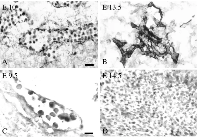
3.1 Endostatin Binding to Benign and Malignant Bladder, Prostate and Kidney Tissue 42 3.2 Expression pattern of collagenXVIII/endostatin in the embryo 45
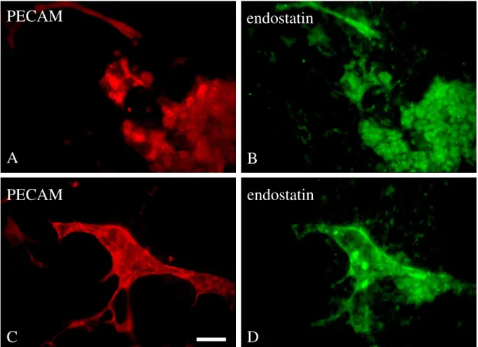
3.3 Embryonic stem cell-derived vessel bind endostatin 48
3.4 Role of endostatin during embryonic vessel growth 49
3.5 Endostatin influences the morphology of endothelial cells and tube formation 57 3.6 Role of endostatin, restin and the heparin mutant form of endostatin during wound
healing 60
3.7 Do restin and heparin mutant endostatin influence the morphology of endothelial cells and
vessels? 63
3.8 Do endostatin, restin and heparin mutant endostatin influence signal transduction? 66
3.9 Is the effect of endostatin NO/cGMP dependent? 73
3.10 Are further pathways concerned? 76
3.11 Is the reduction of sGC a consequence of transcriptional modification? 78
4. Discussion 79 4.1 The heparin-binding site is not necessary for endostatin action 79 4.2 Reason for contrasting results regarding the endostatin-mediated effects 79 4.3 Binding of endostatin, restin and heparin mutant endostatn to tumour and embryonic
vessels 79
4.4 Cell biological aspects during embryonic vessel growth under influence of
Endostatin 81
4.5 Endothelial cell biological aspects during wound healing under influence of
Endostatin 83
4.6 Endostatin, restin and heparin mutant Endostatin influence endothelial morphology 84 4.7 Signaltransduction under influence of Endostatin, restin and heparin mutant
Endostatin 85
5 Conclusion 87
6 Outlook 88
7 Literature 89
8 Explanation (Erklärung) 109
9 Curriculum vitae 110
Abbreviations
AP alkaline Phosphatase
bFGF basic Fibroblast Growth Factor
bNOS brain NO-synthase
BPH benign prostatic hyperplasia
BSA Bovine Serum Albumin
CD31/34 Cluster Determinant 31/34
°C degree Celsius
cm centimetre
cGMP cyclic Guanosin-mono-phosphate
CO2 carbon dioxide
C-PAE Bovine Pulmonary Arterial Cell Line
DAB Diaminobenzidin
DIG Digoxigenin
DMEM Dulbecco`s Modified Eagle Medium
E embryonic day
EB Embryoid Body
EBs Embryoid Bodies
eNOS endothelial NO-synthase
ERK 1/2 Extracellular Signal-regulated Kinase 1/2
ES-cells embryonic stem cells
FCS Foetal Calves Serum
Fig. figure
FGF Fibroblast Growth Factor
Flk-1 Foetal Liver Kinase-1
Flt-1 Fms-like tyrosine kinase-1 receptor
GFP Green Fluorescent Protein
HRP Horseradish Peroxidase
HSPGs Heparansulfat-Proteoglycane
iNOS inducible NO-synthase
kDa Kilodalton
KDR Kinase Insert Domain
MACS Magnetic Associated Cell Sorting
MAPK Microtubule Associated Protein Kinase
µg Microgram
µl Microlitre
µM Micromolar
mA Milliampere
mg Milligram
ml Millilitre
mm Millimetre
mM Millimolar
ms Millisecond
min. Minute
M Molar
MMP Matrix-Metalloprotease
MW or MG Molecular weight
NC-1 Non Collagenous-1
ng Nanogram
nm Nanometre
NO nitrogen monoxide
ODQ 1H-[1,2,4]oxadiazolo[4,3-a]quinoxalin-1-one
PBS Phosphate Buffered Saline, PBS-buffer
PECAM Platelet Endothelial Cell Adhesion Molecule
peNOS phosphorylated/activated eNOS
PFA Paraformaldehyd
PVDF Polyvinylidene Fluoride
RA R158/R184/R270A(heparin mutant endostatin, AS R>A)
rpm Rounds per minute
SDS Sodium Dodecyl Sulphate
Ser1177 Serin1177
SGC soluble Guanylate Cyclase
TBS Tris Buffered Saline, TBS-Puffer
TEMED N,N,N’,N’-tetramethylethylenediamine
U/ml Units per Millilitre
U/µl Units per Microlitre
V Volt
VEGF Vascular Endothelial Growth Factor
VEGFR Vascular Endothelial Growth Factor Receptor
Zusammenfassung
Endostatin und Restin sind proteolytische Spatprodukte von Kollagen XVIII bzw. XV. Sie sind in der Literatur als anti-angiogenetische und aufgrund dessen tumor-inhibierende Proteine beschrieben. Die Verteilung von Endostatin und Restin sowie die Verteilung ihrer Bindungsstellen im Embryoid Body (EB), im Embryo und bei der Wundheilung deutet darauf hin, dass sie eine wichtige Rolle bei der Gefäßentwicklung spielen. Bezogen auf die Gefäßentwicklung differenzierter embryonaler Stammzellen zeigten diese beiden Faktoren sowohl eine angiogenetische als auch eine anti-angiogentische Wirkung, da sie auf für die Gefäßentwicklung und Gefäßneubildung entscheidende Prozesse, wie Proliferation, Migration der Endothelzellen einen steigernden Einfluss nehmen. Andererseits jedoch induzieren sie Apoptose in Endothelzellen. Die Wirkung von Endostatin und Restin auf die Gefäßneubildung kann somit als angio-modulatorisch bezeichnet werden. Endostatin und Restin nehmen jedoch nicht nur Einfluss auf die endotheliale Proliferation, Migration und Apoptose, sondern beeinflussen auch die Morphogenese der Gefäßanlagen in Form einer Kontraktion oder Retraktion, was sowohl mittels vitalmikroskopischer Beobachtung und Dekonvolutionsmikroskopie von embryonalen Gefäßen deutlich wird, als auch bei der elektronenmikroskopischen Betrachtung neugebildeter Gefäße während der Wundheilung ersichtlich ist. Die Beeinflussung der Morphogenese führt zur Frage, welche Signaltransduktionswege einbezogen sind. Insbesondere für die Morphogenese legen Vorbefunde nahe, dass eine die Regulation über den VEGF-Signalwegs oder von Molekülen des VEGF-Signalwegs geschieht. Eine Betrachtung der Endostatin- und Restin-Wirkung auf zellulärer Ebene brachte zum Vorschein, dass diese Faktoren tatsächlich Einfluss auf Signalmoleküle in Endothelzellen nehmen. Und zwar kommt es unter Einfluss von Endostatin und Restin zu einer Dephosphorylierung der aktivierten ERK1/2-Kinase sowie zu einer Runterregulation der löslichen Guanylat Cyclase (sGC) innerhalb eines Zeitraums von 6 Stunden. Diese endostatin-vermittelte Beeinflussung der Signaltransduktion ist über die Protein-Phosphatase 2A (PP2A) induziert, da eine Hemmung der PP2A sowohl eine Dephosphorylierung der aktivierten ERK1/2-Kinase als auch die Runterregulation der löslichen Guanylat Cyclase verhindert. Die Dephosphorylierung der aktivierten ERK1/2- Kinase ist mit Hilfe von zusätzlichem cGMP aufhebbar. Modulatorische Versuche unter Verwendung von cGMP und ODQ legen nahe, dass cGMP unter definierten Bedingungen diese Dephosphorylierung der aktivierten ERK1/2-Kinase stabilisieren kann.
Summary
Endostatin and restin are proteolytic fragments of collagen XVIII and XV. They are described in literature as anti-angiogenic and consequently tumour-inhibitory proteins. The observation of endostatin and restin distribution as well as the distribution of their binding sites in the Embryoid Body (EB), in the embryo and during wound healing, suggests that they have an important role during vessel development. During vessel development of differentiated embryonic stem cells this two factors show both, an angiogenic and an anti-angiogenic effect by increasing endothelial proliferation and migration, processes which play an important role for vessel development and neovascularisation. On the other hand they induce endothelial apoptosis. The effect if endostatin and restin on neovascularisation and vessel development can therefore be described as an angio-modulatory. But endostatin and restin do not only influence endothelial proliferation, migration and apoptosis, they also influence endothelial morphogenesis by inducing a contraction or retraction of vessels. This phenomena was observed by vital-microscopy and deconvolution microscopy of embryonic vessels as well as during wound healing. The influence on morphogenesis leads to the question to which signaling pathways are involved. Especially for the morphogenesis our results imply that these processes are regulated by the VEGF signaling pathway or by molecules of the VEGF- signaling pathway. On cellulary level endostatin and restin show an influence of signaling molecules in endothelial cells. They lead to a dephosphorylation of the activated ERK1/2- kinase as well as to a down-regulation of the solouble Guanylate Cyclase (sGC) in a time course of 6 hours. The modulation of signaltransduction is induced by the protein phosphatase 2A (PP2A) because of the fact that inhibition of PP2A prevents dephosphorylation of activated ERK1/2-kinase and down-regulation of the sGC. The dephosphorylation of the activated ERK1/2-kinase is preventable by additional cGMP. Modulatory experiments by using cGMP and ODQ suggest that cGMP, under definitive conditions is able to stabilise the phosphorylation of the activated ERK1/2-kinase.
1. Introduction
1.1 Vessel Development
1.1.1 Angiogenesis and vasculogenesis
Already in 1508 Leonardo da Vinci speculated that human circulation and vessel system develop in analogy to the botanical example: the tree, the heart with sprouting roots, the liver capillary network, and a trunk with major branches, the aorta and arteries. Later it emerged that the vascular system is determined before the heart starts beating (Risau 1997). Today it is known that the early vascular network is formed from mesoderm by differentiation of angioblasts. Angioblasts form primitive blood vessels. The cell biological mechanism which is responsible for this process is called vasculogenesis. The differentiation of angioblasts from mesoderm and formation of primitive blood vessels by angioblasts are two different steps during early embryonic vasculogenesis (Risau and Flamme 1995). However angioblasts do not only form blood vessels at places of genesis, rather they migrate over long distances, forming a vascular plexus at a distant site (Noden 1991). After a primary vascular network is formed, more endothelial cells are generated, which can form new capillaries by sprouting or by splitting from their vessels of origin in a process termed angiogenesis (Risau 1997). The sprouting angiogenesis occurs mainly in the yolk sac and in the embryo and includes the proteolytic degradation of extra cellular matrix. Afterwards a chemotactic migration and proliferation of endothelial cells, with lumen formation and functional maturation of the endothelium follow (Risau 1997). During non-sprouting angiogenesis vessels arise by splitting pre-existing vessels from transcapillary pillars or posts of extra cellular matrix (Short 1950). In vivo non-sprouting angiogenesis can occur by proliferation of endothelial cells inside a vessel, producing a wide lumen that can be split by transcapillary pillars, or fusion and splitting of capillaries (Pardanaud et al. 1989). The emerging vascular plexus is rapidly remodelled to resemble a mature system with larger and smaller vessels. The responsible process is termed pruning (Risau 1997). The maturation of big vessels includes the differentiation of smooth muscle cells and the formation of the lamina elastica interna.
Formation of a luminal tubus is the key event during endothelial cell maturation and includes differentiation of polarised and functional different cell surfaces (Risau 1995).
1.1.2 Stimulators of angiogenesis
Processes like angiogenesis and vascular remodelling involve endothelial proliferation as well as regression of endothelial cells and they are thereby determined by different growth factors.
For example bFGF (basic fibroblast growth factor) and VEGF (vascular endothelial growth factor) contribute synergistically to the regulation of blood vessel formation (Pepper et al.
1992) and are very important factors regarding neovascularisation and vessel development as well as for endothelial morphogenesis. They are suggested to induce proliferation, migration and differentiation of angioblasts in vitro. BFGF seems to play a major role in the induction of angioblasts whereas VEGF appears to determine the initiation of morphogenesis leading to the initial vascular pattern. Moreover, VEGF has been reported to augment the population of endothelial precursor cells (EPCs) in vivo (Kalka et al. 2000). Kazemi et al. could show that bFGF and VEGF influence survival of angioblasts and that VEGF cause differentiation of angioblasts whereas bFGF does not suggesting a differential role of these factors for vasculogenesis (Kazemi et al. 2002). Capillary endothelial cells of embryonic tissue and organs do not express perceptible amounts of FGF-receptor coding mRNA (Heuer et al. 1990, Wanaka et al. 1991, Peters et al. 1992). Endothelial cells of bigger vessels however express FGF-receptor and react on FGF treatment in vivo (Peters et al. 1992, Lindner et al. 1990, Liaw and Schwartz 1993). Flk-1 (VEGFR-2) is the first endothelial receptor to be expressed in angioblast precursors in the primitive mesoderm of the mouse and is required for angioblast differentiation, followed shortly later by Flt-1 (VEGFR-1), which is required for assembly of blood vessels (Breier et al. 1997).
1.1.3 Inhibitors of angiogenesis
Vasculogenesis and angiogenesis underlay a stringent control of stimulatory and inhibitory factors (Hanahan and Folkman 1996). Many tumours are able to produce angiogenic and anti- angiogenic factors (Folkman 1990, Chen et al. 1995, Gately et al. 1996). In the past numerous angiogenesis inhibitors were identified and characterised. Among them the most important:
Thrombospondin-1 is an adhesive glycoprotein with a size of 160 kDa and was found in the α –granula of thrombocytes. Presumably it is involved in the stabilisation of thrombocyte aggregates (Phillips et al. 1980). In a series of studies it has been shown that thrombospondin- 1 suppress neovascularisation (Good et al. 1990). Also it inhibits angiogenesis and migration of endothelial cells whereas an inhibition of endothelial proliferation could not be shown (Klagsbrun and D’Amore 1991). Thrombospondin-1 is regulated by the tumour suppressor
protein p53 (Good et al. 1990). The complete thrombospondin-1 as well as its specific fragments are able to suppress angiogenesis (Good et al. 1990, Tolsma et al. 1993). On the contrary the potent angiogenesis inhibitors like endostatin and angiostatin are fragments of common extra cellular proteins, which by themselves do not participate in regulation of angiogenesis (Hohenester et al. 1998). Also the angiogenesis inhibitor platelet factor IV was found in the α–granula of thrombocytes and it is released during thrombocytes aggregation. It is a big tetrameric protein with a size of 28 kDa and was initially characterised by its high affinity to heparin (Walz and Hung 1985). Platelet factor IV was characterised as a potent anti-angiogenic factor (Taylor and Folkman 1982). It prevents angiogenesis and endothelial cell proliferation, whereas an inhibition of endothelial migration was not observed (Klagsbrun and D’Amore 1991). Therefore this inhibitor shows a comparable biological action to endostatin. Interferon-γ, a 50 kDa big protein inhibits like endostatin angiogenesis, endothelial migration and proliferation (Klagsbrun and D’Amore 1991). The angiogenesis inhibitor angiostatin is a fragment of plasminogen and has a size of 38 kDa. It suppresses angiogenesis and formation of metastasis (O’Reilly et al. 1994).
1.1.4 Tumour vascularisation
The proliferation of blood vessels is a necessary process for normal tissue growth (Folkman 1971). Vasculogenesis and angiogenesis are subject to a stringent control of stimulatory and inhibitory factors (Hanahan and Folkman 1996). When in normal adult tissue vasculogenesis rests, the balance is lying presumably on the side of inhibitors, however during tumour expansion the balance is disturbed (Chang et al. 1999). In adult organisms angiogenesis appears rarely and it is part of a body internal reparation/regeneration system during healing of wounds or part of the female reproduction cycles (Klagsbrun and D’Amore 1991). Opposite to that an angiogenesis out of balance is often part of a pathological event, like during angiogenesis induced tumour growth (Folkman 1972). Tumour growth is angiogenesis dependent (Folkman 1971). An insufficient tumour vascularisation has the consequence that tumour cells become necrotic and/or apoptotic (Holmgren et al. 1995, Parangi et al. 1996).
Following to a tumour genesis every increase in tumour cell population precedes an increase of vessel growth (Folkman 1990). Experiments regarding tumour growth and angiogenesis show that the growth rate of an implanted, subcutan tumour is low before vascularisation, however high and nearly exponential after vascularisation (Algire 1945). In correlation with an increased angiogenesis in tumours it was observed that many tumours are able to produce
angiogenic factors, which leads to an increased tumour expansion (Relf et al. 1997). Some malign tumours also produce angiogenic inhibitors (Chen et al. 1995, Gately et al. 1996). The increased angiogenesis during tumour growth could lead to an increase of metastases because neovascularisation could permit and support spreading of cells of the primary tumour (Liotta et al. 1974). On the other side a decrease of angiogenesis is associated with decreased metastasis (Starkey et al. 1988). The vascularisation of a tumour and the resulting metastasis are demonstrated in the following:
Fig. 1.1: Vascular components of tumour metastasis (Zetter 1998). The steps of metastasis pathway that involve interactions with blood vessels: (a) small primary tumours (<2 mm) remain avascular until they (b) invade the local epithelia basement membrane. If the tumour cells produce angiogenic factors (c) angiogenesis will occur, allowing expansion of the primary tumour. (d) The new blood vessel provides a route of entry into the bloodstream and the tumour cells circulate until they die or (e) attach specifically to endothelial cells in the vessels (usually venules) of downstream organs. (f) The tumour cells extravasate through the vessel wall and the (g) migrate to sites proximal to arterioles where their growth is enhanced. (h) Micrometastases can remain dormant for extended time periods during which angiogenesis is suppressed. (i) Initiation of angiogenesis at the secondary site releases the metastatic colonies from dormancy and allows rapid growth.
g Migration i Metastasation
f Extravasation
e Adhesion to the vessel
h Micrometastases Primary
Tumour Basment- membran
a b Invasion c Angiogenesis
d
1.1.5 Wound Healing
Wound healing is traditional divided into three phases-the inflammatory phase, the proliferation phase, and the remodelling phase. Injury to adult tissues initiates a series of events including inflammation, new tissue formation, and tissue remodelling (Clark 1996, Martin 1997). The formation of new tissue is initiated by migration and proliferation of keratinocytes at the wound edge within 1 day after injury. New stroma, often called granulation tissue, begins to form after a delay of 2 or 3 days. This tissue consists of inflammatory cells, fibroblasts. Loose connective tissue, and numerous capillaries that endow the neostroma with its granular appearance. The new capillaries are generated by sprouting from pre-existing vessels, in a process called angiogenesis. The later involves migration and proliferation of endothelial cells, lumen formation, and stabilisation of the new vessel by recruitment of associated supporting cells (Risau 1997, Augustin 1998). Wound angiogenesis is essential to support the regenerating tissue with oxygen and nutrition (Clark 1996).
1.2 Signaltransduction
Changes of the vascular system induced by growth factors, are mostly linked to receptor- mediated signalling pathways. An exemplary pathway for such processes, but very important for this work is the VEGF/Flk-1/eNOS/NO/sGC/cGMP/ERK1/2 pathway. Vascular endothelial growth factor (VEGF) is a secreted protein that is a specific growth factor for endothelial cells (Ferrara and Henzel 1989, Keck et al. 1989). It is angiogenic in vivo and in vitro assays (Breier et al. 1992, Goto et al. 1993) and its physiological importance in vasculogenesis is well documented (Fong et al. 1995, Shalaby et al. 1995). The action of VEGF is regulated by two receptor belonging to the tyrosine kinase family Flt-1 and Flk-1 (De Vries et al. 1992, Terman et al. 1992). Flt-1, which has higher affinity for VEGF than Flk-1, is required for endothelial cell morphogenesis, whereas Flk-1 is involved in primarily mitogenesis (Fong et al. 1995, Shalaby et al. 1995, Waltenberger et al. 1994, Keyt et al. 1996).
VEGF effects on permeability (Wu et al. 1996) and vascular tone (Ku et al. 1993) are coupled to nitric oxide (NO) production. Consistent with these findings, in the past it was demonstrated that NO production and cGMP elevation contribute to the angiogenic effect of VEGF (Morbidelli et al. 1996, Ziche et al. 1997). The activation of mitogen-activated protein kinase (MAPK) cascade by VEGF has been recently demonstrated (D’Angelo et al. 1995).
MAPKs are important intermediates in signal transduction pathways and they are stimulated
by a variety of agents (Seger and Krebs 1995). The 44- and 42-kDa MAPK (ERK1/2) isoforms are ubiquitously expressed (Blenis 1993, Davis 1993). Finally, Parenti et al. were able to describe the postreceptor signalling pathway underlying VEGF actions on endothelial cells as Flk-1/eNOS/NO/sGC/cGMP/ERK1/2 cascade activated by VEGF (Parenti et al.
1998). An example for the regulation of eNOS and consequently for the regulation of this pathway is the activation of eNOS by phosphorylation of eNOS at Ser1177. This activation leads to an increased NO-production, whereas the phosphorylation of the eNOS at Ser495 decreases the NO-production (Dimmeler et al. 1999).
1.3 Endostatin
In the year 1997 endostatin was identified first by a Harvard group and was described as an endogen inhibitor of angiogenesis and tumour growth (O’Reilly et al. 1997). It is a proteolytic fragment of the C-terminal end of collagen XVIII (α1(XVIII) collagen) with a size of 20-22 kDa (O’Reilly et al. 1997). The α1(XVIII) collagen is a usual collagen, characterised by 10 domains of tripelhelical repetitions which are separated by non-tripelhelical repetitions (Oh et al. 1994A and B, Rhen and Pihlajaniemi 1994). α1(XVIII) collagen contains a non- collagenous domain (NC-1) which consists of 300 amino acids. The angiogenesis inhibitor endostatin is a component of this NC-1 domain and consists of 184 amino acids (Hohenester et al. 1998). By analysing the crystal structure of the proteolytic fragment of the NC1-domain of collagen XVIII, (Hohenester et al. 1998) a heparin-binding site was detected with the presence of an extensive basic patch formed by 11 arginine residues. Therefore Hohenester et al. suspected that endostatin inhibits angiogenesis by a competitive binding to heparan sulphate proteoglycans (HSPGs) to which normally angiogenesis stimulating factors bind.
Also the heparin binding capability of endostatin was already used for its isolation from hemangioendothelioma-conditioned medium (O’Reilly et al. 1997). However Chang et al.
could show, that binding of endostatin to components of the extracellular matrix, contrary to that of pro-angiogenic factors, is resistant to a treatment with heparinase. Further it has been shown that there is no competition between endostatin and pro-angiogenic factors for binding sites of the extra cellular matrix (Chang et al. 1999). Therefore the anti-angiogenic effect of endostatin could not be a consequence of displacement of pro-angiogenic factors (Chang et al.
1999). The findings regarding heparin-binding capacity of endostatin and heparin necessity were mainly analysed biochemically by solid phase binding (Sasaki et al. 1998/99). This analysis led to the conclusion that the heparin-binding site of endostatin is necessary for its anti-angiogenic activity. On contrary Yamaguchi et al., analysing endothelial migration showed, that heparin binding is not necessary for endostatin’s anti-angiogenic effect. It is still unclear, though, whether the heparin binding site is necessary for the anti-angiogenic activity of endostatin (Sasaki et al. 1998, Sasaki et al. 1999, Karumanchi et al. 2001) or not (Chang et al. 1999, Yamaguchi et al. 1999). Further discussed binding sites for endostatin are glypican and tropomyosin, binding sites with a varying affinity (Karumanchi et al. 2001, MacDonalds et al. 2001).
Recombinant endostatin emerged as a specific inhibitor of endothelial cell proliferation in doses dependent manner. O’Reilly et al. could show that a systemic treatment of mice with 0.3mg/kg/day endostatin leads to a prevention of metastasis and simultaneously to an inhibition of lewis-lung-carcinoma growth (O’Reilly et al. 1997), on the contrary endostatin treatment did not influence resting endothelial cells and did not induce drug resistance (Boehm et al. 1997). On the other hand mice deficient in the endostatin precursor collagen XVIII develop normally with limited evidence of disturbed vascular morphogenesis (O’Reilly et al. 1997, Fukai et al. 2002). Thus, collagen XVIII itself does not appear a relevant regulator of vascular development but releases its activity after proteolytic cleavage. Another effect of endostatin is the induction of apoptosis in endothelial cells (Dhanabal et al. 1999A, Dixelius et al. 2000). In endothelial cells a reduction of the anti-apoptotic protein Bcl-2 and Bcl-xL could be observed while the pro-apoptotic protein Bax remained unchanged (Dhanabal et al.
1999A). Proliferation (Dhanabal et al. 1999B, Yamaguchi et al. 1999) and migration (Dhanabal et al. 1999B and C, Shichiri and Hirata 2001) of growth factor stimulated human umbilical vein endothelial cells (HUVEC), porcine pulmonary artery endothelial cells (PAEC), bovine aortic endothelial cells (BAEC) was also inhibited under influence of endostatin. For endostatin two different binding sites with varying affinity have been described. These binding sites are glypican and tropomyosin (Karumanchi et al. 2001, MacDonalds et al. 2001).
Endostatin influences morphology of endothelial cells and vessels (Dixelius et al. 2000), wound healing (Bloch et al. 2000, Mundhenke et al. 2001), of tumour vessels (Mundhenke et al. 2001, Read et al. 2001) as well as of embryonic stem cell derived endothelial cells and tubes.
Endostatin, which influences signalling molecules in endothelial cells, leads to a down- regulation of MAP-kinase (ERK-1/2) (Knebelmann et al. 1999), influences signalling molecules c-myc and c-fos (Shichiri and Hirata 2001), activates PKA and increases intracellular cAMP (Shichiri and Hirata 2001), induces Shb tyrosine phosphorylation (Dixelius et al. 2000). It increases Ca2+-influx (Jiang et al. 2001) and influences VEGF- receptors as well as phosphorylation of 38 MAKP and 125FAK (Kim et al. 2002). Endostatin has also an effect on the activity of endothelial NO-synthase (eNOS) by influence the
phosphorylation of Ser1177 (Urbich et al. 2002), which probably leads to a reduction of cGMP (Verma et al. 2002). Recently it has been shown that endostatin down-regulated the VEGF-expression in tumour cells (Hajitou et al. 2002).
1.4 Restin
Restin, a proteolytic fragment of collagen XV is described as an anti-angiogenic protein that inhibits tumour growth and endothelial migration (Ramchandran et al. 1999). Ramchendran et al. have shown that restin has 60% homology to endostatin. It decreases tumour volume by 30% and did not bind to heparin. Collagen XV is like collagen XVIII a basement membrane collagen (Sasaki et al. 2002). Both are members of the mulitplexin family (Oh et al. 1994A and B, Sasaki et al. 2002).
1.5 Embryonic Stem Cells (ES-cells)
The embryoid body as in vitro system gives the opportunity to observe comprehensively the endothelial differentiation program and it is therefore an adequate system to analyse the molecular mechanisms, which are involved in vasculogenesis and angiogenesis (Vittet et al.
1996). Embryonic stem cells are non-transformed cell lines, which are extracted directly out of pluripotent foundation tissue of mice embryo, the epiblast (Brook and Gardner 1997, Evans and Kaufman 1981, Martin 1981). It has been shown that they have the potential to generate all embryonic cell lineages when they undergo differentiation (Smith 1992). ES-cells can be maintained in their pluripotential state if cultured in the presence of leukaemia inhibition factor (LIF), which inhibits their differentiation. When LIF is removed, ES cells spontaneously differentiate into cyst-like structure, termed embryoid bodies (EBs), which contain derivatives of the three primitive germ layers (Smith 1992). The appearance of blood island-like structures that consist out of immature hematopoietic cells surrounded by endothelial cells and the formation of vascular-like channels have been reported on the surface of cystic EBs, suggesting that ES-cells produce all factors which are necessary for the induction of vasculogenesis (Doetschman et al. 1985, Risau and Lemmon 1988, Wang et al.
1992, Doetschman et al. 1993, Young et al. 1995). The aggregation of ES-cells induces the formation of several cell types (Bain et al. 1995, Okabe et al. 1996, Fraichard et al. 1995, Strubing et al. 1995). To receive EBs of equal size, which therefore are comparable, a defined
amount of ES-cells were taken to perform EBs by a cultivation technique called „hanging drop“ method (Fasseler et al. 1996) (fig. 2.1). After attachment, EBs begun to grow out centrifugal from the central cell accumulation. The area around the point of attachment shows strongest proliferation but low differentiation. Due to the centrifugal outgrowth in the periphery of EBs only a thin cell layer could be found, however with the strongest differentiation. By differentiation of pluripotent ES-cells inside of EBs several different cells types could be found, like endothelial cells (Wang et al. 1992, Keller 1995), muscle cells (Keller 1995, Drab et al. 1997), neuronal cells (Bain et al. 1995, Okabe et al. 1996, Fraichard et al. 1995, Strubing et al. 1995, Keller 1995), cardiac muscle cells (Wobus et al. 1991, Maltsev et al. 1994, Hescheler et al. 1997), keratinocytes (Bagutti et al. 1996), heamatopoetic cells (Keller 1995) and so on. During the formation of vascular structures in EBs several different endothelial specific marker appear, like PECAM-1 (Platelet Endothelial Cell Adhesion Molecule-1) (DeLisser et al. 1994), VE-(vascular endothelial)cadherin (Lampugnani et al. 1992) and growth factor receptors like Flk-1 (Yamaguchi et al. 1993, Oelrichs et al. 1993 and tie-1/tie-2 (Sato et al. 1993, Schnurch and Risau 1993). The expression pattern of endothelial specific markers, found in EBs, seems to be very similar to that of embryonic angio- and vasculogenesis (Baldwin et al. 1994, Yamaguchi et al. 1993, Sato et al. 1993, Breier et al. 1996, Dumont et al. 1995).
1.6 Target of the Work
The target of this work is to analyse the influence of the extracellular matrix derived factors endostatin and restin on the endothelial cell biology during vascular development. This examination should be performed by a model system of embryonic vessel development, the Embryoid Body (EB) as well as by a model system of regenerative neovascularisation, the physiological wound healing. The following questions should be answered:
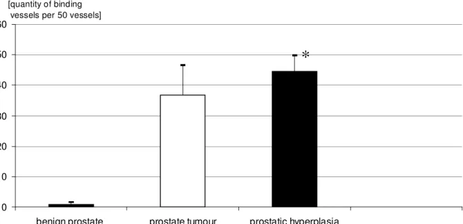
Give the existence and distribution of binding sites for endostatin and restin, evidence for an anti-angiogenic influence in human urological tumours?
Are embryonic vessels accessible for endostatin and restin, do they express and accumulate these molecules and show binding sites for them?
Do endostatin and restin influence the embryonic vessel development and the regenerative neovascularisation?
Is the heparin-binding site decisive for the effects of endostatin and restin?
Is the morphogenesis of vessels influenced by endostatin and restin?
Do this factors influence the endothelial signaltransduction?
2. Material and Methods
2.1 Used chemicals and materials
The following chemicals were obtained from Gibco BRL Gaithersburg:
Dulbecco`s Modified Eagle Medium (DMEM) Foetal Calves Serum (FCS)
L-Glutamin
MEM (non-essential amino acids) Penicillin
Streptomycin
The following chemicals were obtained from Merck, Darmstadt, Germany:
Boron acid (crystal/H3BO3) Di-Sodium-carbonate (Na2CO3)
Di-Sodiumhydrogenphosphate-di-hydrate (Na2HPO4) EDTA (Ethylendinitroilotetraaceticacid/C10H14N2Na2O8)
HEPES (N-[2-Hydroxyethyl]-piperazineN‘-(2-ethansulfonacid/C8H18N2O4S) Potassium-Chloride (KCl)
Magnesium-Chloride (MgCl2) Sodium-Chloride (NaCl)
Sodium-di-hydrogenphosphatemonohydrate (NaH2PO4) Sodium-hydrogen-carbonate (NaHCO3)
Paraformaldehyd ((CH2O)n) Proteinase K
Saccharine (C12H22O11)
Tri-Sodium-Citrate (C6H5Na3O7)
Tris (Trishydroxymethylaminomethan/C4H11NO3) Tween 20
Chloroform (CHCl3)
Acetic acid (C2H4O2) Methanol (CH4O)
The following materials were obtained from Sigma Aldrich, Taufkirchen, Germany:
Acrylamid (C3H5NO) Ammoniumchlorid (NH4Cl) Amoniumpersulfat ((NH4)2S2O8) Bovine Serum Albumin (BSA) DAB (3-3‘Diaminobenzidin)
DMSO (Dimethylsulfoxide/C2H6OS) DTT (Dithiothreitol)
Gelatine
Glycerin (C3H8O3)
N’N‘-Methylenbisacrylamid (C7H10N2O2)
Pipes (Piperazine-N,N‘-bis[2-ethanesulfonicacid]/C8H18N2O6S2) PMSF (Phenylmethylsulfonyl Fluoride/C7H7FO2S)
Salmon Sperm DNA
SDS (Sodium Dodecyl Sulfat/C12H25O4SNa)
TEMED (N,N,N‘,N‘-Tetramethylenediamine/C6H16N2) Trypsin
Yeast tRNA
The following materials were obtained from Serva, Heidelberg, Germany:
Bromphenolblue Mitomycin C
PG 6000 (Polyethylen Glycol) Triton X-100
Glycin (C2H5NO2) Fulka Chemie AG, Buchs, Switzerland Formamid (CH3NO) Fulka Chemie AG, Buchs, Switzerland
Coomassie Brilliant Blue R 250 BioRad Laboratories Munich, Germany
Powdered milk (fatty free): Glücksklee, Nestle, Germany
Paraffin Sherwood Medical (Paraplast Plus), Ireland
Ethanol Firm Hofmann, Düsseldorf, Germany
The following materials were obtained from Falcon (Becton Dickinson), Heidelberg, Germany:
Bacteriological tissue culture dishes (6 cm, 10 cm) Multiwellplates
Centrifugation tubes
The following equipment was obtained from Heraeus Instruments, Hanau, Germany:
CO2-Incubator Lamina
The following materials were obtained from BioRad Laboratories, Munich, Germany:
Gel chamber PowerPac 300
The following materials were obtained from B. Braun Biotech International, Melsungen, Germany:
Micro-Dismembranator U Teflon-container
Wolfram carbide-bead
The following equipment was obtained from Zeiss, Oberkochen, Germany:
Deconvolution-Microscope, Axiovert Deconvolution-Software KS 400 3.0 Fluorescence-Microscope, Axiophot
Glass-Teflon-Homogenisator (Potter), IKA Laborwerke, Hannover, Germany
Micro pipettes/Variopipettes Eppendorf, Hamburg, Germany
All aqueous solutions were prepared with distilled water from our own distiller Milli-RX 20 from Millipore.
Humane kidney, bladder and prostate tissue was obtained from „Urologischen Poliklinik“
University cologne.
2.2 Technical Basics
2.2.1 Cell Culture
For culturing cells were kept in a CO2-incubator surrounded by 5% CO2, 37°C and 95%
humidity. All steps were taking place under sterile conditions, working under laminar air with sterilised materials and media.
2.2.1.1 Stem Cell Culturing - the D3 cell line
Mouse blastocyst-derived embryonic stem cells of the D3 cell line were maintained and cultured as described by Doetscham et al. 1985. The adherent grown cells were passaged all 2- 3 days to prevent a high cell density. A high density would have induced cell differentiation of the pluripotent embryonic stem cells. For passaging cells were washed wit 0.1 M PBS. Then cells were removed from the bottom by using the synthetic enzyme accutase, which has both characteristics of collagenase and trypsin, but in a mild manner. Cell suspension was centrifuged (5 min., 800 rpm, Heraeus Instruments, Labofuge 400, Hanau). After rejecting of supernatant the cell pellet was resuspended in DMEM-medium containing 15% FCS. After counting, 106 cells were plated in a 6 cm dish containing an inactivated feeder layer. The feeder layer provides the possibility to attach but more important the feeder cells produce leukaemia inhibition factor (LIF), which prevents the differentiation of embryonic stem cells.
Stem cells medium was changed every day.
0.1 M PBS: 81mM Di-Sodiumhydrogenphosphate-di-hydrate 19 mM Sodium-di-hydrogenphosphatmonohydrate 150 mM Sodium chloride, pH 7.4
2.2.1.2 Preparation of mouse embryos to gain feeder cells
To gain feeder cells 14-16 days pregnant mice were required. After killing, the uterus was cut out incubated in a petridish containing 0.1 M PBS. Then the embryos were preparated out of uterus and amniotic sac and incubated in fresh PBS. The thorax was opened; organs and the head were removed. The embryos were chopped up with scalpels in a dish with 0.1% trypsin and 0.02% EDTA. After homogenisation the cell suspension were filtered by using a sieve and distributed in petridishes (10 cm) containing DMEM-medium (10% FCS).
2.2.1.3 Culturing of feeder cells
Regularly feeder cells were passaged in the same way as described for the embryonic stem cells. Because of a lower metabolism rate, feeder cell passage and medium exchange was effected once a week. Feeder cells were cultured in DMEM-medium containing 10% FCS.
2.2.1.4 Inactivation of feeder cells
A layer of feeder cells constitutes the basis for embryonic stem cell growth. But to prevent their proliferation they were inactivated using the cell toxin mitomycin C. Adherent cells were treated for 2 hours (37°C) to prevent mitosis activity with a concentration of 10 µg/ml. After inactivation cells were washed three times with PBS and stored in DMEM-medium containing 10% FCS till use.
2.2.1.5 Preparation of hanging drops
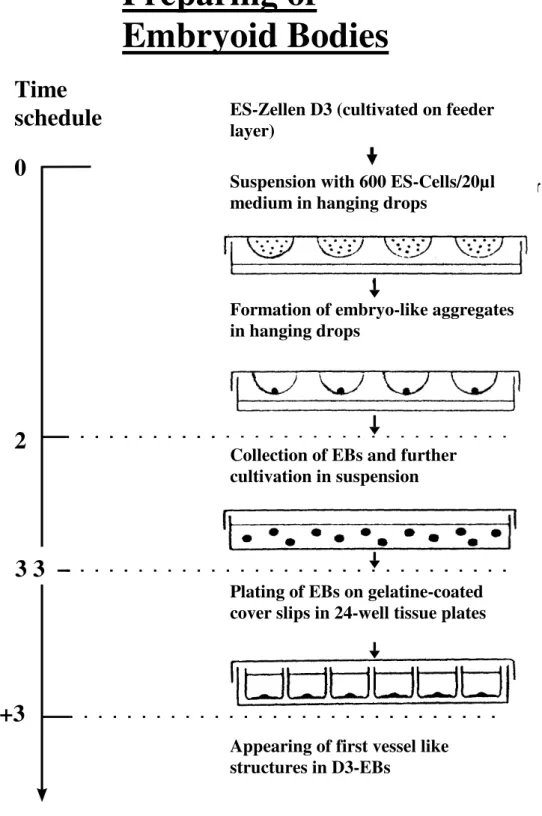
Embryonic stem cells were cultured in hanging drops (fig. 2.1) as described by Fassler et al 1996. Therefore adherent cells were removed and pelleted before counting them. The volume of a hanging drop should grasp 20 µl. Every drop should contain 600 cells. After dilution of the cell suspension in an appropriate manner (DMEM+20% FCS) 50-60 drops were prepared on the inside of the lid. The bottom of this bacteriological petridish was filled with 10 ml PBS to prevent dry out of hanging drops. Then the lid was turned over and placed on the bottom of the dish. Within the following two days the single cells join together to form aggregates.
2.2.1.6 Culturing of aggregates in suspension
Two days after preparing hanging drops, the aggregates (embryoid bodies) were rinsed and cultured in suspension (10 ml DMEM-medium plus 15% FCS) for the next three days. For this purpose bacteriological (non-adhesive) petridishes were used. During these three days cells proliferate but do not differentiate.
Fig. 2.1 Culturing of embryonic stem cells.
Preparing of
Embryoid Bodies
ES-Zellen D3 (cultivated on feeder layer)
3 3 2
5+3 0
Appearing of first vessel like structures in D3-EBs
Time schedule
Suspension with 600 ES-Cells/20µl medium in hanging drops
Formation of embryo-like aggregates in hanging drops
Collection of EBs and further cultivation in suspension
Plating of EBs on gelatine-coated cover slips in 24-well tissue plates
2.2.1.7 Gelatine coating and plating of embryoid bodies
Differentiation of the embryoid bodies (EBs) was induced by plating them on gelatine-coated cover slips. Therefore cover slips (12 mm diameter) were placed in every well of a 24-multi well plate and covered with 0.1% gelatine in 0.1 M PBS. Cover slips were incubated in gelatine solution over night. Before plating, the gelatine solution was removed, cover slips were air-dried and every well was filled with 1 ml medium (DMEM+15% FCS). Each embryoid body was taken out of suspension and placed in the middle of a gelatine-coated cover slip. Medium was changed the first time after three days to let EBs attach.
2.2.1.8 Components of DMEM-medium
DMEM-medium: DMEM (Dulbecco’s Modified Eagle Medium)
50 U/ml Penicillin 50 U/ml Streptomycin 200 µM L-Glutanine
100 µM β–mercaptoethanol
1% MEM (non-essential amino acids)
15% or 20% Foetal Calves Serum (FCS, v/v) Before usage the foetal calves serum was heat inactivated for 30 min. at 56°C.
2.2.2 Tissue Processing
2.2.2.1 Fixation of Tissue
Cells and tissue, received fresh from operation room, were chemically fixed by using 4%
paraformaldehyde (4% PFA) in 0.1 M PBS. During fixation the NH2-groups of cellular and membrane proteins get connected by aldehyde bindings. The duration of fixation procedure depended on tissue size, due to the fact that PFA saturate tissue with a speed of 0.5 mm per hour. The tissue was then washed several times with 0.1 M PBS. For electron microscopy tissue was fixed in 2% paraformaldehyde/2% glutaraldehyde in 0.1 M cacodylate buffer pH 7.2 over night. After washing in cacodylate buffer tissue was treated with 1% uranyl acetate.
4% PFA 4% parformaldehyde, 0.1 M PBS, pH 7.4
0.1 M PBS: 81mM Di-Sodiumhydrogenphosphate-di-hydrate 19 mM Sodium-di-hydrogenphosphatmonohydrate 150 mM Sodium chloride, pH 7.4
0.1 M Cacodylate-buffer 0.1 M Dimethylarsinicacid sodium salt trihydrate
2.2.2.2 Tissue embedding for electron microscopy
For the preparation of thin and ultrathin sections, tissues were fixed as described above. The sections were then incubated in 70% ethanol for 8 hours. Thereafter dehydration was performed in a series of graded ethanol, and specimens were then embedded in araldite. Thin sections of plastic embedded tissue were cut with a glass knife on a Reichert ultramicrotome (Reichert, Bensheim, Germany) and stained with methylene blue. Ultrathin sections (30-60 nm) for electron microscopic observation were processed on the same microtome with a diamond knife and placed on copper grids. The transmission electron microscopy was performed with a Zeiss 902A electron microscope (Zeiss, Oberkochem, Germany).
2.2.2.3 Tissue embedding in paraffin
For paraffin embedding tissue must be dehydrated. Therefore tissue was incubated for 24 hours in 30%, 50%, 70%, 90%, 96% ethanol and two times in 100% isopropanol. As inter medium chloroform was used for 24 hours. Thereafter tissue was incubated two times for 2-3 hours and following over night in paraffin (60°C). At the end tissue was lined up in a block, filled with fresh paraffin before cooled down to solidify. For optimal cutting conditions the paraffin block was cooled.
2.2.2.4 Tissue embedding for cryostat sections
To perform cryostat sections the tissue was fixed and washed as described above. Thereafter tissue was incubated in tissue freezing medium (Jung, Nussloch, Germany) covered by tin foil and store in –80°C. Before cutting tissue was stored for 30 min. at –30°C. The cryostat temperature during cutting was –30°C.
2.2.2.5 Shock freezing of tissue
Tissue for SDS-PAGE and western blotting received freshly from operation room was directly incubated in fluid nitrogen and stored in –80°C.
2.2.3 Immunohistochemistry
During the procedure of immunohistochemistry proteins and enzymes were specifically marked and detected by using corresponding antibodies. Immunohistochemistry with the following course is a standard procedure, however here only the principal will be described.
The specific antibody choice and arrangement will be detailed individually for each experiment.
2.2.3.1 Procedure of immunohistochemistry
The permeabilisation of cells is an important point to facilitate the antibody binding to inner cell proteins. Therefore tissue sections and cells were incubated 10 min. with 0.25% Triton-X- 100 and 0.5 M ammonium chloride in 0.05 M TBS. The detergent Triton-X-100 permeabilises the cell membrane while ammonium chloride reacts with free aldehyde groups to prevent an unspecific binding of the antibody. Following the cells were washed with TBS (3 x 10 min.).
To prevent unspecific bindings due to loading differences, sections and cells were incubated with 5% BSA in 0.05 M TBS. The following incubation with the primary antibody occurred over night at 4°C. After several washings the incubation with the corresponding secondary antibody lasted 1 hour at room temperature. If the secondary antibody was conjugated with biotin instead of the fluorochrom CY2 or CY3, an incubation with streptavidin-conjugated CY2 or CY3 followed. For light microscopic observation a development based on an enzymatic reaction was performed by using DAB-solution. Therefore a streptavidin horseradish-peroxidase-complex was used after incubation with the biotin-conjugated secondary antibody. The development with DAB was controlled under the light microscope.
0.05 M TBS-buffer 50 mM Trishydroxymethylaminoethan (tris) 150 mM Sodium chloride, pH 7.6
DAB-solution: 0.1 M PB, 1% DAB, 0.75% H2O2
0.1 M PB: 81 mM Di-Sodiumhydrogenphosphate-di-hydrate,
19 mM Sodium-di-
hydrogenphosphatemonohydrate, pH 7.4
2.3 Bladder, Kidney and Prostate Tissues
Malignant and benign bladder tissue specimens were obtained from 12 patients. Eight patients underwent transurethral resection and four patients underwent radical cystectomy for bladder cancer (mean age 67.9 years, median 69 years). In final histological staging, there were two patients with stage pTaG1, four with pT1G2, two with pT1G3, three with pT2G2, and one with pT3G3. In this study, prostate samples from 24 patients were tested. Prostate biopsies were collected from 12 patients with prostate cancer (mean age 69.7 years, median 70 years) before they underwent external beam radiation and HDR brachytherapy. In this prostate cancer group there were three pT2G1 cases, eight pT2G2 cases and one pT2G3 case. Normal benign prostate tissue specimens from 12 further patients with benign disease (mean age 69.7 years, median 70 years) served as controls. None of the prostate cancer patients in the first group had received preoperative hormonal therapy, radiation, or chemotherapy. The 21 patients benign prostatic hyperplasia underwent suprapubic prostatectomy.
Kidney tissues (malignant and benign) were obtained from twelve patients (average 63.8 years, median 64 years). All patients underwent nephrectomy for kidney tumours. In final histological staging, there were three patients with stage pT2G2, four with pT1G2, two with pT2G3, and three with pT3G2.
2.4 Animals
Balb/c mice and B2D2F2 mice were obtained from the animal care facility of the Max- Planck-Institute of Biochemistry, or from RCC (Füllinsdorf, Switzerland). They were housed and fed according to federal guidelines, and all procedures were approved by local authorities.
2.5 Wounding and preparation of wound tissue
Wounding and preparation of wound tissue was performed by Sabine Werner (Institute for cell biology, ETH-Zurich, Switzerland). Mice were anaesthetised with a single intreperitoneal injection of ketamine/xylazine. The hair on the animals’ back was cut and the skin was wiped with 70% ethanol. Four full-thickness excisional wounds (6 mm diameter, 3-4 mm apart, two
wounds on each side of the spinal cord) were generated on the back of each animal by excising skin and panniculus carnous as described by Munz et al. 1999. The wounds were allowed to dry to form a scab. Animals were killed at various time points after injury.
Prepared wound tissue was fixed as described above.
2.6 Different kinds of endostatin
For the following experiments three different kinds of endostatin were used. Mouse- endostatin M-ES(4), human endostatin (human ES(2)), restin, a proteolytic fragment of collagen XV as well as R158/R184/R270A (RA) a heparin binding mutant form of endostatin (A kindly gift of Dr. R. Timpl, MPI for biochemistry, Martinsried). This heparin binding mutant form of endostatin was described in detail before (Sasaki et al. 1999). The purification of recombinant mouse endostatin from transfected human kidney cells and a radioimmunoinhibition assay for quantitation have been described before (Sasaki et al. 1998).
The extraction of tissue was performed as recently described (Miosge et al. 1999)
2.7 Endostatin treatment of cells and mice
Embryoid bodies (EBs) were treated with 50 ng/ml and 200 ng/ml endostatin, restin as well as the heparin mutant form of endostatin R158/R184/R270A (RA).
Female Balb/c mice or B2D2F2 mice (12 weeks of age) were injected with 0.3 mg/kg s.c.
murine endostatin, restin and R158/R184/R270A in 100 µl 0.9% saline/0.02M ammonium acetate pH 6.8. Control animals were injected with solvent alone. Injection was performed daily at 11:00 a.m. for 2 days before wounding, immediately after injury and for 5 consecutive days. Mice were killed at day 5 after wounding.
2.8 Binding-Assay
2.8.1 Biotinylation of endostatin
The biotinylation of endostatin was performed as described previously by Friedl et al. 1997.
Briefly, the protein endostatin, restin as well as the heparin mutant form of endostatin (R158/R184/R270A) were diluted in boron buffer (pH 8.8) up to a concentration of 1 mg/ml.
Biotin-X-NHS (MW 454.5, Calbiochem, Schwalbach, Germany) was dissolved in DMSO (1 mg/ml). 12.5% (v/v) of the biotin solutions were incubated with the endostatin solution at 4°C for 4 hours. Subsequently, surplus biotin was removed by filtration against boron buffer. The efficiency of the biotinylation procedure was analysed by detecting endostatin and biotin- conjugated endostatin through western blotting.
Boron buffer: 10 mM Boron acid (crystal 150 mM
Sodium Chloride, pH 8.8
2.8.2 Fluoresceinisothiocyanat-(FITC-)conjugation of endostatin
FITC (Isomer I, on Celite, Sigma, Chemical Company, St. Louis, MO, USA) mixed with silica gel (N-HR, Macherey Nagel Düren, Germany) 1:10 (w/w). The FITC/ silica gel-mixture was mixed with the endostatin solution 1:3 (w/v) and incubated for 25 min. at room temperature. Then the solution was centrifuged to pellet the silica gel (5 min. at 2000 rpm, Heraeus Instruments, Labofuge 400, Hanau, Germany). The supernatant was dialysed against sodium-carbonate/bicarbonate-buffer (pH 9.5) to remove surplus FITC.
Sodium-carbonate/bicarbonate-buffer: 1 M sodium-hydrogen-carbonate 1 M Di-sodium-carbonate, pH 9.5
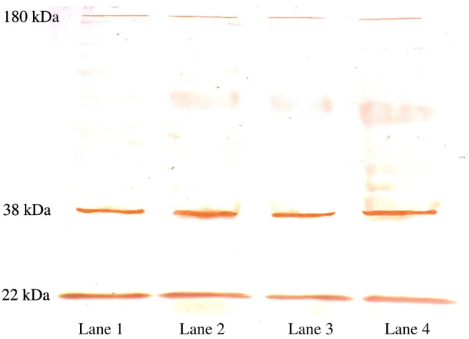
2.8.3 Western -Blot to examine the biotin-conjugation
In order to examine the biotinylation of endostatin a western-blot was performed as described below. The slots were filled either with biotin-conjugated endostatin or non-conjugated endostatin. One half of the blot was treated with a rabbit-anti-endostatin-antibody (polyclonal, 1:2000, a kind gift of Dr. R. Timpl MPI for biochemistry, Martinsried), the other half was treated with streptavidin-biotinylated-horseradish-peroxidase-complex (Amersham, LIFE SCIENCE, Little Chalfont, Buckinghamshire, England, 1:200). Streptavidin binds to biotin.
Afterward the blot was developed with DAB-solution.
DAB-solution: 0.1 M PB, 1% DAB, 0.75% H2O2
0.1 M PB: 81 mM Di-Sodiumhydrogenphosphate-di-hydrate,
19 mM Sodium-di-hydrogenphosphatemono hydrate, pH 7.4
2.8.4 Incubation with biotin-conjugated endostatin
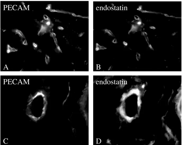
Embryoid bodies and tissue slices were incubated over night at 4°C with 50 ng/ml biotinylated endostatin, biotinylated restin and biotinylated heparin mutant endostatin respectively in 0.05 M TBS. After several washings bounded biotinylated endostatin was detected by using streptavidin conjugated CY3. Following an immunohistochemical detection of endothelial cells and vessels was performed as described below.
2.8.5 Ultrastructural Cytochemistry
Tumour tissues were fixed immediately after surgical removal in a fixative, containing freshly prepared 4% paraformaldehyde, in 0.1 M phosphate buffered saline (PBS, pH 7.4). After 4 hours of immersion fixation, tissue sections of 50-70 µm thickness were prepared on a vibratome (Leica VT 1000) and washed carefully in PBS. The endostatin-biotin binding was conducted for 12 hours at 4°C in 0.05 M tris buffered saline (TBS) followed by five washing cycles in TBS. Finally, the biotin-detection was carried out with Extravidin, coupled to 15 nm colloidal gold (Sigma-Aldrich, Taufkirchen, Germany) in TBS containing 0.8% bovine serum albumin (BSA) overnight at 4°C. After rinsing in TBS, the incubated tissue sections were stabilised with 2% glutaraldehyde in cacodylate buffer and post-fixed with osmiumtetroxide.
After „en bloc„ counter staining with 1% uranylic acetate, the samples were dehydrated with ethanol and embedded flat in Araldite epoxy resin, using propyleneoxide as intermedium.
Thin sections of silver-grey interference colour were cut on an ultramicrotome (Reichert Ultracut UCT) and collected on 150 mesh formvar coated copper grids, which were not further stained in order to intercept the gold signal in the electron microscopy. Also thin sections were collected on 100 mesh formvar coated nickel grids for preparing a pre- immunogold staining. Sections were washed several times in TBS, permeabelized with 5%
HCl, blocked with 1% powdered milk and 0.1% Tween-20 in TBS for 30 min. at room temperature and incubated with Extravidin coupled with 15 nm colloidal gold (1:20, Zymed Laboratories, INC., San Francisco, CA, USA) for 2h at room temperature. Gold staining, to detect bound, biotinylated endostatin was stabilised with 2% glutaraldehyde for 10 min. and counter stained with uranylic acetate for 20 min. The sections were evaluated with a Zeiss EM 902A electron microscope at 80 kV.
2.9 Detection of endothelial cells and vessels
The immunohistochemical procedure was performed as described above. To mark endothelial cells and vessel like structures the antibody rat anti-mouse-PECAM-1 (CD31) was used (1:800, mAb, Pharmingen, San Diego, CA, USA). The relevant secondary antibody was sheep anti-rat Ig biotinylated (1:400, Amersham, LIFE SCIENCE, Little Chalfont, Buckinghamshire, England). This biotinylated antibody was detected with Extravidin conjugated CY 3 (1:1000, Sigma Chemicals, St. Louis, MO, USA; absorption 552 nm, emission 565 nm) or Alexa Fluor™ conjugated 488 (Mo Bi Tec, Göttingen), a fluorochrom that absorbs blue light with a wavelength of 495 nm and emit green light with a wavelength of 519 nm. For evaluation every vessel-like, PECAM-positive structure of at least six EBs from endostatin treated as well as untreated were counted.
2.10 Proliferation-Assay
In order to analyse the influence of endostatin on the proliferation an immunohistochemistry was performed with an antibody against Ki67 (Kosco-Vilbois et al. 1997, Traut et al. 1998, Heidebrecht et al. 1996). Ki67 is active in all cell cycles phases (G1, S, G2 and mitosis). In resting cells (G0-phase) Ki67 lacks. After identifying the endothelial cells as described, proliferating cells were detected with the primary antibody rabbit anti-mouse-Ki67 (1:150, pAb, Dianova, Hamburg, Germany). The relevant secondary antibody was goat anti-rabbit conjugated CY2 (1:200, Dianova, Hamburg, Germany; absorption 495 nm, emission 519 nm).
For utilisation proliferating endothelial cells of vessel-like structures were counted (n=6) with an Axiophot fluorescence microscope (Zeiss, Oberkochen, Germany) by using a fluorescent double filter.
2.11 BrdU cell proliferation
Proliferation of EBs cultured with and without endostatin, restin as well as the heparin mutant form (R158/R184/R270A) were analysed by using a BrdU-Assay (n=4) (for detail see Bloch et al. 1997).
2.12 Apoptosis-Assay
The apoptotic effect of endostatin (O’Reilly et al. 1997, Dhanabal et al. 1999A) was analysed immunohistochemically focusing on caspase-3 and anti-Poly(ADP- Ribose)Polymeras(PARP)p85-fragment (Ellis et al. 1991, Steller 1995, Burek and Oppenheim 1996, Johnson et al. 1996, Cohen et al. 1992, Arends et al. 1990, Trucco et al. 1998, Smulson et al. 1998, Duriez and Shah 1997, Simbulan-Rosenthal et al. 1998, Saldeen and Welsh 1998, Patel et al. 1996, Alnemri et al. 1996, Fujita and Tsuruo 1998). Apoptotic cells were detected with a rabbit anti-active-caspase 3 antibody (1:500, pAb, Pharmingen, San Diego, CA, USA) or a rabbit anti-Poly(ADP-Ribose)Polymeras (PARP) antibody p85 fragment (1:250, pAb, Promega, Madison, WI, USA). The secondary antibody goat anti-rabbit conjugated CY 2 was used and the number of apoptotic endothelial cells were counted under a fluorescence microscope (n=6).
2.13 Migration-Assay
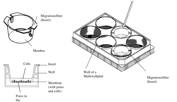
Migration-Assays were performed in a modified Boyden-Chamber by using a 24-well HTS Fluoro Blok TM insert system (fig. 2.2) (Falcon Becton Dickinson GmbH, Heidelberg, Germany). This insert is made out of a polyethylene membrane with 8.0 µm pores and it blocks >99% of the light transmission in a wavelength region from 490 till 700 nm. Inserts were coated with 0.1% gelatine solution (in 0.1 M PBS) for 1 hour. EBs (5+12) were dissociated with Accutase (PAA Laboratories GmbH, Linz, Austria). Afterwards 104 single cells were put on top of an insert and incubated for 8 hours in DMEM with 15% FCS for control or additionally with endostatin, restin as well as the heparin mutant form (R158/R184/R270A) in a concentration of 50 and 200 ng/ml. Then, cells were fixed and immunohistochemically stained with PECAM-1 antibody. Afterwards, the membrane was cut out of the insert and covered in DAPI mounting medium (4´,6-diamidino-2-phenyl-indole, Vectashield, Vector Laboratories, Burlingame, CA, USA; absorption 360 nm, emission 460 nm) between two thin cover slips. The total number of migrated cells was counted as well as the number of migrated endothelial cells (n=6). It was not possible to perform a chemotaxis chamber with different gradient of endostatin as described by Dhanabal et al. 1999B and C
and Yamaguchi et al. 1999 because of its high diffusion rate trough the chamber. Difference in concentration would be balanced over a period of a few minutes.
Fig. 2.2 Migration. Schematic depiction of a modified Boyden chamber
2.14 Signaltransduction
2.14.1Treatments
Embryoid bodies (EBs) were treated with 8bromo-cGMP (5 x 10⁻⁴M, Sigma, Deisenhofen, Germany) for immunohistochemical analysis and for vital microscopy as well as with ODQ (10-6M, Alexis, Grünberg, Germany), NONOate (10-5M, Alexis, Grünberg, Germany), PD98059 (5 x 10⁻⁴M, Calbiochem, Darmstadt, Germany) and with PP2A inhibitor, ocadaic acid (10-10M, Calbiochem, Schwalbach, Germany) for immunohistochemical analysis.
2.14.2 Immunohistochemistry
The primary antibodies anti-active ERK1/2-kinase (1:400, m mAb, Sigma, Sant Louis, USA), anti sGC (1:1000, rb pAb, Alexis Biochemicals, Grünberg, Germany), anti-active AKT-kinase (1:500, rb pAb, upstate, Hamburg, Germany). anti-ERK1/2-kinase (1:400,rb, pAb, upstate, Hamburg, Germany), anti-AKT-kinase (1:500, rb, pAb, upstate, Hamburg, Germany), anti- cGMP (1:600, rb, pAb, Biogenesis, Poole, England), anti-iNOS (1:1000, rb, pAb, Biomol,
Insert Well Membran (with pores and cells) Membra
Migrationsfilter (Insert)
Well of a
Multiwellplatt Migrationsfilter
(Insert)
Pores in the
Cells