of the inhibitory effect of Liprin- α 3 on mDia1 function
Inaugural - Dissertation zur
Erlangung des Doktorgrades
der Mathematisch-Naturwissenschaftlichen Fakultät der Universität zu Köln
vorgelegt von
Julian Brenig aus Köln
Hundt Druck GmbH, Köln
2016
Structural and mechanistic studies of the inhibitory effect of
Liprin- α 3 on mDia1 function
I n a u g u r a l - D i s s e r t a t i o n zur
Erlangung des Doktorgrades
der Mathematisch-Naturwissenschaftlichen Fakultät der Universität zu Köln
vorgelegt von
Julian Brenig
aus Köln
Tag der letzten mündlichen Prüfung: 04.12.2015
Z usammenfassung
Diaphanous-related formins(DRFs) sind Multidomänen-Proteine, die an der Nukleation und dem Zusammenbau von Aktinfilamenten beteiligt sind. Sie werden durch eine autoinhibitorische Interaktion der C-terminalen Domäne (Diaphanous-autoregulatory do- main, mDiaDAD) mit der N-terminalen Domäne (Diaphanous-inhibitory domain, mDiaDID) reguliert. Durch die Bindung von aktivem RhoA wird dieser inhibitorische Zustand aufgelöst und das Formin gleichzeitig zur Plasmamembran rekrutiert. Eine aktuelle Studie indentifizierte Liprin-α3 als einen weiteren Interaktionspartner des Formins mDia1. Es konnte gezeigt werden, dass die Überexpression von Liprin-α3 zu einer Ver- ringerung in der Menge an zellulärem Aktin sowie zu der Aufhebung der Lokalisierung von mDia1 an der Plasmamembran führte. Das Ziel dieser Arbeit war es, die Bindung von Liprin-α3 an mDia1 strukturell und mechanistisch zu charakterisieren, und zu analysieren welchen Einfluss dies auf die Aktivität von mDia1 hat.
Umfassende ITC Studien wurden durchgeführt, um ein minimales Liprin-α3 Fragment zu definieren, das noch immer dazu in der Lage ist an mDia1 zu binden und die Menge an zellulärem Aktin zu verringern. Dieses Fragment wurde dazu verwendet, die Kristallstruktur des mDia1•Liprin-α3 Komplexes zu lösen. Mit Hilfe von ther- modynamischen und kinetischen Analysen konnte gezeigt werden, dass Liprin-α3 die Bindung von RhoA an mDiaN effizienter inhibiert als die Bindung von mDiaDAD an mDiaN. Zusätzlich deuten die strukturellen Daten darauf hin, dass die Dissozi- ation von Liprin-α3 von mDia1 durch RhoA allosterisch erfolgt, während mDiaDAD und Liprin-α3 um eine überlagernde Bindestelle an mDia1 konkurrieren. Überex- pressionsstudien mit Liprin-α3 in HeLa und N2a Zellen zeigten Unterschiede in der Lokalisierung von Liprin-α3 und dem inhibitorischen Potential von Liprin-α3 auf die Funktion von mDia1. Diese Ergebnisse lassen auf Zelltyp abhängige Mechanismen schließen, welche die Funktion von Liprin-α3 regulieren.
Basierend auf den in der Studie gewonnenen Daten werden die folgenden Mechanis- men bezüglich der Inhibierung von mDia1 durch Liprin-α3 postuliert. Erstens, die Wiederherstellung des autoinhibitorischen Zustandes wird unterstützt durch die verän- derten Bindungsaffinitäten von RhoA und mDiaDAD an mDiaN. Zweitens, RhoGAPs sind in der Lage effektiver mit mDiaN um die Bindung von RhoA zu konkurrieren.
Dadurch wird die Hydrolyse von gebundenem GTP in RhoA verstärkt, was zu einer Inaktivierung von RhoA und anschließend von mDia1 führt. Drittens, Liprin-α3 schwächt die Interaktion mit weiteren mDia1 aktivierenden Proteinen und ist in der
durch Liprin-α3 und verdeutlicht die Bedeutung vom Gerüstprotein Liprin-α3 für die Regulierung des Aktinzytoskeletts.
”Men love to wonder, and that is the seed of science“
Ralph Waldo Emerson, Philosopher
Diaphanous-related formins (DRFs) are multi-domain proteins, that are involved in the nucleation and assembly of actin filaments. They are regulated by an autoinhibitory interaction of the C-terminal Diaphanous-autoregulatory domain (mDiaDAD) with the N-terminal Diaphanous-inhibitory domain (mDiaDID). Binding of active Rho proteins to the N-terminal domain leads to the release of the inhibitory state and plasma membrane localization of DRFs. Most recently, Liprin-α3 has been identified as a novel interaction partner of mDia1. It was shown, that overexpression of Liprin-α3 leads to a reduction in the amount of cellular F-actin and the translocation of mDia1 from the plasma membrane. The aim of this thesis was to structurally and mechanistically characterize the binding of mDia1 and Liprin-α3 and to investigate how the presence of Liprin-α3 influences the activity of mDia1.
Comprehensivein vitrostudies were performed to define a minimal Liprin-α3 fragment (Liprin-core region, LCR), that still binds to mDia1 and is able to induce a reduction in the amount of cellular F-actin. Using this fragment the interaction of Liprin-α3 and mDia1 was also characterized structurally. Presence of Liprin-α3 reduced the binding affinity of RhoA to mDiaN more efficiently than the binding of mDiaDAD
to mDiaN, as determined by thermodynamic and kinetic analysis. Furthermore, the structural data revealed, that the dissociation of Liprin-α3 from mDia1 by RhoA is mediated allosterically, while mDiaDAD dissociates Liprin-α3 by competing for the a highly overlapping binding site on mDiaN. Additionally, overexpression of Liprin-α3 in HeLa and N2a cells displayed differences in Liprin-α3 localization and the reduction of the cellular amount of F-actin. This indicated the importance of cell-type dependent mechanisms that regulate the function of Liprin-α3.
Based on the data obtained in this study the following mechanisms explaining the inhibition of mDia1 by Liprin-α3 were postulated. Firstly, the re-establishment of the mDia1 autoinhibitory state is supported by the altered binding affinities of RhoA and mDiaDAD for mDiaN. Secondly, RhoGAPs can compete more efficiently with mDiaN for RhoA binding. This leads to the downregulation of RhoA activity due to enhanced GTP hydrolysis and subsequently the inactivation of mDia1. Thirdly, Liprin-α3 inhibits the interactions of mDia1 with additional activating proteins, besides RhoA, and is able to recruit further regulatory proteins of mDia1 function. In conclusion, this thesis presents a model for the inhibition of mDia1 function by Liprin-α3, emphasizing the impact of the scaffold protein Liprin-α3 on the regulation of the actin cytoskeleton.
T able of content
Abstract . . . i
List of figures . . . v
List of tables . . . vii
1. Introduction . . . 1
1.1. Actin cytoskeleton . . . 1
1.1.1. F-actin polymerization . . . 2
1.1.2. Actin filament regulation and remodeling . . . 4
1.2. Diaphanous-related formins . . . 5
1.2.1. Autoinhibition and activation of DRFs by Rho GTPases . . . 8
1.2.2. Formin induced actin nucleation and elongation . . . 12
1.2.3. mDia1 localization and interacting proteins . . . 16
1.3. Guanine nucleotide binding proteins . . . 17
1.3.1. Small GTPases of the Rho-family . . . 19
1.3.2. The G-domain of the Ras-superfamily . . . 20
1.3.3. Regulatory cycle . . . 21
1.3.4. RhoGAPs . . . 22
1.3.5. Activation by guanine nucleotide exchange factors . . . 23
1.4. Scaffold protein Liprin-α3 . . . 25
1.4.1. Domain organization and interaction partner of Liprin-α . . . . 25
1.4.2. Liprin-αfunctions . . . 26
2. Aim of this thesis . . . 29
3. Materials and Methods . . . 31
3.1. Materials . . . 31
3.1.1. Cell strains . . . 31
3.1.2. Antibiotics . . . 31
3.1.3. Vectors . . . 31
3.1.4. Buffers and solutions . . . 32
3.1.5. Media . . . 33
3.1.6. Crystallization Screens . . . 33
3.1.7. Antibodies and actin filament staining . . . 34
3.1.8. Enzymes . . . 34
3.1.9. Chromatography columns . . . 34
3.1.10. Cloning primer . . . 35
3.2. Molecular Biological Methods . . . 38
3.2.1. Cloning . . . 38
3.2.2. Site-directed mutagenesis . . . 39
3.2.3. Purification of plasmid DNA . . . 40
3.2.4. Denaturing SDS-polyacrylamid gelelectrophoresis . . . 41
3.2.5. Analysis of protein expression by immunoblotting . . . 41
3.2.6. Peptide synthesis . . . 42
3.3. E.colitransformation and cryopreservation . . . 42
3.3.1. Preparation of competent E.colicells . . . 42
3.3.2. Transformation . . . 42
3.3.3. E. colicryopreservation . . . 43
3.4. Protein Purification . . . 43
3.4.1. Expression of recombinant proteins . . . 43
3.4.2. Cell lysis . . . 43
3.4.3. Affinity purification . . . 43
3.4.4. Size exclusion chromatography . . . 44
3.4.5. Determination of protein concentration . . . 45
3.4.6. Nucleotide exchange on RhoA . . . 45
3.5. Biophysical Methods . . . 46
3.5.1. Isothermal Titration Calorimetry . . . 46
3.5.2. Fluorescence polarization assay . . . 47
3.5.3. Stopped-flow fluorescence spectroscopy . . . 48
3.6. Protein structure determination . . . 50
3.6.1. Crystallization . . . 50
3.6.2. Data collection and processing . . . 51
3.6.3. Model building and refinement . . . 53
3.7. Immunocytochemistry . . . 53
3.7.1. Cell cultivation . . . 53
3.7.2. Cell transfection and staining . . . 54
3.7.3. Microscopy and quantification . . . 54
4. Results . . . 55
4.1. Definition of the interaction sites of mDia1 and Liprin-α3 . . . 55
4.1.1. Design and examination of soluble mDia1 and Liprin-α3 fragments . . . 55
4.1.2. Purification of mDia1 and Liprin-α3 peptides . . . 57
4.1.3. Definition of the mDia1-lip binding site by isothermal titration calorimetry . . . 59
4.1.4. Oligomeric states of Liprin-α3 and mDia1 . . . 61
4.2. Crystal structure of the mDia1•Liprin-α3 complex . . . 63
4.2.1. Crystallization attempts of different mDia1•Liprin-α3 complexes . . . 63
4.2.2. Data Collection and refinement . . . 63
4.2.3. Structural analysis . . . 65
4.2.4. Mutational analysis . . . 67
4.2.5. Binding specificity of mDia . . . 68
4.2.6. Liprin-α3 specificity and regulation by posttranslational phosphorylation . . . 70
4.3. Binding Mechanism . . . 72
4.3.1. Effect of Liprin-α3 on the RhoA•mDia1 interaction . . . 73
4.3.2. Effect of Liprin-α3 on the mDiaDAD•mDiaN interaction . . . 79
4.4. Effect of Liprin-α3 on F-actin formation in different cell types . . . 82
4.4.1. Transiently transfected HeLa cells . . . 82
TABLE OF CONTENT iii
4.4.2. Transiently transfected N2a cells . . . 84
4.4.3. Localization of Liprin-α3 fragments in different cell types . . . . 86
5. Discussion . . . 87
5.1. Summary of Results . . . 87
5.2. Binding specificity of the mDia1-Liprin-α3 interaction . . . 88
5.3. Liprin-α3 interferes with mDiaDAD and RhoA binding . . . 90
5.3.1. Interplay of mDiaDAD and Liprin-α3 . . . 90
5.3.2. Possible mechanisms leading to the displacement of Liprin-α3 from mDia1 by RhoA . . . 91
5.4. Regulation of the inhibitory potency of Liprin-α3 . . . 94
5.5. Mechanisms explaining the inhibition of F-actin formation by Liprin-α3 . . . 95
5.6. Physiological relevance of the mDia1-Liprin-α3 interaction . . . 97
5.7. Conclusion and Outlook . . . 99
A. Appendix . . . 101
Literature . . . 109
Abbreviations . . . 127
Acknowledgments . . . 129
1.1 Ribbon representation of a G-actin monomer . . . 2
1.2 Three phases ofin vitro actin polymerization . . . 2
1.3 Treadmilling model after Wegner . . . 3
1.4 Modular domain organization of human formins . . . 7
1.5 Model of the autoregulatory mechanism of mDia1 . . . 8
1.6 Crystal structure of the mDiaDID•mDiaDAD complex . . . 9
1.7 Interaction between mDia1 and RhoC . . . 11
1.8 mDia1 binding interface of DAD and RhoC . . . 12
1.9 Dimeric FH2 structure in complex with actin . . . 13
1.10 ”Stepping second“ mechanism of actin elongation . . . 15
1.11 Impact of RhoA, Rac and Cdc42 on the actin cytoskeleton . . . 20
1.12 Superposition of several GNBP G-domains in their GTP- and GDP-bound state . . . 21
1.13 Regulatory cycle of Rho GTPases . . . 22
1.14 Differences in the GAP-accelerated GTP-hydrolysis of Rho proteins . . . . 23
1.15 Schematic presentation of the “push and pull” mechanism of GEFs . . . 24
3.1 Structure of 2‘/3‘-O-(N-Methylanthraniloyl)-GppNHp . . . 45
3.2 Crystallization phase diagram . . . 50
4.1 Example of the two-step purification protocol using affinity columns and size exclusion chromatography . . . 57
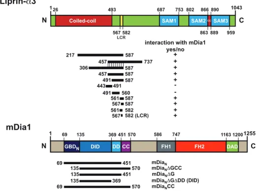
4.2 Overview of the mDia1 and Liprin-α3 domains and the used constructs 58 4.3 SDS-PAGE of the mDia1 and Liprin-α3 fragments used for further studies 58 4.4 Determination of the Liprin-α3-mDia1 binding site by ITC . . . 60
4.5 Coiled-coil predictions and oligomeric states of mDia1 and Liprin-α3 fragments . . . 62
4.6 Protein crystals of Liprin-α3•mDia1 complexes . . . 63
4.7 Crystal structure of the mDiaDID•Lip567–587 complex . . . 65
4.8 ITC measurements with Liprin-α3 fragments visible in the protein structure . . . 66
4.9 Binding interface of mDiaDID and Lip567–587 . . . 67
4.10 Binding specificity of mDia to Liprin-α3 . . . 69
4.11 Regulation of Liprin-α3•mDia1 binding by posttranslational phosphorylation . . . 70
4.12 Superposition of putative Liprin-α3•mDiaDAD/RhoA complex . . . 72
4.13 ITC analysis of the influence of Liprin-α3 on the mDiaN-RhoA binding thermodynamics . . . 74
4.14 Polarization assays of putative ternary mDia1•RhoA•Liprin-α3 complexes . . . 76
4.15 Stopped-flow analysis of the influence of Liprin-α3 on the mDia1•RhoA interaction dynamics . . . 77
vi LIST OF FIGURES 4.16 ITC analysis of the influence of mDiaDAD on the mDiaN- Liprin-α3
binding thermodynamics . . . 79
4.17 ITC experiments to analyze the influence of Liprin-α3 on the mDiaDID•mDiaDAD binding thermodynamics . . . 80
4.18 Size exclusion chromatography and SDS-PAGE analysis of the putative ternary mDiaDID•Lip561–587•mDiaDAD complex . . . 81
4.19 The effect of different Liprin-α3 fragments on the amount of F-actin in RhoA overexpressing HeLa cells . . . 83
4.20 The effect of different Liprin-α3 fragments on the amount of F-actin in RhoA overexpressing N2a cells . . . 84
4.21 Comparison of the transfection efficiency and protein expression levels in HeLa and N2a cells . . . 85
5.1 Secondary structure predictions of the mDia1-binding region of Liprin-α isoforms . . . 89
5.2 Structural comparison of the mDiaN in complex with different interaction partners . . . 92
5.3 Electrostatic surface potential of mDiaDID in a putative ternary complex with RhoC and Liprin-α3 . . . 93
5.4 Model for the inhibition of mDia1 function by Liprin-α3 . . . 97
A.1 Mutational analysis of the mDia1-Liprin-α3 interaction I . . . 101
A.2 Mutational analysis of the mDia1-Liprin-α3 interaction II . . . 102
A.3 Interaction of mDia1 and Liprin-α3 as determined by ITC I . . . 103
A.4 Interaction of mDia1 and Liprin-α3 as determined by ITC II . . . 104
A.5 Interaction of mDia1 and Liprin-α3 as determined by ITC III . . . 105
A.6 Analytical size exclusion chromatography of Liprin-α3 and mDia1 mutants . . . 105
A.7 Ramachandran plots by residue type . . . 106
A.8 ITC experiments to analyze the influence of Lip457–587 on the mDiaN∆GCC•mDiaDAD binding thermodynamics . . . 107 A.9 Primary data of the association-rates determined by stopped-flow kinetics 108
1.1 DRFs and their GTPases . . . 10
1.2 The five major subfamilies of the Ras superfamily . . . 18
1.3 The Rho subfamily of Ras proteins . . . 19
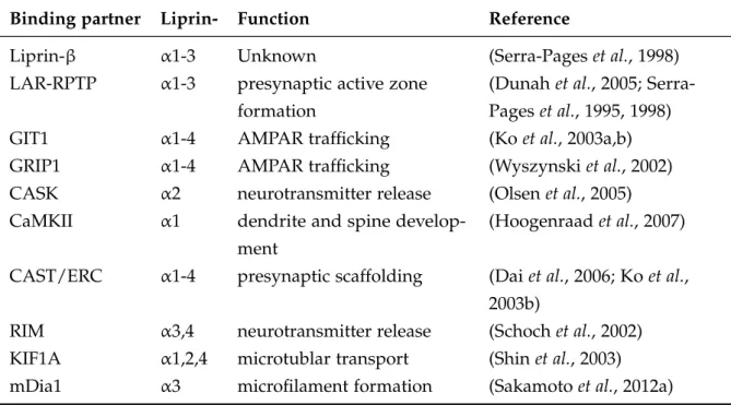
1.4 Overview of Liprin-αbinding proteins . . . 26
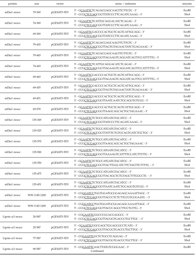
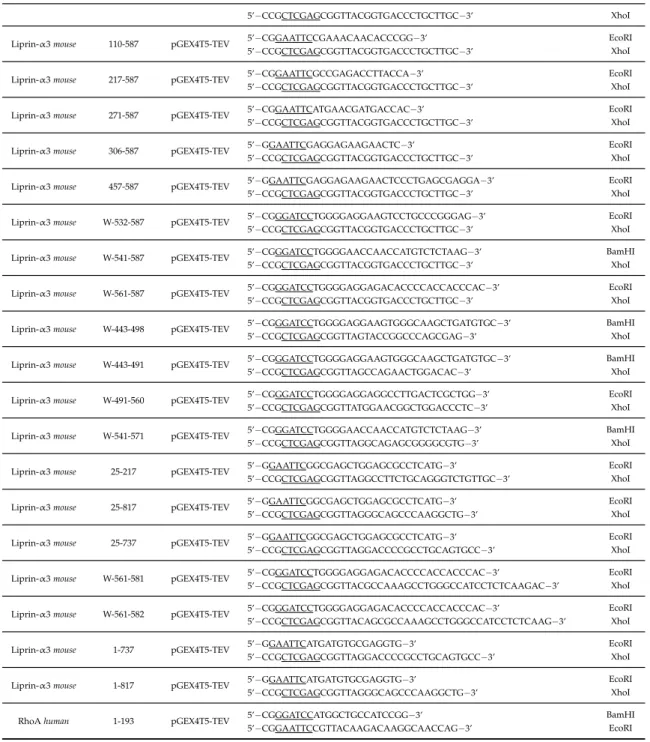
3.1 Primers used for cloning into the pGEX4T5-TEV vector . . . 35
3.2 Primers used for cloning into the pEGFP-N3 and mCherry-C1 vectors . . . 36
3.3 Primers used for site-directed mutagenesis . . . 37
3.4 PCR Settings . . . 38
3.5 QuikChange PCR cycler settings . . . 40
3.6 SDS-PAGE gel composition . . . 41
3.7 mDia1•Liprin-α3 complexes used for crystallization screens . . . 51
4.1 Cloned and purified fragments of mDia1 and Liprin-α3 . . . 56
4.2 ITC measurements for the identification of the mDia1–Liprin binding-site 61 4.3 Data collection, refinement and structure validation of the mDiaDID•Lip567–587 structure . . . 64
4.4 ITC data of the mutational characterization of the mDia1-Liprin-α3 binding interface . . . 68
4.5 Phosphomimetic Liprin-α3 ITC measurements . . . 71
4.6 Rho-Liprin-α3 ITC competition measurements . . . 75
4.7 Effect of different Liprin-α3-fragments on the RhoA-mDia1 interaction dynamics as determined by stopped-flow measurements . . . 78
4.8 mDiaDAD-Liprin-α3 competition ITC measurements . . . 81
A.1 Validation of the Lip567–587 •mDiaN structure . . . 107
1. I ntroduction
1.1. Actin cytoskeleton
The cytoskeleton is essential for many cellular processes such as cell migration, mitosis, cell division, cell polarity and many more (Clarke & Spudich, 1977; Pollard, 1976).
In vertebrates it can be composed of microfilaments, microtubules and intermediate filaments. All three work together to form highly organized networks and are thereby able to respond to and to mediate internal and external signals (Zigmond, 1996).
Microtubules are polymeric structures that consist of α- and β-tubulin dimers. They are important for the establishment of the spindle apparatus, intracellular transport processes and macromolecular assemblies, such as flagella or cilia (Petry et al., 2013;
Vale, 2003). Important for the biological function is their polar organization. At their plus-end only β-subunits are present, whereas the minus-end is composed of α-subunits. The elongation rate is distinct faster at the plus-end, compared to the minus-end (Walker et al., 1988). Intermediate filaments are assembled of two parallel coiled-coil dimers, that align in an anti-parallel orientation. In difference to microtubules and microfilaments this leads to a nonpolar organization, of so-called A11-tetramers (Herrmann et al., 1996). The second notable difference is their distinct specificity for tissues and organisms (Block et al., 2015). Based on their sequence homology, they are classified in five subfamilies. They all have in common to provide the cell with mechanical resilience and stability (Schopfereret al., 2009). Well described members are Keratin, Desmin and Vimentin (Linet al., 2010; Pawelzyket al., 2014).
Microfilaments or filamentous actin (F-actin) are composed of polar, globular actin (G-actin) monomers. G-actin has a molecular weight of approximately 42 kDa and is the most abundant protein in eukaryotic cells (Figure 1.1) (Pollard, 1986). Similar to microtubules the filaments exhibit a structural polarity, accomplished by a unidi- rectional orientation of G-actin molecules. Based on this polarity the filament end where increased elongation occurs, is termed ”plus-end“ and the opposite site where the depolymerizaion happens is called ”minus-end“ (Small et al., 1978; Woodrum et al., 1975). The ”plus-end“ is also termed ”barbed end“ and the ”minus-end“ is also referred to as ”pointed end“. These annotations derive from their appearance in electron microscopy experiments using myosin decoration of microfilaments (Huxley, 1969).
I II
4
3
2
1 flexible
loop ATP
Mg2+
Figure 1.1 Ribbon representation of a G- actin monomer.
Crystal structure of a G-actin monomer of stri- ated muscle tissue of a rabbit in complex with ATP and Mg2+. The monomer is orientated with the minus-end on top and the plus-end at the bottom. The two domains I (subdomain 1 and 2, blue) and domain II (subdomain 3 and 4, purple) are depicted, as well as the bound ATP molecule in the middle of these domains.
The linkerα-helix is shown in green. Modified from Graceffa & Dominguez (2003).
Despite the globular structure of G-actin it is composed of two domains consisting of two subdomains each (Figure 1.1). The nucleotide binding pocket is at the interface of these four domains. Upon binding of ATP, ADP or in the nucleotide free state the monomer cycles between different structural conformations and can adapt open and closed states. Another special feature is the flexible DNase I binding loop in subdomain 2, that it most likely transitioned into anα-helix, upon ATP hydrolysis and the release of theγ-phosphate (Otterbein et al., 2001).
1.1.1 F-actin polymerization
The polymerization of actin filaments is characterized by three different phases (Fig- ure 1.2). Based on the instability of actin dimers and trimers spontaneous filament polymerization is an unlikely event (lag-period or nucleation phase). However, once a stable nucleus of three or more G-actin monomers has been formed the addition of further actin monomers occurs (elongation phase). The third phase (steady-state
Figure 1.2 Three phases ofin vitroactin polymerization.
Modified from Lodishet al.(2000).
1.1. Actin cytoskeleton 3 phase) is defined by the decreasing concentration of free G-actin during the elongation process. At some point the concentration reaches a level at which the simultaneously polymerization equals the dissociation and no net change in filament length takes place. This indicates the importance of actin nucleation as a rate limiting step.
The ratio of the dissociation rate constant (kdiss) and the association rate constant (kass) of ATP-G-actin monomers to the filaments ends describes the so-called critical concentration Cc or equilibrium dissociation constant (KD). At concentrations higher than Cc G-actin polymerizes. Polymerization assays have determined a Cc for the plus-end of 0.12 µM and 0.6 µM for the minus-end (Figure 1.3) (Wegner, 1976; Wegner
& Engel, 1975). This shows that lower concentrations of free actin are needed for the elongation at the plus-end, compared to the minus-end. In consequence, during the steady-state actin elongation happens at the barbed-end while actin dissociation occurs at the pointed-end, resulting in a movement of bound G-actin filaments to the pointed-end. This effect is also calledtreadmillingeffect (Kirschner, 1980; Wegner, 1976).
ATP ATP
KD = 2.0 KD = 2.0
KD = 0.6 KD = 0.12
barbed-end pointed-end (+)
(-)
Figure 1.3 Treadmilling model after Wegner.
Association and dissociation rates at actin filaments. Association rates are depicted as µM-1s-1, dissociation rates have the unit s-1 and the equilibrium dissociation constant is shown in µM.
Modified from Pollard & Borisy (2003).
Additional to the polarity of the filaments, the nucleotide state of the G-actin monomer influences the actin assembly (Figure 1.3). While the incorporation of ATP-G-actin at the barbed-end is highly driven by the increased association rate constant compared to the pointed-end, the equilibrium constant for ADP-G-actin is the same at both filament ends. Following the incorporation, the bound ATP is rapidly and irreversible hy- drolyzed (half time 2 s) (Blanchoin & Pollard, 2002; Carlieret al., 1988). The subsequent phosphate dissociation is distinctly slower (half time 350 s), determining the filament lifetime. (Carlier & Pantaloni, 1986). ADP-Pi-actin intermediates in the filaments have similar properties as ATP-G-actin. Although the ATP-hydrolysis is not needed for the polymerization of actin filaments itself, it is a crucial step for the treadmilling effect, due to the altered kinetics at both ends of the filament. In the steady-state phase the elongation at the barbed-end depends on the amount of free G-actin, and is thereby limited by the dissociation at the pointed-send. Under these conditions the
filament growth is limited to approximately 0.04 µm min-1. However, cell movement of 10 µm min-1 has been shown, indicating the requirement of additional regulatory mechanisms (Pollard & Borisy, 2003). One example is the conformational change of the G-actin monomers upon binding of cations, which increases the polymerization (Maruyma & Tsukagoshi, 1984; Seldenet al., 1983). Furthermore, more than 60 protein classes exist, that modulate the F-actin formation (Vale & Kreis, 1999). Elongation can be inhibited by F-actin capping proteins, that bind to the barbed end (Cooper &
Schafer, 2000), or by binding of Thymosin β4 to ATP-G-actin preventing binding to the filaments (Carlier et al., 1993). Another actin binding protein named Profilin binds to ADP-actin monomers and enhances the exchange of ADP to ATP (Lu & Pollard, 2001). Secondly, Profilin bound ATP-G-actin is recruited to prolin rich regions of pro- teins, such as WASP family members and formins. This local increase of ATP-G-actin enhances the nucleus formation (Watanabe et al., 1997). The protein class of formin homology proteins, especially mDia1 is introduced in greater detail in 1.2. Furthermore a range of microfilament regulatory processes involving Arp2/3-complexes and their activating proteins are introduced in 1.1.2.
1.1.2 Actin filament regulation and remodeling
As mentioned before the nucleation is a crucial step in the actin filament formation.
Hitherto, three protein classes have been identified to aid in nucleation and promote polymerization of new actin filaments. These proteins belong to the actin-related protein 2/3 (Arp2/3) complex, formins and a protein class containing tandem repeats of G-actin binding motifs, such as Spire, Cordon-bleu or Leiomodin (Ahuja et al., 2007;
Goley & Welch, 2006). The Arp2/3 complex consists of seven subunits, including the Arp2 and Arp3 subfamilies (Machesky & Insall, 1998), and possesses a low nucleation activity on its own (Mullins et al., 1998). So-called nucleation promotion factors (NPFs) are of importance for the activity of Arp2/3. These NPFs can be divided into two classes.
Class I NPFs mediate the actin nucleation via their C-terminally located Verprolin homology (V), central (C) and acidic (A) regions (Macheskyet al., 1999). The V-domain (also called W or WH2 for WASp homology 2) binds to actin monomers and the C and A motifs interact with multiple subunits of the Arp2/3 complex. Thereby, NPFs recruit actin monomers to the Arp2/3 complex. The Arp2 and Arp3 subunits form a trimeric structure with the actin monomer, inducing a different conformation of Arp2/3. This leads to more potent state to promote the actin nucleation step for new filaments (Chereauet al., 2005; Marchandet al., 2001).
1.2. Diaphanous-related formins 5 Class I NPFs can be further classified into five groups
1. Wiskott-Aldrich Syndrome protein (WASP) and neuronal-enriched homologue of WASP (N-WASP)
2. WASP family Verprolin-homologous (WAVE) proteins (Suppressor of cAMP receptor [Scar])
3. WASP and Scar homologue (WASH)
4. WASP homologue associated with actin, membranes and microtubules (WHAMM)
5. Junction-mediating regulatory protein (JMY)
Class II NPFs contain proteins, such as Cortactin another activating protein of the Arp2/3 complex. However, Cortactin displays a weaker effect on the nucleation, compared to class I NPFs. Instead, it mainly aids in the stabilization of branch junctions and inhibits debranching (Weaveret al., 2001).
In contrast to the class of Diaphanous-related formins (see 1.2) the activated Arp2/3 complex initiates 70◦ branching of existing actin filaments. In this process the new daughter filament is anchored by Arp2/3 with its pointed end to the mother filament (Rouiller et al., 2008). As a result, Arp2/3 mediates the formation of a so-called dendritic network of branched actin filaments (Blanchoinet al., 2000), and is thereby regulating the cell morphology. The branching of actin filaments has been shown to be crucial for many cellular processes, including cell migration and adhesion (DeMali et al., 2002; Macheskyet al., 1997), phagocytosis (Mayet al., 2000) and trafficking events (Stamnes, 2002).
1.2. Diaphanous-related formins
Formin homology (FH) proteins, or short formins, are multidomain proteins consisting of more than 1000 amino acids. The first formin genes (FMN1/2) were identified by mutations of the limb deformity gene locus in mouse, that induced severe defects cell polarity and morphogenesis (Maas et al., 1990). Although, more recent studies ascribed the phenotype to gremlin, a gene located on the same chromosomal locus (Zuniga et al., 2004). Subsequent studies showed, that mutations of the Drosophila genedialead to impaired cytokinesis, with a gene product (Diaphanous) homologous to formin (Castrillon & Wasserman, 1994). Additionally, studies in budding yeast (Saccharomyces cerevisiae) displayed the importance of the formin Bni1 for the assembly of microfilaments (Evangelistaet al., 2002). A comparison of the FMN1/2 proteins of
mice, Diaphanous of flies and Bni1 of yeast showed a high sequence homology of two regions termed formin-homology FH1 and FH2. These domains are present, albeit in different sizes, in all known formins with the exception of ForC in Dictyostelium discoideum, which consists only of the FH2 domain (Castrillon & Wasserman, 1994).
The conservation of these domains indicates their functional importance in formins.
Indeed, both domains are involved in the regulation and formation of microfilaments and microtubules during a variety of cellular processes (Palazzoet al., 2001; Wallar &
Alberts, 2003).
Hitherto, in mammals 15 different formins are described, that can be classified into eight groups: (Breitsprecher & Goode, 2013; Schönichen & Geyer, 2010)
1. Dia (Diaphanous homolog formin)
2. DAAM (dishevelled-associated activator of morphogenesis) 3. FMNL (formin-like protein)
4. WHIF (WH2 domain-containing formin) 5. INF (inverted formin)
6. FHOD (formin homology domain-containing) 7. Delphilin
8. FMN (formin)
Besides the conserved FH1 and FH2 domains a third domain FH3 was identified at the N-terminus of Fus1 in Schizosaccharomyces pombe(Petersenet al., 1998). The FH3 has been further divided into the diaphanous-inhibitory domain (DID) and the subsequent dimerization-domain (DD), and is a special feature of a formin class that is regulated by autoinhibition. These Diaphanous-related formins (DRFs) are autoregulated by an intramolecular interaction between the C-terminal Diaphanous-autoregulatory domain (DAD) and the N-terminal DID domain (Alberts, 2001; Li & Higgs, 2003; Nezamiet al., 2006; Otomo et al., 2010). Another characteristic domain of DRFs is the N-terminally located GTPase-binding domain (GBD), which is located C-terminally of the DID (Rose et al., 2005). Upon binding of active Rho GTPases to the GBD the DID-DAD interaction and thereby autoinhibition is relieved (Lammerset al., 2005; Rose et al., 2005; Sethet al., 2006; Westendorf, 2001).
1.2. Diaphanous-related formins 7 An overview of the domain organization of one formin of each class is shown in Figure 1.4.
Figure 1.4 Modular domain organization of human formins.
Overview of the domain organization of one example for each formin class. Show- ing the GTPase binding domain (GBD), Diaphanous-inhibitory domain (DID), dimeriza- tion domain, coiled-coil domain (CC), formin homology domains 1,2,3 (FH1, FH2, FH3), Diaphanous- autoregulatory domain (DAD), Wiskott-Aldrich syndrome homology region 2 (WH2), microtubule-binding domain (MTBD) and PDZ homology domain. Modified from Schönichen & Geyer (2010).
The DRFs Dia1, Daam1, FMNL1 and FHOD1 display a typical GTPase binding domain and a Diaphanous-inhibitory domain (DID) at their N-terminus, followed by the FH1 and FH2 domains and the C-terminal Diaphanous-autoregulatory domain (DAD). It has been shown, that GBD/DAD deletion mutants of DRFs are constitutively active and induce the formation of actin stress fibers, in presence of ROCK (Rho associated coiled-coil kinase) (Copeland & Treisman, 2002; Watanabeet al., 1999). Delphilin is the only formin containing PDZ domains, that are required for binding of the glutamate receptor delta 2 subunit (GluRδ2) (Miyagi et al., 2002), thus shown to be important
for the localization at dendritic spines of Purkinje cells (Matsuda et al., 2006). INF1 is able to interact directly with microtubules via a C-terminal microtubule-binding domain (MTBD), which leads to co-alignment of microtubules with actin filaments (Young et al., 2008). Instead of DAD the formin WHIF1 (INF2) has an C-terminally located WH2 (WASP homology 2) domain. Similar to the DAD the WH2 domain binds to the DID, but additionally binds to actin monomers, accelerating F-actin nucleation (Chhabra & Higgs, 2006). Interestingly, the binding of WH2 to the DID solely impacts actin polymerization and not nucleation processes (Chhabra et al., 2009).
Since formins are able to interact with small GTPases of the Rho-family and simultane- ously with proteins regulating the actin cytoskeleton (e.g. F-BAR proteins, Profilin) they display important effector proteins that mediate signal transduction. The reg- ulation of DRFs and their impact on F-actin nucleation and polymerization will be discussed in the subsequent sections.
1.2.1 Autoinhibition and activation of DRFs by Rho GTPases
Although the autoinhibitory interaction of the Diaphanous-inhibitory domain (DID) and the Diaphanous-autoregulatory domain (DAD) is a common feature of all Diaphanous-related formins (DRFs) (Alberts, 2001; Liu et al., 2008; Vaillant et al., 2008), so far only the structure of the mDia1 inhibition has been solved by X-ray crys- tallography (Lammerset al., 2005; Nezamiet al., 2006; Otomoet al., 2010). A simplified model of the autoinhibitory state of mDia1 is depicted in Figure 1.5. The DAD binds to the DID adjacent to the GTPase binding domain (GBDN), leading to the formation of a closed state and the inactivation of the formin.
N
C
open conformation
GBDN DID DD FH1 FH2 DAD
autoinhibited conformation
N
Figure 1.5 Model of the au- toregulatory mechanism of mDia1.
Presented are the open and closed (autoinhibited) confor- mation of mDia1. Shown are the GTPase-binding domain (GBDN), the Diaphanous- inhibitory domain (DID), dimerization domain (DD), Formin-homology domain (FH1,2) and the Diaphanous- autoinhibitory domain (DAD).
1.2. Diaphanous-related formins 9
dimerization domain (DD)
Diaphanous-inhibitory domain (DID) Diaphanous-autoregulatory
domain (DAD)
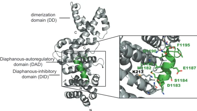
Figure 1.6 Crystal structure of the mDiaDID•mDiaDADcomplex.
Left:The ribbon presentation shows the mDiaDIDdomain in complex with the mDiaDAD core region exhibiting the amino acids 1180-1195 (PBD: 2BAP). Right: Zoom in on the binding interface, depicting some of the major amino acids involved in the binding. Modified from Lammerset al.(2005).
The DAD of mDia1 can be divided into the DAD core region (DCR; aa 1175-1195) and a subsequent basic region, which varies in length and sequence (Lammerset al., 2005;
Wallar et al., 2006). In the first solved crystal structure of the DID•DAD complex, only the DCR is visible forming an amphipathic helix that binds to the hydrophobic concave side of the armadillo repeat region (ARR) of the DID (Figure 1.6) (Lammers et al., 2005). Although the subsequent amino acids of the DAD are not visible in the structure they are important for the binding affinity (Wallar et al., 2006). It has been postulated that the C-terminal part of the DAD contacts the negatively charged patches on mDiaN along theα-helix (interdomain helix, α17), connecting the DID domain with the dimerization domain (DD) (Nezamiet al., 2010; Otomoet al., 2010). This binding mechanism seems to be similar in all DRFs sharing the following consensus sequence for DAD (G/A)(V/A)MDXLLEXL(K/R/Q)X(G/A)(S/G/A)(A/P) (Alberts, 2001).
Binding of active Rho GTPases to the regulatory N-terminus (mDiaN, GBDN-DID-CC) leads to the release of the DID-DAD interaction and resolves the autoinhibited state.
The formin p140mDia was the first to be identified as effector protein of small GTPases (Watanabeet al., 1997). In recent years the binding specificity and the interaction has been studied intensively with biochemical, structural and cell dependent methods.
Especially, the activation of mDia1 isoforms and their interaction with various GTPases
has been characterized in great detail (Lammerset al., 2008). Described interactions of formins with specific Rho GTPases are summarized in Table 1.1.
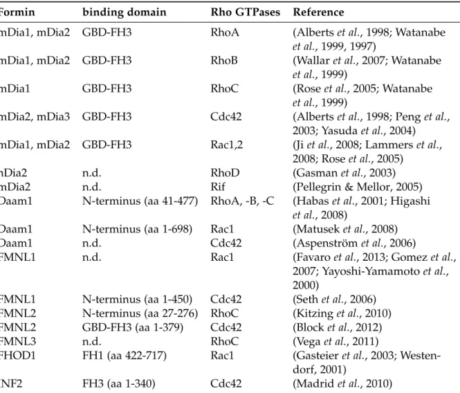
Table 1.1 DRFs and their GTPases. Modified from Kühn & Geyer (2014).
Formin binding domain Rho GTPases Reference
mDia1, mDia2 GBD-FH3 RhoA (Albertset al., 1998; Watanabe et al., 1999, 1997)
mDia1, mDia2 GBD-FH3 RhoB (Wallaret al., 2007; Watanabe et al., 1999)
mDia1 GBD-FH3 RhoC (Roseet al., 2005; Watanabe
et al., 1999)
mDia2, mDia3 GBD-FH3 Cdc42 (Albertset al., 1998; Penget al., 2003; Yasudaet al., 2004) mDia1, mDia2 GBD-FH3 Rac1,2 (Jiet al., 2008; Lammerset al.,
2008; Roseet al., 2005)
hDia2 n.d. RhoD (Gasmanet al., 2003)
mDia2 n.d. Rif (Pellegrin & Mellor, 2005)
Daam1 N-terminus (aa 41-477) RhoA, -B, -C (Habas et al., 2001; Higashi et al., 2008)
Daam1 N-terminus (aa 1-698) Rac1 (Matuseket al., 2008)
Daam1 n.d. Cdc42 (Aspenström et al., 2006)
FMNL1 n.d. Rac1 (Favaroet al., 2013; Gomezet al.,
2007; Yayoshi-Yamamotoet al., 2000)
FMNL1 N-terminus (aa 1-450) Cdc42 (Sethet al., 2006) FMNL2 N-terminus (aa 27-276) RhoC (Kitzinget al., 2010) FMNL2 GBD-FH3 (aa 1-379) Cdc42 (Blocket al., 2012)
FMNL3 n.d. RhoC (Vegaet al., 2011)
FHOD1 FH1 (aa 422-717) Rac1 (Gasteieret al., 2003; Westen- dorf, 2001)
INF2 FH3 (aa 1-340) Cdc42 (Madridet al., 2010)
The first crystal structure of a GTPase in complex with a formin was solved by the group of Wittinghofer (Figure 1.7) (Rose et al., 2005). In accordance with other effector proteins, these data revealed that active, GppNHp loaded RhoC binds mainly via switch I and II. While switch I binds interacts with mDia1 via the GBDN, switch II contacts residues in the ARR of the DID. Moreover, the Rho-insert helix also locates in close approximation to the fifth armadillo repeat of the DID. Notably, the residues located in the switch I and II regions involved in formin binding, are conserved throughout the Rho GTPases (Kühn & Geyer, 2014). A major contribution to the specificity of the mDia1-GTPase interaction seems to be created by the triple asparagine motif in the loop connecting the third helix of ARM1 and the first helix of ARM2 in mDiaDID. Mutating the motif to the residues TSH (Thr, Ser, His) of mDia2 and mDia3, increases
1.2. Diaphanous-related formins 11 the affinity of mDia1 for Cdc42. Further specificity of the DRF-GTPase interaction is created by the Rho-insert helix (Lammerset al., 2008). The specific interactions of the switch I and II region of RhoC with the ARR and GBDN are shown in Figure 1.7.
switch II
switch I GppNHp insert helix
GBDN dimerization domain
(DD)
interdomain helix (ID)
Diaphanous-inhibitory domain (DID)
RhoC
A B
Figure 1.7 Interaction between mDia1 and RhoC. A: Ribbon presentation of the mDia1•RhoC•GppNHp complex of PDB: 1Z2C. B: Interactions created between Rho and the armadillo repeat region (ARR, DID) and the GTPase binding domain (GBDN) of mDia.
Modified from Roseet al.(2005).
As depicted in Figure 1.8 the binding interfaces of mDiaDAD and RhoC on mDiaN
only partially overlap. However, the formation of a putative ternary complex of mDiaDID•mDiaDAD•Rho has been experimentally ruled out in several studies (Lam- merset al., 2008, 2005; Nezamiet al., 2006; Otomoet al., 2005a). A two step mechanism for the dissociation of mDiaDAD from mDiaDID by RhoA has been suggested, that includes an initial weak binding of RhoA to mDia1, followed by a tighter association.
This would subsequently lead to the dissociation of mDiaDAD from mDiaDID by steric interference and additional charge-charge repulsion (Lammers et al., 2005).
Whether the initial loose binding is initiated by the Rho-insert helix and is thereby also important the specificity, or if the RhoA first binds loosely to the GBDN and then forms stronger contacts to the mDiaDID remains unclear.
A B
DID DID
GBDN GBDN
N217 N217
DAD binding interface RhoC binding interface
K213
K107
Figure 1.8 mDia1 binding interface of DAD and RhoC .
A: mDiaDAD(mDia Diaphanous-autoinhibitory domain) binding interface on mDia1. Shown are the residues of the mDiaDID(mDia Diaphanous-inhibitory domain) involved in the binding of mDiaDAD (PDB: 2F31) (Nezamiet al., 2006). Depicted in light blue are the residues N217, N310 and Q352 that mediate hydrogen bonds with mDiaDAD. K213 of mDiaDID, which forms a salt bridge with D1183 of mDiaDADis shown in blue. Hydrophobic interactions of I222, K252, L253, A256, I259, L260, Q307, A311, T314, V351, and V355 are colored yellow. B: RhoC binding interface on mDia1. Shown are the residues of the GBDN-DID involved in the binding of RhoC (PDB: 1Z2C). Hydrophobic interactions of M90, M94, N95, L96, P103, L104, and M115 are depicted in yellow, polar interactions of K100 and Q118 (GBD) and N164, N165, N166 and N217 (mDiaDID) in light blue. The created salt bridge of K107 is marked in blue color. Modified from Kühn & Geyer (2014).
1.2.2 Formin induced actin nucleation and elongation
Mandatory for the activity of Diaphanous-related formins (DRFs) are the release of the autoinhibitory state by Rho GTPase binding and the distinct features of the dimeric formin structure. DRFs accelerate F-actin formation by enhancing the nucleation process and by supporting the elongation at the barbed end. These effects are mainly driven by their FH1 and FH2 domain.
Actin nucleation
It has been shown, that the isolated FH2 domain is able to nucleate actin filaments in vitro (Pring et al., 2003; Pruyneet al., 2002). Based on structural data it was postulated that the nucleation is accelerated by the recruitment and stabilization of actin dimers and trimers (Figure 1.9). The crystal structure of the FH2 domain of the formin Bni1p of S. cerevisiaedisplays the completeα-helical composition. The monomers are arranged head-to-tail in the dimer, forming a donut-shaped ring (Xu et al., 2004). Important for the dimerization of the FH2 is the N-terminally located region, also referred
1.2. Diaphanous-related formins 13 to as lasso, which contacts the post domain of the other monomer (Xu et al., 2004) (Figure 1.9). Furthermore, this region displays minor differences between the structure of Bni1p and mDia1. Additionally, Otomo et al.could show by crystallization with tetramethylrhodamine-actin (TMR-actin), that the dimeric FH2 domain contacts three actin molecules, while each FH2 monomer exhibits two actin binding sites. (Otomo et al., 2005b). Crucial for the actin binding are a conserved isoleucine (in Bni1p I1431) and lysine (in Bni1p K1601) (Luet al., 2007; Otomoet al., 2005b) and mutations of these residues results in reduced activity of the formin (Bartolini et al., 2008; Ramabhadran et al., 2012).
A B
post
coiled- coil
knob linker
lasso
knob post
Figure 1.9 Dimeric FH2 structure in complex with actin. A: Crystal structure of the FH2 domain of theS. cerevisiaeformin Bni1p showing the residues 1350-1760 (PDB: 1UX5, Xuet al.
(2004)). The FH2 monomers are presented in green and purple. Labeled are the lasso, linker, knob, coiled-coil, and post regions of the green FH2 subunit. B: Ribbon representation of the FH2 domain of Bni1p (residues 1350-1760) in complex with muscle actin (PDB: 1Y64, Otomo et al.(2005b). The three actin subunits of the polymer are depicted as surface representation in blue and gray, labeled 1 to 3 from the barbed- to the pointed-end. Modified from Paul &
Pollard (2009b).
The knob region of FH2 domains contact the actin molecules in the hydrophobic groove between the subdomains 1 and 3. Additionally, the post site of the FH2 forms electrostatic contacts along subdomain 1 of actin subunits (Figure 1.9 B). However, the reported binding affinities of the FH2 domain for single G-actin monomers are rather low (>5 µM) (Evangelista et al., 2003; Zigmond, 2004) or not present at all (Chesarone et al., 2010). On the other hand the affinity for the barbed end is very high (low nanomolar KD, Moseleyet al.(2004)). Together with the slowin vitronucleation
rate of the FH2 domain it is therefore hypothesized that the FH2 is needed for the stabilization of spontaneously formed actin dimers or trimersin vitroand additional regions of formins are involved in actin nucleation in vivo. An alternative nucleation mechanism has been postulated involving the FH1 domain. The FH1 domain, which is predicted to be mostly unstructured contains several proline-rich motifs (Kovaret al., 2003; Michelot et al., 2005). These motifs are known to be recognition areas for Profilin and SH3 (Src-homology 3) and WW domains (WWP repeating motif) (Bedford et al., 1997; Imamuraet al., 1997; Macias et al., 2002). Especially the binding and recruitment of Profilin bound ATP-G-actin at the FH1 was believed to initiate nucleation (Sagot et al., 2002). More recent studies indicate that the interaction of the FH1 domain and Profilin•G-actin might only play minor role in the nucleation (Paul & Pollard, 2008).
Latest data reveal the emerging importance of the formins tail regions. Sequences located C-terminally of the FH2 domain bind actin monomers and thereby effectively enhance nucleation (Gould et al., 2011; Heimsath & Higgs, 2012). In addition, an increasing number of regulatory proteins, that bind to the tail regions have been identified (Grazianoet al., 2011; Okada et al., 2010). These proteins include so-called nucleation promoting factors (NPFs) ( 1.2.3).
Actin elongation
Formins fulfill several functions, that lead to a regulated elongation at the barbed end of actin filaments. They prevent the binding of capping proteins, which would block further elongation (Harris et al., 2004; Kovar et al., 2005; Zigmond et al., 2003), they prevent the annealing of barbed filament ends to pointed ends (Kovaret al., 2003) and finally they recruit actin monomers to the growing barbed end. It has been emphasized, that the FH1 domain is needed for Profilin•G-actin recruitment to the FH2 domain, resulting in accelerated elongation at the barbed-end (Paul & Pollard, 2009a). Early electron micrographs and additional kinetic assays indicated that formins stay bound to the barbed end during the processive elongation (Pruyne et al., 2002; Zigmondet al., 2003). This would require an intensively debated translocation of the FH2 domain for each actin monomer that is added to the barbed end. The initial theory explaining this mechanism is known as ”stair stepping“ model. (Otomo et al., 2005b; Xu et al., 2004). These models were based on structural information of the FH2 domain and indicated that formins act as leaky capping proteins. Due to conformational changes of the FH2 dimer, formins can switch between an ”open state“ and a ”closed state“.
During the closed state both parts of the dimer bind tightly to the actin subunits at the barbed end, which are orientated in a planar structure with a 180◦ rotation between consecutive subunits. This unfavorable conformation presents no contacts for further actin subunits. Partial dissociation of the FH2 (step off) would lead to a more
1.2. Diaphanous-related formins 15 relaxed rotation of 167◦ of actin subunits and enable one free G-actin in the solution to bind to the exposed FH2, as well as the barbed end. Assembly of the actin monomer subsequently leads to the re-establishment of the closed state (Kozlov & Bershadsky, 2004; Paul & Pollard, 2009a; Vavyloniset al., 2006; Zigmondet al., 2003). The time span between the alternating open and close states is defined as the ”gating factor“ (Paul &
Pollard, 2009b). This value varies from almost 1 (uninhibited) for mDia1 to nearly 0 (capping) for Cdc12 in S. pombe (Kovaret al., 2006) and could explain differences in the elongation rates of different formins (Blocket al., 2012; Kovaret al., 2003; Neidtet al., 2008).
1 2 3
4 5
Figure 1.10 ”Stepping second“ mechanism of actin elongation. This model shows the hy- pothesized FH2 induced 5 step elongation of actin at the barbed end in regard of the FH2 dimer translocation. In green depicted is the leading half of the FH2 dimer and the trailing part is shown in magenta. K, P refer to the knob and post site of the FH2 domain. Actin monomers are blue and silver and interactions with the knob or post site are indicated as (+) or (-). The 5 steps are illustrated as the side view of the filament with the barbed end orientated downwards (upper image) and the top view of the barbed end (lower image). The closed and open state of the FH2 dimer is indicated by the green and red angle symbols. Images 1,2 and 4,5 are equivalent images of the cycle. Modified from Paul & Pollard (2009b).
However, the initial ”stair stepping“ model did not take the helical twist of F-actin into account. Induced by the helical structure the FH2 needs to rotate relatively to the filament in an angle of 14◦per elongation cycle. Therefore, the stair stepping model was expanded by the ”screw mode“ (Shemeshet al., 2005) and also the ”stepping second”
hypothesis has been emphasized (Paul & Pollard, 2009b). The latter is displayed in Figure 1.10.
The ”stepping second“ model is based on the new structure data of incorporated actin subunits provided by Oda et al.(2009). Upon incorporation into the microfilament the actin monomers become flattened. This lead to the conclusion that the bound FH2 dimer might influence the orientation as well as the conformation of the actin subunits andvice versa(Paul & Pollard, 2009b). Paul and Pollard propose, that following each open state and actin subunit insertion, the formin subunit is transiently bound to the interior actin in the microfilament. Subsequently, the tense FH2 domain translocates to the newly build end and shifts into the closed state, lowering its free energy (Paul &
Pollard, 2008). Similar to the ”stair stepping“ model the ”stepping second“ mechanism includes several steps of partially dissociation of the FH2 domain. In comparison to the ”stair stepping“ model, during the ”stepping second“ model the dissociation and translocation occurs after the incorporation of actin monomers. Notably, recent studies using single-molecule fluorescence polarization could confirm the rotational movement of mDia1 during the elongation of actin filaments (Mizunoet al., 2011).
1.2.3 mDia1 localization and interacting proteins
Besides the activation, the localization of DRFs is also influenced by Rho GTPases.
This has been shown for many different formins and GTPases (Evangelistaet al., 1997;
Martin et al., 2007; Tolliday et al., 2002). The prenylated GTPases bind to the GBD domain and recruit the activated DRFs to the membrane. Notably, recent studies identified several new mechanisms mediating the plasma membrane localization of formins, that are independent of Rho GTPases. Another protein, that binds to the mDiaN region upon the release of the autoinhibitory state is the scaffolding protein IQGAP1. The interaction with IQGAP1 is mandatory for the proper localization of mDia1 in phagocytic cup formation and phagocytosis (Brandtet al., 2007). Furthermore, scaffolding proteins containing membrane associated BAR domains could have an impact on formin localization (Frost et al., 2009). These interactions have been found for mDia1, which binds with the FH1 domain to the SH3 domain of IRSp53 (BAR- domain protein) (Fujiwara et al., 2000; Gohet al., 2012) and Daam1, which interacts with SH3 domain of Cip4 (Aspenströmet al., 2006). Other sequences, important for the localization of formins, have been postulated for Bnr1 inS. cerevisiae and Cdc12 inS.
1.3. Guanine nucleotide binding proteins 17 pombe (Gaoet al., 2010). These regions are also located at the N-terminus of the formin and interact with septin-associated kinases, regulating the function of Bnr1 (Buttery et al., 2012).
Alongside the phosphorylation by septin-associated kinases a variety of post transla- tional modification (PTM) has been described for formins. Phosphorylation by Prk1p of both tails is involved in the release of the autoinhibitory state of Bni1 inS. cerevisiae (Wang et al., 2009). The formins mDia2 (Diaphanous homolog 3) and FHOD1 are activated upon interaction with the Rho-associated protein kinase (ROCK) (Deanet al., 2011; Hannemann et al., 2008; Takeya et al., 2008). Additionally, the formin mDia3 is phosphorylated and regulated by the kinase Aurora B (Cheng et al., 2011) and the formins FHOD1 and FHOD3 are targets of CK2 (casein kinase 2 subunitα) and PRKG1 (cGMP-dependent protein kinase 1) (Iskratschet al., 2010, 2013). The described farnesylation and myristoylation of INF2 (inverted formin-2) and FMNL2 (formin-like protein 2), have a more direct impact on the membrane localization (Blocket al., 2012;
Chhabraet al., 2009)
Notably, in mDia1 and mDia2 polybasic clusters at the N- and C-terminus have been identified, that might directly associate with phospholipids through electrostatic in- teractions (Gorelik et al., 2011; Ramalingamet al., 2010). The direct interaction with phospholipids has also been confirmed for the plant formins AFH1, formin1 and class II formins (Cheunget al., 2010; Martiniereet al., 2011; van Gisbergenet al., 2012), indicating a general mechanism of several species.
Another protein, that binds to the N-terminal region of mDia1 and is involved in F-actin regulation is is the scaffolding protein Liprin-α3 (Sakamotoet al., 2012a). This new class of mDia1 interaction proteins will be further introduced in 1.4.
1.3. Guanine nucleotide binding proteins
The signal transduction of extracellular stimuli to the intracellular compartments is an important step for many cellular functions. These processes are often controlled and mediated by guanine nucleotide binding proteins (GNBP), also named G-Proteins.
Based on their structural and sequential similarities they can be divided into TRAFAC (translation factor) and SIMIBI (signal recognition GTPases, MinD and BioD) (Leipe et al., 2002). Well known examples of the TRAFAC GNBPs are the translation factor IF-2, the Ras-superfamily (e.g.Rho, Ras, CDC42) and the myosin-kinesin superfamily.
Proteins associated with the SIMBI-GNBPs are often involved in protein localization and targeting. Besides their structural categorization GNBPs can also be classified regarding their function, which results in five distinct groups.
1. α-subunits of heterodimeric G-proteins (Gαs) 2. Translation factors (e. g. IF-2, elongation factor Tu) 3. Ras superfamily (e. g. Rho, Ras, Cdc42)
4. Signal recognition particle (SRP) and SRP-receptor 5. Large GNBPs (e. g. Dynamin)
Hitherto, in humans more than 150 members of the Ras superfamily are known.
Most of them are structurally and functionally described in great detail. They have a molecular weight of approx. 20-25 kDa and can be further divided into five major subfamilies based on structural and functional similarities (Table 1.2). The first member and eponym of the superfamily, Ras (rat sarcoma) , was identified by Chienet al. (1979) as the product of a proto-oncogene.
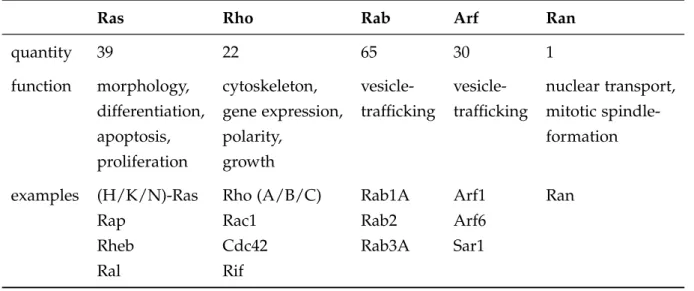
Table 1.2 The five major subfamilies of the Ras superfamily. Classification based on Wenner- berg et al.(2005) and Rojaset al.(2012).
Ras Rho Rab Arf Ran
quantity 39 22 65 30 1
function morphology, cytoskeleton, vesicle- vesicle- nuclear transport, differentiation, gene expression, trafficking trafficking mitotic spindle-
apoptosis, polarity, formation
proliferation growth
examples (H/K/N)-Ras Rho (A/B/C) Rab1A Arf1 Ran
Rap Rac1 Rab2 Arf6
Rheb Cdc42 Rab3A Sar1
Ral Rif
Proteins of the Ras-subfamily mediate cell-proliferation, -apoptosis, -morphology and -differentiation (Vojtek & Der, 1998). Concerning their diverse functions it is not surprising, that mutations of Ras can be found in diverse tumors and in fact 20-30 % of all human tumors carry a Ras-activating mutation (Prioret al., 2012). Rho-proteins (Ras- homologous) play important roles in the regulation of the cytoskeleton, cell growth and also gene expression (Kitayamaet al., 1989; Sander & Collard, 1999). Regulation of vesicular transport is controlled by ADP-ribosylation factor (Arf) family proteins and Ras-like proteins in brain (Rab) (Moss & Vaughan, 1998; Schimmöller et al., 1998). The ran-related nuclear (Ran) proteins are involved in the nucleocytoplasmic transport of proteins and RNA (Weis, 2003).
1.3. Guanine nucleotide binding proteins 19 Their common feature is the ability to cycle between an active GTP-bound state and an inactive GDP-bound state (1.3.3), which is determined by conformational changes of the G-domain (1.3.2). Only in the GTP-bound form effector proteins can bind, which leads to their activation (e.g.DRFs) and signal transduction events occur. The state of GPTases is highly regulated by the hydrolysis (1.3.4) and the exchange (1.3.5) of the guanine nucleotide.
1.3.1 Small GTPases of the Rho-family
Based on their sequence homology and functional similarities proteins of the Rho- family can be subdivided into six major groups (Table 1.3) (Wennerberg & Der, 2004). In mammals the first described Rho proteins of the Rho-family were the isoforms RhoA, B and C (Madaule & Axel, 1985). Today, the most noted and well characterized members are RhoA (ras homologous A), Rac1 (ras-related C3 botulinum toxin substrate) and Cdc42 (cell division cycle 42) (Hall, 1998).
Table 1.3 The Rho subfamily of Ras proteins. Classification based on Wennerberg & Der (2004).
Rho Rac Cdc42 Rnd Miro RhoBTB
RhoA Rac1 Cdc42 Rnd1 Miro1 RhoBTB1 RhoB Rac1b TCL Rnd2 Miro2 RhoBTB2
RhoC Rac2 Wrch1 Rnd3 RhoBTB3
Rac3 Chp RhoG TC10
The small GTPases of the Rho-family are involved in a wide range of cellular processes, such as cell-cell adhesion, cell polarity, cell migration and gene transcription. Many of these processes are mediated by the activation of effector proteins, involved in the regulation of the actin cytoskeleton (Chimini & Chavrier, 2000; Kaibuchiet al., 1999;
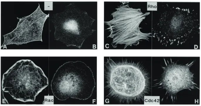
Van Aelst & DSouza-Schorey, 1997). RhoA, Cdc42 and Rac1 display distinct effects on the actin cytoskeleton if activated in cells (Figure 1.11).
Figure 1.11 Impact of RhoA, Rac and Cdc42 on the actin cytoskeleton.
Quiescent, serum-starved Swiss 3T3 fibroblasts cells with activated RhoA by lysophosphatidic acid addition (C,D), microinjection of constitutively active Rac (E,F) and microinjection of the Cdc42 exchange factor FGD1 (G,H). Staining of actin in (A), (C), (E) and (G) vinculin staining in (B), (D), (F) and (H). Modified from Hall (1998).
Activation of RhoA leads to an increase in the amount of F-actin (Figure 1.11 C) and to the formation of focal adhesions (Figure 1.11 D). The effector protein mDia1, involved in acceleration of actin nucleation and elongation, is activated upon RhoA binding (Hill et al., 1995). In comparison, Cdc42 mediates filopodia formation (Figure 1.11 G) and Rac induces the formation of lamellipodia, by the activation of the Arp2/3 complex through binding to NPFs (e.g. WASP, WAVE)(Figure 1.11 E) (Bishop & Hall, 2000;
Ridley et al., 1992). Moreover, the small GTPases are regulated by a cross-talk between the Rho proteins. It has been shown that Cdc42 can also activate Rac, while Rac in turn is able to activate Rho (Nobes & Hall, 1995; Ridleyet al., 1992)
1.3.2 The G-domain of the Ras-superfamily
The G-domain is the central structural motif of Ras-superfamily proteins and de- termines nucleotide and effector protein binding (Figure 1.12 A). It consists of six β-sheets and five adjacentα-helices, which are linked by ten loop regions. A distinct feature of Rho proteins is the insert helix (α3), that has been shown to be important for the activation of ROCK (Rho-associated protein kinase) (Zong et al., 2001). Five of the loop regions (G1-G5) hold the key elements for specific nucleotide and effector binding (Bourne et al., 1991; Johnet al., 1990; Schmidtet al., 1996; Via et al., 2000). The
1.3. Guanine nucleotide binding proteins 21
A B
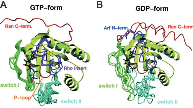
Figure 1.12 Superposition of several GNBP G-domains in their GTP- and GDP-bound state.
The ribbon presentation displays the difference of the switch I and II region, dependent on the guanine nucleotide bound state. A: In the GTP-bound active state the switch I and II adopt a rigid conformation. B: In the Inactive GDP-bound state the switches are more flexible.
Additional, structural features of Ran (red), Arf (blue) and Rho (purple) are highlighted.
Modified from Vetter & Wittinghofer (2001).
P-loop (G1) with the consensus sequence GxxxxGK(S/T) mediates the high affinity binding of the nucleotide by interactions with theβ- andγ-phosphates (Saraste et al., 1990). Interaction with effector and regulatory proteins depend on the switch I and switch II regions (G2, G3). They bind to the γ-phosphate of GTP and Mg2+. Upon hydrolysis the binding is disrupted and conformational changes occur explaining the molecular switch mechanism (Figure 1.12 B) (Spoerneret al., 2001; Wittinghofer & Pal, 1991). Guanine-binding specificity is created by the G4 and G5 loop motifs due to the formation of specific hydrogen bonds with the guanine base (Zhong et al., 1995).
1.3.3 Regulatory cycle
Small GNBPs are binary molecular switches with nucleotide dependent conformations.
They are able to cycle between a GTP- and GDP-bound form (Figure 1.13). In the GTP- bound active state the conformation of the switch I and switch II regions are stabilized by the interaction with theγ-phosphate. The intrinsic hydrolysis rate and nucleotide dissociation of small GTPases are extremely low. Without additional proteins that support and regulate these processes, GTPases would not be a molecular switch of biological significance.
Figure 1.13 Regulatory cycle of Rho GTPases.
Small GNBPs are regulated by three distinct protein classes. The guanine nucleotide exchange is mediated by GEFs (guanine nucleotide exchange factors), the hydrolysis of GTP to GDP is accelerated by GAPs (GTPase activating proteins) and GDIs (guanine nucleotide dissociation inhibitors) extract the inactive prenylated GTPases from the membrane. Modified from Etienne- Manneville & Hall (2002).
Active GTP-bound Rho GTPases are inhibited by RhoGAPs (1.3.4). These proteins accelerate the hydrolysis rate of GTP to GDP. The activation of Rho GTPases is mediated by so-called GEFs (guanine-nucleotide exchange factors), that accelerate the nucleotide dissociation (1.3.5). In addition the localization of GTPases is regulated by GDIs (guanine nucleotide dissociation inhibitors), which are able to extract prenylated Rho proteins from lipid bilayers (Figure 1.13).
1.3.4 RhoGAPs
RhoGAPs play a crucial role to switch off signaling transduction pathways. Without RhoGAPs the slow intrinsic GTP-hydrolysis rate of GNBPs would lead to an ongoing downstream signaling, which would result in drastic consequences for cell functions.
Since the discovery of the first RhoGAP more then 25 years ago (Trahey & McCormick, 1987), over 70 members of yeast to human RhoGAPs have been identified today and some of them are structurally and functionally characterized (Bernards, 2003; Pecket al., 2002). The RhoGAPs of specific GTPases are conserved, however among the different Rho subfamilies they display different mechanisms to accelerate to the GTP-hydrolysis (Scheffzeket al., 1997; Seewald et al., 2002). Three of these mechanisms are depicted in Figure 1.14.
1.3. Guanine nucleotide binding proteins 23
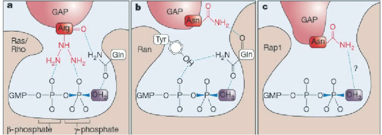
Figure 1.14 Differences in the GAP-accelerated GTP-hydrolysis of Rho proteins.
Essential for the effectiveness of the different GAPs is the positioning of a H2O molecule for the nucleophilic attack on theγ-phosphate. a: The catalytic glutamine of Ras/Rho is positioned by the arginine of RasGAP (Scheffzeket al., 1997). b: RanGAPs use an asparagine to stabilize the glutamine (Seewaldet al., 2002). c: Rap1 specific GAPs use an asparagine to position the water molecule directly (Daumkeet al., 2004). Illustration from Rehmann & Bos (2004).
The successful crystallizations of several small GTPases in complex with GAPs made it possible to gain further insights into the catalytic mechanisms. Ras- and RhoGAPs insert a positively charged arginine (”arginine finger“) into the active site. One effect is the neutralization of the negative charge of the phosphates. As a second important function the arginine positions the glutamine (position 61 in Ras and 63 in RhoA) in trans. This glutamine on the other hand, orients the water molecule that attacks the γ-phosphate (Figure 1.14 a). RhoA exhibits an intrinsic hydrolysis rate of 0.022 min-1, which is increased to nearly 100 min-1 by p190RhoGap (Zhang & Zheng, 1998). Despite their structural differences, a similar mechanism seems to be the basis for all RhoGAP catalyzed hydrolysis reactions in GTPases. In RanGAPs the arginine is replaced by a asparagine, that stabilizes the glutamine of Ran, while the catalytic effect remains basically the same (Figure 1.14 b). Furthermore, the asparagine releases the inhibitory effect of the tyrosine 39 in Ran, that contacts theβ-phosphate and orients the catalytic glutamine incis (Bruckeret al., 2010). A distinct mechanisms is used for Rap1, which does not possess a catalytic glutamine. In Rap proteins the H2O molecule is positioned directly by the asparagine of the GAP (”asparagine thumb“), as postulated by Daumke et al. (2004) and Scrimaet al. (2008).
1.3.5 Activation by guanine nucleotide exchange factors
The intrinsically slow dissociation of bound nucleotides is the rate determining step in the transition of GNBPs into the active GTP-bound conformation. Hence, the
dissociation rate is increased by guanine nucleotide exchange factors (GEFs). Although, the different GEFs display structural differences the general reaction mechanism is functionally similar. Induced by GEFs the guanine nucleotide dissociation rate is accelerated by several orders of magnitude (Hutchinson & Eccleston, 2000; Klebe et al., 1995). The concentration of GTP in the cell is approximately tenfold increased compared to GDP. Thus, upon dissociation of GDP, the GTPase is preferentially binding GTP instead of GDP. The GEFs p190RhoGEF and PDZ-RhoGEF increase the nucleotide exchange from 4.8 x 10⁻⁴min-1 to 1930 x 10-4min-1 (van Horcket al., 2001), and from 5.5 x 10⁻⁴min-1 to 1179 x 10-4min-1, respectively (Gasmi-Seabrooket al., 2010).
Figure 1.15 Schematic presentation of the “push and pull” mechanism of GEFs.
GEFs reduce the binding affinity of nucleotides to GNBPs. The inser- tion of several side chains into the active site impair the positioning of the P-loop and Mg2+. Furthermore the switch I region is pulled out and the switch II region is pushed in the catalytic site. Modified from Vetter &
Wittinghofer (2001).
During the course of the so-called “push and pull” mechanism the switch I region is pulled out and the switch II region is pushed in the nucleotide binding site (Vetteret al., 1999). The insertion of additional amino acids into the nucleotide binding site by GEFs alters the conformation of the P-loop and displaces the Mg2+-Ion, which is needed for the high nucleotide binding affinity (Gasper et al., 2008; Lenzenet al., 1998). This ultimately lowers the nucleotide affinity. Upon GEF-binding the P-loop lysine (K16 in Ras; K18 in Rho) is not able to interact with the negative charges of the phosphates but instead is orientated to the switch II (Figure 1.15). Notably, GEFs have no influence on the binding selectivity of GTPases regarding GTP or GDP. The insertion of guanine nucleotides is only driven by their intracellular concentration.