Analysis of Signaling Mechanisms Essential to Mature B Cell Viability
D i s s e r t a t i o n
zur Erlangung des akademischen Grades d o c t o r r e r u m n a t u r a l i u m
(Dr. rer. nat.) im Fach Biologie eingereicht an der
Mathematisch-Naturwissenschaftlichen Fakultät I der Humboldt-Universität zu Berlin
von
Diplom-Biologin Alina Patke geboren am 29. März 1976 in Berlin
Präsident der Humboldt-Universität zu Berlin Prof. Dr. Christoph Markschies
Dekan der Mathematisch-Naturwissenschaftlichen Fakultät I Prof. Dr. Christian Limberg
Gutachter: 1. Prof. Dr. Peter-Michael Kloetzel Gutachter: 2. Prof. Dr. Wolfgang Lockau Gutachter: 3. Prof. Dr. Alexander Tarakhovsky
Tag der mündlichen Prüfung: 17.9.2007
Abstract
The maintenance of mature peripheral B cells depends on at least two survival cues, tonic signaling from the B cell receptor (BCR) complex and the extracellular cytokine B cell activating factor of the TNF family (BAFF). In addition to enhancing viability, BAFF controls the functional efficiency of the peripheral B cell pool by regulating complex physiological processes including cell growth, metabolism, energy homeostasis and entry into the cell cycle.
BAFF-mediated induction of two molecular mechanisms, namely activation of the Akt signal transduction pathway and upregulation of the oncogenic kinase Pim-2 results in the modification of effector proteins including transcription factors and regulators of protein synthesis which are capable of executing the observed cellular physiological changes. The classic protein kinase C β is instrumental in BAFF-induced Akt-activation and PKCβ-deficient B cells and mice show signs of partial refractiveness to BAFF. The protein tyrosine kinase Syk plays a role in early B cell development and is activated in mature B cells by immunogenic BCR-stimulation. Inducible ablation of Syk in mice results in the loss mature B cells from the peripheral lymphoid organs and reveals an indispensable function for Syk in tonic BCR survival signaling.
Kurze Zusammenfassung
Die Langlebigkeit reifer periphärer B Zellen ist abhängig von mindestens zwei Überlebenssignalen, einem tonischen Signal, welches vom B Zellrezeptor ausgeht und dem Zytokin B Zell aktivierender Faktor der TNF-Familie (BAFF).
BAFF fördert nicht nur das Überleben von reifen B Zellen, sondern kontrolliert auch deren Funktionstüchtigkeit, indem es vielschichtige physiologische Prozesse wie Zellwachstum und –metabolismus, Energiehaushalt und Eintritt in den Zellzyklus reguliert. Zwei BAFF-induzierte molekulare Mechanismen, zum einen die Aktivierung des Akt Signaltransduktionsweges sowie die erhöhte Expression der onkogenen kinase Pim-2 zum anderen, führen zu Veränderungen in Effektorproteinen welche in der Lage sind diese physiologischen Zellveränderungen auszulösen. Die BAFF-induzierte Aktivierung von Akt hängt von der klassischen Proteinkinase C (PKC) β ab und sowohl PKCβ-defiziente B Zellen als auch Mäuse zeigen Anzeichen von Unsensitivität gegenüber BAFF-Stimulation. Die Proteintyrosinkinase Syk spielt eine Rolle während der frühen B Zellentwicklung und wird in reifen B Zellen durch Stimulation des B Zellrezeptors aktiviert. Induzierbare Inaktivierung von Syk in Mäusen führt zum Verschwinden reifer B Zellen aus den periphären lymphoiden Organen, was auf eine unverzichtbare Funktion von Syk in der Vermittlung des tonischen B Zellrezeptorsignals schliessen läßt.
FOR MY PARENTS
ABSTRACT II KURZE ZUSAMMENFASSUNG III ABBREVIATIONS IV
1 INTRODUCTION 1
1.1 Brief overview of B cell development 1
1.2 Mature B cell survival through tonic BCR signaling 3 1.2.1 Composition of the BCR-complex and its signaling mechanisms 3
1.2.2 The role of Syk in B cell biology 8
1.3 Mature B cell survival through BAFF signaling 13
1.3.1 The BAFF-related protein network 13
1.3.2 The functions of BAFF and BAFF-R in B cell survival and beyond 15 1.3.3 The functions of TACI, BCMA and APRIL 18 1.3.4 The molecular consequences of BAFF-signaling 19 1.4 Lymphocyte survival through growth factor signaling 22 1.4.1 The association between the trophic state of a cell and its viability 22 1.4.2 Growth factor signaling is transduced by the PI3K-Akt pathway 23 1.4.3 Association between the PI3K pathway and cell transformation 28
1.5 The aims of this study 29
2 MATERIALS AND METHODS 30
2.1 Materials 30
2.1.1 Equipment and consumables 30
2.1.2 Standard buffers 31
2.1.3 Oligonucleotides 32
2.1.4 Antibodies 34
2.2 Methods 37
2.2.1 Molecular Biology 37
2.2.2 Cellular Methods 39
2.2.3 Biochemical Methods 45
3 RESULTS 53 3.1 Part 1: BAFF acts as a survival and growth factor on B cells 53 3.1.1 BAFF promotes B cell growth and metabolic fitness 53 3.1.2 BAFF activates the PI3K-Akt signaling pathway 59 3.1.3 PKCβ controls BAFF-mediated Akt-activation 64 3.1.4 Other BAFF-induced signaling events are PKCβ-independent 68 3.1.5 PKCβ-deficiency impairs BAFF-induced cellular responses and
causes a defect in peripheral B cell maturation 70 3.2 Part 2: The role of Syk in B cell survival 74 3.2.1 Arrested B cell development in Syk-deficient mice 74 3.2.2 Aberrant B cell development in the absence of Syk is a B cell intrinsic
phenomenon 76 3.2.3 Syk is indispensable for mature B cell survival 79 3.2.4 Development of an experimental system for inducible Syk-ablation in
vitro 82
4 DISCUSSION AND FUTURE PERSPECTIVE 93 4.1 BAFF-mediated effects on B cell physiology 94 4.2 A novel BAFF-induced signaling mechanism 97 4.3 The role of Syk in mature B cell survival 104 4.4 Development of a novel protein knockout system for Syk inactivation in
B cells 106 4.5 Other potential methods for rapid inducible Syk inactivation 110 4.6 B cell survival mechanisms which could represent targets of the Syk-
mediated tonic BCR signal 112
5 SUMMARY 116
6 ZUSAMMENFASSUNG 117
7 REFERENCES 119
8 ACKNOWLEDGEMENTS 160
9 ERKLÄRUNG 161
10 PUBLICATIONS 162
11 APPENDIX 163
Abbreviations
Standard abbreviations used throughout this work include chemical symbols, SI units as well as one and three letter amino acid codes.
Non-standard abbreviations are explained in the text upon first citation and are listed in the following:
4E-BP1 4E binding protein 1 A1 BCL2-related protein A1 Act1 NF-κB activator 1
AMP Adenosine monophosphate AMPK AMP-activated protein kinase APRIL a proliferation-inducing ligand ATM ataxia telangiectasia mutated ATP Adenosine triphosphate Bad Bcl-2-associated death promoter
BAFF B cell activating factor of the TNF family BAFF-R BAFF receptor
Bak BCL2-antagonist/killer Bax Bcl2-associated X protein Bcl-2 B-cell lymphoma 2
Bcl-xL Bcl2-like 1 Bcl10 B-cell lymphoma 10 BCMA B-Cell Maturation Antigen BCR B cell receptor
Bid BH3 interacting domain death agonist Bim Bcl-2 interacting mediator of cell death BLAST basic logical alignment search tool BLNK B cell linker protein
BrdU bromodeoxyuridine BSS Hanks Balanced Salt Solution Btk Bruton’s tyrosine kinase
Carma1 caspase-recruitment domain membrane-associated guanylate kinase protein 1
Cbl Casitas B-lineage lymphoma
CCCP carbonyl cyanide m-chlorophenyl hydrazone
CD Cluster of Differentiation Cdc cell division cycle
Cdk cyclin-dependent kinase
CFDA-SE carboxyfluorescein diacetate succinimidyl ester CFSE carboxyfluorescein succinimidyl ester
CHX cycloheximide
CLL chronic lymphocytic leukemia CRD Cystein-Rich Domain Cre Cyclization recombination Csk C-terminal src-kinase DAG diacylglycerol
DNA deoxyribonucleic acid DNA-PK DNA-dependent protein kinase EDTA ethylenediamine tetraacetic acid EGTA ethylene glycol tetraacetic acid
eIF4E eukaryotic translation initiation factor 4E Erk extracellular signal-regulated kinase
ES embryonic stem
FACS fluorescence activated cell sorting FBS fetal bovine serum
Fc fragment crystallizable FITC fluorescein isothiocyanate FoxO Forkhead box O
G1 Gap1
GAP GTPase activating protein
GC germinal center
GFP green fluorescent protein Gsk-3 gycogen synthase kinase 3 Gst Glutathione S-transferase HA hemagglutinin
HEPES 4-(2-hydroxyethyl)-1-piperazineethanesulfonic acid HK hexokinase
Ig immunoglobuline
IκB inhibitor of nuclear factor-κB IKK IκB kinase
IP3 inositol-1,4,5-trisphosphate IPTG isopropyl-β-D-thiogalactopyranoside
ITAM immunoreceptor tyrosine-based activation motif ITIM immunoreceptor tyrosine-based inhibitory motif Jnk Jun N-terminal kinase
LB Luria-Bertani loxP locus of X-over of P1 MACS magnetic cell sorting
MALT1 (mucosa-associated-lymphoid-tissue lymphoma-translocation gene 1
MAPK mitogen-activated protein kinase Mcl-1 myeloid cell leukemia sequence 1 Mcm minichromosome maintenance deficient mTOR mammalian target of rapamycin
MZ marginal zone
neo neomycin
NFAT nuclear factor of activated T cells NF-κB nuclear factor-κB
PBS phosphate-buffered saline PCR polymerase chain reaction
PIP3 phosphatidylinositol-3,4,5-triphosphate PDK1 3-phosphoinositide-dependent kinase 1 PE Phycoerythrin
PH pleckstrin homology
PHLPP PH domain leucine-rich repeat phosphatases PI3K phosphoinositide-3 kinase
PIKK phosphoinositide-3-kinase-related kinase PIP3 phosphatidylinositol (3,4,5)-trisphosphate PKC protein kinase C
PLC phospholipase C Plk Polo-related kinase PRAS40 proline-rich Akt substrate 40kD
PTEN phosphatase and tensin homolog deleted from chromosome 10
RA rheumatoid arthritis Rb retinoblastoma
Rheb Ras-homolog enriched in brain RNA ribonucleic acid
ROS reactive oxygen species S6 ribosomal S6 protein S6K ribosomal S6 kinase 1
SDS sodium dodecyl sulfate
SLE systemic lupus erythematosus
SH2 Src-homology 2
SHP-1 SH2-domain containing protein tyrosine phosphatase 1 Sos Son of sevenless
Src Rous sarcoma oncogene Syk spleen tyrosine kinase
T1 transitional 1
T2 transitional 2
TACI Transmembrane Activator and CAML Interactor TCA trichloroacetic acid
TCR T cell receptor TEV Tobacco Etch Virus
THD TNF-homology domain TNF tumor necrosis factor
Tk thymidine kinase
TMRE tetramethylrhodamine ethyl ester TRAF TNF receptor associated factor
TSC tuberous sclerosis
ZAP-70 zeta-chain associated protein kinase, 70kD
1 Introduction
The pool of peripheral B lymphocytes in mice consists of approximately a hundred million cells, the majority of which are mature, follicular B cells with a life-span ranging from several weeks to months (Forster and Rajewsky, 1990). Most of these cells are circulating the periphery in a quiescent state without actively contributing to an acute immunogenic response. This vast number of resting cells has to be sustained in order to assure the presence of a diverse B cell repertoire with the capacity to recognize foreign antigens.
Lasting B cell persistence in the periphery has to be actively maintained and is dependent on survival signals which are transduced by cell surface receptors. An indispensable survival function has been established for two independent signals, the B cell receptor (BCR) complex and the cytokine B cell activating factor of the TNF-family (BAFF). The aim of this study was to illuminate the mechanism by which these signals accomplish the long-term survival and functional efficiency of mature B cells in the periphery.
1.1 Brief overview of B cell development
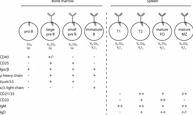
The pool of mature peripheral B cells in adult mice is replenished by ongoing lymphopoesis in the bone marrow. The purpose of this process is the release of cells, each carrying a single type of functional antigen receptor whose specificity is unique among all others within the B cell repertoire. This aim is accomplished in an astounding combination of stochastic yet tightly coordinated events encompassing the random generation of diversity and allelic exclusion (for a comprehensive review, see (Janeway, et al., 2001). B lineage commitment begins with the differentiation of pro B cells from hematopoietic progenitors (Hardy and Hayakawa, 2001). Pro B cells are characterized by expression of the BCR accessory chains immunoglobuline α (Igα) and Igβ and by ongoing rearrangement at the Ig µ heavy chain locus (figure 1). Immunoglobuline gene recombination begins with diversity (D)- to junction (JH)- gene segment fusion followed by rearrangement of DJ to variable (V ) gene segments. Productive V DJ recombination
H
H H H inhibits
potential processing of the second heavy chain allele and allows for the presentation of the transmembrane heavy chain molecule on the cell surface in conjunction with a surrogate light chain, VpreB or λ5, forming the pre-BCR.
An adequate signal from the pre-BCR induces pre-B cell expansion and allows for VL- to JL- rearrangement of κ and, if necessary, λ light chain genes.
Once a functional BCR, composed of a µ heavy and κ or λ light chains and lacking self-reactive specificity, is expressed on the surface, the newly-formed B cells exit the bone marrow into the periphery where they have to compete with the existing B cells for entry into the stable pool of mature naive B lymphocytes in the secondary lymphoid organs. In the spleen, B cells newly immigrated from the bone marrow undergo further maturation through distinct stages which can be distinguished by surface marker expression (Loder, et al., 1999) and figure 1). With few exceptions, cell surface molecules on leukocytes are named after the CD nomenclature where CD denotes “Cluster of Differentiation” (Morse, 1992). Newly immigrated splenic B cells go from the IgM high CD21/35-negative (IgMhi CD21/35-) transitional 1 stage (T1) to the IgM high CD21/35 high (IgMhi CD21/35hi) T2 stage, whereas the mature follicular B cell compartment (M) is characterized by an intermediate expression level of both surface proteins (IgMint CD21/35int). A second
Figure 1: Overview of murine B cell development and maturation in the bone marrow and spleen. Diagrams represent different B cell developmental stages including the composition of the respective BCR. Abbreviations underneath indicate the state of the heavy (top) and light (bottom) chain immunoglobuline regions. The expression of selected surface markers commonly used to distinguish the different B cell
subtype of splenic B cells which resides in the marginal zone (MZ) surrounding the lymphatic follicles and is hence named MZ B cells is also CD21/35hi IgMhi, but differs from T2 cells in the low expression of the surface marker CD23. The lineage commitment of the latter splenic B cell subset has not been entirely resolved, but it appears increasingly likely that follicular and MZ B cells originate from a common transitional B cell precursor (Pillai, et al., 2005).
While signals originating from the surface expressed BCR or pre-BCR regulate the survival of B cells throughout their development, it is at the T2 stage that they become simultaneously sensitive to as well as dependent on the cytokine BAFF for viability (see below).
The developmental pathway described above applies for conventional B-2 lineage cells which constitute the vast majority of the mature peripheral B cell pool and represent the B cell component of the adaptive immune system. In contrast, B-1 cells originate from a different source whose nature is still largely elusive. The fraction of B-1 cells is low in lymphoid compartments including spleen, lymph nodes and peripheral blood, but they constitute a significant portion of lymphocytes in the peritoneum and an important component of the innate immune response (Hardy, 2006).
1.2 Mature B cell survival through tonic BCR signaling 1.2.1 Composition of the BCR-complex and its signaling
mechanisms
Signaling from the BCR or pre-BCR has the astounding capacity to induce dramatically different cell fates. In mature naive B cells, BCR engagement with a foreign antigen represents the first step towards the induction of an immunogenic program which comprises clonal expansion, BCR maturation through class-switch recombination and somatic hypermutation as well as B cell differentiation into memory B or antibody-secreting plasma cells (Janeway, et al., 2001). Conversely, triggering of the same type of receptor by an auto-antigen has the opposite effect. Depending on the developmental stage and co-stimulatory signals, B cell encounter with a self-antigen leads to deletion, tolerance induction or alteration of receptor specificity through receptor editing (Healy and Goodnow, 1998). Importantly, some crucial signals originate from the BCR even in the absence of a discernable receptor ligand. This so-called tonic BCR-signal is critical to the survival of mature
naive B cells in the periphery (see below).
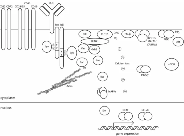
In mature naive B cells, the BCR complex is composed of a transmembrane Ig heavy chain of the µ or δ type (IgM or IgD) which is covalently linked to Ig light chains. Together, heavy and light chains form the antigen-binding unit of the BCR whose specificity is identical between all the receptors of a single B cell, but unique among all other receptors in the peripheral B cell pool. The BCR’s signal transduction capacity is conveyed through the non-covalently associated BCR-accessory chains Igα and Igβ. Each chain contains a single immunoreceptor tyrosine-based activation motif (ITAM) in which two tyrosine residues are surrounded by a characteristic consensus sequence. Receptor cross-linking causes phosphorylation of the Igα-Igβ heterodimer on ITAM- tyrosines by cytoplasmic protein tyrosine kinases, creating docking sites for a plethora of Src homology 2 (SH2)-domain containing proteins (figure 2 and (Kurosaki, 2002: ; Monroe, 2006). Ligand-induced ITAM-phosphorylation is carried out by cytoplasmic protein tyrosine kinases of the Src- and Syk-family.
In B cells, the most prominent members of theses families are Lyn and Syk, respectively. Although Lyn is placed upstream of Syk in most BCR-signaling models, both are capable of ITAM-phosphorylation in vitro and in vivo (see below). In any case, both appear to play a crucial role since the sole absence of either one compromises BCR-mediated signal transduction (Takata, et al., 1994). The kinase activities of Syk and Lyn are themselves stimulated by binding of phospho-ITAMs to their respective SH2-domains. ITAM- phosphorylation and proximal tyrosine kinase activation lead to the formation of multi-protein signaling complexes, sometimes called signalosomes, which ultimately relay the signal to changes in cellular architecture and gene expression. The adaptor protein B cell linker protein (BLNK, also known as SLP-65) has a major role in orchestrating the activation of Bruton’s tyrosine kinase (Btk) and phospholipase C γ 2 (PLCγ2) and thus in the generation of second messengers inositol-1,4,5-trisphosphate (IP3) and diacylglycerol (DAG). BLNK also functions in the activation of guanine nucleotide exchange factors Vav and Son of sevenless (Sos) which are instrumental for cytoskeletal rearrangements and for triggering of the mitogen-activated protein kinase (MAPK) pathway, respectively. IP3 signals the release of Ca2+- ions from intracellular stores and mediates activation of the Ca2+-sensitive transcription factor nuclear factor of activated T cells (NFAT) and the Ca2+-
composed of BCL-10 (B-cell lymphoma 10), CARMA1 (caspase-recruitment domain membrane-associated guanylate kinase protein 1) and MALT1 (mucosa-associated-lymphoid-tissue lymphoma-translocation gene 1) functions in the activation of the IKK (inhibitor of nuclear factor-κB (IκB) kinase) complex, a prerequisite for NF-κB-activation. BCR-ligation also enhances the formation of the lipid second messenger phosphatidylinositol (3,4,5)-trisphosphate (PIP3) through activation of phosphoinositide-3 kinase (PI3K). This is a key event for the activation of many pleckstrin homology (PH)-domain-containing signaling proteins including the oncogenic kinase Akt (see below).
Figure 2: Overview of BCR-signaling. See text for details (modified from Monroe, 2006).
In addition to the BCR core elements surface Ig and Igα-Igβ heterodimer, the complex can contain several co-receptors which have the capacity to modulate BCR-induced signaling (Nitschke, 2005: ; Poe, et al., 2001).
Examples for important co-stimulants are CD19, CD21/35 and CD45, whereas CD22 and CD72 transduce inhibitory signals through motives termed immunoreceptor tyrosine-based inhibitory motifs (ITIMs) in analogy to the ITAMs. ITIMs function in the recruitment of SH2-domain containing protein tyrosine phosphatases which are not only important in the down-regulation
and termination of the BCR-induced response, but which also possess the important capacity to modulate the strength of the BCR-mediated signal by directly opposing the action of proximal protein tyrosine kinases.
Signaling form the BCR or pre-BCR can direct dramatically different cell fates ranging from survival and proliferation to anergy and cell death. Despite the wealth of information on BCR-induced signaling pathways, establishing a certain cellular outcome as the consequence of specific molecular events has proven challenging. It is likely that key signaling factors are important for several potential cell fates, but there could be specific differences in their respective regulation through feedback interactions or signaling input from additional receptors. Conversely, some components of the BCR-related signaling web could function as unique features in promoting a specific cellular destiny. Most likely, a combination of both quantitative and qualitative differences enables the diversification from a single inducer to a range of cellular outcomes. For example, the MAPK Erk (extracellular signal-regulated kinase) and the transcription factor NFAT are responsive to antigenic stimulation in both naive and auto-reactive B cells, albeit to a different extend, whereas activation of the MAPK Jnk (Jun N-terminal kinase) and the transcription factor NF-κB occurs only in naive cells (Healy and Goodnow, 1998). These unique BCR-induced signaling profiles coincide with opposing cell fates, namely the clonal expansion of activated versus the inactivation and deletion of auto-reactive cells.
Several lines of evidence strongly suggest that the BCR complex has a critical signaling function even in the absence of ligand binding. Loss of the Ig portion of the BCR complex on mature B cells causes their demise and disappearance from the peripheral B cell pool (Lam, et al., 1997). The same is true for ablation of the ITAM-containing signaling portion of the Igα and Igβ accessory chains (Kraus, et al., 2004: ; Meffre and Nussenzweig, 2002).
Conversely, expression of a plasma membrane targeted chimeric Igα-Igβ protein in bone marrow progenitor cells which lack a functional Ig heavy chain coding region, is sufficient to generate peripheral follicular B cells which persist in the absence of surface BCR-expression (Bannish, et al., 2001).
These studies demonstrate that the maintenance of mature resting B cells in the periphery requires a constitutive survival signal which originates from the signaling portion of the BCR-complex and the expression of this segment is
Several hypotheses have been put forward to explain the nature of the so- called tonic BCR-mediated survival signal(Monroe, 2006). It has been proposed that a potential oligomeric assembly of BCR-complexes on the cell surface even in the absence of a cross-linking agent may be sufficient to generate a transducible event through the physical vicinity of signaling modules (Schamel and Reth, 2000). However, most of the current data suggest that BCR-complexes are rather dispersed throughout the plasma membrane in a monomeric state in resting naive cells, whereas stimulation causes their clustering, cap formation and eventual internalization (Tolar, et al., 2005). An alternative model focuses on BCR-localization in specialized ganglioside-rich membrane domains called lipid rafts. These membrane micro-domains are believed to function as signaling platforms and BCR- aggregation into lipid-raft localized clusters occurs upon receptor cross- linking. As a fraction of BCR-complexes is lipid-raft associated in resting unstimulated B cells, they might represent the source of the tonic BCR-signal (Guo, et al., 2000). This model is complicated by the observation that many of the signaling proteins believed to be essential for BCR-mediated signaling, including Syk and PLCγ2, only localize to lipid rafts upon receptor cross- linking. Furthermore, BCR-localization in lipid rafts appears to be dispensable for its tonic signaling to occur (Fuentes-Panana, et al., 2005).
Finally, Monroe and others have proposed the homeostatic equilibrium model.
Tonic BCR-signaling in this model represents a steady-state balance between stochastic phosphorylation of BCR-ITAMs by receptor-associated protein tyrosine kinases and its reversal by protein tyrosine phosphatases recruited by inhibitory BCR co-receptors. More specifically, a basal enzymatic activity of Lyn and Syk could account for random transient BCR-ITAM-phosphorylation which constitutes the origin of the tonic BCR survival signal. At the same time, full-blown receptor signaling is prevented by the activity of CD22-associated SH2-domain containing protein tyrosine phosphatase 1 (SHP-1). This model is in line with the effects of CD22- or SHP-1-deficiency on BCR-signaling (Cyster and Goodnow, 1995: ; Pani, et al., 1995), although the actual mechanism in vivo may be more complex and involve additional factors. BCR- stimulation with antigen would tip the balance between positive and negative signaling events within the complex towards productive phosphorylation. This could be achieved through the physical exclusion of inhibitory receptor components from the lipid-raft localized BCR-clusters and/or by transient inactivation of protein tyrosine phosphatases through the generation of reactive oxygen species. While this model makes a very coherent suggestion
regarding the initiation of tonic versus immunogenic BCR signaling, the nature of the downstream events continues to be unknown. Specifically, it remains to be determined how transient ITAM-phosphorylation is translated to cell survival versus the multitude of cellular changes in response to BCR- stimulation. This could be due to a quantitative difference in the signaling strength or a qualitative difference involving a distinct set of signaling factors or a combination of both. To date, any specific differences between tonic and ligand-induced BCR-signaling on a molecular level, qualitatively or quantitatively, have remained elusive.
1.2.2 The role of Syk in B cell biology
Syk was first purified as a tyrosine kinase activity from porcine spleen, hence the name for spleen tyrosine (Y) kinase (Kobayashi, et al., 1990: ; Sakai, et al., 1988: ; Taniguchi, et al., 1991). It forms a subgroup of cytoplasmic tyrosine kinases together with the closely related protein ZAP-70 (zeta-chain associated protein kinase, 70kD). Syk is expressed widely throughout the lymphoid and myeloid lineage as well as in some non-hematopoietic cell types including fibroblasts and epithelial cells (Turner, et al., 2000: ; Yanagi, et al., 2001).
Syk plays a critical role in signaling downstream of various receptors in different cell types. In B cells, Syk is rapidly activated upon cross-linking of the BCR (Hutchcroft, et al., 1991: ; Law, et al., 1994: ; Yamada, et al., 1993).
Furthermore, BCR-induced signaling responses are abrogated in a Syk- deficient variant of the chicken B cell line DT40, indicating an indispensable function of Syk in BCR-mediated signal transduction (Takata, et al., 1994). In addition to its function in B cells, Syk plays a role in signaling from various types of Fc receptors through which immune cells bind and respond to soluble antibodies. Fc stands for “fragment, crystallizable” and corresponds to the constant region of an antibody molecule (Janeway, et al., 2001). In mast cells, Syk mediates the response to IgE via FcεRI and is required for degranulation and cytokine release (Costello, et al., 1996: ; Taylor, et al., 1995: ; Zhang, et al., 1996). Similarly, Syk is involved in antibody-dependent cell-mediated cytotoxicity in response to FcγRIII cross-linking in natural killer (NK) cells (Brumbaugh, et al., 1997: ; Colucci, et al., 1999) and FcγR-mediated
interact with the phosphorylated TCRζ chain in thymocytes and Jurkat T cells (Chan, et al., 1994: ; Thome, et al., 1995: ; van Oers and Weiss, 1995). A major role for Syk in mature T cells is unlikely based on its low expression level (Chu, et al., 1999). However, enforced Syk-expression is able to restore the impaired T cell development ensuing from a lack of ZAP-70 in mice (Gong, et al., 1997), indicating that the functions of Syk and ZAP-70 may be partially interchangeable and differences may mainly be bestowed by a differential expression pattern. In support of this hypothesis, enforced expression of ZAP-70 restores BCR-signaling in Syk-deficient DT40 B cells (Kong, et al., 1995).
Both Syk and ZAP-70 are composed of an N-terminal tandem SH2-domain followed by a linker region and a C-terminal kinase domain (figure 3). The main difference between the two proteins is found in the linker region, which contains a 23 residues insertion in Syk as compared to ZAP-70. Interestingly, a deletion of the same region occurs in SykB which arises from alternative splicing of the full-length Syk transcript. This difference has functional consequences as full-length Syk displays a higher affinity to phosphorylated ITAMs and mediates more efficient signaling from the FcεRI receptor (Latour, et al., 1998).
Figure 3: Domain structure of Syk, SykB and ZAP-70. The positions of the structural domains as well as of tyrosine residues are indicated. Residue numbering is based on the full-length murine Syk protein. Asterisks denote residues whose phosphorylation has only been observed in vitro (modified from Turner et. al. 2000).
Syk-activation downstream of the BCR, pre-TCR and Fc-receptors most likely occurs through a shared mechanism which depends on the ITAMs present in all these receptor complexes. Syk has the capacity to bind phosphorylated ITAMs via its tandem SH2-domain (Johnson, et al., 1995: ; Law, et al., 1994: ; Law, et al., 1993: ; Saouaf, et al., 1994). This induces a conformational
change in the protein which likely enables kinase activity, resulting in the phosphorylation of
downstream targets as well as Syk itself (Kimura, et al., 1996: ; Rowley, et al., 1995). Initial BCR ITAM-phosphorylation was originally believed to be accomplished by Src-family tyrosine kinases, although later evidence suggested that Syk-activation does not require prior Src-kinase activity and Syk itself is capable of ITAM-phosphorylation (Kurosaki, et al., 1994: ; Zoller, et al., 1997). The order of proximal signaling events downstream of the BCR is therefore not entirely clear and it is not known, how potential Syk-activity on BCR-ITAMs is regulated in the absence of stimulation. Finally, alternative ITAM-independent mechanisms of Syk activation may exist as Syk also plays in role in signaling downstream of non-ITAM receptors, for example integrins, although it is possible that such receptors require ITAM-containing adaptor molecules (Gao, et al., 1997: ; Mocsai, et al., 2006: ; Woodside, et al., 2001).
Syk contains multiple tyrosine residues whose phosphorylation has been observed in vitro and in vivo (Furlong, et al., 1997: ; Keshvara, et al., 1998).
Upon BCR-cross linking, Syk-phosphorylation can be detected on Y130 located between the two SH2-domains, the linker region residues Y317, Y342 and Y346 as well as Y519 and Y520 within the kinase domain (figure 3).
Phosphorylation of Y130 and the kinase domain tyrosines is accomplished through auto-phosphorylation. Y519 and Y520 autophosphorylation is essential to Syk catalytic activity whereas the modification of Y130 appears to play a role in modulating Syk binding to the BCR (Couture, et al., 1997: ; Keshvara, et al., 1997: ; Kurosaki, et al., 1995). Phosphorylation of the linker region tyrosines is presumably mediated by Lyn and serves important biological functions. On the one hand, it appears to play a role in the binding of Syk to downstream targets such as PLCγ1 and Vav (Deckert, et al., 1996: ; Law, et al., 1996). On the other hand, phosphorylation of Y317 is critical for Syk recognition by the ubiquitin-ligase Cbl (Casitas B-lineage lymphoma).
Cbl-mediated proteolytic degradation of Syk constitutes an important mechanism in the down regulation of Syk-activity (Lupher, et al., 1998: ; Sohn, et al., 2003: ; Yankee, et al., 1999). In addition to the sites mentioned above, Syk-phosphorylation on Y290, Y358, Y624 and Y625 has been observed in vitro (Furlong, et al., 1997). The potential modification of the C-terminal residues Y624 and Y625 is particularly intriguing due to a potentially
of Syk C-terminal tyrosine residues to phenylalanines creates a gain-of- function Syk mutant (Zeitlmann, et al., 1998). Yet, neither a mechanism of Syk-inactivation through C-terminal phosphorylation nor a potential C-terminal Syk kinase has been described to date.
Syk-deficiency in mice results in perinatal lethality due to excessive hemorrhaging (Cheng, et al., 1995: ; Turner, et al., 1995). This is caused by a failure in the segregation of lymphatic and blood vessels during embryonic development (Abtahian, et al., 2003). The potential function of Syk in the immune system has been assessed through adoptive transfer of fetal liver which is the site of fetal hematopoiesis, from Syk-deficient embryos into immunodeficient hosts. The resulting chimeric mice exhibit a grossly normal T cell compartment and various functional deficits in myeloid cell types. The most dramatic effect is observed in the B cell compartment. Lack of Syk causes a severe block in early B cell development in the bone marrow at the transition from the pro B to the pre B stage, resulting in a near absence of B cells in the peripheral lymphoid organs. This demonstrates an obligatory role for Syk at the onset of B lymphopoiesis. A similar phenotype in mutant mice which are genetically unable to express a functional BCR can be overcome by the expression of a rearranged BCR-transgene, but this is not possible in the absence of Syk (Rajewsky, 1996); (Cornall, et al., 2000: ; Turner, et al., 1997).
In the latter case, the fraction of IgM-positive cells in the bone marrow is increased by expression of a transgenic BCR. However, these cells remain phenotypically immature, are refractive to BCR-stimulation and are unable to exit the bone marrow and persist in the periphery. Similarly, extension of B cell survival by ectopic expression of B-cell lymphoma 2 (Bcl-2) does not rescue B cell maturation in the absence of Syk (Turner, et al., 1997). These results argue for an indispensable signaling function for Syk downstream of the BCR.
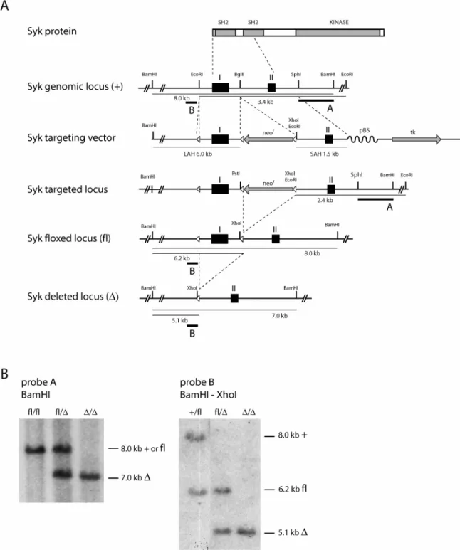
The early developmental block in the absence of Syk precludes an assessment of its function at later B cell stages including the survival, activation and differentiation of mature naive B cells. In an attempt to circumvent this problem, a conditional Syk knock-out was generated (Saijo, et al., 2003) and figure 4A). A targeting vector was constructed from genomic DNA of 129/Sv origin encompassing the first two exons of the murine Syk locus. The first exon was flanked with a single loxP site on the one side and with a loxP-flanked neomycin resistance cassette (neor) on the other. E14.1 embryonic stem (ES) cells were transfected with the targeting vector and screened for homologous recombination by Southern blotting of genomic DNA
Figure 4: Conditional Syk inactivation in mice. A. Gene targeting strategy for conditional Syk inactivation. The domain structure of the Syk protein is shown together with part of the Syk genomic locus, the targeting vector and part of the Syk locus before and after Cre-mediated recombination. Black squares denote exons I and II. Open triangles represent loxP sites. Gray bars denote expression cassettes for the neomycin resistance (neor) and herpes simplex thymidine kinase (tk) genes with the
between endonuclease restriction sites. LAH = long arm of homology, SAH = short arm of homology, pBS = pBluescript. Modified from Saijo et. al., 2003. B. Southern blot analysis of genomic DNA isolated from mouse tail tips. left: floxed (fl, 8 kb) and deleted (∆, 7 kb) alleles using BamHI restriction digestion and probe A; right: wild-type (+, 8 kb), floxed (fl, 6.2 kb) and deleted (∆, 5.1 kb) alleles using BamHI-XhoI double digestion and probe B.
after EcoRI digestion using the external probe A. The neor cassette was removed from positive clones through transfection with a Cre-recombinase expression vector and newly neomycin-sensitive clones were screened for correct excision of only the neor-cassette by Southern blot using BamHI-XhoI double digestion and probe B. ES cell clones containing the correctly targeted locus were injected into C57BL/6 blastocysts and the resulting chimeras were bred to C57BL/6 mice for germline transmission of the floxed Syk allele. The floxed Syk allele is functionally indistinguishable from a wild-type allele (my unpublished observation) and can be converted to a null allele through Cre- recombinase mediated excision of the first exon which contains the translation start codon. Genotyping of mice or cells is feasible through two different Southern blot strategies: use of probe A on BamHI-digested genomic DNA distinguishes the deleted allele from the wild-type or floxed allele, whereas probe B on BamHI-XhoI double-digested genomic DNA separates all three different alleles (figure 4B). The spatiotemporal regulation of Syk inactivation is determined by the pattern of Cre-expression. Different mouse Cre-strains are available in which Cre-expression is inducible by an external signal or targeted to a specific cell type.
1.3 Mature B cell survival through BAFF signaling 1.3.1 The BAFF-related protein network
It was long believed that tonic BCR signaling is the principle or even sole factor in mature B cell maintenance until a cytokine was discovered which dramatically increased the survival of B cells in vitro and in vivo (Batten, et al., 2000: ; Do, et al., 2000: ; Gross, et al., 2000: ; Khare, et al., 2000: ; Mackay, et al., 1999: ; Moore, et al., 1999: ; Mukhopadhyay, et al., 1999: ; Rolink, et al., 2002: ; Schneider, et al., 1999: ; Shu, et al., 1999: ; Thompson, et al., 2000). BAFF, also known as BLyS, TALL-1, THANK, zTNF4 and TNFSF 13b, increases B cell viability in culture and causes B cell hyperplasia and
autoimmunity when ectopically expressed in mice. In contrast to potent B cell mitogenic stimuli such as BCR cross-linking, CD40-ligand or endotoxin, BAFF exclusively enhances B cell viability without inducing proliferation.
Together with a related protein called a proliferation-inducing ligand (APRIL) BAFF forms a subgroup within the tumor necrosis factor (TNF) family. As such, BAFF is synthesized as a type II transmembrane protein which homotrimerizes via its C-terminal TNF-homology domain (THD). Soluble BAFF is the result of cleavage by furin family proteases, releasing a 138 residues product from the full-length (309 residues) pre-curser (Schneider, et al., 1999). BAFF contains putative N-linked glycosylation sites in its extracellular domain and its sensitivity to treatment with peptide N-glycanase indicates BAFF-glycosylation in vivo. Mouse and human BAFF are 59%
identical in their amino acid sequence, the main difference being a 31 residues insertion in mouse BAFF between the proteolytic cleavage site and the conserved THD-fold. Heterotrimers between BAFF and ARPIL are possible when the two proteins are co-expressed and have been found in the serum of patients with systemic rheumatic disease (Roschke, et al., 2002). A second BAFF isoform termed ∆BAFF arises from alternative splicing of the BAFF-transcript and can form multimers together with full-length BAFF. The biochemical properties of ∆BAFF vary significantly from the full-length protein in its poor release through protease-mediated cleavage and its decreased receptor affinity (Gavin, et al., 2003). Accordingly, ∆BAFF opposes full-length BAFF activity and cannot functionally substitute for loss of BAFF in vivo (Gavin, et al., 2005). BAFF-trimers can assemble into a 60-mer which forms a virus-like structure (Cachero, et al., 2006: ; Liu, et al., 2002). This form of BAFF is biologically active, although the significance of this particular assembly remains unknown (Zhukovsky, et al., 2004).
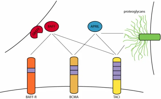
Three TNF receptor proteins have been found to bind BAFF although their ligand specificity partially overlaps with APRIL (figure 5). Transmembrane Activator and CAML Interactor (TACI) and B-Cell Maturation Antigen (BCMA) bind both BAFF and APRIL while BAFF is the sole ligand for BAFF receptor (BAFF-R) (reviewed in (Bodmer, et al., 2002: ; Bossen and Schneider, 2006: ; Mackay, et al., 2003). All three receptor proteins are untypical members of the TNF receptor family in that they are type III transmembrane proteins and their intracellular portion lacks a death domain. The extracellular domains contain
the differential affinity of certain members to proteogycans (see below and figure 5).
Figure 5: The BAFF protein family. Ligands BAFF, ∆BAFF and APRIL are shown together with the receptor proteins BAFF-R, BCMA and TACI as well as the proteoglycan moiety of other cell surface-expressed proteins. Boxes in the receptors denote cystein rich domains (CRDs). Ligand-receptor interactions are indicated by thin black lines. Note that the oligo- or multimeric nature of ligand and receptor complexes is not represented in this diagram.
1.3.2 The functions of BAFF and BAFF-R in B cell survival and beyond
The function of the BAFF-related protein network has been investigated using mouse models which lack individual components of the ligand-receptor system. In line with its dramatic impact on B cell viability, loss of BAFF diminishes the peripheral B cell compartment in mice. While early B cell development in the bone marrow proceeds largely unimpaired, a characteristic block ensues during peripheral B cell maturation in the spleen from the T1 to the T2 stage. Consequently, mature B cells are largely absent from the periphery of BAFF-deficient mice (Gross, et al., 2001: ; Schiemann, et al., 2001: ; Schneider, et al., 2001). A very similar phenotype, albeit somewhat less severe, is observed in the natural mouse mutant A/WySnJ (Lentz, et al., 1996: ; Miller and Hayes, 1991). This lead to the identification of BAFF-R as the principle receptor protein involved in BAFF-mediated B cell
survival. A/WySnJ mice harbor a transposon insertion in the BAFF-R locus which alters the C-terminal eight amino acids of the receptor and presumably impairs its signaling capacity (Thompson, et al., 2001: ; Yan, et al., 2001).
Subsequent BAFF-R ablation by gene targeting provided additional support for the vital role of BAFF-R in BAFF-mediated survival of peripheral B cells from the T2 stage on (Sasaki, et al., 2004: ; Shulga-Morskaya, et al., 2004).
This BAFF-dependency is not only restricted to conventional follicular B cells, but likely includes MZ B cells. The analysis of this question has been complicated by the fact that BAFF regulates the expression of CD21/35 and CD23 which are commonly used surface markers in the identification of splenic B cell subsets (Gorelik, et al., 2004). It appears that some MZ B cells are present in BAFF null and A/WySnJ mice, whereas none are detected in BAFF-R null mice and those expressing a TACI-Ig transgene (Amanna, et al., 2003: ; Gorelik, et al., 2004: ; Sasaki, et al., 2004: ; Tardivel, et al., 2004).
Overall, these results indicate that some level of BAFF-signaling is required for MZ B lymphopoiesis or maintenance or both. This conclusion is also supported by an expansion of the MZ B cell compartment in BAFF-transgenic mice (Gross, et al., 2000: ; Khare, et al., 2000: ; Mackay, et al., 1999). Quite surprisingly, despite the near absence of mature B cells in the absence of BAFF signaling, it is not required for some secondary B cell immune responses including the formation of germinal centers (GCs) and BCR affinity maturation. Yet, it does contribute to GC stability and BCR class-switch recombination to selected isotypes (Kalled, 2006). Most reports have concluded that BAFF signaling does not have an impact on the B-1 subset of B cells, although one study documented a modulation of B-1 cell numbers by decrease or increase in BAFF (Gavin, et al., 2005). In addition to its most prominent role in B cell biology, BAFF modulates the T cell compartment in several ways (reviewed in (Mackay and Leung, 2006). BAFF can directly stimulate BAFF-R-expressing T cell subpopulations including activated and regulatory T cells. BAFF provides a direct co-stimulatory signal to anti-CD3- triggered T cells through an autocrine loop by which activated T cells synthesize BAFF and increase BAFF-R expression. BAFF-costimulation of T cells enhances cytokine release and their differentiation into effector T cells.
In vivo, BAFF-influences T cell-mediated allograft rejection, although it is not established whether this is due to direct BAFF-signaling on T cells.
systemic autoimmunity (Gross, et al., 2000: ; Khare, et al., 2000: ; Mackay, et al., 1999). Correspondingly, elevated levels of BAFF have been measured in the serum of autoimmune-prone mice and in patients suffering form various autoimmune disorders including systemic lupus erythematosus (SLE), rheumatoid arthritis (RA) and primary Sjogren's syndrome (pSS) (reviewed in (Mackay, et al., 2005). A mechanistic basis for BAFF’s involvement in the negative selection of auto-reactive cells stems from the fact that the latter require higher BAFF-concentrations for survival than normal naive B cells (Lesley, et al., 2004: ; Thien, et al., 2004). Given that the amount of available BAFF is limited in a normal physiological context, this differential BAFF- requirement ensures the exclusion of auto-reactive cells from the stable peripheral B cell pool. Conversely, excess BAFF enables the persistence of B cells with a self-reactive receptor specificity in the periphery with the potential to cause tissue damage. This model fits very well with the observed clinical correlation between BAFF titers and autoimmune symptoms in humans and mice. Also in line with this model is the observation that ablation of two BAFF- regulated negative players in B cell survival, namely PKCδ and Bcl-2 interacting mediator of cell death (Bim), renders B cell survival BAFF- independent, ablates B cell tolerance and causes autoimmunity (see below).
The dual B cell pathologies resulting from insufficient as well as excess BAFF- signaling illustrate that the amount of biologically active BAFF has to be very tightly regulated to ensure the maintenance of a physiological peripheral B cell pool. BAFF-synthesis has been attributed to radiation-resistant stromal cells in the spleen, different myeloid cell types including monocytes, macrophages, neutrophils and dendritic cells, as well as activated T cells, follicular dendritic cells, astrocytes, osteoclasts and others (Craxton, et al., 2003: ; Gorelik, et al., 2003: ; Kalled, 2006: ; Litinskiy, et al., 2002: ; Nardelli, et al., 2001: ; Scapini, et al., 2005). While stromal cells could be a constitutive source of BAFF which is mainly responsible for the maintenance of physiological BAFF serum titers, BAFF-production by monocytes, macrophages, dendritic cells or neutrophils is externally regulated by other cytokines and could serve the modulation of BAFF-concentrations in specialized micro-environments such as inflammatory sites. Some instances of BAFF-production have been related to a pathological state, such as BAFF-synthesis by fibroblast-like synoviocytes in rheumatic joints, by ductal epithelial cells in the salivary gland of Sjogren’s syndrome patients and by nurse-like cells in chronic lymphocytic leukemia (CLL) (Groom, et al., 2002: ; Ittah, et al., 2006: ; Nishio, et al., 2005: ; Ohata, et al., 2005). Despite the identification of numerous BAFF-producing cell types, the
mechanism accounting for the precise regulation of BAFF levels remains unknown.
In addition to the available amount of BAFF, the level of BAFF-R surface expression seems to impact the strength of BAFF-signaling. This is evidenced by the intermediate B cell phenotype of heterozygous A/WySnJ mice compared to homozygous A/WySnJ mice and the parental A/J strain and indicates a gene dosage effect (Harless, et al., 2001: ; Lentz, et al., 1996: ; Miller and Hayes, 1991).
1.3.3 The functions of TACI, BCMA and APRIL
Despite certain structural similarities between the ligands BAFF and APRIL as well as the receptors TACI, BCMA and BAFF-R, the biological functions of the individual proteins vary dramatically. As described above, mature B cell survival is predominantly accomplished by BAFF-binding to BAFF-R whereas a similar role for the other proteins is less likely based on the phenotype of the respective knock-out mice. Lack of BCMA does not have a discernable effect on mature B cell viability (Xu and Lam, 2001). Yet, it plays a role in the survival of long-lived plasma cells in the bone marrow (Avery, et al., 2003: ; O'Connor, et al., 2004).
The function of TACI in B cell biology appears to be fairly complex and may not be entirely conserved between mice and humans. On the one hand, TACI- deficiency in mice renders B cells hyper-responsive to mitogenic stimuli and leads to an SLE-like disorder, B cell hyperplasia and even spontaneous development of lymphoma in a significant fraction of mutant animals (Seshasayee, et al., 2003: ; von Bulow, et al., 2001: ; Yan, et al., 2001).
These symptoms argue for a predominantly inhibitory role for TACI in B cell activation in mice. On the other hand, TACI is required for a proper immune response to type II T cell-independent antigens and plays a role in class switch recombination to IgG1 and IgA, a process which is promoted by both BAFF and APRIL through BAFF-R and TACI (Castigli, et al., 2004: ; Castigli, et al., 2005: ; Litinskiy, et al., 2002: ; Sakurai, et al., 2006). Furthermore, mutations in TACI are associated with an immunodeficiency syndrome in humans (Castigli, et al., 2005: ; Salzer, et al., 2005). Interestingly, in addition to the ligands BAFF and APRIL, TACI was shown to interact with the heparan
2006). Collectively, TACI-signaling appears to effect B cell functions both negatively and positively. The precise impact may be related to the B cell maturation stage, the type of ligand or even the species.
APRIL-deficiency does not appear to influence the survival of mature B cells, but as mentioned above, it can contribute to class-switch recombination by signaling through TACI and promotes plasma cell survival through BCMA (Avery, et al., 2003: ; O'Connor, et al., 2004). Intriguingly, in addition to binding to the TNF-family receptors TACI and BCMA, APRIL displays an affinity for the glycosaminoglycan side chains of proteoglycans (Ingold, et al., 2005). APRIL has also been implicated in the survival of lymphatic malignancies including B-cell chronic lymphoid leukemia (B-CLL), multiple myeloma and non-Hodgkin's lymphoma (Dillon, et al., 2006).
In addition to their individual functions, APRIL, TACI and BCMA can have a dramatic effect on B cell survival by titrating the available amounts of biologically active BAFF and BAFF-R. For example, TACI- or BCMA-fusion proteins function as decoy BAFF receptors when injected in mice or expressed from a transgene, depleting the peripheral B cell compartment (Gross, et al., 2001: ; Pelletier, et al., 2003: ; Schneider, et al., 2001: ; Thompson, et al., 2000). Also, chimeric receptor proteins containing the intracellular portion of BAFF-R, TACI or BCMA, respectively, elicit partially overlapping outcomes in terms of cell survival and molecular responses when expressed and triggered in B cell lines (Craxton, et al., 2005). In this experimental system, all three proteins boost cell survival while BAFF-R and TACI but not BCMA cause Bim-phosphorylation and only BAFF-R induces NF-κB processing (see below). In summary, there appears to be an intricate web of interactions between the ligands BAFF and APRIL, the receptors BAFF-R, TACI and BCMA and possibly also the proteoglycan moieties of cell surface molecules such as members of the syndecan family. While individual functions have been assigned to specific contacts, the full scope of potential cross-communication may continue to unfold.
1.3.4 The molecular consequences of BAFF-signaling
Considering the enormous interest and the wealth of knowledge on the function of BAFF in B cell physiology and disease, the level of insight into its signaling mechanism is surprisingly modest. A search for proximal signaling factors which directly interact with BAFF-R has so far yielded a single protein,
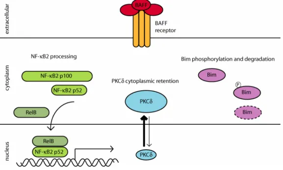
TRAF3. A physical and functional association has been proposed between BAFF-R, TRAF3 and the adaptor protein NF-κB activator 1 (Act1) which dampens BAFF-induced cellular responses (Qian, et al., 2004: ; Xu and Shu, 2002). Despite limited knowledge on the direct proximal signaling events upon B cell stimulation with BAFF, several molecular changes have been observed which likely contribute to its B cell survival effect (figure 6). First, it was discovered that BAFF induces activation of the transcription factor NF-κB which is known to play a central role in lymphocyte survival. NF-κB in mammals comprises a family of five related proteins (p50, p52, p65, RelB and c-Rel) which function in transcriptional regulation as hetero- or homodimers (Bonizzi and Karin, 2004). p50 and p52 originate from the processing of precursor proteins p105 (NF-κB1) and p100 (NF-κB2), respectively. The mechanism of NF-κB activation has been well studied for many years and relies on the rapid IKK-mediated phosphorylation and subsequent proteolytic degradation of IκB proteins which allows for the nuclear translocation of transcriptionally active NF-κB dimers. An example for this type of NF-κB activation is B cell stimulation through its antigen receptor. By contrast, BAFF primarily elicits NF-κB activation through an alternative mechanism in which NF-κB2 p100 is processed to mature p52 and subsequently forms transcriptionally active dimers with RelB (Claudio, et al., 2002: ; Kayagaki, et al., 2002: ; Senftleben, et al., 2001: ; Xiao, et al., 2001). The transcriptional targets of BAFF-induced NF-κB activation are not well characterized.
Upregulated expression of some pro-survival members of the Bcl-2 family has been observed in some instances but not in others (Claudio, et al., 2002: ; Hatada, et al., 2003: ; Hsu, et al., 2002: ; Lesley, et al., 2004: ; Thomas, et al., 2005: ; Trescol-Biemont, et al., 2004) and my own unpublished observations).
In addition to NF-κB activity, BAFF controls the subcellular localization of PKCδ. PKCδ is an important negative regulator of B cell viability as evidenced by B cell hyperplasia, autoimmunity and lack of B cell tolerance in PKCδ- deficient mice (Mecklenbrauker, et al., 2002). Furthermore, mature B cell survival becomes BAFF-independent in the absence of PKCδ (Mecklenbrauker, et al., 2004). The pro-apoptotic function of PKCδ is associated with its translocation to the nucleus and capacity for histone modification. BAFF promotes the cytoplasmic retention of PKCδ, thus containing its pro-apoptotic potential.
Figure 6: BAFF-induced changes in cellular signaling. BAFF binding to its receptor induces NF-κB activation by enhancing the processing of NF-κB2 p100 to mature p52 which regulates gene transcription in conjunction with RelB. BAFF-mediated cytoplasmic retention of PKCδ is indicated by the symbol and arrow size. Regulation of Bim by BAFF involves its phosphorylation and degradation.
key function for Bim in B cell homeostasis is demonstrated by elevated numbers of B cells and impaired deletion of auto-reactive cells in Bim- deficient mice as well as by the extended survival of Bim-deficient B cells in vitro (Bouillet, et al., 1999: ; Enders, et al., 2003). As in the case of PKCδ- deficiency, absence of Bim alleviates the BAFF-dependency of mature B cells for survival (Oliver, et al., 2006). BAFF-treatment also has a direct effect on Bim expression as a consequence of BCR-cross linking which it is able to suppress (Craxton, et al., 2005). This coincides well with the augmentation of BCR-induced proliferation and survival upon co-stimulation with BAFF. Also, BAFF promotes Erk-mediated phosphorylation of Bim which curbs its pro- apoptotic function.
The transcription factor c-Myb has been identified as another regulator of BAFF-mediated B cell survival (Thomas, et al., 2005). Lack of c-Myb reduces the frequency of mature peripheral B cells in mice and c-Myb-deficient B cells are partially refractive to BAFF-mediated responses, possibly due to suboptimal BAFF-R expression. Lack of c-Myb impairs BAFF-induced survival and cytoplasmic retention of PKCδ but not p100 processing. Impaired BAFF- mediated B cell survival and defective BAFF-R expression also occur in the
absence of the GTPases Rac1 and Rac2, suggesting that these proteins may play a role in the regulation of BAFF-signaling (Walmsley, et al., 2003).
BAFF-mediated effects on NF-κB activation, PKCδ localization and Bim expression are observed in a range of 12 to 24 hours after stimulation, raising the question, whether they represent direct BAFF-induced signaling events.
Alternatively, diminished p100 processing, nuclear translocation of PKCδ and upregulation of Bim in the absence of BAFF could merely reflect early signs of cellular atrophy which are prevented by BAFF’s effect on B cell viability. In either scenario, all the described molecular changes are most likely involved in enhancing B cell survival in the presence of BAFF. Indeed, artificial augmentation of B cell viability by ectopic expression of Bcl-2, Bcl-xL (Bcl2- like 1) or constitutively active NF-κB as well as removal of PKCδ is sufficient to restore peripheral B cell survival in BAFF-R-mutant mice (Amanna, et al., 2003: ; Gorelik, et al., 2004: ; Mecklenbrauker, et al., 2004: ; Rahman and Manser, 2004: ; Sasaki, et al., 2004: ; Sasaki, et al., 2006: ; Tardivel, et al., 2004).
1.4 Lymphocyte survival through growth factor signaling
1.4.1 The association between the trophic state of a cell and its viability
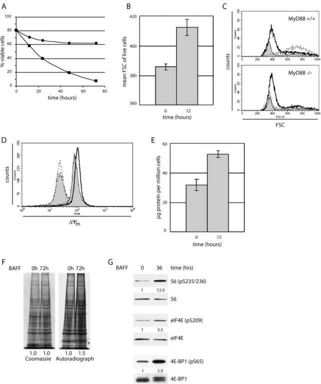
The supply of nutrients from the extracellular environment is indispensable to the viability, growth, division and differentiation of all cell types. At the same time, it is imperative for multicellular organisms to regulate these processes not only based on nutrient availability, but on additional extracellular cues whose concerted action ensures the proper development and function of the organism as a whole. Lymphocytes are a prime example for a cell type whose survival and expansion is, among others, dependent on extracellular nutrients as well as trophic cytokines or growth factors. The latter afford cells the ability to not only ingest nutrients, but to optimize nutrient metabolism for cellular growth and energy derivation. For example, stimulation with trophic cytokines promotes the cell surface expression of glucose and amino acid transporters, thus enabling cellular nutrient uptake (Edinger and Thompson, 2002). At the
membrane where access to intramitochondrial ATP enhance the enzyme’s kinetics, while the activity of phosphofructokinase is sensitive to growth factor- dependent phosphorylation (Bertrand, et al., 1999: ; Deprez, et al., 1997: ; Marsin, et al., 2000: ; Robey and Hay, 2006). Glycolytic glucose-utilization and energy derivation feed back to mitochondrial respiration by supplying critical intermediates of the electron transport chain. Cellular deprivation from growth factors creates a shortage of electron transport substrates, leading to a drop in mitochondrial membrane potential and a subsequent closure of voltage-dependent anion channels, preventing the exchange of metabolites between mitochondria and the cytoplasm (Plas, et al., 2002: ; Plas and Thompson, 2002). Atrophy as a consequence of growth factor withdrawal in lymphoid and hematopoietic cells is characterized by impaired nutrient transporter expression and nutrient uptake, decreased glycolytic rates and a drop in the mitochondrial membrane potential (Barata, et al., 2004: ; Rathmell, et al., 2000: ; Vander Heiden, et al., 2001).
An important consequence of trophic cytokine mediated nutrient uptake is the cells’ ability to maintain or increase their size. Cell growth is obviously a prerequisite for proliferative expansion and has to precede cell division unless daughter cells should become progressively smaller. At the same time, cell growth should not proceed beyond a certain checkpoint, unless in preparation for subsequent cell division. Demonstrating the trophic function of growth factors, cells deprived of nutrients or growth factors will become progressively smaller before they collapse (Plas, et al., 2001: ; Rathmell, et al., 2000). While constitutive ectopic expression of anti-apoptotic Bcl-2-family proteins prevents the death of such cells, it does not restore size maintenance, nutrient uptake or energy homeostasis. This indicates that cell survival mediated by Bcl-2- family proteins or growth factors constitute separate non-redundant mechanisms.
1.4.2 Growth factor signaling is transduced by the PI3K-Akt pathway
In contrast to Bcl-2-family members, components of the PI3K pathway are able to avert the decline in cellular metabolism as a consequence of growth factor deprivation, indicating that this pathway plays a vital role in growth factor signaling (Plas, et al., 2001). PI3K can act on lipids as well as proteins, but its best characterized and presumably most important function is the phosphorylation of inositol phospholipids on the D3-position of the inositol ring
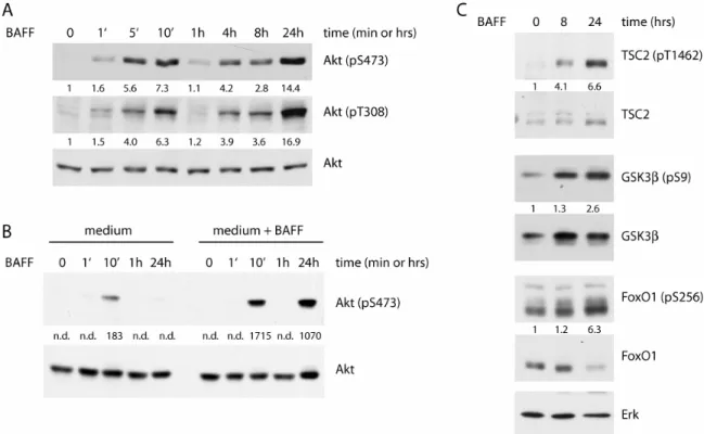
to create, depending on the substrate, phosphatidylinositol-3,4-bisphosphate (PIP2) and phosphatidylinositol-3,4,5-triphosphate (PIP3) (Fruman, 2004: ; Okkenhaug and Vanhaesebroeck, 2003). The most common PI3K isoform in B cells belongs to the class IA subgroup and is composed of a catalytic subunit p110δ and a regulatory subunit p85α. PI3K-activation occurs by binding of the regulatory subunit’s SH2-domain to phosphorylated tyrosine residues in upstream regulatory proteins which result from tyrosine kinase activation by growth factor stimulation. p85-binding of upstream activators induces a conformational change in the protein complex which allows for enzymatic activity of the constitutively associated catalytic subunit. PI3K- mediated PIP3-accumulation at the plasma membrane creates a docking site for signaling proteins bearing a PH-domain. In terms of growth factor signaling, the most important of these is the oncogenic serine-threonine kinase Akt (figure 7).
Figure 7: The PI3K-Akt signaling pathway. See text for details.
Akt, sometimes also called protein kinase B (PKB), is known as a master regulator of cell survival, growth and cell cycle progression (Massague, 2004:
; Plas and Thompson, 2005). It directly influences cell viability through down- regulation of Bcl-2-associated death promoter (Bad), a member of the pro- apoptotic BH3-only sub-family of Bcl-2 proteins (Datta, et al., 1997: ; del Peso,
family of transcription factors (Burgering and Kops, 2002). In a process which is evolutionary conserved from Caenorhabditis elegans through to mammals, FoxO transcription factors protect cells from oxidative stress and signal cell cycle exit under glucose limiting conditions. Apoptosis and cell cycle exit are promoted by FoxO proteins through upregulation of Bim and the cyclin- dependent kinase (Cdk) inibitors p21 and p27 (Seoane, et al., 2004: ; Stahl, et al., 2002). Collectively, FoxO-proteins antagonize growth factor-induced cellular responses. Accordingly, direct FoxO-phosphorylation by growth factor-activated Akt inhibits FoxO-mediated transcriptional regulation in the nucleus by creating a binding site for 14-3-3 proteins and subsequent sequestration of FoxO-proteins in the cytosol (Brunet, et al., 1999). In addition to this immediate mechanism of FoxO-inactivation, prolonged Akt-activity has the potential to permanently modify the cellular transcriptional program by targeting FoxO-proteins for proteolytic degradation (Plas and Thompson, 2003). In mature B cells, mitogenic stimulation leads to FoxO1- phosphorylation in a PI3K-dependent manner and over-expression of a mutant FoxO-protein which is resistant to Akt-mediated inactivation, causes cell cycle arrest and apoptosis concomitant with an induction of p27 and Bim in activated lymphocytes (Stahl, et al., 2002: ; Yusuf, et al., 2004).
One of the most important targets of Akt-signaling is the mammalian target of rapamycin (mTOR), a kinase of the phosphoinositide-3-kinase-related kinase (PIKK) family which also includes ATM (ataxia telangiectasia mutated), DNA- PK (DNA-dependent protein kinase) and others (Martin and Hall, 2005: ; Wullschleger, et al., 2006). Although mTOR has been a subject of close investigation for many years, its regulation and function appear to be incredibly complex and are still incompletely understood. mTOR exists in complex with accessory proteins raptor (mTORC1) or rictor (mTORC2). The two complexes serve distinct cellular functions and are further distinguished by their differential sensitivity to the immunosuppressant rapamycin (Bhaskar and Hay, 2007). While mTORC2 functions in actin-polymerization and is indifferent to rapamycin treatment, mTORC1 represents the “classic“ mTOR complex which acts in protein synthesis, metabolism and growth in a rapamycin-sensitive fashion. The following description of mTOR function and regulation will apply to mTORC1 unless otherwise stated. Under growth- favorable conditions, mTOR has a direct impact on protein synthesis through phosphorylation of ribosomal S6 kinase 1 (S6K1) and 4E binding protein 1 (4E-BP1) (Ruggero and Sonenberg, 2005). S6K1-mediated phosphorylation of ribosomal S6 protein is considered a hallmark of active protein synthesis,
whereas 4E-BP1 is an inhibitor of translational initiation through its sequestration of eukaryotic translation initiation factor 4E (eIF4E).
Phosphorylation of 4E-BP1 disrupts its association with eIF4E, thus enabling the latter’s participation in the translation initiation complex. mTOR-activity also contributes to cell growth by promoting ribosome biogenesis as well as the expression of many metabolically relevant genes (Martin and Hall, 2005: ; Peng, et al., 2002). Furthermore, mTOR negatively impacts protein degradation via autophagy (Lum, et al., 2005). Finally, organization of the cytoskeleton which is important in growth related events including establishment of cell polarity and cell division is regulated by mTORC2 (Jacinto, et al., 2004: ; Sarbassov, et al., 2004).
Given that the mTOR-dependent cellular events are overwhelmingly energy- consuming, it is probably as appropriate as it is necessary that mTOR functions as an integrator for a multitude of signaling inputs including growth factors, nutrients, energy and stress, all of which can impact mTOR-activity.
The metabolic function of mTOR is dependent on the small G-protein Rheb (Ras homolog enriched in brain) which is subject to inactivation by the GTPase activating protein (GAP) Tuberous sclerosis 2 (TSC2) (Tee and Blenis, 2005). Mutations in the TSC2 gene are the leading cause for the disease Tuberous sclerosis, a hereditary condition characterized by the frequent appearance of benign tumors in multiple organs. Very recently, PRAS40 (proline-rich Akt substrate 40kD) has been identified as another major mTOR inhibitor (Sancak, et al., 2007: ; Vander Haar, et al., 2007).
Under conditions unfavorable for mTOR-activity and growth, PRAS40 binds to mTOR and prevents its activation. One important characteristic of unfavorable growth conditions is a low ATP to AMP ratio which is directly sensed by the AMP-activated protein kinase (AMPK) (Hardie, 2005). AMPK-activity has a dual negative effect on mTOR-signaling: it enhances TSC2’s GAP-activity through direct phosphorylation, thus constraining mTOR activity, while at the same time inhibiting activation of the mTOR targets 4E-BP1 and S6K. Growth factor-induced activation of the PI3K-Akt pathway has a profound effect on mTOR activity at multiple levels. First, Akt phosphorylates and inactivates TSC2, thus enabling Rheb·GTP-dependent mTOR activity. As has been discovered very recently, Akt also alleviates PRAS40-dependent suppression of mTOR activity (Kovacina, et al., 2003: ; Sancak, et al., 2007: ; Vander