Isolated ectopic cilia in an 11-year-old girl
Abstract
Ectopic cilia (EC) are a very rare condition with only few cases reported in literature. Many associations were seen with ectopic cilia which in-
Hosny Ahmed Zein
1M. Tarek A. Moustafa
1clude distichiasis, choristoma and aberrant lacrimal gland, hypochromic nevus, atopic eczema and others. We are reporting a case of an 11- year-old girl with isolated left upper lid ectopic cilia, which was confirmed by surgical removal and histopathological study.
1 Department of Ophthalmology, Minia University, Minia, Egypt Keywords:eyelids, cilia, pathology
Introduction
Ectopic cilia (EC) are a very rare condition with only a few reported cases in the literature [1], [2], [3]. The most common presentations are the abnormal growth of eye- lashes on the external, lateral quadrant of the upper eyelid, or the internal, conjunctival surface of the eyelid.
There is usually a negative family history, no ocular symptoms but significant cosmetic disfigurement. The ectopic cilia present as a tuft of eyelash follicles on the temporal, upper eyelid. After excision, the pathologic diagnosis is confirmed by the presence of pilosebaceous follicle (PSF) with sebaceous and sweat glands in the specimen.
Case description
An 11-year-old girl was seen on September 20 2015 in the ophthalmology outpatient clinic at Minia university hospital, with a chief complaint of a hair tuft projecting between the left upper eyelid lashes (Figure 1). The hair tuft had been present early after birth and epilation had been performed three times. However, the hair tuft re- curred within one month after treatment. The ophthalmo- logy examination was normal except for a group of abnor- mal lashes about 4 mm above the upper lid margin. The patient was otherwise healthy and a dermatological con- sultation showed no systemic abnormalities with respect to her hair or skin pigmentation. The family history was negative for hair or skin abnormalities.
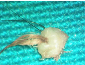
After discussing the surgery with her parents, an informed consent was obtained and an excisional biopsy was per- formed in the operating room under complete aseptic conditions. The left upper eyelid was injected locally with a 1:1 mixture of Xylocaine (2%) and Epinephrine (1:100,000). The tissue specimen was removed as a circular mass with a diameter of 8 mm that included skin, subcutaneous tissue with a centrally situated hair tuft, and some normal lashes located on one side of the ab- normal hair tuft (Figure 2). The specimen was sent for histopathology and the report described both sebaceous
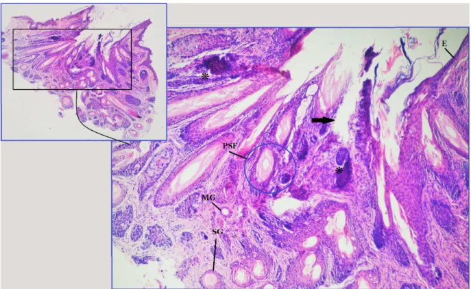
glands (SG) and epithelial lined apocrine Molls glands (MG) adjacent to subcutaneous pilosebaceous follicle (PSF) (Figure 3 and Figure 4). No other glands could be identified. The patient was examined at 1 week, 4 weeks and 3 months postoperatively and no evidence of local recurrence was found.
Figure 1: Clinical photograph of ectopic cilia
Figure 2: Clinical photograph of the specimen after surgical excision
1/3 GMS Ophthalmology Cases 2016, Vol. 6, ISSN 2193-1496
Case Report
OPEN ACCESS
Figure 3: Photomicrograph shows lash follicle (*), its fibrous tract (arrow), epidermis (E), Molls gland (MG), sebaceous gland (SG) and pilosebaceous follicle (PSF).
Figure 4: Photomicrograph shows sebaceous gland lobules (*) and epithelial-lined sweat gland (arrow).
2/3 GMS Ophthalmology Cases 2016, Vol. 6, ISSN 2193-1496
Zein et al.: Isolated ectopic cilia in an 11-year-old girl
Discussion
Ectopic cilia are rarely found in humans with only about 20 cases reported in the literature. The origin of these abnormally situated lashes is not yet clear. Previous re- ported cases described either congenital ectopic cilia originating from the anterior temporal aspect of the upper tarsal plate [1], [4], [5] or acquired post-inflammatory cilia located in the tarsal conjunctiva [6]. We believe that our case belongs to the first group.
The ectopic cilia are not usually isolated but are often associated with distichiasis [7], choristoma and aberrant lacrimal glands [8], hypochromic nevus [3], sebum accu- mulation [9], atopic eczema [10], nail-patella syndrome [11], and a combination of orbital dermoid cyst and sinus tract [2]. However, in our case we could not find any eye- related or systemic abnormalities.
The embryologic origin of the ectopic lashes has been thought to be a deformity of the upper lid glands, with complete or partial replacement of the meibomian glands with skin glands [5]. The anatomical location, as well as the congenital development of Tessier Type 9 facial cleft at the same site support the theory that ectopic cilia is a congenital anomaly, as this site is embryologically related to the watershed area of two angisomes, the superficial temporal artery and the termination of the facial artery [12].
Summary
Ectopic cilia are rare and can present either as an isolated event or in association with other ocular and systemic abnormalities. After surgical removal, the histopathologic- al identification of pilosebaceous follicle, Molls and se- baceous glands are essential for the diagnosis.
Notes
Competing interests
The authors declare that they have no competing in- terests.
References
1. Jakobiec FA, Yoon MK. Histopathologic proof for the origin of ectopic cilia of the eyelid skin. Graefes Arch Clin Exp Ophthalmol.
2013 Mar;251(3):985-8. DOI: 10.1007/s00417-012-2224-0 2. Krahulík D, Karhanová M, Vaverka M, Brychtová S, Pospíšilová
D. Ectopic cilia associated with an orbital dermoid cyst and sinus tract: case report. J Neurosurg Pediatr. 2015 Aug;16(2):203-6.
DOI: 10.3171/2014.12.PEDS14512
3. da Fonseca FL, Yamanaka PK, Lima PP, Matayoshi S. A 6-year- old girl with ectopic cilia and hypochromic nevus. Clin Ophthalmol.
2014;8:1259-61. DOI: 10.2147/OPTH.S63313
4. Baghestani S, Banihashemi SA. Ectopic cilia in a 14-year-old boy.
Pediatr Dermatol. 2011 Jan-Feb;28(1):55-6. DOI:
10.1111/j.1525-1470.2010.01354.x
5. Chen TS, Mathes EF, Gilliam AE. “Ectopic eyelashes” (ectopic cilia) in a 2-year-old girl: brief report and discussion of possible embryologic origin. Pediatr Dermatol. 2007 Jul-Aug;24(4):433- 5. DOI: 10.1111/j.1525-1470.2007.00473.x
6. Hase K, Kase S, Noda M, Ohashi T, Shinkuma S, Ishida S. Ectopic cilia: a histopathological study. Case Rep Dermatol. 2012 Jan;4(1):37-40. DOI: 10.1159/000336887
7. Bader A. Aplasia congenitalis glandularum Meibomi palpebrae inferioris [Congenital aplasia of the meibomiam glands of the lower eyelid]. Albrecht Von Graefes Arch Ophthalmol.
1950;150(3-4):411-3. DOI: 10.1007/BF00681437
8. Gordon AJ, Patrinely JR, Knupp JA, Font RL. Complex choristoma of the eyelid containing ectopic cilia and lacrimal gland.
Ophthalmology. 1991 Oct;98(10):1547-50. DOI:
10.1016/S0161-6420(91)32090-6
9. Chappell MC, Spencer W, Day SH, Silkiss RZ. Congenital ectopic cilia of the upper eyelid. Ophthal Plast Reconstr Surg. 2011 Mar- Apr;27(2):e42-4. DOI: 10.1097/IOP.0b013e3181e17501 10. Möhrenschlager M, Köhler LD, Ring J. Ectopic cilia in a Caucasian
girl with atopic eczema. Acta Derm Venereol. 1998 Mar;78(2):146-7. DOI: 10.1080/000155598433520 11. Edmunds MR, Kipioti A, Colloby PS, Reuser TT. A case of ectopic
cilia in nail-patella syndrome. Int Ophthalmol. 2012 Jun;32(3):289-92. DOI: 10.1007/s10792-012-9552-2 12. MacQuillan A, Hamilton S, Grobbelaar A. Angiosomes, clefts, and
eyelashes. Plast Reconstr Surg. 2004 Apr;113(5):1400-3. DOI:
10.1097/01.PRS.0000112793.45806.7A
Corresponding author:
M. Tarek A. Moustafa, M.D., PhD, FRCS
Department of Ophthalmology, Minia University, Minia, Egypt, Phone: +1 949-627-5034, Fax: +1 949-824-9626 mohamedtarek@mu.edu.eg
Please cite as
Zein HA, Moustafa MT. Isolated ectopic cilia in an 11-year-old girl. GMS Ophthalmol Cases. 2016;6:Doc13.
DOI: 10.3205/oc000050, URN: urn:nbn:de:0183-oc0000509
This article is freely available from
http://www.egms.de/en/journals/oc/2016-6/oc000050.shtml Published:2016-10-12
Copyright
©2016 Zein et al. This is an Open Access article distributed under the terms of the Creative Commons Attribution 4.0 License. See license information at http://creativecommons.org/licenses/by/4.0/.
3/3 GMS Ophthalmology Cases 2016, Vol. 6, ISSN 2193-1496
Zein et al.: Isolated ectopic cilia in an 11-year-old girl