Surgical Neurology International
Editor:James I. Ausman, MD, PhD University of California, Los Angeles, CA, USAOPEN ACCESS
For entire Editorial Board visit : http://www.surgicalneurologyint.com
Case Report
Decompressive craniectomy and early cranioplasty in a 15‑year‑old boy with N. meningitidis meningitis
Julius Hoehne, Monika Friedrich, Alexander Brawanski, Michael Melter
1, Karl‑Michael Schebesch
Departments of Neurosurgery, 1Pediatrics, University Medical Center Regensburg, Franz‑Josef‑Strauss Allee 11, 93053 Regensburg, Germany E‑mail: *Julius Hoehne ‑ julius.hoehne@ukr.de; Monika Friedrich ‑ monika.friedrich@ukr.de; Alexander Brawanski ‑ alexander.brawanski@ukr.de;
Michael Melter ‑ michael.melter@ukr.de; Karl‑Michael Schebesch ‑ karl‑michael.schebesch@ukr.de
*Corresponding author
Received: 23 August 14 Accepted: 19 December 14 Published: 09 April 15
Abstract
Background: Intracranial hypertension is a well‑known life‑threatening complication of bacterial meningitis. Investigations on decompressive craniectomy after failure of conservative management are scarce, but this surgical treatment should be considered and performed expeditiously, as it lowers the intracranial pressure and improves brain tissue oxygenation. Early cranioplasty can further aid the rehabilitation.
Case Description: A 15‑year‑old boy was admitted to our emergency department because of sudden onset of neurologic decline and consecutive loss of consciousness. Clinical examination and imaging showed elevated intracranial pressure, leading to the suspected diagnosis of meningitis. Intracranial pressure monitoring was installed, but the initiated conservative management failed. Finally, the patient underwent bilateral decompressive craniectomy. The microbiological test showed growth of Neisseria meningitidis. After full neurologic recovery, cranioplasty with two CAD/CAM titanium implants was conducted successfully.
Conclusions: This unique report shows that decompressive craniotomy with duroplasty may be a crucial therapeutic approach in bacterial meningitis with refractory increased intracranial pressure and brainstem compression. Early cranioplasty with a patient‑specific implant allowed the early and full reintegration of the patient.
Key Words: Cranioplasty, computer‑aided design/computer‑aided manufacturing, encephalitis, intracranial pressure, meningococcal disease, meningitis, Neisseria meningitidis, W/Y‑135
CASE REPORT
A 15‑year‑old Turkish boy was admitted to the emergency department after loss of consciousness.
3 days prior to admission, he had experienced fever and ague with a gradual decline of his general condition.
When presenting to his primary care physician, he was treated with nonsteroidal antiinflammatory drugs (NSAIDs) and antitussives for a suspected viral
infection. Because his condition had not improved and he had developed nausea and vomiting, the patient presented to an emergency physician the night prior to hospital admission. This time, paracetamol and antiemetics were administered. During the day he was tired, disoriented, and somnolent. The next morning, he was found unconscious and was subsequently admitted to the emergency department. No clinical signs of epileptic seizures could be detected.
This article may be cited as:
Hoehne J, Friedrich M, Brawanski A, Melter M, Schebesch KM. Decompressive craniectomy and early cranioplasty in a 15-year-old boy with N. meningitidis meningitis. Surg Neurol Int 2015;6:58.
Available FREE in open access from: http://www.surgicalneurologyint.com/text.asp?2015/6/1/58/154776
Copyright: © 2015 Hoehne J. This is an open-access article distributed under the terms of the Creative Commons Attribution License, which permits unrestricted use, distribution, and reproduction in any medium, provided the original author and source are credited.
Access this article online Website:
www.surgicalneurologyint.com DOI:
10.4103/2152-7806.154776 Quick Response Code:
[Downloaded free from http://www.surgicalneurologyint.com on Monday, May 11, 2015, IP: 132.199.144.70]
Surgical Neurology International 2015, 6:58 http://www.surgicalneurologyint.com/content/6/1/58 On admission, the patient was not adequately responsive,
showing a Glasgow Coma Score of 9 including neck stiffness.
The body core temperature was 36.5°C. Pupils were equal, round, as well as reactive to light and accommodation. No additional neurological abnormality was present.
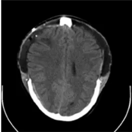
The initial computed tomography (CT)‑scan showed generalized brain edema [Figure 1]. Consequently, no lumbar puncture was conducted.
At the neurological department, the patient was initially treated with corticosteroids (10 mg dexamethasone), antibiotics (2 g ceftriaxone and 400 mg ampicillin), and antiviral medication (750 mg aciclovir) because of suspected meningitis. Relatives and people in close contact with the patient were treated accordingly. An intracranial pressure (ICP) probe was implanted for ICP monitoring.
Despite maximal osmotic management, the patient developed intractable ICP with values up to 36 mmHg.
Because the patient failed to response to maximal conservative treatment, the team of pediatrics, neurologists, and neurosurgeons decided to conduct bilateral craniectomy and durotomy to control the extensively elevated ICP.
Immediately after decompressive surgery, the ICP decreased significantly but increased again few hours after the first surgery, while the right pupil became dilated consecutively. The immediate CT‑scan showed an epidural hematoma and subgaleal blood collection. The hemorrhage was evacuated instantly [Figures 2 and 3]. During the following days, the ICP remained stable within normal limits, and the ICP‑probe was removed. Radiologic follow‑up by means of magnetic resonance imaging (MRI) on day 7 after ictus showed diffusion impairment in parts of the insular region and around the temporal horn. On day 13 after surgery, a further radiological follow‑up by means of a CT scan confirmed bilateral hygroma that were drained by bilateral subdural drainages. A lumbar drainage was implanted for 5 days. The electroencephalogram showed no signs of
epilepsy; brainstem auditory evoked potentials could not be obtained on the right, whereas somatosensory evoked potentials were positive.
Despite the initial antibiotic and antiviral treatment without prior lumbar puncture, microbial testing of cerebrospinal fluid at a later stage showed Neisseria meningitidis type W/Y‑135; the polymerase chain reaction (PCR) test for herpes simplex (HS) virus type 1/2 was negative. Uncomplicated pneumonia was treated successfully with antibiotics but prolonged the intubation period. Two weeks after admission, inflammatory hematological parameters had improved significantly, and the boy was extubated. Rapidly, the patient’s general condition improved, hence he was discharged on day 45 without any neurologic deficit and soon afterwards returned to school.
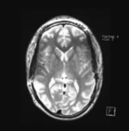
After 4 weeks, patient specific CAD/CAM titanium cranioplasties (CL instruments, Attendorn, Germany) were implanted bilaterally. Wound healing was uneventful and postoperative imaging showed accurate fitting without relevant artifacts [Figure 4].
DISCUSSION
Basically, permanent neurological deterioration due to meningitis is reported for up to 16% of affected children with neurologic sequelae; this rate refers to survivors in developed countries, irrespective of maximal conservative treatment.[2] Neurological and neuropsychological sequelae have been mainly reported in cases of bacterial meningitis.[13] In patients with bacterial meningitis, brain edema has been observed in up to 24% of patients.[15]
Monitoring of ICP has been proposed by Barnett et al.
as a useful adjunct for therapy and provided indication for prognosis in an outcome analysis of 10 patients with encephalitis.[3] Direct intraparenchymal ICP monitoring is accurate, low risk, and routine procedure in pediatric
Figure 1: Initial CT-scan with generalized brain edema Figure 2: Postoperative CT-scan demonstrating adequate bilateral decompression
[Downloaded free from http://www.surgicalneurologyint.com on Monday, May 11, 2015, IP: 132.199.144.70]
Surgical Neurology International 2015, 6:58 http://www.surgicalneurologyint.com/content/6/1/58
neurosurgery[20] that is superior to radiological evaluation alone.[8] Only anecdotal evidence is available for decompressive craniectomy as a treatment option for bacterial meningitis with intractable ICP. A 28‑year‑old female with bacterial N. meningitidis infection underwent decompressive craniectomy, after diagnostic lumbar puncture lead to temporal herniation and raised ICP could not be managed conservatively.[4] A 17‑year‑old boy with meningitis after exacerbated pansinusitis developed brain edema with consecutive unilateral anisocoria and underwent bilateral decompressive craniectomy.
Seven weeks later, the autologous bone was successfully reimplanted without any neurological deficit.[14] A 34‑year‑old female suffered from Streptococcus pneumonia meningitis and developed intractable intracranial hypertension exceeding 30 mmHg with a GCS of 3.
Bilateral decompression was performed and the patient was at discharge, apart from bilateral hearing loss, fully independent.[12] In a further report two patients with S. pneumonia meningitis underwent surgical intervention.
First, a 35‑year‑old male with chronic pansinusitis neurologically deteriorated. Subsequently late left hemispheric craniectomy was performed but the patient died due to the sequelae of territorial ischemia. The other patient, a 19‑year‑old male, suffering from meningitis as a result of prolonged and exacerbated maxillary sinusitis, was treated successfully with bifrontal craniectomy and recovered without any deficit.[7] Finally, in a 56‑year‑old female with bacterial meningitis, treatment with bilateral decompressive craniectomy and guidance by cerebral microdialysis and brain tissue oxygen pressure measurement resulted in a favorable outcome.[6]
Most other patients described in the literature had viral infections.[1,17,18] The treatment of elevated ICP – refractory to conservative and osmotic management – is not new. Supporting evidence mainly stems from studies on ischemic stroke and traumatic
brain injuries, in which early decompressive craniectomy was beneficial for survival and neurological outcome.[9,11,19]
Contrary to ischemic brain injury, decompressive surgery in traumatic brain injury is still controversial.
The titanium implant offers complete restoration of cosmesis and good defect closure to prevent neurologic sequelae.[10] Early cranioplasty in children by means of the CAD/CAM technique has not been reported before.
In our patient, the interval between craniectomy and cranioplasty was 4 weeks, whereas longer waiting periods have been proposed. The recommended time span before implantation ranges from as early as 2 weeks to more than 2 months.[5,16]
CONCLUSION
Decompressive craniectomy has been shown beneficial in certain conditions other than severe traumatic brain injury resulting in severe refractory ICP. In this unique case of a 15‑year‑old boy suffering from bacterial meningitis with N. meningitidis, bilateral decompressive craniectomy was conducted as last resort because of generalized brain edema and massively increased ICP that was refractory to any conservative and osmotic treatment. Bilateral preformed titanium cranioplasty implants were used at an early stage, and neurological recovery was excellent.
REFERENCES
1. Adamo MA, Deshaies EM. Emergency decompressive craniectomy for fulminating infectious encephalitis. J Neurosurg 2008;108:174-6.
2. Baraff LJ, Lee SI, Schriger DL. Outcomes of bacterial meningitis in children:
A meta-analysis. Pediatr Infect Dis J 1993;12:389-94.
3. Barnett GH, Ropper AH, Romeo J. Intracranial pressure and outcome in adult encephalitis. J Neurosurg 1988;68:585-8.
4. Baussart B, Cheisson G, Compain M, Leblanc PE, Tadie M, Benhamou D, et al. Multimodal cerebral monitoring and decompressive surgery for the treatment of severe bacterial meningitis with increased intracranial pressure.
Acta Anaesthesiol Scand 2006;50:762-5.
Figure 3: 3D-reconstruction of postoperative CT-scan after bilateral
decompression Figure 4: MRI after cranioplasty with bilateral titanium implants
[Downloaded free from http://www.surgicalneurologyint.com on Monday, May 11, 2015, IP: 132.199.144.70]
Surgical Neurology International 2015, 6:58 http://www.surgicalneurologyint.com/content/6/1/58 5. Beauchamp KM, Kashuk J, Moore EE, Bolles G, Rabb C, Seinfeld J, et al.
Cranioplasty after postinjury decompressive craniectomy: Is timing of the essence? J Trauma 2010;69:270-4.
6. Bordes J, Boret H, Lacroix G, Prunet B, Meaudre E, Kaiser E. Decompressive craniectomy guided by cerebral microdialysis and brain tissue oxygenation in a patient with meningitis. Acta Anaesthesiol Scand 2011;55:130-3.
7. Di Rienzo A, Iacoangeli M, Rychlicki F, Veccia S, Scerrati M. Decompressive craniectomy for medically refractory intracranial hypertension due to meningoencephalitis: Report of three patients. Acta Neurochir (Wien) 2008;150:1057-65.
8. Hirsch W, Beck R, Behrmann C, Schobess A, Spielmann RP. Reliability of cranial CT versus intracerebral pressure measurement for the evaluation of generalised cerebral oedema in children. Pediatr Radiol 2000;30:439-43.
9. Hofmeijer J, Kappelle LJ, Algra A, Amelink GJ, van Gijn J, van der Worp HB, et al. Surgical decompression for space-occupying cerebral infarction (the Hemicraniectomy After Middle Cerebral Artery infarction with Life-threatening Edema Trial [HAMLET]): A multicentre, open, randomised trial. Lancet Neurol 2009;8:326-33.
10. Hohne J, Brawanski A, Gassner HG, Schebesch KM. Feasibility of the custom-made titanium cranioplasty CRANIOTOP((R)). Surg Neurol Int 2013;4:88.
11. Juttler E, Schwab S, Schmiedek P, Unterberg A, Hennerici M, Woitzik J, et al.
Decompressive Surgery for the Treatment of Malignant Infarction of the Middle Cerebral Artery (DESTINY): A randomized, controlled trial. Stroke 2007;38:2518-25.
12. Perin A, Nascimben E, Longatti P. Decompressive craniectomy in a case of
intractable intracranial hypertension due to pneumococcal meningitis. Acta Neurochir (Wien) 2008;150:837-42.
13. Pfister HW, Feiden W, Einhaupl KM. Spectrum of complications during bacterial meningitis in adults. Results of a prospective clinical study. Arch Neurol 1993;50:575-81.
14. Raffelsieper B, Merten C, Mennel HD, Hedde HP, Menzel J, Bewermeyer H.
Decompressive craniectomy for severe intracranial hypertension due to cerebral infarction or meningoencephalitis. Anasthesiol Intensivmed Notfallmed Schmerzther 2002;37:157-62.
15. Schmidt H, Heimann B, Djukic M, Mazurek C, Fels C, Wallesch CW, et al.
Neuropsychological sequelae of bacterial and viral meningitis. Brain 2006;129:333-45.
16. Schuss P, Vatter H, Marquardt G, Imohl L, Ulrich CT, Seifert V, et al.
Cranioplasty after decompressive craniectomy: The effect of timing on postoperative complications. J Neurotrauma 2012;29:1090-5.
17. Schwab S, Junger E, Spranger M, Dorfler A, Albert F, Steiner HH, et al.
Craniectomy: An aggressive treatment approach in severe encephalitis.
Neurology 1997;48:412-7.
18. Taferner E, Pfausler B, Kofler A, Spiss H, Engelhardt K, Kampfl A, et al.
Craniectomy in severe, life-threatening encephalitis: A report on outcome and long-term prognosis of four cases. Intensive Care Med 2001;27:1426-8.
19. Vahedi K, Vicaut E, Mateo J, Kurtz A, Orabi M, Guichard JP, et al.
Sequential-design, multicenter, randomized, controlled trial of early decompressive craniectomy in malignant middle cerebral artery infarction (DECIMAL Trial). Stroke 2007;38:2506-17.
20. Wiegand C, Richards P. Measurement of intracranial pressure in children:
A critical review of current methods. Dev Med Child Neurol 2007;49:935-41.
[Downloaded free from http://www.surgicalneurologyint.com on Monday, May 11, 2015, IP: 132.199.144.70]