protein and its biological functions in the mammalian vascular endothelium
Dissertation
Zur Erlangung des Doktorgrades der Naturwissenschaften (Dr. rer. nat.) der Fakultät der Biologie und Vorklinischen Medizin
der Universität Regensburg
Durchgeführt am Lehrstuhl für Embryologie und Humananatomie der Universität Regensburg
vorgelegt von Leonie Herrnberger
aus Cham
2014
Das Promotionsgesuch wurde eingereicht am: 18.02.2014
Die Arbeit wurde angeleitet von: Prof. Dr. Ernst R. Tamm
Unterschrift:
Für meine Familie und Christian
Manuscripts included in this thesis:
Herrnberger L, Seitz R, Kuespert S, Bösl MR, Fuchshofer R, Tamm ER. Lack of endothelial diaphragms in fenestrae and caveolae of mutant Plvap-deficient mice. Histochem Cell Biol. 2012, 138(5):709-724
Leonie Herrnberger, Kathrin Ebner, Benjamin Junglas, Ernst R. Tamm. The role of plasmalemma vesicle-associated protein (PLVAP) in endothelial cells of Schlemm's canal and ocular capillaries. Exp Eye Res. 2012, 105:27-33
Leonie Herrnberger, Robert Hennig, Werner Kremer, Claus Hellerbrand, Achim Göpferich, Hans Robert Kalbitzer, and Ernst R Tamm. Pore formation in the endothelial wall of liver sinusoids depends on plasmalemma vesicle-associated protein (PLVAP) and is critically required for the passage of lipoproteins. Submitted
Authors contribution:
The data submitted in this thesis are the results of my own investigation, unless stated otherwise. Experiments performed by other persons are noted in the figure legends.
_______________________________
Leonie Herrnberger
Table of Content
Chapter 1
1 General introduction ... 10
1.1 Transport mechanisms across endothelial cells ... 11
1.1.1 Paracellular transport ... 11
1.1.2 Transcellular transport ... 11
1.1.2.1 Caveolae ... 12
1.1.2.2 Transendothelial channels ... 13
1.1.2.3 Vesiculo-vacuolar organelles (VVOs) ... 14
1.2 Endothelial Heterogeneity ... 14
1.2.1 Discontinuous endothelium ... 14
1.2.2 Continuous endothelium ... 15
1.2.3 Fenestrated endothelium ... 15
1.2.3.1 Fenestrae ... 16
1.3 Plasmalemma vesicle-associated protein ... 17
1.3.1 Ultrastructure and molecular composition of PLVAP ... 17
1.3.2 Localization of PLVAP and its participation in fenestrae formation ... 18
1.3.3 Structure of diaphragms and integration of PLVAP ... 19
1.4 Aim of the study ... 20
Chapter 2 2 Lack of endothelial diaphragms in fenestrae and caveloae of mutant Plvap-deficient mice ... 23
2.1 Abstract ... 23
2.2 Introduction ... 24
2.3 Materials and methods... 26
2.3.1 Generation of Plvap-deficient mice ... 26
2.3.2 RNA analysis ... 26
2.3.3 Western blot analysis ... 27
2.3.4 Light microscopy ... 28
2.3.5 Transmission electron microscopy ... 29
2.3.6 Staining for β-galactosidase activity ... 29
2.4 Results ... 30
2.4.1 Targeted removal of Plvap leads to embryonic death ... 30
2.4.2 Plvap-deficient embryos suffer from subcutaneous edemas and hemorrhages ... 31
2.4.3 Cardiac defects in Plvap -/- embryos ... 35
2.4.4 Plvap -/- mice in a mixed C57BL/6N/FVB-N background are viable and do not form fenestrae with diaphragm ... 37
2.5 Discussion ... 43
Chapter 3 3 The role of plasmalemma vesicle-associated protein (PLVAP) in endothelial cells of Schlemm's canal and ocular capillaries ... 47
3.1 Abstract ... 47
3.2 Introduction ... 48
3.3 Materials and methods... 50
3.3.1 Tissues and animals ... 50
3.3.2 Immunohistochemistry ... 50
3.3.3 Staining for β -galactosidase activity ... 51
3.3.4 Transmission electron microscopy ... 51
3.3.5 Quantitative analysis ... 52
3.4 Results ... 53
3.5 Discussion ... 59
Chapter 4 4 Pore formation in the endothelial wall of liver sinusoids depends on plasmalemma vesicle-associated protein (PLVAP) and is critically required for the passage of lipoproteins ... 62
4.1 Abstract ... 62
4.2 Introduction ... 63
4.3 Materials and Methods ... 65
4.3.1 Animals ... 65
4.3.2 Electron microscopy ... 65
4.3.3 Perfusion studies ... 66
4.3.4 Light microscopy ... 67
4.3.5 Immunohistochemistry ... 68
4.3.6 TUNEL staining ... 68
4.3.7 Plasma analysis ... 69
4.3.8 NMR spectroscopy ... 69
4.3.9 Quantitative analysis ... 70
4.4 Results ... 71
4.4.1 The loss of PLVAP in sinusoidal endothelial cells is associated with a lack of fenestrations ... 71
4.4.2 The lack of fenestrations leads to a decrease in the permeability of sinusoidal endothelial cells ... 74
4.4.3 The lack of sinusoidal fenestrae in Plvap-deficient mice causes hyperlipoproteinemia and steatosis ... 76
4.4.4 Increase in plasma chylomicron remnants in Plvap-deficient mice ... 78
4.4.5 Steatosis in Plvap-deficient mice progresses to necrosis and fibrosis ... 80
4.5 Discussion ... 83
Chapter 5 5 General discussion ... 87
5.1 PLVAP is required for the formation of diaphragms ... 88
5.2 Lack of PLVAP leads to embryonic or perinatal lethality ... 88
5.3 Role of PLVAP in Schlemm's canal endothelial cells and ocular capillaries ... 90
5.4 Lack of PLVAP causes a substantial reduction in the number of fenestrae in fenestrated endothelial cells ... 92
5.5 PLVAP expression in liver sinusoidal endothelial cells ... 93
5.6 The loss of PLVAP in sinusoidal endothelial cells is associated with a lack of fenestrations ... 93
5.7 The lack of fenestrations leads to a decrease in the permeability of sinusoidal endothelial cells ... 94
5.8 Lack of sinusoidal fenestrae in Plvap-deficient mice causes
hyperlipoproteinemia and steatosis ... 94
6 Summary ... 97
7 Table of figures ... 98
8 List of tables ... 99
9 References ... 100
10 Abbreviations ... 117
11 Acknowledgements-Danksagung ... 119
12 Curriculum vitae ... Fehler! Textmarke nicht definiert.
Chapter 1
General Introduction
1 General introduction
In 1628, the English physician William Harvey published his seminal book, Exercitatio Anatomica de Motu Cordis et Sanguinis in Animalibus. He could prove by a simple experiment that blood circulates throughout the body by a connected system of capillaries and is constantly pumped by the heart. His opus replaced the previous theory of the Greek physician Galen of Pergamon, who claimed that arterial blood originates in the heart and venous blood in the liver, from where it gets distributed to the rest of the body.
The functionality of the organism rests on the adequate supply of oxygen and nutrients to the different organs and parts of the body, assuming a constant flow and circulation of blood.
Beyond that, the advanced understanding of blood flow, vasculature, and endothelial lining points at something far more complex than Harvey's theory.
The inner lining of blood vessels and lymphatics consists of a thin monolayer of polygonal flattened endothelial cells. In adults, the amount of endothelial cells is approximately 1 to 6 x 10¹³. In summary they weigh 1 kg and have a total surface of 1 to 7 m2 (Cines et al., 1998).
Endothelial cells exhibit a barrier between the blood flow and the surrounding tissue and play a crucial and active role in the maintenance of the vascular homeostasis. They regulate a variety of complex biological processes, such as hemostasis, blood coagulation, regulation of vascular smooth muscle tone, angiogenesis, immune response, and inflammatory processes including migration of leukocytes. Endothelial cell function requires the constant response to physical and chemical stimuli of circulating blood cells, and the release of a broad spectrum of paracrine and autocrine factors (Sumpio et al., 2002; Verma et al., 2003).
Another key function of vascular endothelial cells is the selective and bidirectional exchange of macromolecules, small molecules, and water between the luminal and abluminal side of capillaries (Verma et al., 2003). The size-specific selection is necessary to establish a protein gradient which is in turn essential for the maintenance of the fluid balance between blood and tissue, and depends on both the molecular radius of the transported molecules and the composition of the particular endothelial barrier (Mehta and Malik, 2006). One plasma protein turns out to be especially important in this process, namely albumin, given that this protein facilitates the co-transport of other hydrophobic molecules such as lipids and hormones across the endothelial wall due to its molecular structure and electric charge (Mehta and Malik, 2006). In general, different strategies arise for endothelial cells to accomplish such a specific transport of molecules.
1.1 Transport mechanisms across endothelial cells
1.1.1 Paracellular transport
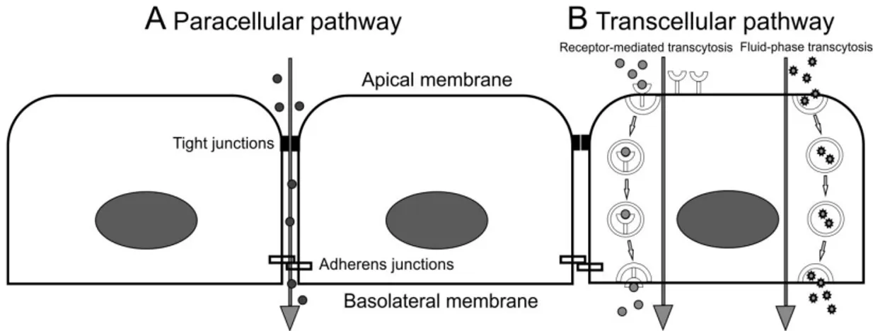
The paracellular transport describes the passage of small molecules, water and ions between neighboring cells across the endothelial barrier (Figure 1). This restricted permeability is sustained by interendothelial junctions, such as tight junctions, gap junctions, and adherens junctions. Tight junctions and adherens junctions impede the uncontrolled penetration of large molecules by attaching neighboring cells, whereas gap junctions form paracellular channels which allow and facilitate the passage of small molecules, water and ions (Komarova and Malik, 2010).
Figure 1: Paracellular and transcellular pathways to overcome the endothelial barrier. A, Small molecules, such as water and ions are transported via the paracellular route from the apical side to the basolateral side of the cell. Tight junctions and adherens junctions allow the passage of small molecules between neighboring cells. B, Transcellular transport involves membrane-bound vesicles which detach from the plasma membrane and transport molecules from the apical membrane to the basolateral side were they fuse again with the plasma membrane and release their cargo. This transport can be either by receptor-mediated transcytosis or by fluid- phase transcytosis.
1.1.2 Transcellular transport
The process of transcytosis was first described by Palade in the year 1950 during his studies on the permeability of blood capillaries. However, the term transcytosis was characterized by Simionescu in the year 1979 (Tuma and Hubbard, 2003). Transcytosis is defined by the process of transporting albumin from the luminal side of a capillary to the subendothelial
space (Minshall et al., 2002; Tuma and Hubbard, 2003). As already mentioned, besides albumin, other macromolecules such as albumin-binding ligands, insulin, lipids, and hormones are transported via transcytosis (Frank et al., 2003a; Minshall et al., 2002).
Soluble plasma molecules can be taken up together with blood plasma, a process called fluid-phase transcytosis, or by receptor-mediated transcytosis which requires a specific binding of the molecule to a membrane-bound receptor (Tuma and Hubbard, 2003) (Figure 1). Transcytosis is an energy-dependent transport which takes place by means of membrane-bound vesicles. To transport molecules within the cell, they need to get internalized at the plasma membrane. Specialized cells, commonly referred to as phagocytes, such as macrophages, monocytes, neutrophils, and dendritic cells can internalize substances via phagocytosis, an important mechanism in terms of defense against infectious agents (i.e. bacteria and viruses). However, in most cell types the main mechanism to internalize molecules is via endocytosis. Endocytosis is defined as the process in which molecules get engulfed in coated vesicles that are formed at the plasma membrane. So far, there are three different types of coated vesicles, the Clathrin-coated, the CopI-coated, and the CopII-coated vesicles. Among those, Clathrin-coated vesicles are the best studied type so far. After formation at the plasma membrane, the vesicles detach from the membrane along with the engulfed molecules. Shortly after detaching, the clathrin- covering gets discarded and the transport vesicles migrate to their target compartment and fuse with the membrane. First they reach the early endosome than the late endosome and finally the lysosome.
Lately, the spotlight has been put on a clathrin-independent mechanism as well, namely the caveolae-mediated endocytosis (Pelkmans and Helenius, 2002).
1.1.2.1 Caveolae
In turn, the pioneering work was done by Palade and Yamada. During his observations of semithin sections from continuous lung endothelia, Palade discovered the existence of cell invaginations with a "cave-like" morphology in the early 1950s. The "cave-like" appearance gave this newly discovered cellular structure the name caveolae.
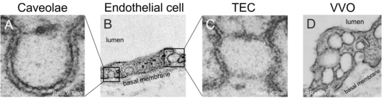
Caveolae are spherical, omega-shaped cell invaginations of the plasma membrane with a mean diameter of 50-80 nm (Figure 2A and B). They are also called non-coated or smooth- coated plasmalemmal invaginations, because they lack a cytoplasmic coat which is a typical feature of clathrin-coated pits (Gumbleton et al., 2000). Caveolae are thought to arise from lipid rafts, areas at the plasma membrane which are rich in cholesterin, glycosphingolipids, and GPI-anchoring proteins. Caveolae are very common and appear in many highly
differentiated mammalian cell types with some exceptions such as red blood cells, thrombocytes, lymphocytes, and neuronal cells (Fra et al., 1995). Caveolae tend to appear in clusters, but they are also found as single pits. Recent studies have shown that caveolae are involved in cellular transport and human diseases such as cancer, muscular dystrophy, Alzheimer's disease, and atherosclerosis (Engelman et al., 1998; Frank, 2010). A big step in the research of caveolae was the discovery of caveolin, the main structural protein of caveolae (Rothberg et al., 1992). Caveolin, now referred to as caveolin-1 is an integral membrane protein which is part of a protein family with at least three members in vertebrates: caveolin-1, caveolin-2, and caveolin-3 (Glenney, 1992; Rothberg et al., 1992;
Scherer et al., 1996; Tang et al., 1996). Caveolin-1 and caveolin-2 are highly expressed in endothelial cells, adipocytes, and type I pneumocytes, Caveolin-3 is only expressed in cells of skeletal and cardiac muscle (Scherer et al., 1996, 1994; Tang et al., 1996), whereas in smooth muscle cells all protein members are present (Tang et al., 1996). Like clathrin-coated vesicles, caveolae are capable of engulfing molecules and detaching from the plasma membrane. Unlike clathrin-coated vesicles, caveolae can migrate from the luminal to the abluminal side of the cell where they release their cargo, a scenario which has been shown by perfusion with varying tracer molecules (Predescu et al., 1997; Simionescu et al., 1975).
In addition, similar to phagocytes, caveolae are able to take up viruses and bacteria, and to deliver them from the cell surface to intracellular compartments for degradation (Anderson et al., 1996; Montesano et al., 1982; Shin et al., 2000). In most endothelial cells, caveolae are more common than clathrin-coated pits. The highest number of caveolae are found in endothelial cells of heart, lung and skeletal muscle (Aird, 2007a).
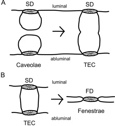
1.1.2.2 Transendothelial channels
Transendothelial channels (TECs) are openings which span the entire cell from the luminal to the abluminal side (Figure 2B and C). They are most likely formed either by the fusion of a caveola with the luminal and abluminal plasma membrane, or by the fusion of 2 to 4 caveolae (Stan, 2000). After application of VEGF, Chen et al. demonstrated an enrichment of caveolae in HUVEC (human umbilical vein endothelial cells), followed by the fusion of caveolae to transendothelial channels (Chen et al., 2002).
1.1.2.3 Vesiculo-vacuolar organelles (VVOs)
The term vesicular-vacuolar organelles (VVOs) represents the fusion or cluster of several caveolae (Figure 2D). VVOs are evident in endothelial cells of arterioles, capillaries, and most commonly in venular endothelium (Feng et al., 2002), are capable of spanning the entire thickness of the cell and can form groups of more than 100 single caveolae (Dvorak and Feng, 2001).
Figure 2: Endothelial cell organelles. Transmission electron micrographs of endothelial subcellular structures.
A, Caveola with a stomatal diaphragm. B, Endothelial cell showing caveolae on the abluminal side of the cell (boxed area on the left) and a transendothelial channel (boxed area on the right). C, Electron micrograph of a transendothelial channel (TEC) with two diaphragms on both sides. D, Endothelial cell presenting vesiculo- vacuolar organelles. Pictures in A, C and D are taken from Radu V Stan, 2007.
1.2 Endothelial Heterogeneity
Vascular endothelial cells of different vascular beds are remarkably heterogeneous with respect to their structural and functional properties. This heterogeneity is based on the different needs for permeability of water and solutes in the different vascular beds (Aird, 2007a, 2007b). Endothelial cells are either discontinuous or continuous.
1.2.1 Discontinuous endothelium
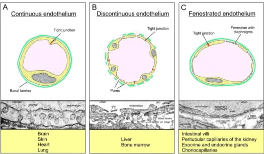
Discontinuous endothelial cells are evident in vascular beds such as the bone marrow, the spleen, but most notably the liver (Figure 3B). They are characterized by large circular openings with mean diameters of 100-300nm - depending on the species - and the absence of a basal lamina (Braet and Wisse, 2002). In the liver, sinusoidal pores function as a
dynamic biofilter, which separates macromolecules of different sizes, mainly lipoproteins, between the sinusoidal lumen and space of Disse (Fraser et al., 1995).
Figure 3: Endothelial heterogeneity. Schematic cross sections of the three basic types of blood capillaries. A, Continuous endothelium with endothelial cells linked by tight junctions and underlying continuous basal membrane. Continuous endothelial cells are found in brain tissue, skin, heart and lung and typically contain numerous caveolae. B, Discontinuous endothelium is characterized by the presence of large circular open pores with a mean diameter of 100-300 nm and by the absence of a basal lamina. Discontinuous endothelial cells are evident in vascular beds such as liver and bone marrow. C, Characteristic for fenestrated endothelial cells are transcellular pores with a mean diameter of 62-68 nm which are, depending on the vascular bed, bridged by a fenestral diaphragm. (Modified after Benninghoff and Drenckhahn, 2003).
1.2.2 Continuous endothelium
Continuous endothelial cells can be further classified into non-fenestrated or fenestrated endothelia (Tse and Stan, 2010). Non-fenestrated continuous endothelial cells are commonly found in capillaries of the brain, skin, heart, and lung (Figure 3A). The endothelial cells are connected by tight junctions and supported by an underlining continuous basal membrane.
1.2.3 Fenestrated endothelium
Fenestrated endothelial cells (Figure 3C) are also associated with a continuous basal membrane and are located in capillaries of the intestinal villi, peritubular capillaries of the
kidney, exocrine and endocrine glands, or the choriocapillaris in the eye, all organs with a substantial need for filtration and transendothelial transport. The structural hallmark is the presence of pores or fenestrae.
1.2.3.1 Fenestrae
Fenestrae are transcellular circular openings with a constant diameter of 62-68 nm which extend the entire thickness of the cells (Clementi and Palade, 1969a, 1969b; Simionescu et al., 1974). Fenestrae are arranged in so called sieve plates, large clusters of individual fenestrae. The density of fenestrae varies depending on the vascular bed. For example, in the kidney cortex 8.35 % of total endothelial surface is occupied by fenestrations, in contrast to the exocrine pancreas with only 3.68 % (Milici et al., 1985).
Depending on the particular vascular bed, caveolae, VVOs, TECs, and fenestrae are characteristically bridged by a thin 5 to 6 nm non-membranous diaphragm (Clementi and Palade, 1969c; Friederici, 1969, 1968a). Caveolae and TECs are covered by a stomatal diaphragm, fenestrae by a fenestral diaphragm (Figure 3C), both similar in regard to their structure. The diaphragm may qualify these structures with increased size and charge selectivity, allowing only small molecules and limited amounts of plasma to pass (Aird, 2007a; Palade et al., 1981; Simionescu et al., 1981b, 1981c). In vitro studies could show that factors like vascular endothelial growth factor (VEGF), extracellular signals (i.e. TGFβ, serotonin, retinoic acid), phorbol myristate acetate (PMA), cytochalasin B, calcium ions, or extracellular matrix components influence fenestrae formation (Carley et al., 1988; Esser et al., 1998a; Gatmaitan et al., 1996; Lombardi et al., 1988, 1986; McGuire et al., 1992; Milici et al., 1985; Steffan et al., 1987). Especially VEGF has been shown to be able to induce fenestrae formation. For instance, Roberts and Palade have shown that the topical application of VEGF to the rat cremaster muscle or subcutaneous injection in the skin induced the formation of fenestrae in these tissues, which do not contain fenestrae naturally.
Furthermore, fenestrae induction was reported to appear after only 10 min exposure in situ, indicating that the molecular "ingredients" for the formation of diaphragms are already available, given that exposure time was too short for de novo protein synthesis (Roberts and Palade, 1995). However, the importance of VEGF in fenestrae formation is not generally accepted. Several studies in recent decades presented controversial results (Hofman et al., 2000; Kohn et al., 1992; Vasile et al., 1999).
Besides fenestrated vascular beds, also endothelial cells of Schlemm's canal endothelium possess fenestrae-like pores. The inner wall of Schlemm's canal (SC) shows one of the highest hydraulic conductivities throughout the body allowing the aqueous humor to exit the eye by passing the trabecular meshwork and entering the lumen of Schlemm's canal. The high hydraulic conductivity requires the formation of large intracellular (I-pores) and intercellular pores (B-pores) with diameters of 0.1 to 2 µm (Johnson, 2006). Solitary minipores which were found in SC endothelial cells of monkey and human eyes (Inomata et al., 1972; Tamm, 2009a) are very similar to diaphragmed fenestrae of fenestrated capillaries related to their size and structure, but are much more rarely formed in endothelial cells of SC endothelium than in those of fenestrated endothelia. Bill and Mäepea hypothesized that I- pores of SC develop from those minipores (Inomata et al., 1972), a scenario quite similar to that seen in liver sinusoids and glomerular capillaries during embryonic development (Bankston and Pino, 1980; Braet and Wisse, 2002; Ichimura et al., 2008).
1.3 Plasmalemma vesicle-associated protein
The only protein known so far to be localized to diaphragms of fenestral and stomatal diaphragms is plasmalemma vesicle-associated protein (PLVAP, synonyms: MECA32, PV-1) a cationic single span type II integral membrane protein which is specifically expressed in endothelial cells (Stan et al., 1999a, 1999b). PLVAP is encoded by Plasmalemma vesicle- associated Protein gene in humans, but the protein has also been documented in several other mammalian species including mouse, rat, chicken and bovine (Stan, 2005).
1.3.1 Ultrastructure and molecular composition of PLVAP
Protein sequences and cDNA of Plvap are highly conserved across species. The monomer has a calculated molecular mass of about 50 KDa in the unglycosylated form and about 60 KDa in the glycosylated form and tends to form homodimers in situ. The human PLVAP consists of a short N-terminal cytoplasmatic domain, a single span transmembrane domain, and a long C-terminal extracellular domain (Figure 4). The intracellular domain contains a short sequence near the transmembrane region with a putative caveolin-1 binding site (Stan, 2005). The extracellular domain holds nine cysteines, four consensus N-glycosylation sites, a proline-rich region near the C-terminus, and two coiled-coil domains. The presence of the two
coiled-coil domains suggests the secondary structure of PLVAP to be mostly alpha helical (Hallmann et al., 1995; Stan et al., 1999b).
Figure 4: Schematic drawing of the PLVAP monomer. PLVAP consists of a short N-terminal cytoplasmatic domain, a single span transmembrane domain, and a long extracellular C-terminal domain. The extracellular domain holds nine cysteines, four consensus N-glycosylation sites, a proline-rich region near the C-terminus, and two coiled-coil domains. TM=transmembrane domain. Cyt=cytoplasmic (modified after Stan, 2007)
1.3.2 Localization of PLVAP and its participation in fenestrae formation
PLVAP is highly expressed in lung tissue confirmed on both mRNA and protein level, but it is also expressed in several other vascular beds such as kidney, liver, spleen, heart and skeletal muscle, but at much lower levels. Within those tissues, PLVAP is exclusively found at stomatal diaphragms of caveolae and TECs, and fenestral diaphragms of fenestrae, as shown by immunolocalization of PLVAP in rat lung (Stan et al., 1999a).
Stan and colleagues were able to show the de novo formation of stomatal diaphragms in caveolae and TECs, as well as fenestral diaphragms in fenestrae by upregulation of Plvap expression in human umbilical vein endothelial cells (HUVEC) with phorbol myristate acetate.
Downregulation of Plvap by RNA interference resulted in the loss of diaphragms in caveolae.
In addition, no fenestrae and transendothelial channels were observed (Stan et al., 2004). In turn, Ioannidou et al. found only a 70 % reduction in the amount of PLVAP in siRNA-treated cells. Furthermore, the partial reduction in PLVAP did not cause a significant reduction in the number of fenestrae. However, a population of fenestrae did not form any diaphragm and those fenestrae had enlarged and variable diameters and disorganized sieve plates, implicating that PLVAP is necessary in fenestral morphogenesis and arrangement in sieve plates (Ioannidou et al., 2006).
1.3.3 Structure of diaphragms and integration of PLVAP
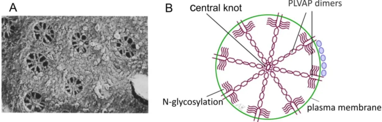
Based on a quick-freeze, deep-etch study from Bearer and Orci in the year 1985 (Bearer and Orci, 1985), Radu Stan postulated a working hypothesis for the structure of stomatal and fenestral diaphragms and the integration of PLVAP in diaphragms (Stan, 2007). He assumes that both stomatal and fenestral diaphragms are built of a scaffold of radial fibrils which are on one side anchored in the rim of the pore and on the other side interweave in a central knot, resulting in a structure similar to a spoke (Figure 5). He reckons that the fibrils consist of PLVAP dimers whose C termini form the central point of the diaphragm and the intense glycosylation near the N-terminal side of the protein (Figure 4) might help to keep the dimer
"afloat". Furthermore, the diaphragm could be stabilized by interactions between the C termini of neighboring PLVAP dimers or by the cooperation of an additional stabilizing extracellular protein. In general, stomatal and fenestral diaphragms are very similar with respect to their structure, but behave differently. Studies with non-specific probes such as cationic molecules (Palade et al., 1981; Simionescu et al., 1984, 1982, 1981b) or lectins (Bankston and Milici, 1983; Bankston et al., 1991) showed that stomatal diaphragms can bind lectins and lack anionic sites. In contrast, fenestral diaphragms do hardly bind lectins (Furuya, 1990) but possess multiple anionic sites instead, which are conferred by heparan sulfate proteoglycans (HSPGs) (Simionescu et al., 1981c).
Figure 5: Molecular structure of PLVAP. A, Quick-freeze, deep-etch scanning electron micrograph of a peritubular capillary in the rat kidney cortex presenting numerous fenestrae with fenestral diaphragms. Many interweaving fibrils which converge in a central point are observable. B, Working hypothesis for the integration of PLVAP in endothelial diaphragms. Fibrils composed of PLVAP dimers are anchored in the rim of the pore (via the N-terminal domain) and meet in the midpoint in a central mesh (via the C-terminal domain). The glycosylation sites near the N-terminal side of the protein helps to keep the dimer "afloat" by preventing the collapse on the plasma membrane. Picture in A is taken from Bearer and Orci, 1985. Picture in B is modified after Stan, 2007.
1.4 Aim of the study
Since stomatal and fenestral diaphragms are thought to play a crucial role in vascular permeability and might be involved in pore formation in Schlemm's canal endothelium, the overall goal of the current study was to molecularly dissect the biological function of PLVAP.
Specifically, the plan involved to find answers to some pivotal questions in endothelial cell biology.
What happens with stomatal and fenestral diaphragms in fenestrated endothelial cells if PLVAP is absent?
Are there any general effects on endothelial cell permeability?
Is there any impact on Schlemm's canal physiology?
Furthermore, scientists recently gained some insight into the molecular composition and formation of caveolae (Fra et al., 1995; Stan et al., 1999a, 1999b, 1997; Vogel et al., 1998), whereas the precise mechanisms for the formation of TECs and fenestrae still need to be explored. According to a current hypothesis, fenestrae might evolve from the fusion of caveolae during endothelial thinning, followed by the approach of the membranes (Figure 6).
Figure 6: Theory of fenestrae formation via fusion of caveolae. Schematic drawing of the potential onset of fenestrae formation. A, During endothelial thinning, caveolae could get in contact and fuse to transendothelial channels with two diaphragms on both sides. B, Further thinning is supposed to lead to a collapse of the transendothelial channel followed by the loss of one diaphragm. Modified after Kathrin Ebner, 2008.
Still, several findings argue against this hypothesis. For instance, caveolin-1 has been shown to be absent in fenestrae of glomerular endothelial cells in vivo (Sörensson et al., 2002) and adrenal cortical microvascular endothelial cells in culture (Esser et al., 1998b). In addition, neither the distribution, amount or phosphorylation of caveolin-1 is altered by VEGF (Esser et al., 1998b). Furthermore, the fact that fenestrations in kidney and liver endothelial cells in caveolin-1 knockout mice (which do not form caveolae) do not show any alterations argues strongly against this hypothesis (Sörensson et al., 2002; Warren et al., 2010). These data provoked additional questions, which were followed up in this thesis:
Is PLVAP involved during the process of caveolae, TEC, and fenestrae formation?
Is PLVAP essential for their formation?
Due to the fact that fenestrae are bridged by diaphragms during embryonic development of liver sinusoids and glomerular capillaries (Bankston and Pino, 1980; Ichimura et al., 2008), I investigated liver sinusoidal endothelial cells in more detail.
The general concept of my work was based on the assumption that the deletion of PLVAP would provide an opportunity to answer those questions based on data obtained in vivo.
In particular, following goals were pursued:
1) The generation of a knockout mouse line, resulting in a complete deletion of PLVAP
2) The phenotype analysis of Plvap-deficient mice during embryonic and postnatal development in 2 different genetic backgrounds
3) The analysis of the general expression pattern of PLVAP in fenestrated endothelial cells of Schlemm's canal endothelium and ocular capillaries
4) The investigation of fenestrated vascular beds of Plvap-deficient animals in greater depth (i.e. peritubular capillaries of the kidney, capillaries of the pancreas, and Schlemm's canal endothelium)
5) The investigation of sinusoidal endothelial cells in livers of Plvap-deficient animals
Chapter 2
Lack of endothelial diaphragms in fenestrae and caveloae of mutant Plvap-deficient mice
(adapted from: Herrnberger L, Seitz R, Kuespert S, Bösl MR, Fuchshofer R, Tamm ER.
Lack of endothelial diaphragms in fenestrae and caveolae of mutant Plvap-deficient mice. Histochem Cell Biol. 2012, 138(5):709-724)
2 Lack of endothelial diaphragms in fenestrae and caveloae of mutant Plvap-deficient mice
2.1 Abstract
Plasmalemma vesicle-associated protein (PLVAP, PV-1) is specifically expressed in endothelial cells in which it localizes to diaphragms of fenestrae, caveolae, and transendothelial channels. To learn more about its function, we generated mutant mice that lack PLVAP. In a C57BL/6N genetic background, homozygous Plvap-deficient embryos die before birth and suffer from subcutaneous edema, hemorrhages, and defects in the vascular wall of subcutaneous capillaries. In addition, hearts of Plvap -/- embryos show ventricular septal defects and thinner ventricular walls. In wild-type embryos, PLVAP and caveolae with a stomatal diaphragm are present in endothelial cells of subcutaneous capillaries and endocardium, while a diaphragm is missing in caveolae of Plvap -/- littermates.
Plvap -/- mice in a mixed C57BL/6N/FVB-N genetic background are born and survive at the most for 4 weeks. Capillaries of exocrine and endocrine pancreas and kidney peritubular interstitium were investigated in more detail as examples of fenestrated capillaries. In these vascular beds, Plvap -/- mice show a complete absence of diaphragms in fenestrae, caveloae, and transendothelial channels, findings which are associated with a substantial decrease in the number of endothelial fenestrae. The changes in the capillary phenotype correlate with a considerable retardation of postnatal growth and anemia. Plvap -/- mice provide an animal model to clarify the specific functional role of endothelial fenestrae and their contribution to passage of water and solutes in different organs.
2.2 Introduction
The inner surface of blood vessels and lymphatics are lined by endothelial cells which critically contribute to the various physiological requirements of circulation. Since the different vascular beds vary considerably with respect to their specific functions, vascular endothelial cells are remarkably heterogeneous with regard to their structural and functional properties, and their expression pattern of characteristic molecules (Aird, 2007a, 2007b). Endothelial cell heterogeneity is especially evident in capillaries in which different needs for permeability of water and solutes require different structural phenotypes of endothelial cells. As a consequence, capillary endothelia may be of a discontinuous, fenestrated, or continuous type (Tse and Stan, 2010). Discontinuous capillary endothelial cells such as in the sinusoids of the liver form large circular pores that are 100-200 nm in diameter. Such openings are missing in continuous endothelial cells which are commonly found in capillaries of the brain, skin, heart, and lung. In contrast, fenestrated endothelia are observed in capillaries of exocrine and endocrine glands, intestinal villi, peritubular interstitium of the kidney, or the choriocapillaris in the eye, all organs or tissues that have in common the need for a substantial filtration and transendothelial transport.
The structural hallmarks of endothelial cells in fenestrated capillaries are transcellular pores or fenestrae with a remarkably constant diameter of 62-68 nm, which extend through the full thickness of the endothelial cell. Fenestrae are characteristically bridged across their opening by a thin 5-6 nm non-membranous diaphragm (Clementi and Palade, 1969a; Friederici, 1969, 1968a). Results from quick-freeze deep-etch electron microscopy suggest that the diaphragm consists of radial fibrils which extend from the peripheral rim of the cellular pore to meet and interweave in a central mesh (Bearer and Orci, 1985). It has been suggested that the presence of a diaphragm may provide fenestrae with increased size and charge selectivity allowing only small molecules and limited amounts of plasma protein to pass (Aird, 2007a; Palade et al., 1981; Simionescu et al., 1981b, 1981c).
Up until now, the only protein that has been found to be directly localized to fenestral diaphragms is plasmalemma vesicle-associated protein (PLVAP, synonyms: MECA32, PV-1), a cationic, integral membrane glycoprotein that is specifically expressed in endothelial cells (Stan et al., 1999a, 1999b). In addition to its presence in diaphragms of endothelial fenestrae, PLVAP was also found to be localized in the stomatal diaphragms of caveolae and the diaphragms of transendothelial channels (Stan, 2005; Stan et al., 2004). Moreover, in vitro studies in cultured cells have shown that the knock-down of Plvap expression by RNA interference impairs the formation of diaphragms in fenestrae, caveolae, and transendothelial
channels (Ioannidou et al., 2006; Stan et al., 2004). Caveolae which are defined as spherical invaginations of the plasma membrane of regular shape and size (about 70 nm) occur in all types of endothelia as well as in most vertebrate cell types. Still, caveolae with stomatal diaphragms are only found in some selected endothelia of the continuous type (i.e., in the capillaries of the lung) and in all fenestrated and sinusoidal endothelia (Bruns and Palade, 1968; Stan, 2005). The stomatal diaphragm of caveolae appears to be quite comparable in structure to the diaphragms of fenestrae (Bearer and Orci, 1985). Transendothelial channels are pores through the entire endothelial cell provided with two diaphragms, one luminal and one abluminal, which are found predominantly in fenestrated endothelia (Milici et al., 1985). It has been hypothesized that caveolae with stomatal diaphragms fuse from luminal and abluminal sides of endothelial cells to form transendothelial channels which may then collapse to form fenestrae (Roberts and Palade, 2000). Still, the observation that in mutant caveolin-1-deficient mice, which do not form endothelial caveolae (Drab et al., 2001; Razani et al., 2001), the number of diaphragmed fenestrae and transendothelial channels is largely unchanged (Tkachenko et al., 2012; Zhao et al., 2002) argues against this hypothesis and leaves the functional role of the stomatal diaphragm in caveolae unresolved.
In order to shed light on the in vivo function of PLVAP and its role in the formation of diaphragms in fenestrae, caveolae, and transendothelial channels, we generated mutant mice that lack PLVAP. We show that in the living organism such diaphragms do not form in the absence of PLVAP. Depending on the genetic background, the lack of diaphragms interferes with cardiac morphogenesis, the integrity of embryonic vessels, and the formation of fenestrated endothelial layers. Our mouse model will provide an ideal tool to study the in vivo functional properties of diaphragms in capillary endothelial cells.
2.3 Materials and methods
2.3.1 Generation of Plvap-deficient mice
Embryonic stem cell clones (HEPD0550_4_E12) were obtained through the International Knockout Mouse Consortium (Projekt 31505) (Skarnes et al., 2011). The targeted ES cells were injected into C57Bl/6N blastocysts to generate chimeras. Chimeric males transmitted the targeted allele to their offspring. Genotyping was routinely performed by PCR analysis, using two upstream primers located in intron 1 of Plvap (5’-AGAGCCTTCTCTGCCAAGTG- 3’) or in the inserted targeting cassette (5’-TCTCATGCTGGACTTCTTCG-3’), and a downstream primer located in intron 1 downstream of the cassette (5’- GGCTAGCCTGAGCTACAGAGG-3’) resulting in a 672 bp PCR fragment for the wild-type allele and a 552 bp fragment for the targeted allele. DNA was obtained from tail tips. PCR analysis was performed in 15 µl reaction volumes containing standard buffer, 0.1 µM of each primer, 1 mM dNTPs, 2.5 mM MgCl2 , 8% glycerol, 0.2 mM cresol red sodium salt (Sigma, Taufkirchen, Germany) and 0.25 U Taq-Polymerase (New England Biolabs, Taufkirchen, Germany). The cycling conditions consisted of an initial 3-min denaturing step at 94°C, followed by 34 cycles for 30 s at 94°C, for 1 min at 65°C, and for 1 min 15 s at 72°C.
Tg (TIE2GFP) 287Sato/J mice (Stock number 003658) (Motoike et al., 2000) were obtained from The Jackson Laboratory (Bar Harbor, Maine). Genotyping was routinely performed by PCR analysis, using a forward primer (5’-ATTCTCGTGGAACTGGATGG-3’) and a reverse primer (5’-GGACAGGTAATGGTTGTCTGG-3’), resulting in a 567bp fragment. DNA was obtained from tail tips. PCR was performed in 15 µl reaction volumes containing standard buffer, 0.1 µM of each primer, 1 mM dNTPs, 2.5 mM MgCl2 , 20% glycerol, 0.2 mM cresol red sodium salt (Sigma) and 0.25 U Taq-Polymerase (New England Biolabs). The cycling conditions consisted of an initial 3 min denaturing step at 94°C, followed by 31 cycles for 30 s at 94°C, for 1 min at 60°C, and for 1 min at 72°C.
2.3.2 RNA analysis
Total RNA from embryos at embryonal (E) day 13.5 or postnatal (P) day 13 was extracted with peqGold Trifast (Peqlab, Erlangen, Germany) according to manufacturer`s instructions.
First strand cDNA from total RNA was generated using the iScript cDNA Synthesis Kit (BioRad, München, Germany) according to the manufacturer`s recommendations. RNA that
was not reverse transcribed into cDNA served as negative control for real-time RT-PCR.
Real-time RT-PCR was performed on a BioRad iQ5 Real-time PCR Detection System (BioRad) with the temperature profile as follows: 40 cycles of 10 s melting at 95°C and 40 s of annealing and extension at 60°C. Primer pairs (Table 1) were purchased from Invitrogen (Darmstadt, Germany) and were designed to extend over exon-intron boundaries. In initial experiments, the potential housekeeping genes for real-time RT-PCR were identified for each tissue and best results were obtained for GAPDH for whole embryos at E13.5 and GNB2L for mouse tissue (P13). Quantification was performed using BioRad iQ5 Standard-Edition (Version 2.0.148.60623) software (BioRad).
Primer Sequence
mPlvap fwd 5’-TCAACAAGACCTGCGAAGC-3’
mPlvap rev 5’-AGCACACTGCCTTCTCCTTG-3’
mGNB2L fwd 5’-TCTGCAAGTACACGGTCCAG-3’
mGNB2L rev 5’-GAGACGATGATAGGGTTGCTG-3’
mGAPDH fwd 5’-TGTCCGTCGTGGATCTGAC-3’
mGAPDH rev 5’-CCTGCTTCACCACCTTCTTG-3
Table 1. Primer used for real-time RT-PCR
2.3.3 Western blot analysis
Tissues were homogenized in RIPA buffer (150 mM NaCl, 1% NP-40, 0.5% deoxycholic acid, 0.1% SDS, and 50 mM Tris), and insoluble constituents were removed by centrifugation. For Western blot analysis of PLVAP up to 30 µg of protein was subjected to an 8% SDS-PAGE and transferred onto a PVDF membrane (Roche Diagnostics GmbH, Mannheim, Germany) by semidry blotting. After blocking with 5% low-fat milk in TBS-T, membranes were incubated overnight with rat anti-mouse pan ECA (MECA-32) IgG2a antibodies (Santa Cruz Biotechnology, Santa Cruz, CA), diluted 1:500 in 0.5 % low-fat milk in TBS-T. After washing in TBS-T, membranes were hybridized with HRP-conjugated chicken anti-rat antibodies (SantaCruz), diluted 1:1000 in 0.5% low-fat milk in TBS-T. For visualization, membranes were incubated in Luminata Forte Western HRP Substrate (Millipore Corporation, Billerica,
MA) and visualized on a LAS 3000 Imager work station (Fujifilm, Düsseldorf, Germany). As loading control membranes were stained with Coomassie Blue.
2.3.4 Light microscopy
Embryos were obtained from timed mating with noon of the day of vaginal plug discovery designated as 0.5 days of gestation (E 0.5). Embryos and tissues were collected, rinsed in PBS and fixed in 4 % PFA for 4 h. After washing in 0.1 M phosphate buffer the tissue samples were processed through several graded alcohols and xylenes, and embedded in paraffin. Sections were stained with hematoxylin and eosin (H and E) and analyzed using a Zeiss Axio Imager microscope (Carl Zeiss AG, Oberkochen, Germany). Blood smears were generated with blood collected from tail veins. The slides were fixed in 100% methanol for 10 min, washed in H2O and stained with 300 µl May-Grünwald-Giemsa solution (Fluka Chemika, Buchs, Switzerland) in 10 ml H2O. For Immunohistochemistry, embryos and tissue samples were fixed in Carnoy’s fixative (60 % methanol, 30 % chloroform, and 10 % acetic acid) for 4 h, washed in 0.1 M phosphate buffer and embedded in paraffin. For frozen sections, the tissue was equilibrated in 10, 20, and 30 % sucrose for 4 h and embedded in Tissue-Tek optimal cooling temperature (OCT) compound (Sakura Finetek Europe B.V., Zoeterwoude, NL) and cooled at -20 °C. After removal of Tissue-Tek and paraffin, the sections were blocked with 0.2 % cold water fish gelatin (Aurion, Wageningen, Netherlands), 1 % BSA and 0.1 % Triton-X (all in 0.1 M PBS) for 1 h at room temperature. After blocking, the sections were incubated with anti-pan ECA (MECA-32) IgG2a (1:50, Santa Cruz) and anti- CD31/PECAM-1 IgG (1:20, R&D Systems, Wiesbaden, Germany) for double labeling. Alexa Fluor 488 (1:1000) rabbit anti-goat IgG (Invitrogen) and donkey anti-rat IgG (1:2000) conjugated to Cy3 (Jackson Immuno Research) were used as secondary antibodies and incubated for 1 h at room temperature. 4′,6-Diamidino-2-phenylindole (DAPI) was used to counterstain nuclear DNA. Slides were mounted in a medium containing DAPI (Vectashield;
Vector Laboratories, Burlington, CA, USA) and analyzed under a fluorescence microscope (Zeiss Axio Imager). For negative controls, primary antibodies were omitted and the slides were incubated with secondary antibodies only.
2.3.5 Transmission electron microscopy
Tissue samples were fixed in 2.5 % glutaraldehyde in 0.1 M cacodylate buffer (pH 7.4) for 4 h. Samples were washed, postfixed with 1 % OsO4, 0.8 % K4[Fe(CN)6] in 0.1 M cacodylate buffer for 1.5 h, dehydrated with graded ethanol solutions and embedded in Epon (Roth, Karlsruhe, Germany). Semithin sections were stained with Richardson’ stain (Richardson et al., 2009). Ultrathin sections were stained with uranyl acetate and lead citrate, and analyzed on a transmission electron microscope (Libra, Zeiss).
2.3.6 Staining for β-galactosidase activity
Embryos and tissues were fixed in 2.5 % glutaraldehyde in 5 mM EGTA (pH 7.3) and 2 mM MgCl2 dissolved in 0.1 M PBS for 4 h on ice with shaking. After three 30 min rinses in washing buffer (2 mM MgCl2, 0.01 % sodium deoxycholate, 0.02 % Nonidet-P40 in 0.1 M PBS), β-galactosidase activity was visualized in X-Gal staining solution (0.1 M PBS, 2 mM MgCl2, 0.01 % sodium deoxycholate, 0.02 % Nonidet-P40, 5 mM potassium ferrocyanide, 5 mM potassium ferricyanide, 1 mg/ml X-Gal). Tissues were stained for 24 h at 37 °C in the dark, washed 3 times for 5 min in washing buffer and embedded in paraffin or in Tissue-Tek for frozen sections.
2.4 Results
2.4.1 Targeted removal of Plvap leads to embryonic death
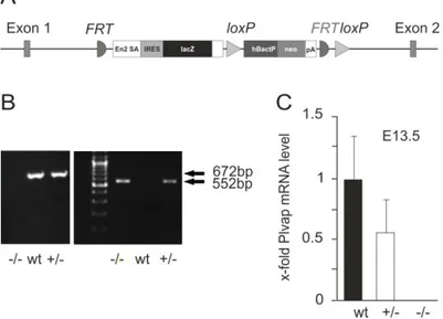
To disrupt gene function, an IRES:lacZ trapping cassette and a promoter-driven neo cassette (Skarnes et al., 2011) were inserted between exons 1 and 2 of Plvap (Figure 7A). Following germ-line transmission, genotyping of newborn pups was performed by PCR. In the C57BL/6N genetic background that was used for initial breeding, only heterozygous Plvap- deficient mice or wild-type littermates were detected, strongly indicating that the complete deletion of Plvap is lethal during prenatal development. Next, heterozygous Plvap-deficient mice were crossed with each other to obtain embryos for genotyping. We were now able to detect homozygous Plvap-deficient embryos in addition to embryos with Plvap +/- or wild-type genotype when litters at embryonic (E) days E 13.5 to E 17.5 were analyzed (Figure 7B). By real-time RT-PCR, we observed in Plvap +/- embryos a reduction of approximately 50% in the amount of Plvap mRNA when compared with wild-type littermates, while no mRNA was detected in Plvap -/- embryos (Figure 7C).
Figure 7: Generation of mutant Plvap-deficient mice. A, Schematic representation of the targeted allele. B, Genotyping by PCR using a template DNA from embryos following mating of heterozygous Plvap-deficient mice.
A 552-bp fragment illustrates the presence of the mutated allele which replaces the wild-type locus normally represented as a 672-bp fragment. C, Real-time RT-PCR analysis for Plvap mRNA in total RNA from whole embryos at E 13.5. The mean value obtained with RNA from wild-type embryos was set to 1. GAPDH was used as a reference gene.
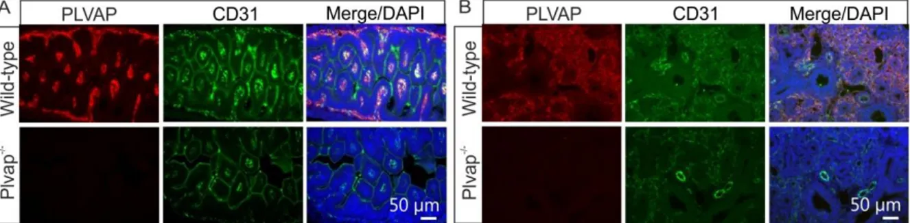
Lack of PLVAP was further confirmed by immunohistochemistry. In E 16.5 wild-type embryos, double immunohistochemistry with antibodies against PLVAP and the endothelial cell marker CD31 showed the presence of PLVAP in capillary endothelial cells of small intestine (Figure 8A) and lung (Figure 8B). In contrast, no labeling for PLVAP was observed in small intestine and lung of Plvap -/- littermates (Figure 8A, B).
Figure 8: Generation of mutant Plvap-deficient mice. A and B, Double immunohistochemistry with antibodies against PLVAP and the endothelial cell marker CD31 shows the presence of PLVAP in capillary endothelial cells of small intestine (A) or lung (B) in E 16.5 wild-type animals, but not in Plvap -/- littermates.
2.4.2 Plvap-deficient embryos suffer from subcutaneous edemas and hemorrhages
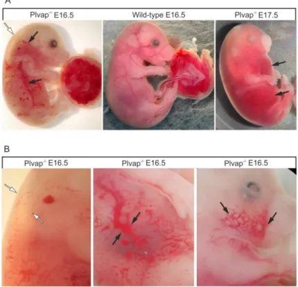
E 16.5 Plvap -/- embryos were typically presented with a pronounced subcutaneous edema that extended from the neck to the lower back (Figure 9A, B). In addition, focal hemorrhages were present in various subcutaneous regions of trunk, extremities, head, and neck (Figure 9A, B). In areas not affected by subcutaneous hemorrhages, the skin was quite pale when compared with that of wild-type littermates indicating anemia (Figure 9A). Moreover, blood filled intact vessels that shined through the skin of body or umbilical cord in wild-type animals were not visible in Plvap -/- embryos (Figure 9A). Neither subcutaneous edemas nor hemorrhages were observed in wild-type littermates (Figure 9A). At E 17.5, homozygous Plvap-deficient embryos suffered from severe total body edema and extensive non-specific focal hemorrhages (Figure 9A, B). While homozygous Plvap-deficient embryos were still viable at E 17.5 as evidenced by cardiac pulsation and spontaneous movements, no Plvap -/- embryos were observed to be alive at later stages.
Figure 9: Phenotype of Plvap-deficient embryos in C57BL/6N background. A, E 16.5 Plvap -/- embryo with a pronounced subcutaneous edema that extends from the neck to the lower back (white arrow). In addition, focal hemorrhages are present in various subcutaneous regions of trunk, extremities, head, and neck (black arrows). In areas not affected by subcutaneous hemorrhages, the skin is pale when compared with that of the wild-type littermate. In addition, blood-filled intact vessels that shine through the skin of the body or umbilical cord in the wild-type animal are not visible in the Plvap -/- embryo. Neither subcutaneous edema nor hemorrhages are seen in the wild-type embryo. The E 17.5 Plvap -/- embryo shows severe total body edema and extensive non-specific focal hemorrhages (black arrows). B, Higher magnification of edema in the dorsal region of an E 16.5 Plvap -/- embryo (white arrows) and of subcutaneous hemorrhages (black arrows).
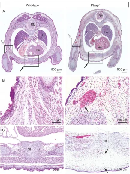
Subcutaneous edemas and hemorrhages were similarly detected in horizontal sections through E 16.5 Plvap -/- embryos, which showed accumulations of extracellular erythrocytes as well as a marked widening of the extracellular spaces beneath the skin in the anterior and dorsal regions of the trunk (Figure 10A-C). In contrast, neither subcutaneous edemas nor hemorrhages were observed in wild-type littermates (Figure 10A-C).
Figure 10: Histological characterization of Plvap-deficient embryos in C57BL/6N background. A, Transversal paraffin sections through the thorax of E 16.5 embryos stained with H and E. The Plvap -/- embryo displays edema in the anterior region of the trunk (black arrow) and hemorrhages (white arrow) beneath the skin, which are not seen in the wild-type littermate. B and C, Higher magnification of boxed areas in A. B, In the Plvap -
/- embryo, but not in the wild-type littermate, numerous erythrocytes (white arrows) can be seen outside of capillaries in the surrounding tissue, indicating open blood vessels (black arrow). C, Massive widening of the extracellular spaces indicating subcutaneous edema in the Plvap-deficient (black arrows). Sc spinal cord, Lu lung, He heart, St sternum.
To follow up on the question, if lack of PLVAP in subcutaneous blood vessels might be responsible for subcutaneous edemas and hemorrhages, we analyzed the expression and the localization of PLVAP in this region.
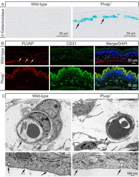
When staining for β-galactosidase was performed at E 15.5 to detect expression of the reporter gene lacZ and activity of the Plvap promoter, positive staining was observed in endothelial cells of subcutaneous capillaries in Plvap-deficient embryos, but was absent throughout the entire subcutaneous region of wild-type littermates (Figure 11A).
Subsequently, we performed immunostaining with antibodies against PLVAP and the
endothelial cell marker CD31 to find immunoreactivity for PLVAP in endothelial cells of the vast majority of mouse subcutaneous capillaries at E 16.5 (Figure 11B). In contrast, no specific immunoreactivity for PLVAP was observed in homozygous Plvap-deficient embryos (Figure 11B). Finally, we investigated endothelial cells of subcutaneous capillaries at E 17.5 by transmission electron microscopy. In both wild-type and Plvap-deficient embryos, the endothelium of subcutaneous capillaries was continuous without fenestrae (Figure 11C).
Upon higher magnification, endothelial cells of wild-type capillaries showed numerous caveolae with stomatal diaphragms on both their luminal and abluminal surfaces.
Accordingly, the neck or stroma of the caveolae was bridged by a distinct, 5- to 7-nm thick, electron-dense diaphragm (Figure 11C). Caveolae with stomatal diaphragms were invariably absent in capillaries of Plvap -/- embryos. Here, the neck of caveolae was not bridged by a diaphragm, but contained some ill-defined, fluffy electron-dense material instead (Figure 11C).
Figure 11: Expression and localization of PLVAP. A, β-galactosidase staining of sagittal frozen sections through the back skin and adjacent subcutaneous tissue of a wild-type embryo and a Plvap -/- littermate at E 15.5.
Positive staining (arrows) is seen in the endothelial cells of subcutaneous capillaries in the Plvap -/- embryo, whereas no staining is detectable in the wild-type littermate. B, Double labeling of transverse paraffin sections through the back skin and adjacent subcutaneous tissue of an E 16.5 wild-type embryo with antibodies against PLVAP (red) and CD31 (green) shows immunoreactivity for PLVAP in the majority of subcutaneous capillaries (arrows) and largely colocalizes with CD31 as seen in the merged picture. No specific staining for PLVAP is observed in the Plvap-deficient embryo. C, Transmission electron microscopy of subcutaneous capillaries at E 17.5. Both wild-type and Plvap -/- subcutaneous capillaries exhibit a continuous endothelium without fenestrae (arrows). At higher magnification, wild-type endothelial cells show numerous caveolae with a distinct 5- to 7-nm thick stomatal diaphragm (arrows). Caveolae are not bridged by a diaphragm in the endothelial cell membrane of the Plvap -/- embryo, but contain fluffy electron-dense material instead (arrows).
When areas with subcutaneous hemorrhages were more closely investigated, capillaries were observed in which the endothelial wall appeared to be incomplete (Figure 12A).
Transmission electron microscopy confirmed the lack of endothelial integrity and revealed extensive defects in the endothelial lining of capillaries. In places, the defects were bridged by degranulated thrombocytes (Figure 12B, C).
Figure 12: Loss of endothelial integrity in Plvap -/- embryos. A, Semithin section (Richardson's stain) through an area with subcutaneous hemorrhage in an E 16.5 Plvap -/- embryo. Erythrocytes are seen within and outside a capillary with a fragmented wall that contains large openings (arrows). B and C, Transmission electron
microscopy of a capillary with a large opening (arrows) that is bridged by processes of degranulated thrombocytes (Tr). C, Higher magnification of B.
2.4.3 Cardiac defects in Plvap -/- embryos
Since total body edema and extensive hemorrhages in mouse embryos are very often associated with defects in cardiac morphogenesis (Clark et al., 1999; Dumont et al., 1994;
Nasser et al., 2008; Patan, 1998; Peng et al., 2008; Puri et al., 1995; Ranger et al., 1998;
Sato et al., 1995; Wu et al., 2000), we next investigated the hearts of Plvap -/- embryos and of their wild-type littermates. E 16.5 embryos were cut in the transverse plane and examined in serial sections from the cephalic to the caudal aspects of the specimens. In each of the three
Plvap -/- embryos that were investigated, histological serial sections stained with H and E showed a distinct muscular ventricular septal defect (Figure 13A). In the affected embryos, the cardiac ventricular walls were also thinner than those of the wild-type embryos (Figure 13A). By immunohistochemistry, specific immunoreactivity for PLVAP was observed in the endocardium of wild-type mice, but not in that of Plvap -/- embryos (Figure 13A). Moreover, the endocardium of cardiac ventricle and atrium as well as the endothelial cells of the associated cardiac vessels were stained for β-galactosidase indicating activity of the Plvap promoter (Figure 13B).
Figure 13: Cardiac and blood abnormalities in Plvap -/- embryos. A, H and E stained transverse paraffin sections through the thorax of wild-type and Plvap -/- embryos at E 16.5. The heart (He) of the Plvap -/- embryo shows a muscular ventricular septal defect (black arrows) and a thinner ventricular wall (white arrow) when compared with the wild-type control. Immunohistochemistry for PLVAP (red) shows positiv immuoreactivity in the
ventricular endocardium of the wild-type heart (white arrows), but not in that of the Plvap -/- embryo. Nuclear DNA is labeled with DAPI (blue). B, β-galactosidase staining in an E 15.5 Plvap -/- embryo shows activity of the Plvap promoter in ventricular and atrial endocardium, and in the endothelium of associated cardiac vessels. C, Smears of blood (May-Grünwald-Giemsa stain) collected from tail veins at E 16.5 show more nucleated red blood cells from the Plvap -/- embryo compared with the wild-type littermate. D, Ratio of nucleated erythrocytes to enucleated erythrocytes at different embryonic stages (E 14.5, E 16.5, E 17.5). SC spinal cord, Lu Lung.
Finally, we investigated the percentage of nucleated erythrocytes in blood smears from embryonic blood (Figure 13C), as we had observed in Plvap -/- embryos multiple nucleated erythrocytes in tissue sections of hemorrhages (Figure 10B, Figure 12A). In the blood smear of an E 14.5 wild-type embryo, 21 % of erythrocytes were found to be nucleated, a number that dropped to 1 % in an E 16.5 embryo, while essentially no nucleated erythrocytes were observed at E 17.5. In contrast, in Plvap -/- embryos, 61 % of erythrocytes were nucleated at E 14.5, 11 % at E 16.5 and still 2 % at E 17.5 (Figure 13D).
2.4.4 Plvap -/- mice in a mixed C57BL/6N/FVB-N background are viable and do not form fenestrae with diaphragm
In the course of our studies, we crossed Plvap-deficient mice that had been bred in a C57BL/6N genetic background with TIE2GFP mice that are in a FVB-N genetic background.
While the initial goal was to take advantage of the GFP-labeling in endothelial cells of TIE2GFP mice, we observed that in the mixed C57BL/6N/FVB-N background, Plvap -/- animals are viable after birth and survive at the most up to an age of 4 weeks. E 17.5 embryos in the mixed background showed edema in the neck and back, but no visible hemorrhages (Figure 14A).
Figure 14: Phenotype of Plvap -/- animals in mixed C57BL/6N/FVB-N background. A, An E 17.5 Plvap -/- embryo shows edema in neck and back (white arrow) which is not seen in the wild-type littermate. B, A 15-day- old Plvap -/- animal with a kink in the tail (arrow) shows a marked reduction in body size when compared with its wild-type littermate. C, Real-time RT-PCR for Plvap mRNA in RNA from eye, pancreas, lung, and kidney of wild- type (wt), heterozygous (+/-) and homozygous (-/-) Plvap-deficient animals at 4 weeks of age. The mean value of wild-type RNA was set at 1. GNB2L was used as a reference gene. D, Western blot analysis for PLVAP in proteins isolated from kidneys of homozygous Plvap-deficient mice and their wild-type littermate at P 13. (E, F) Kidney (E), spleen (black arrows, F) and pancreas (white arrows, F) are smaller in a 3-week-old Plvap -/- animal when compared with the wild-type littermate and were very pale indicating anemia. Experiment in C performed by Sabrina Küspert. Experiment in D performed by Roswitha Seitz.
Postnatal, Plvap -/- mice show a marked reduction in body size when compared with wild-type littermates (Figure 14B). In addition, Plvap -/- mice typically show kinks in their tails that are not seen in wild-type littermates (Figure 14B). No ventricular septal defect was observed in tangential sections through the heart of 3-week-old mice (data not shown). When the expression of Plvap was analyzed by real-time RT-PCR at 4 weeks of age in organs known