Original article:
TREATMENT WITH PLATELET LYSATE INDUCES ENDOTHELIAL DIFFERENTIATION OF BONE MARROW MESENCHYMAL STEM CELLS UNDER FLUID SHEAR STRESS Farshad Homayouni Moghadam1,2,6*,Tahereh Tayebi1, Alireza Moradi3, Hamid Nadri3, Kazem Barzegar4, Gilda Eslami5
Shahid Sadoughi University of Medical Sciences, Yazd, Iran
1 Department of Physiology, School of Medicine
2 Neurobiomedical Research Center, School of Medicine
3 Department of Medicinal Chemistry, School of Pharmacy
4 English Language Department, School of Medicine
5 Department of Medical Parasitology and Mycology, School of Medicine
6 Yazd Cardiovascular Research Center
* Corresponding author: Farshad Homayouni Moghadam, Shahid Sadoughi University of Medical Sciences, Shohadaye Gomnam Blv , 8915173143, Yazd, Iran,
Tel.: +98 351 8202630; Fax: +98 351 8203411; E-mail: f.homayounie@gmail.com
ABSTRACT
By considering stem cell-based therapies as a new hope for the treatment of some tragic dis- eases, marrow stromal cells or marrow mesenchymal stem cells (MSCs) were considered as a suitable and safe multipotential cell source for this new therapeutic approach. For this pur- pose, many investigations have been performed on differentiation of MSCs toward specific cell lines to overcome the demand for providing the organ specific cells for cell therapy or preparation of engineered tissues. In the present study, differentiation of MSCs to endothelial cells (ECs) by mechanical and chemical stimulation was evaluated. Fluid shear stress (FSS) was used as mechanical inducer, while platelet lysate (PL) and estradiol (E) were used as chemical induction factors. MSCs were placed under FSS with different forces (2, 5 and 10 dyn/cm2) for different periods (6, 12 and 24 hours). In some groups, PL and E were added to the culture media to evaluate their effect on expression of EC specific markers. This inves- tigation revealed that FSS with low tension (2.5-5 dyn/cm2) for a long time (24 hours) or high tension (10 dyn/cm2) in short time (6 hours) in the presence of PL could differentiate MSCs toward ECs. The presence of PL was necessary for initiation of endothelial differentiation, and in the absence of PL, there was not any expression of CD34 and Cadherin5 (Cdh5) among cells. Adding E to the culture medium did not change the rate of endothelial differenti- ation under FSS. Generated endothelial progenitors could produce von Willebrand factor (vWF) after two weeks culture and also they formed tubular structures after culture on mat- rigel.
Keywords: Endothelial cell, differentiation, platelet lysate, fluid shear stress, mesenchymal stem cell
INTRODUCTION
Marrow stromal cells also known as mar- row mesenchymal stem cells (MSCs) are multipotential cells and considered to be a suitable candidate for regenerative medicine and tissue engineering (Das et al., 2013).
Studying the differentiation capacity of MSCs to endothelium could provide useful data about capacity of these cells to contrib- ute in the healing process of damaged endo- thelium (Pati et al., 2011); furthermore, pro- vided endothelial cells (ECs) can be used in the field of tissue engineering for preparation of better functional engineered vessels (Bertanha et al., 2014).
Differentiation of MSCs toward different cell types such as ECs has already been re- ported by many researchers (Janeczek Portalska et al., 2012; Jiang et al., 2002).
Furthermore, some researchers could pro- duce small vessels from MSCs by using suit- able scaffolds (Duttenhoefer et al., 2013).
Although there has been extensive scientific progress in these fields till now, develop- ment of protocols in both areas, production of suitable scaffolds and preparation of best cells by modification of cell culture methods are still under progressive investigation. Re- garding ECs, it is clear that MSCs are in close relationship with blood cells and are under the influence of physiological and bio- physical parameters of blood (Liu et al., 2009). There are also reports about their con- tribution in normal function of vasculature by secreting von Willebrand factor (vWF) (Sanada et al., 2013). Studying the behavior of MSCs under the influence of some of these parameters can identify the possible mechanism of their differentiation toward ECs. If they respond to these physiological conditions of circulation, then researchers could produce better engineered vessels by application of these conditions on cultured cells.
One of these important physiological conditions affecting normal endothelium is fluid shear stress (FSS) (Obi et al., 2012, 2009). Several studies have been performed to evaluate the effect of FSS on MSCs to
find the best protocol for endothelial differ- entiation (Stolberg and McCloskey, 2009).
Other studies along them showed that culture of MSCs in the medium containing growth factors such as VEGF can differentiate them toward endothelial cells (Oswald et al., 2004;
Pankajakshan et al., 2013). Some studies mixed them together and reported that a combination of FSS with VEGF had more effect on EC differentiation (Janeczek Por- talska et al., 2012). Regarding the effect of FSS intensity on endothelial differentiation, it has been reported that low levels of FSS can differentiate MSCs toward ECs (below 10 dyn/cm2) while vigorous ones can differ- entiate them toward smooth muscle cells (Kim et al., 2011). Besides, there are some reports indicating that FSS can differentiate Mesenchymal stem cells toward cardiomyo- cytes and osteocytes (Govey et al., 2013;
Huang et al., 2010; Liu et al., 2010).
In the present study, we evaluated the endothelial differentiation of MSCs by using FSS as a mechanical inducer and platelet ly- sate (PL) and estradiol (E) as chemical in- duction factors. We used PL because it has high concentrations of growth factors and especially VEGF, platelet-derived growth factor (PDGF), and platelet-derived endothe- lial cell growth factor (PD-ECGF). It was re- ported that all of them can direct the fate of stem cells toward ECs (Baik et al., 2014;
Crespo-Diaz et al., 2011; Hamdan et al., 2014; Hemalatha et al., 2009; Kryza et al., 2014). Furthermore, PL contains basic fibro- blast growth factor (bFGF) and transforming growth factor (TGF β1), both being potent growth factors and differentiation inducers (Burnouf et al., 2010; Meyer et al., 2012).
Since preparation of PL is easier than prepa- ration of recombinant growth factors, PL could be a suitable alternative for growth and differentiation induction of stem cells. For- mer studies revealed that PL can be used as a substitute for fetal bovine serum in culture medium and somehow could increase the rate of proliferation of MSCs because of its high epidermal growth factor (EGF) and bFGF content (Doucet et al., 2005).
In addition, it was reported by many studies that E can affect the function of ECs (Imanishi et al., 2005; Zhang and Lü, 2013), and in the present study, we investigated its effect on endothelial differentiation when combined with FSS and PL.
MATERIALS AND METHODS Cell culture
Bone marrows were isolated from tibia and femur of adult male Wistar rats by flush- ing method by some modifications (Lennon and Caplan, 2006). After cutting the ends of each bone, the bone marrow rinsed out from the bone canal by flushing the DMEM medi- um using an insulin syringe. After pipetting the harvested bone marrow for several times, blood clots were solved and the resulted ho- mogenous liquid was filtered through a 40 µm filter. Then, the filtrate was trans- ferred into the 10 cm culture dish containing DMEM high glucose + 15 % FCS + Penicil- lin/Streptomycin (All from Gibco). After 48 hours of culture in 10 cm dishes, the cells were washed and their medium changed. As a result of washing, unattached cells were removed and just attached MSCs remained in the culture dish. Passage of cells contin- ued until confluence and cells were trypsi- nated and cultured again. After 3 serial pas- sages, P3 and MSCs were used for study as- says. All of the procedures were performed under laminar hood in sterile conditions. All procedures on animals and also in-vitro stud- ies were approved by ethical committee of Shahid Sadoughi Yazd University of Medi- cal Sciences.
Shear stress application
Shear stress was applied using an orbital shaker with orbit radius of 1 cm. Shear stress force was calculated with the equation:
ז = a ηρ(2πf)3
where ז is the shear stress (dyn/cm2), a is the orbital radius of rotation of the shaker (1 cm), η is the density of the culture medi- um (0.9985 g/mL), ρ is the viscosity of the medium (0.0103 poise measured with a vis- cometer), and f is the frequency of rotation (rotation/sec).
For finding the best results, different shear stress rates and culture media were ex- amined step by step in three serial experi- ments.
Experiment 1
Shear stress induction: at this part, differ- ent FSS rates were applied on cells with 0, 2, 5 and 10 dyn/cm2 in the presence of 5 % fe- tal bovine serum (FBS, Gibco) and durations of exposure to FSS were 6, 12, 24 hours. In the next step explained above, shear stresses were examined in the presence of PL (5 %) with the same durations. In each step, ex- pression of early endothelial mRNAs (CD34) was evaluated by real time PCR. To make the RTPCR results reliable, 24 hours after expression of mRNAs, immunofluores- cence staining against CD34 protein was per- formed and CD34 stained cells were counted in 20 images captured randomly from differ- ent sites of each well of the 6-well culture plate. The best one of them was selected for the remaining parts of the study. The specifi- cations of groups are presented in Table 1.
Table 1: Groups studied in experiment 1
Experiment 1
Fetal calf serum 5 %
Hours under fluid shear stress 6 12 24
Shear stress rate (dyn/cm2) 0 2 5 10 0 2 5 10 0 2 5 10
Group number 1 2 3 4 5 6 7 8 9 10 11 12
Platelet lysate 5 %
Hours under fluid shear stress 6 12 24
Shear stress rate 0 2 5 10 0 2 5 10 0 2 5 10
Group number 13 14 15 16 17 18 19 20 21 22 23 24
Experiment 2
After finding the best FSS rate and cul- ture medium in the experiment above, the ef- fect of estradiol (conjugated estradiol, FER- RER, Spain) on expression rate of CD34 markers (as explained in Exp.1) was evaluat- ed; in this step, different concentrations of estradiol (10 ng/ml, 100 ng/ml, 1 μg/ml) were added into the culture medium as shown in Table 2.
Table 2: Groups studied in experiment 2 Experiment 2
Platelet lysate 5 %
Hours under fluid shear stress 6
Shear stress rate 10
Estradiol concentration (μg/ml) 0.01 0.1 1
Experiment 3
The best method from previous experi- ments was selected and cells were cultured based on that method for 2 weeks to evaluate the production of mature endothelial cells by estimation of expression of von Willebrand factor (vWF) and platelet-endothelial cell adhesion molecule (Pecam1) mRNAs using RTPCR. Also, for evaluation of endothelial specific protein expression, the presence of vWF was determined among cells by im- munocytochemical staining. Cells cultured in FBS 5 % (group 4) were assumed as the con- trol group. In all of the experiments ex- plained above, cells were cultured on 6-well culture plates and experiments repeated 3 times for each group.
Production of platelet lysate (PL)
For the production of PL EDTA-treated rat, the blood was transferred to 15 ml tubes and centrifuged at 80 g for 10 minutes. The buffy coat area was removed by Pasteur pi- pette and placed at -80 °C. Then, platelet ly- sis was performed by thawing frozen tubes in a 37 °C water bath. After centrifugation for 20 minutes at 400 g, supernatants (PL so- lution) were collected, filtered through a 40 µm cell strainer and one unit/ml heparin was added to it to prevent clot formation (Jonsdottir-Buch et al., 2013).
Tube formation assay
To evaluate the tube formation property of produced cells, we investigated their tube formation ability by culturing them on Ma- trigel. Tube formation was evaluated be- tween two types of cells (groups 4 and 16) with different regimens: In group 4, cells were exposed to FSS (10 dyn/cm2) in the medium containing FCS (5 %) and then cul- tured in the same medium for 1 week, and in group 16, MSCs were exposed to FSS (10 dyn/cm2) in the medium containing PL (5 %) and then cultured in the same medium for 1 week. The cultured cells explained above, were treated with 0.25 % trypsin- EDTA were collected, counted, and resus- pended (250000 cells/well) in culture medi- um with 2 % FCS in a 6-well tissue culture plate coated with growth factor-reduced Mat- rigel (BD Labware, Bedford, Mass). The cells were incubated at 37 °C in a 5 % CO incubator for 6 hours. Tube formation was calculated in phase-contrast images (20 im- ages with 10X magnification) that were cap- tured from cells. Image J software was used to determine the total length of tube-like networks and branching points of networks in images.
Real time PCR
Total RNA of cultured cells was extract- ed by using the GF-1 RNA Extraction Kit (Vivantis) according to the manufacturer’s instructions. The extracted RNA was then reverse-transcribed into single stranded cDNA synthesis using MMLV Reverse Transcriptase and Oligo (dT)15 Primers ac- cording to the manufacturer’s instructions (Vivantis) at 65 °C for 5 min, 25 °C for 10 min, 50 °C for 60 min, and 70 °C for 10 min and then samples were chilled on ice.
For gene expression analysis, relative quantitation PCR (qPCR) was performed us- ing SYBR-Green-based protocols in Ro- torgen (Qiagen) system and software. The studied genes were CD34, Pecam1 (platelet and endothelial cell adhesion molecule), Cdh5 and Gapdh (Glyceraldehyde-3-phos- phate dehydrogenase, as housekeeping
gene). For the amplification reactions, the qPCR SYBR Master Mix was used (Qiagen SYBR Green PCR Master Mix). The oligo- nucleotide primer sequences are provided in Table 3.
The qPCR conditions were 10 min at 95 °C, followed by 35 cycles at 95 °C for 15 seconds, 52 °C for 1 min. The expression levels of the target genes in each sample were calculated by the comparative method after being normalized to the Ct value of the Gapdh (housekeeping gene).
Immunofluorescence staining
Cells were stained against vWF and CD34 using primary antibodies from rabbit (abcam ab6994 and ab1289, respectively) and their nuclei counterstained by 4,6- diamidino-2-phenylindole dihydrochloride (DAPI, Sigma, D9542). Secondary antibody was goat anti rabbit (abcam, ab96885;
Dylight 549). Before staining against vWF, cells were incubated for 5 minutes in hista- mine (100 µm) to stimulate the secretion of vWF from endothelial cells. The cells were fixed by cold 2 % paraformaldehyde for 10 min, rinsed 3 times with Phosphate buffered saline (PBS) with 0.05 % tween 20 (washing buffer). Then, they were incubated with PBS plus 0.1 % triton X100, 0.05 % tween20, 2 % goat sera and 0.05 % sodium azide for 30 min that followed by three times rinse and then cells were incubated with primary anti- body solved in PBS and 1 % bovine serum albumin (BSA), 0.05 % tween20, 0.05 % so- dium azide (antibody dilution buffer) for 12 hours. The next day, the cells were rinsed five times with wash buffer and incubated in secondary antibody solved in antibody dilu- tion buffer in room temperature for 2 hours.
After that, the cells were rinsed five times again and incubated for 20 minutes with 1 μg/ml of DAPI solved in PBS. Finally, the cells were rinsed three times and viewed un- der inverted fluorescence microscope and images were captured by digital camera (Ni- kon). Washing buffers and supplements were purchased from Sigma.
Flow cytometry assay
Cultured cells from groups 1 and 16 were incubated with monoclonal antibody against CD34 (abcam, ab1289), followed by incuba- tion with secondary goat anti rabbit (abcam, ab96885; Dylight 549) antibody. Cell count- ing was performed by using a CyFlow SL (Partec).
Statistical analysis
Data analysis among groups was per- formed using paired t student test using SPSS software. P-values less than 0.05 were assumed statistically significant. The data were represented as mean ± standard devia- tion.
RESULTS Results of experiment 1
The goal of this set of experiments was finding the best and fastest way for produc- tion of endothelial or endothelial progenitor cells under FSS. Different culture media containing 5 % FBS or 5 % PL were exam- ined under different rates of FSS 0, 2, 5 and 10 dyn/cm2 to identify the most successful method among them. Some groups cultured under no FSS were assumed as control groups.
Table 3: The name of genes, their ID in Genbank and their designed primer sequences Gene Name GenBank ID Forward Primer Reverse Primer
CD34 305081 TGCCGTCTGTCAATGTTTCTG GCAGATGCCCTGAGTAAATGG Pecam1 29583 CTTCGTCATTCCTCAGTCTCG TGTCACCTCCTTTTTGTCCAC
vWF 116669 CACTTGCTCCTGTGAGTCCA ACAGTGTGGGTTTCCTCCAG
Cdh5 307618 AGCAACTTCACCCTCATCAACA CACAGGCAGGTAGTGGAACTTG
Gapdh 24383 CTGTAGCCAAATTCGTTGTCATAC GTGGTCTCCTCTGACTTCAACAG
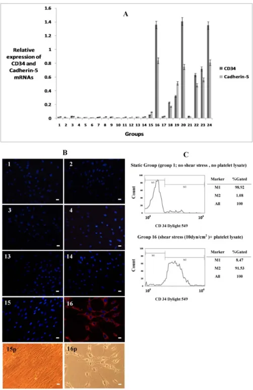
Endothelial differentiation was deter- mined by evaluating the expressions of endo- thelium specific mRNAs (CD34, vWF, Pecam1 and Cdh5 (Figure 1)). After estab- lishment of changes in gene expression, im- munofluoroscent staining against CD34 and vWF was performed to establish the expres- sion of endothelial markers in protein level (Figure 1).
As shown in Figure 1, data of real time PCR showed that in those groups cultured in medium containing FBS, there was not any endothelial specific gene expression even in the presence of high FSS. However, when PL was added to their culture medium in the exchange of FBS, they showed endothelial differentiation. This finding revealed that FSS alone is not sufficient for endothelial differentiation of MSCs and needs a chemi- cal induction factor. After application of 10 dyn/cm2 shear tension for 6 hours, there was apparent CD34 and Cdh5 mRNA ex- pression in 5 % PL treated cells compared to other groups in the same time (groups 1-15, P < 0.001). In groups placed under 2 and 5 dyn/cm2 FSS rates, expressions of CD34 and Cdh5 appeared after 12 hours FSS appli- cation. There was not any apparent increase in mature endothelial markers vWF or Pecam1 at this stage among groups.
Staining of cells was performed in the 8 groups (groups 1-4 and 13-16 ) after 2 days of FSS application and it confirmed that there was higher rate of CD34 positive cells in 10 dyn/cm2 group (group 16) compared with other groups (Figure 1; P < 0.001).
These data revealed that short term high sur- face tension was more potent than low sur- face tension on induction of endothelial dif- ferentiation of MSCs. In the static groups (without FSS, groups 1 and 13), there was not any CD34 and Cdh5 gene expression or CD34 protein synthesis. These findings demonstrated that FSS in combination with PL was effective on induction of differentia- tion of MSCs toward endothelial cells.
The results of flow cytometry assay also showed that in group 16 more than 90 % of cells were CD34 positive while in static
group about 1 % of cells were CD34 positive (Figure 1).
Results of experiment 1 indicated that in all of the groups, there were not any vWF gene or protein expressions and we conclud- ed that it was because CD34 and Cdh5 are endothelial progenitor specific markers while vWF and Pecam1 are known as specific markers for mature endothelial cells.
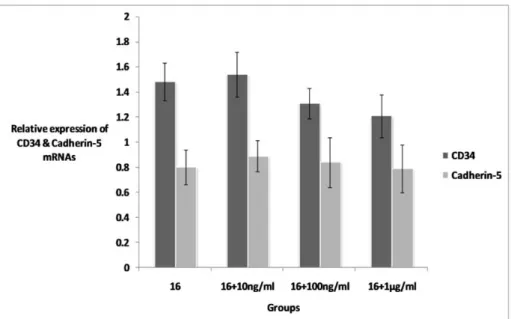
Results of experiment 2
The results of experiment 1 showed that the culture protocol with 10 dyn/cm2 FSS for 6 hours in the presence of 5 % PL (10 dyn/
6 h/5 % pl) has the best productivity of CD34+ cells. For improvement of this proto- col, conjugated estradiol was added to the culture medium with concentrations of 10 ng/ml, 100 ng/ml, 1 μg/ml.
As shown in Figure 2, treatment with es- tradiol did not change the expression rate of endothelial markers CD34 and Cdh5 in treat- ed groups and the results were not signifi- cantly different when compared to group 16.
Results of experiment 3
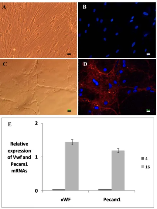
To evaluate the potentiality of produced endothelial progenitor cells for production of mature endothelial cells, we continued cul- ture in PL for two weeks and expressions of vWF and Pecam1 mRNAs were evaluated as the markers of mature endothelial cells. For identification of vWF expression in protein level, cells were also stained by anti vWF antibody. Data of this part of study (Figure 3) showed that after production of endotheli- al progenitor cells they can form mature en- dothelial cells. As shown in Figure 4, cells from groups 16 and 4 were selected and cul- tured for 2 weeks and the results showed that markers of endothelial cells just expressed in group 16.
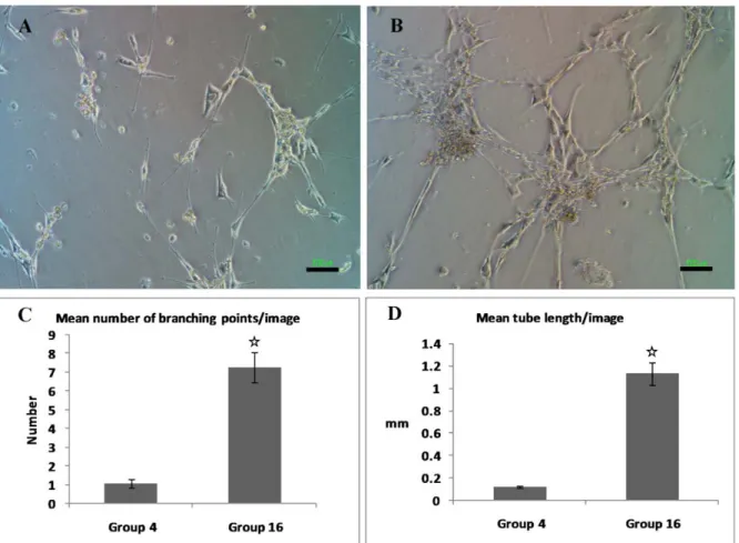
Results of tube formation assay:
The cells that were produced from group 16 (FSS 10dyn/cm2+PL 5 %) and 4 (FSS 10 dyn/cm2 +FBS 5 %) were trypsinated af- ter 48 hours culture in the same medium and were transferred to Matrigel (BD) coated 6-
well culture dishes to evaluate their tube formation potentiality. Results showed that there was a significant difference between
these groups in the length of formed tubes and the number of branching points in the tubes (Figure 4, P < 0.001).
Figure 1: Expression of endothelial specific markers in mesenchymal stem cells (MSCs) treated with fluid shear stress (FSS) and platelet lysate (PL). A: Expression rate of CD34 and Cadherin-5 mRNAs in different groups of study (specifications groups were presented in Table 1). Expression rate of glyceraldehyde-3-phosphate dehydrogenase (Gapdh) mRNA was assumed as a reference marker. B:
staining against CD34 protein in some of the groups, name of each group presented above each im- age. CD 34 stained cells (red) just appeared in group 16, nuclei were stained with DAPI (blue). Phase contrast images from cells of groups 15 and 16 presented in 15p and 16p respectively. C: Results of flowcytometry assay in groups 1 and 16, cells were stained against CD34 antibody, more than 90 % of cells were CD34 stained cells in group 16. Scale bars represent 10 µm.
Figure 2: Expression of CD34 and Cadherin-5 mRNAs in estradiol treated and non-treated groups.
Addition of estrdiol to culture medium did not change the expression rate of CD34 and cadherin-5 in treated groups. Rate of expressions calculated as the fold of expression of each gene against expres- sion rate of housekeeping gene glyceraldehyde-3-phosphate dehydrogenase (Gapdh).
DISCUSSION
The results of this study showed that PL and FSS can induce expression of endotheli- al markers CD34 and cdh5 in MSCs and fur- ther culture of these CD34 cells induced them to express vWF and Pecam1. Also, they formed tubular structures after culture on Matrigel. According to the other studies on the effect of FSS on MSCs, it was report- ed by many of researchers that FSS is a po- tent endothelial induction factor. The effect of FSS in combination with VEGF was stud- ied by Bai et al. (2010) and they found that VEGF presence is very important for endo- thelial differentiation of MSCs. Based on their study, Bai et al. (2010) found that 10 dyn/cm2 FSS for 24 hours in the presence of VEGF had more effect on endothelial dif- ferentiation. They also found that application of higher rates of FSS on MSCs induces them to differentiate toward smooth muscle cells. Further reports from Maul et al. (2011) and Kim et al. (2011) supported the findings of Bai et al. (2011) and Kim et al. (2011) re- ported that low level of FSS for 24 hours leads to an increase in vWF expression while higher rates of FSS (more than 10 dyn/cm2) induces the calponin gene expression which
is a smooth muscle specific protein. Indicat- ing the efficacy of FSS as a potent mechani- cal induction factor, some studies reported that FSS induces differentiation toward other cell types except Ecs. For example, Huang et al. (2010) reported that FSS in the presence of Azacitidine can induce cardiomyogenic differentiation of MSCs. Also, in one study by Zhang et al. (2009) the effect of FSS on MSCs cultured in endothelial specific culture media was evaluated. They reported that 20 dym/cm2 of FSS for 72 hours could differen- tiate MSCs toward smooth muscle cells as it was also reported formerly by Bai et al.
(2010). Furthermore, it was reported by some researchers that intermittent FSS in- duces human MSCs toward osteogenic dif- ferentiation (Liu et al., 2012; Yourek et al., 2010). So, based on the reports above, FSS is a potent induction factor and could generate different types of cells from stem cells in dif- ferent culture conditions and compositions.
According to the reports above on the ef- fect of FSS on endothelial differentiation of MSCs, most reported that FSS in the pres- ence of VEGF was effective on endothelial differentiation. In earlier studies, different FSS forces were applied on MSCs ranging
from 2-20 dyn/cm2 and for different periods of time ranging from 24 hours to 4 days. In almost all of them, endothelial differentiation occurred in shear stress rates equal to or lower than 10 dyn/cm2 and for a period of time near to 24 hours (Bai et al., 2010; Kim et al., 2011; Maul et al., 2011). Yet, accord- ing to the results of the present study, it is the first time that induction of endothelial differentiation has been reported after
6 hours exposure to 10 dyn/cm2 FSS. The major difference between this study and former ones is the use of PL in the place of FBS and VEGF. According to the results, it could be concluded that PL is much stronger that VEGF on induction of endothelial dif- ferentiation of MSCs. This effect of PL could be attributed to the presence of many biochemical active components in it.
Figure 3: Phase contrast and fluorescent images of cells (A and B from group 4 ; C and D from group 16). Cells were stained against von Willebrand factor (vWF), expression of vWF just appeared in group 16 (D). vWF strands are visible in C and D. Relative expressions of platelet-endothelial cell ad- hesion molecule (Pecam1) and vWF have shown in E. Scale bars represent 10 µm.
Figure 4: Generation of tubular structures from treated mesenchymal stem cells after culture on mat- rigel, A represented group 4 and B group 16. C and D: Mean number of branching points and tube length in images (P < 0.001).Scale bars represent 100 µm.
Behfar et al. (2010) reported that treating MSCs with a specific regimen of growth fac- tors could differentiate them toward cardio- myocytes. With careful attention to the fac- tors that they used in their research, it be- comes clear that almost all of them existed in PL altogether. So PL with its complete growth factors has strong potentiality to be used as a complete basement regimen for in- duction of differentiation toward specific cell lines and it could work specifically when some extra chemicals and growth factors or other kinds of differentiation inducers such as FSS are added to it.
In the present study, we introduced PL as a potent induction factor. This is because of its high concentrations of basic fibroblast growth factor (bFGF), platelet-derived growth factor AA (PDGF-AA), platelet- derived growth factor AB/BB (PDGF-
AB/BB) and transforming growth factor-β1 (TGF-β1) (Fekete et al., 2012). Platelet- derived endothelial cell growth factor (PD- ECGF) is another important secretion of platelets that has an effect on endothelial cell maturation and growth (Hemalatha et al., 2009).
About the possible mechanism responsi- ble for the effect of FSS, in one study on mu- rine embryonic mesenchymal progenitor cells (CH3H/10T1/2 cell line), Wang et al.
(2008) reported that FSS down-regulates the growth factors associated with smooth mus- cle cell differentiation. In another study, they reported that FSS acts mainly through down- regulation of TGF β1 to induce the differen- tiation of mouse embryonic mesenchymal progenitor cells to endothelial cells (Wang et al., 2008, 2005). Therefore, the possible ef- fect of combination of FSS and PL on EC
differentiation could be inhibition of smooth muscle differentiation with low level of FSS and directing differentiation toward ECs with growth factors presented in PL.
In conclusion our findings indicated that exertion of 10 dyn/cm2 FSS for 6 hours in combination with 5 % PL on MSCs can di- rect them to differentiate toward CD34 posi- tive cells, indicating the initiation of endo- thelial differentiation of MSCs. Data from groups cultured in media containing fetal bovine serum showed that FSS can’t induce endothelial differentiation without PL. Our data shows that PL could be useful as an en- dothelial induction factor in the presence of FSS. This is because of its growth factors such as PD-ECGF, PDGF and bFGF.
ACKNOWLEDGMENTS
This study was supported financially by headquarter of development and application of stem cell research, vice president of sci- ence and technology, ministry of health and medical education of I.R. of Iran.
CONFLICT OF INTEREST Authors don’t have any conflict of interest.
REFERENCES
Bai K, Huang Y, Jia X, Fan Y, Wang W. Endothelium oriented differentiation of bone marrow mesenchymal stem cells under chemical and mechanical stimulat- ions. J Biomech 2010;43:1176-81.
Baik SY, Lim YA, Kang SJ, Ahn SH, Lee WG, Kim CH. Effects of platelet lysate preparations on the proliferation of HaCaT cells. Ann Lab Med 2014;34:
43-50.
Behfar A, Yamada S, Crespo-Diaz R, Nesbitt JJ, Rowe LA, Perez-Terzic C et al. Guided cardiopoiesis enhances therapeutic benefit of bone marrow human mesenchymal stem cells in chronic myocardial infarction. J Am Coll Cardiol 2010;56:721-34.
Bertanha M, Moroz A, Almeida R, Alves FC, Acorci Valério MJ, Moura R et al. Tissue-engineered blood vessel substitute by reconstruction of endothelium using mesenchymal stem cells induced by platelet growth factors. J Vasc Surg 2014;59:1677-85.
Burnouf PA, Juan PK, Su CY, Kuo YP, Chou ML, Su C et al. A novel virally inactivated human platelet lysate preparation rich in TGF-beta, EGF and IGF, and depleted of PDGF and VEGF. Biotechnol Appl Biochem 2010;56:151-60.
Crespo-Diaz R, Behfar A, Butler GW, Padley DJ, Sarr MG, Bartunek J et al. Platelet lysate consisting of a natural repair proteome supports human mesen- chymal stem cell proliferation and chromosomal stability. Cell Transplant 2011;20:797-811.
Das M, Sundell IB, Koka PS. Adult mesenchymal stem cells and their potency in the cell-based therapy.
J Stem Cells 2013;8:1-16.
Doucet C, Ernou I, Zhang Y, Llense JR, Begot L, Holy X et al. Platelet lysates promote mesenchymal stem cell expansion:a safety substitute for animal serum in cell-based therapy applications. J Cell Physiol 2005;205:228-36.
Duttenhoefer F, Lara de Freitas R, Meury T, Loibl M, Benneker LM, Richards RG et al. 3D scaffolds co- seeded with human endothelial progenitor and mesenchymal stem cells:evidence of prevasculari- sation within 7 days. Eur Cell Mater 2013;26:49-64;
discussion 64-5.
Fekete N, Gadelorge M, Fürst D, Maurer C, Dausend J, Fleury-Cappellesso S et al. Platelet lysate from whole blood-derived pooled platelet concentrates and apheresis-derived platelet concentrates for the isolation and expansion of human bone marrow mesenchymal stromal cells: production process, content and identification of active components. Cyto- therapy 2012;14:540-54.
Govey PM, Loiselle AE, Donahue HJ. Biophysical regulation of stem cell differentiation. Curr Osteo- poros Rep 2013;11:83-91.
Hamdan R, Zhou Z, Kleinerman ES. Blocking SDF- 1α/CXCR4 downregulates PDGF-B and inhibits bone marrow-derived pericyte differentiation and tumor vascular expansion in Ewing tumors. Mol Cancer Ther 2014;13:483-91.
Hemalatha T, Tiwari M, Balachandran C, Manohar BM, Puvanakrishnan R. Platelet-derived endothelial cell growth factor mediates angiogenesis and anti- apoptosis in rat aortic endothelial cells. Biochem Cell Biol 2009;87:883-93.
Huang Y, Jia X, Bai K, Gong X, Fan Y. Effect of fluid shear stress on cardiomyogenic differentiation of rat bone marrow mesenchymal stem cells. Arch Med Res 2010;41:497-505.
Imanishi T, Kobayashi K, Hano T, Nishio I. Effect of estrogen on differentiation and senescence in endothe- lial progenitor cells derived from bone marrow in spontaneously hypertensive rats. Hypertens Res 2005;
28:763-72.
Janeczek Portalska K, Leferink A, Groen N, Fernan- des H, Moroni L, van Blitterswijk C et al. Endothelial differentiation of mesenchymal stromal cells. PLoS One 2012;7:e46842.
Jiang Y, Jahagirdar BN, Reinhardt RL, Schwartz RE, Keene CD, Ortiz-Gonzalez XR et al. Pluripotency of mesenchymal stem cells derived from adult marrow.
Nature 2002;418:41-9.
Jonsdottir-Buch SM, Lieder R, Sigurjonsson OE.
Platelet lysates produced from expired platelet con- centrates support growth and osteogenic differentia- tion of mesenchymal stem cells. PLoS One 2013;
8:e68984.
Kim DH, Heo SJ, Kim SH, Shin JW, Park SH. Shear stress magnitude is critical in regulating the differentiation of mesenchymal stem cells even with endothelial growth medium. Biotechnol Lett 2011;
33:2351-9.
Kryza T, Achard C, Parent C, Marchand-Adam S, Guillon-Munos A, Iochmann S et al. Angiogenesis stimulated by human kallikrein-related peptidase 12 acting via a platelet-derived growth factor B-depend- ent paracrine pathway. FASEB J 2014;28:740-51.
Lennon DP, Caplan AI. Isolation of rat marrow- derived mesenchymal stem cells. Exp Hematol 2006;
34:1606-7.
Liu L, Yuan W, Wang J. Mechanisms for osteogenic differentiation of human mesenchymal stem cells induced by fluid shear stress. Biomech Model Mecha- nobiol 2010;9:659-70.
Liu L, Yu B, Chen J, Tang Z, Zong C, Shen D et al.
Different effects of intermittent and continuous fluid shear stresses on osteogenic differentiation of human mesenchymal stem cells. Biomech Model Mechano- biol 2012;11:391-401.
Liu ZJ, Zhuge Y, Velazquez OC. Trafficking and differentiation of mesenchymal stem cells. J Cell Bio- chem 2009;106:984-91.
Maul TM, Chew DW, Nieponice A, Vorp DA.
Mechanical stimuli differentially control stem cell behavior: morphology, proliferation, and differentia- tion. Biomech Model Mechanobiol 2011;10:939-53.
Meyer A, Wang W, Qu J, Croft L, Degen JL, Coller BS et al. Platelet TGF-β1 contributions to plasma TGF-β1, cardiac fibrosis, and systolic dysfunction in a
mouse model of pressure overload. Blood 2012;119:
1064-74.
Obi S, Yamamoto K, Shimizu N, Kumagaya S, Masu- mura T, Sokabe T et al. Fluid shear stress induces arterial differentiation of endothelial progenitor cells.
J Appl Physiol 2009;106:203-11.
Obi S, Masuda H, Shizuno T, Sato A, Yamamoto K, Ando J et al. Fluid shear stress induces differentiation of circulating phenotype endothelial progenitor cells.
Am J Physiol 2012;303:C595-606.
Oswald J, Boxberger S, Jørgensen B, Feldmann S, Ehninger G, Bornhäuser M et al. Mesenchymal stem cells can be differentiated into endothelial cells in vitro. Stem Cells 2004;22:377-84.
Pankajakshan D, Kansal V, Agrawal DK. In vitro differentiation of bone marrow derived porcine mesenchymal stem cells to endothelial cells. J Tissue Eng Regen Med 2013;7:911-20.
Pati S, Gerber MH, Menge TD, Wataha KA, Zhao Y, Baumgartner JA et al. Bone marrow derived mesen- chymal stem cells inhibit inflammation and preserve vascular endothelial integrity in the lungs after hemorrhagic shock. PLoS One 2011;6:e25171.
Sanada C, Kuo CJ, Colletti EJ, Soland M, Mokhtari S, Knovich MA et al. Mesenchymal stem cells contri- bute to endogenous FVIII:c production. J Cell Physiol 2013;228:1010-6.
Stolberg S, McCloskey KE. Can shear stress direct stem cell fate? Biotechnol Prog 2009;25:10-9.
Wang H, Riha GM, Yan S, Li M, Chai H, Yang H et al. Shear stress induces endothelial differentiation from a murine embryonic mesenchymal progenitor cell line. Arterioscler Thromb Vasc Biol 2005;25:
1817-23.
Wang H, Li M, Lin PH, Yao Q, Chen C. Fluid shear stress regulates the expression of TGF-beta1 and its signaling molecules in mouse embryo mesenchymal progenitor cells. J Surg Res 2008;150:266-70.
Yourek G, McCormick SM, Mao JJ, Reilly GC. Shear stress induces osteogenic differentiation of human mesenchymal stem cells. Regen Med 2010;5:713-24.
Zhang JC, Lü G. Effect of 17β‑estradiol in rat bone marrow‑derived endothelial progenitor cells. Mol Med Rep 2013;8:178-82.
Zhang L, Li Y, Zhang C, Zhang Y, Yang X.
[Differentiation of bone marrow mesenchymal stem cells co-cultured with endothelial cells under shear stress]. Sheng Wu Yi Xue Gong Cheng Xue Za Zhi 2009;26:85-8. [Article in Chinese]