Characterization of key mechanisms involved in transmigration and invasion of mesenchymal stem cells
Improvement of transendothelial migration via focussed ultrasound-mediated microbubble stimulation
Inaugural-Dissertation zur
Erlangung des Doktorgrades
der Mathematisch-Naturwissenschaftlichen Fakultät der Universität zu Köln
vorgelegt von Caroline Steingen
aus Köln
Copy Team Cologne, Köln 2008
Berichterstatter: Prof. Dr. Wilhelm Bloch Prof. Dr. Manolis Pasparakis
Tag der mündlichen Prüfung: 16. April 2008
Meinen Eltern Carin & Rudi Steingen und meiner lieben Oma
Kurzzusammenfassung
Adulte mesenchymale Stammzellen (MSCs) werden bereits für die Therapie verschiedener Krankheiten klinisch eingesetzt. Der erste Teil der vorliegenden Arbeit beschäftigt sich mit der Aufklärung von Schlüsselmechanismen, die an der Transmigration von MSCs über die Endothelbarriere und der Invasion in ein Ziel- gewebe beteiligt sind. Beide Prozesse sind die Grundvoraussetzung für eine erfolgreiche Stammzelltherapie, da MSCs stets das Blutgefäßsystem verlassen müssen, um in umgebendes Gewebe einzuwandern. Versuche mit verschiedenen Modelsystemen sowie in vivo Experimente ergaben, dass MSCs schnell Kontakt zur Endothelbarriere aufnehmen und anschließend die Blutbahn über folgende Schritte verlassen: (1) durch eine Integration in das Endothel, (2) durch die Transmigration über die Endothelbarriere, vermittelt durch die Ausbildung von Zellfortsätzen, (3) durch Penetration der Basalmembran und anschließende Invasion in das umgebende Gewebe.
Zudem konnte gezeigt werden, dass die Transmigration humaner MSCs durch die Interaktion der Oberflächenmoleküle VLA-4 und VCAM-1 vermittelt wird und in einer regionalen Ansammlung von β1-Integrinen resultiert. Desweiteren schütten MSCs aktive Matrix-Metalloproteinase (MMP)-2, nicht aber MMP-9 aus, um in Herzmuskel- gewebe einzudringen.
Die vorliegende Arbeit zeigt, dass sowohl der zeitliche Verlauf als auch die morpho- logischen Aspekte der Transmigration vom Phänotyp der Endothelzellen abhängig sind. Dies deutet auf Unterschiede in der Effizienz der Transmigration innerhalb des Gefäßsystems – abhängig vom Gefäßtyp – hin. Außerdem beschleunigt die Zugabe von Zytokinen, besonders VEGF und EPO, zu Beginn der Transmigration den Ablauf des Prozesses. Auch ergaben Versuche, dass MSCs, sobald sie Kontakt zu Endo- thelzellen aufnehmen, vermehrt Stickstoffmonoxid (NO) und reaktive Sauerstoff- Spezies (ROS) freisetzen. Veränderungen des NO- oder ROS-Haushaltes resultierten in Abweichungen der transmigratorischen Fähigkeit der MSCs.
Der letzte Teil der vorliegenden Arbeit beschäftigt sich mit zwei möglichen Ansätzen, die therapeutische Effizienz von MSCs zu erhöhen. Einerseits zeigte sich, dass eine genetische Modifikation der Stammzellen durch adenovirale Überexpression des Chemokin-Rezeptors CXCR4 keine Verbesserung ihrer transmigratorischen Aktivität zur Folge hat. Andererseits verbesserte eine gezielte Vorbehandlung des Endothels durch eine neuartige und nicht-invasive Technik, die Ultraschall-vermittelte Mikro- bläschen-Stimulierung (UMS), sowohl die Anlockung als auch die Transmigration und Invasion von MSCs in gesundes und ischämisches Herzmuskelgewebe. Diese regionale Verbesserung wurde vermutlich durch die Ultraschall-vermittelte Frei- setzung von Stickstoffmonoxid und Zytokinen, sowie die lokale Aktivierung von Matrix-Metalloproteinasen erzielt. Daher stellt die Ultraschall-vermittelte Mikrobläschen-Stimulierung eine zukunftsweisende Möglichkeit dar, die therapeutische Effizienz von MSCs lokal und nicht-invasiv zu erhöhen.
Abstract
Stem cell therapy using human adult mesenchymal stem cells (MSCs) has emerged as a novel strategy for the treatment of a variety of damaged tissues. For a successful systemic stem cell therapy MSCs have to exit the blood circulation by transmigrating across the endothelium and invading into the target tissue. Elevating our knowledge on these core processes might help to optimize stem cell based therapies. The first part of the present study provides insights into key mechanisms involved in the transmigration and invasion of MSCs. Different model systems as well as in vivo studies revealed that MSCs quickly come into contact with the endothelium and subsequently exit the blood circulation by (1) integrating into the endothelium, (2) transmigrating across the endothelial barrier via the insertion of plasmic podia, (3) penetrating the basement membrane and subsequently invading the surrounding tissue.
Additionally, it was proven that transmigration of human MSCs not only requires the interaction of very late antigen-4 (VLA-4, α4β1 integrin) and its most important ligand vascular cell adhesion molecule-1 (VCAM-1), but also triggers a clustering of β1 integrins. Furthermore, upon invading into cardiac tissue MSCs secrete active matrix metalloproteinase (MMP)-2, but not MMP-9.
This study also demonstrates that both the time course and the morphological aspects of MSC transmigration differ depending on the endothelial phenotype, thus indicating, that a variable capacity for transendothelial migration exists within the vasculature. Furthermore, addition of cytokines, mainly vascular endothelial growth factor (VEGF) and erythropoietin (EPO), accelerate the transmigration of MSCs at early stages. Moreover, nitric oxide (NO) and reactive oxygen species (ROS) are released by MSCs upon contact with endothelial cells; manipulating the NO and ROS system by donors and inhibitors resulted in alterations of the transmigratory capacity of MSCs.
The second part of the study deals with two possible strategies to enhance the transmigration of MSCs and thereby their therapeutic effectiveness. First, the results demonstrate that genetic modification of MSCs using adenoviral overexpression of the chemokine receptor CXCR4 does not lead to an increase in the transmigration efficiency. Second, focussed pretreatment of the endothelium by a novel and non- invasive technique using ultrasound-mediated microbubble stimulation (UMS) induces a targeted improvement of MSC attraction, transmigration and invasion into non-ischemic as well as into ischemic myocardium. This effect was most likely due to the release of nitric oxide, cytokines and the regional activation of proteases. Thus, UMS represents a forward-looking possibility to increase the efficiency of MSC engraftment by modulating the process of transmigration in a targeted and non- invasive manner.
Table of contents
1. Introduction ... 1
1.1 Sources, multipotency and markers of mesenchymal stem cells ... 1
1.2 Therapeutic usage of MSCs... 2
1.3 Open questions regarding transmigration and invasion of MSCs... 4
1.3.1 Adhesion molecules involved ... 4
1.3.2 Role of proteases for the invasion of MSCs ... 5
1.3.3 Role of endothelial phenotype and cytokines... 6
1.3.4 Involvement of nitric oxide and reactive oxygen species... 7
1.4 Approaches to enhance the transmigration of MSCs ... 8
1.4.1 Genetic modification of MSCs ... 9
1.4.2 Ultrasound-mediated microbubble stimulation ... 9
1.5 Aims and experimental design of this study ... 11
2. Material and Methods... 13
2.1 Cell Culture ... 13
2.1.1 Mesenchymal stem cells (MSCs) ... 13
2.1.1.1 Isolation of human MSCs ... 13
2.1.1.2 Culture of human MSCs... 14
2.1.2 Feeder cells... 15
2.1.2.1 Culture of feeder cells ... 15
2.1.2.2 Inactivation of feeder cells... 15
2.1.3 Embryonic stem cells ... 16
2.1.3.1 Culture of embryonic stem cells ... 16
2.1.3.2 Preparation of hanging drops... 16
2.1.3.3 Culturing of aggregates in suspension... 16
2.1.3.4 Gelatine coating and plating of embryoid bodies ... 17
2.1.4 Endothelial cells ... 17
2.1.4.1 Isolation of murine microvascular endothelial cells via Magnet associated cell sorting (MACS) and culture ... 17
2.1.4.2 Culture of human umbilical vein, coronary artery and aortic endothelial cells ... 19
2.2 Quality control of MSCs... 19
2.2.1 Flow cytometry (FACS) ... 19
2.2.2 Colony forming unit-fibroblast (CFU-F) assay ... 20
2.2.3 Differentiation assays... 20
2.2.3.1 Adipogenic differentiation... 20
2.2.3.2 Osteogenic differentiation ... 21
2.2.3.3 Chondrogenic differentiation ... 21
2.2.3.3.1 RNA isolation ... 22
2.2.3.3.2 Semi-quantitative RT-PCR... 22
2.3 Vital staining of cells... 24
2.3.1 PKH26 and PKH67 staining ... 24
2.3.2 CellTracker staining ... 24
2.4 Monolayer co-cultivation experiments ... 24
2.4.1 Co-cultivation ... 25
2.4.2 Cytokines and additive substances ... 25
2.4.3 Blocking experiment... 25
2.5 Three-dimensional model systems for transmigration ... 26
2.5.1 Collagen invasion assay ... 26
2.5.2 Endothelial spheroid model ... 26
2.5.3 Perfusion of isolated mouse hearts ... 26
2.6 Adenoviral infection of MSCs ... 27
2.7 In vivo experiments ... 29
2.7.1 Experimental protocol ... 29
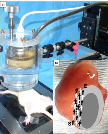
2.7.2 Focussed ultrasound-mediated microbubble stimulation (UMS) ... 31
2.7.3 Transplantation of MSCs via aortic root catheter ... 33
2.8 Immunohistochemistry ... 33
2.9 Migration assay ... 34
2.10 DAF and APF fluorometry ... 35
2.11 TUNEL assay ... 35
2.12 Immuno blot (SDS-Page) ... 36
2.12.1 Preparation of cell and tissue lysates... 36
2.12.2 Measurement of protein content... 37
2.12.3 Separation of proteins via SDS-Polyacrylamid-gelelectrophoresis (PAGE) ... 37
2.12.4 Western blot ... 37
2.12.5 Immunological Detection... 38
2.13 Cytokine Antibody Array... 39
2.14 Enzyme-linked immunosorbent assay (ELISA) ... 39
2.15 In situ zymography ... 40
2.16 Microscopy ... 40
2.16.1 Confocal laser scanning microscopy... 40
2.16.2 Calculation of MSC integration... 41
2.16.3 Quantification of transmigration... 41
2.16.4 Electron microscopy... 42
2.17 Statistical analysis ... 43
3. Results ... 44
3.1 Characterization of human mesenchymal stem cells ... 44
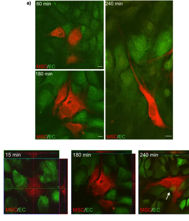
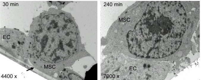
3.2 Characterization of transmigration and invasion: different model systems. 47 3.2.1 Co-cultivation ... 47
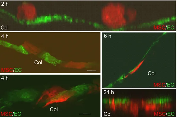
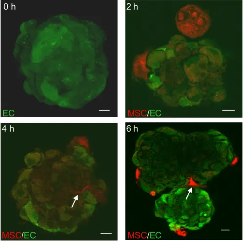
3.2.2 Three-dimensional model systems for transmigration... 50
3.2.2.1 Collagen invasion assay ... 50
3.2.2.2 Endothelial spheroids... 50
3.2.2.3 Isolated mouse heart perfusions ... 50
3.3 Observation of transmigrating MSCs in vivo... 54
3.4 VCAM-1/VLA-4-interaction and β1 integrin clustering are crucial for transendothelial migration ... 55
3.5 MSCs secrete active MMPs at sites of invasion... 56
3.6 Transmigration is dependent on the endothelial phenotype ... 58
3.7 Cytokines influence MSC transmigration... 58
3.8 Role of nitric oxide for the transmigration of MSCs ... 60
3.9 Influence of reactive oxygen species on transmigration of MSCs ... 63
3.10 Influence of adenoviral infection on the transmigration of MSCs... 66
3.10.1 Efficiency of MSC infection... 66
3.10.2 Decrease of migratory and transmigratory activity of MSCs by CXCR4-infection... 68
3.11 Manipulation of MSC transmigration and attraction via focussed ultrasound-mediated microbubble stimulation (UMS)... 70
3.11.1 Placement of catheter ... 70
3.11.2 Efficiency of focussed UMS... 70
3.11.3 Impact of UMS on MSC cell attraction ... 72
3.11.4 Transmigration ... 74
3.11.5 Influence of UMS on endothelial structure, basal lamina, cytokines and apoptosis ... 75
4. Discussion ... 81
4.1 Characterization of MSCs ... 81
4.2 Discussion of the morphological aspects and time course of MSC transmigration... 84
4.2.1 Co-culture experiments ... 84
4.2.2 Three-dimensional model systems... 86
4.2.3 Transmigration of MSCs in vivo... 87
4.3 Integrins and their role in transendothelial migration ... 88
4.4 Role of matrix metalloproteinases for the invasion of MSCs ... 90
4.5 Importance of the endothelial phenotype for transmigration... 92
4.6 Role of cytokines ... 93
4.7 Involvement of nitric oxide and free oxygen radicals ... 94
4.7.1 Nitric oxide ... 94
4.7.2 Reactive oxygen species ... 96
4.8 Genetic manipulation of MSCs via adenoviral infection... 97
4.9 Preconditioning of the endothelium by ultrasound-mediated microbubble stimulation triggers attraction, transmigration and invasion of MSCs ...101
4.9.1 Systemic delivery of MSCs...101
4.9.2 Ultrasound-mediated microbubble stimulation ...104
4.9.3 Effect of UMS on cell attraction and cytokine levels...106
4.9.4 Effect of UMS on transmigration, basement membrane and protease activity ...109
5. Summary ...113
6. References ...115
Table of figures
Figure 1: MSC multi-lineage differentiation potential... 3
Figure 2: Scheme of the isolation procedure of mononuclear cells via density gradient centrifugation... 14
Figure 3: Scheme of the preparation of embryoid bodies... 18
Figure 4: Scheme of the adenoviral shuttle vector pAdTrack-CMV... 29
Figure 5: Scheme of experimental protocol of in vivo experiments ... 31
Figure 6: Focussed ultrasound-mediated microbubble stimulation (UMS) ... 32
Figure 7: Scheme of a modified Boyden Chamber migration assay... 34
Figure 8: Light microscopic picture of a confluent monolayer of MSCs... 44
Figure 9: Flow cytometric analysis of expressed surface antigens of MSCs ... 45
Figure 10: MSCs differentiate to mesenchymal lineages ... 46
Figure 11: MSCs integrate into endothelial monolayer in vitro... 48
Figure 12: Integrity of endothelial monolayer ... 49
Figure 13: Electron micrographs of in vitro co-cultivation ... 49
Figure 14: Collagen gel invasion assay... 51
Figure 15: Co-cultivation of endothelial spheroids with MSCs... 52
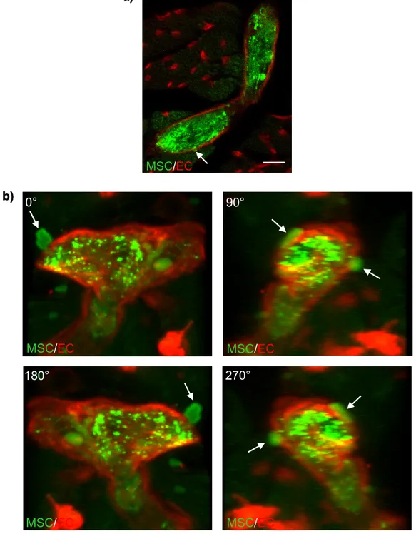
Figure 16: Isolated mouse heart perfusions visualize transmigration and invasion of MSCs ex vivo... 53
Figure 17: MSCs transmigrate and invade in vivo... 54
Figure 18: Transmigration requires VCAM-1/VLA-4... 55
Figure 19: Transmigration triggers clustering of β1 integrins... 56
Figure 20: MSCs secrete MMP-2 ... 57
Figure 21: Active gelatinases are secreted at sites of MSC invasion ... 57
Figure 22: Differences in transmigration capacity depend on endothelial phenotype………59
Figure 23: Cytokines influence the transmigration of MSCs at early time points.. 60
Figure 24: Co-cultivation under the influence of NO-donor and NOS-inhibitor ... 61
Figure 25: Changes in intracellular NO-levels caused by the co-culture of MSCs and endothelial cells ... 62
Figure 26: Representative pictures of DAF measurements... 63
Figure 27: Co-cultivation under the influence of H2O2 and a radical scavenger ... 64
Figure 28: Changes in intracellular ROS-levels caused by co-culture of MSCs and endothelial cells ... 65
Figure 29: Representative pictures of APF measurements ... 66
Figure 30: Efficiency of adenoviral infection of MSCs ... 67
Figure 31: Migratory activity of infected MSCs in the Boyden Chamber... 68
Figure 32: Transmigration of infected MSCs in isolated mouse heart perfusions. 69 Figure 33: Percutaneous myocardial delivery via aortic root catheter ... 71
Figure 34: Focussed ultrasound-mediated microbubble stimulation (UMS) ... 72
Figure 35: Focussed UMS enhanced cell attraction... 73
Figure 36: UMS preconditioning increased MSC cell attraction ... 74
Figure 37: Focussed UMS enhanced transendothelial migration ... 75
Figure 38: Alteration of the basal lamina ... 76
Figure 39: Morphology of the myocardial endothelium... 77
Figure 40: TUNEL assay of myocardium under the influence of UMS ... 79
Figure 41: Induction of gelatinase activity by UMS... 80
List of abbreviations
3D three-dimensional
α-MEM Minimum Essential Medium alpha Modification Ad.CXCR4.GFP CXCR4 and GFP expressing adenovirus Ad.GFP GFP expressing adenovirus
ANOVA one-way analysis of variance
Ap apex
APF 3´-(p-aminophenyl) fluorescein AW anterior wall
bFGF basic fibroblastic growth factor BP bandpass emission filter bp base pair(s)
BSA bovine serum albumin CD cluster of differentiation cDNA complementary DNA
CFU-F colony forming unit-fibroblast assay CMFDA 5-chloromethylfluorescein diacetate CMV cytomegalovirus
CXCR4 CXC chemokine receptor 4 DAB diaminobenzidine
DAF 4-amino-5-methylamino-2`,7`-difluorofluorescein diacetate DAPI 4´,6-diamidino-2-phenyl-indole
DEPC diethylpyrocarbonate DETA diethylenetriamine
Dil-Ac-LDL 1,1'-dioctadecyl-3,3,3',3'-tetramethyl-indocarbocyanine perchlorate acetylated-low density lipoprotein
D-MEM Dublecco`s Modified Eagle Medium DMSO dimethylsulfoxide
DNA deoxyribonucleic acid DNAse deoxyribonuclease
dNTP deoxyribonucleotide triphosphate dT deoxyribosylthymine nucleotide DTT dithiothreitol
e.g. for example EBs embryoid bodies ECD energy-coupled dye ECG electrocardiogram ECs endothelial cells
EDTA ethylene-diamine tetraacetic acid ELISA enzyme-linked immunosorbent assay eNOS endothelial nitric oxide synthase EPO erythropoietin
EtOH ethanol
F forward
FACS fluorescence activated cell sorting FCS fetal calf serum
FITC fluorescein isothiocyanate
Gel ZYM active gelatinases detected by in situ zymography GFP green fluorescent protein
GM-CSF granulocyte-macrophage colony-stimulating factor HCAEC human coronary artery endothelial cells
HEPES 4-(2-hydroxyethyl)-1-piperazineethanesulfonic acid HFT main dichroic beam splitter
HRP horseradish peroxidase HuAoEC human aortic endothelial cells
HUVEC human umbilical vein endothelial cells I/R ischemia followed by reperfusion ICAM intercellular adhesion molecule IgG immunoglobulin
IL interleukin kDa kilodalton
LAD left descending coronary artery LIF leukemia inhibition factor L-NAME N-nitro-L-arginine methyl ester LP longpass emission filter LSM laser scanning microscope LV left-ventricular cavity mAb anti-mouse antibody
MACS magnet associated cell sorting MAP mitogen activated protein MAPK MAP kinase
MI myocardial infarction min minute
M-MLV moloney murine leukemia virus MMP matrix metalloproteinase MOI multiplicity of infection
MOPS 3-morpholinopropane-1-sulfonic acid MSC mesenchymal stem cell
n. s. no significant difference NAc N-acetylcysteine
NFT secondary dichroic beam splitter NO nitric oxide
NOS nitric oxide synthase
PAGE polyacrylamide-gelelectrophoresis PBS phosphate-buffered saline
PC phycoerythrin-cyanine PCR polymerase chain reaction PE tube polyethylene tube
PE phycoerythrin
PECAM platelet-endothelial cell adhesion molecule PFA paraformaldehyde
Pfu plaque-forming units
pH potentia hydrogenii
PVDF polyvinylidene fluoride PW posterior wall
R reverse
RNA ribonucleic acid
ROS reactive oxygen species rpm revolutions per minute
RT-PCR reverse transcription polymerase chain reaction RV right ventricle
SDF stromal-derived growth factor SDS sodium dodecyl sulfate sec seconds
TAE Tris-acetate-EDTA TBS Tris-buffered saline
TBST Tris-buffered saline containing Tween 20 TGF transforming growth factor
TNF-α tumor necrosis factor alpha
Tris 2-Amino-2-(hydroxymethyl)propane-1,3-diol
TUNEL terminal uridine desoxynucleotidyl transferas dUTP nick end labeling
U unit
UMS ultrasound-mediated microbubble stimulation UV ultraviolet light
v/v volume per volume VCAM vascular cell adhesion molecule VE-Cadherin vascular endothelial Cadherin VEGF vascular endothelial growth factor VLA very late antigen
vs. versus
w/v weight per volume
1. Introduction
Bone marrow-derived mesenchymal stem cells (MSCs) are adult stem cells that reside within the bone marrow compartment (Schmidt et al., 2006b). MSCs can be isolated and expanded in culture and are described as multipotent because of their ability, even as clonally isolated cells, to differentiate into a variety of different cells (Tuan et al., 2002; Pittenger et al., 2004). Stem cell therapy using human adult bone marrow derived MSCs has emerged as a novel strategy for the treatment of a variety of damaged tissues as they possess a multi-lineage differentiation potential (Jiang et al., 2002). The use of adult stem cells to regenerate damaged tissue not only circumvents the ethical and technical issues associated with the use of embryonic stem cells, but also allows the transplantation of autologous as well as allogeneic cells. As allogeneic MSCs inhibit T cell proliferation and maturation (Krampera et al., 2003; Le Blanc et al., 2003; Tse et al., 2003b) they are not recognized by responder T cells and therefore not rejected by a recipient host. Thus, allogenic MSCs can be prepared in advance for any patient at the needed time (Pittenger et al., 1999) and can be administered without immunosuppression (Krause et al., 2007).
1.1 Sources, multipotency and markers of mesenchymal stem cells MSCs, first identified by Friedenstein and co-workers in 1976 (Friedenstein et al., 1976), have a fibroblastic morphology, possess a broad ex vivo expansive potential and can maintain an undifferentiated, stable phenotype over many generations (Pittenger et al., 1999). Currently, bone marrow aspirate is considered to be the most accessible and enriched source of MSCs. Nevertheless, MSCs have also been isolated from several other sources, including adipose tissue (Zuk et al., 2002; Lee et al., 2004b), scalp tissue (Shih et al., 2005), dermal tissue (Toma et al., 2001) and peripheral blood (Fernandez et al., 1997). In addition to adult tissues, MSCs were also identified in various fetal tissues, such as in the placenta (In 't Anker et al., 2004), in umbilical veins and umbilical cord blood (Erices et al., 2000). Although MSCs represent a very small fraction of cells – only about 0.001 to 0.01% of the cells isolated from a density gradient of a bone marrow aspirate belong to this population (Pittenger et al., 1999) – they are easy and fast expandable ex vivo. Due to the plastic-adherence of MSCs, non-adherent contaminating hematopoietic cells can be easily removed after isolation. Human MSC populations can be distinguished from
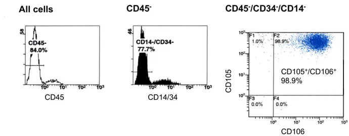
hematopoietic stem cells and leukocytes by distinct cell surface markers. Considering their mesodermal origin, MSCs are known to be negative for the leukocyte common antigen CD45, for the lipopolysaccharide receptor CD14 and for CD34, a marker of human hematopoietic stem and progenitor cells. The collective presence of a combination of other markers, such as CD105 (endoglin or transforming growth factor TGF β1/3-receptor) and CD106 (vascular cell adhesion molecule-1, VCAM-1), albeit not specific to MSCs, defines the MSC cell population (Pittenger et al., 1999).
As depicted in Figure 1, MSCs are capable of differentiating into various tissues of mesenchymal origin such as bone (Friedenstein et al., 1987; Haynesworth et al., 1992; Kuznetsov et al., 1997; Meinel et al., 2004a; Meinel et al., 2004b), skeletal muscle cells and cardiomyocytes (Wang et al., 2000; Goodell et al., 2001; Toma et al., 2002; De Bari et al., 2003), cartilage (Friedenstein et al., 1987; Li et al., 2005b) or vascular endothelial cells (Reyes et al., 2002; Koike et al., 2004), but can also give rise to non-mesenchymal cells like neural cells (Azizi et al., 1998; Kopen et al., 1999).
1.2 Therapeutic usage of MSCs
The ability of MSCs to differentiate into various cells in vivo has understandably aroused great interest for the therapeutic treatment of a variety of diseases. Indeed, clinical benefit has been observed following the intravenous administration of heterogeneous populations of MSCs in patients with osteogenesis imperfecta (Horwitz et al., 1999) or severe acute graft-versus-host disease (Le Blanc et al., 2004). Moreover, due to the very limited ability of myocardial tissue to regenerate after a myocardial infarction, treatment with stem cells also seems to be a promising therapeutic approach in cardiology. Adult cardiomyocytes have essentially no regenerative capacity and although a cardiac stem cell has been described (Anversa et al., 2002), its physiological role in repair after infarction appears minimal (Pittenger and Martin, 2004). Therefore, only an implantation of exogenous cells may allow replacement of damaged cardiac cells. Indeed, cellular transplantation, termed cellular cardiomyoplasty, has been investigated as a potential therapy for myocardial infarction in order to replace cardiomyocytes lost after ischemia (Bittner et al., 1999;
Jackson et al., 2001; Orlic et al., 2001a; Orlic et al., 2001b; Nygren et al., 2004). The first use of bone marrow cells for cardiomyoplasty was reported in 1999 by Tomita and co-workers at the University of Toronto (Tomita et al., 1999). For a successful cardiomyoplasty, MSCs are a promising stem cell type, because they differentiate
into a cardiomyocyte-like phenotype when implanted in healthy myocardium (Pittenger et al., 2000; Toma et al., 2002). Furthermore, MSCs supply growth factors and cytokines, which help to repair the damaged tissue via paracrine effects (Pittenger and Martin, 2004; Mirotsou et al., 2007). Thus, in several ongoing clinical trials, patients are treated with MSCs after a myocardial infarction (for review see (Laflamme et al., 2005; Ohnishi et al., 2007)). However, for a successful clinical application different strategies for transporting MSCs to their target destinations have to be evaluated regarding their applicability and efficiency.
Y
Mesenchymal stem cell (MSC) ProliferationCommitment
Lineage Progression
Differentiation
Maturation
MSC Proliferation
Osteogenesis Chondrogenesis Myogenesis Marrow Stroma
Tendogenesis Ligamentagenesis
Other
Myoblast fusion
Unique Micro-niche
Osteocyte
Hypertrophic
Chondrocyte Myotube Stroma Cells Fibroblast
Adipocyte Dermal Cells Neurons and Other Cells
BONE CARTILAGE MUSCLE MARROW
TENDON/
LIGAMENT
OTHER TISSUE
Figure 1: MSC multi-lineage differentiation potential
MSCs are able to undergo extensive proliferation prior to differentiation into a range of mesenchymal tissue and cell types, including bone, cartilage, muscle, stroma, tendon and adipose tissue. In addition, there is evidence that MSCs possess the ability to differentiate into non-mesenchymal tissues including skin and nervous tissues.
Scheme adapted from Caplan and Bruder, 2001 (Caplan et al., 2001).
Various routes of administration have been tested for delivering MSCs. For example, stem cells were applied during a heart catheter session (Wang et al., 2001; Assmus et al., 2002; Chen et al., 2004; Kang et al., 2004; Wollert et al., 2004; Katritsis et al., 2005; Chen et al., 2006) or via an intramyocardial (Wang et al., 2000; Kawamoto et al., 2003; Li et al., 2005a; Yoon et al., 2005), intraventricular or intravenous injection (Barbash et al., 2003).
However, as lately demonstrated by Breitbach and co-workers (Breitbach et al., 2007), a direct intramyocardial transplantation of bone marrow cells into the infarcted heart carries a considerable risk since MSCs formed encapsulated structures in the infarcted myocardium containing ossifications. Apparently, systemic delivery of MSCs circumvents theses problems associated with site-specific delivery, such as risks of calcification and tissue damage (Fox et al., 2007), because none of these complications have been reported after systemic administration (Saito et al., 2002;
Barbash et al., 2003; Price et al., 2006). Furthermore, systemic cell delivery allows the administration of multiple doses (Fox et al., 2007). Indeed, MSCs have the ability to home to sites of myocardial injury when administered intravenously after acute infarction (Saito et al., 2002; Price et al., 2006). Therefore, for therapeutic purposes systemic infusion of MSCs is not only the most practicable way of administration, but also holds less risk due to its minimal-invasive character. However, during a systemic application MSCs must be attracted to the target tissue, come into contact with the endothelial barrier and subsequently open and transmigrate across the endothelium to exit the blood circulation and to invade their target tissue. To date, most of the underlying morphological and molecular mechanisms involved in transmigration and invasion of MSCs remain unelucidated. However, improving our knowledge on these core processes might help elevate the efficiency of stem cell therapy.
1.3 Open questions regarding transmigration and invasion of MSCs
1.3.1 Adhesion molecules involved
In order to elucidate key steps of transendothelial migration and invasion of MSCs, the diapedesis of leukocytes can serve as a model system. Leukocyte diapedesis is well characterized and divided into four steps: (1) margination, (2) rolling, (3) adherence and finally (4) transmigration (Seely et al., 2003). This multi-step process involves several types of adhesion molecules, proteases and cytokines (De Becker et
al., 2007). Most of the leukocyte adhesion molecules like β2 integrin linked molecules are not expressed on the surface of MSCs (Pittenger and Martin, 2004; Ruster et al., 2006; Docheva et al., 2007). However, very late antigen-4 (VLA-4, α4β1 integrin) and its most important ligand, vascular cell adhesion molecule-1 (VCAM-1), are expressed in MSCs (Minguell et al., 2001; Ruster et al., 2006; Segers et al., 2006). A recent study reported that anti-VCAM-1 antibodies, but not anti-ICAM-1 (intercellular adhesion molecule-1) antibodies, reduce the adhesion of rat MSCs to microvascular endothelial cells (Segers et al., 2006). In addition, it has been shown that β1 integrins are crucial for the rolling and adhesion of human MSCs to endothelial cells (Ruster et al., 2006). Thus far, adhesion molecules involved in the transmigration of MSCs – the final step of exiting the blood circulation – remain to be identified.
1.3.2 Role of proteases for the invasion of MSCs
The second key barrier within vascular walls is the basement membrane. It is composed of a complex network of several extracellular matrix proteins, such as laminins (Sorokin et al., 1994), collagen type IV (Timpl, 1996) and gelatine (Nagase et al., 1999). The vascular basement membrane serves as a significant structural support for endothelial cells, but also provides a distinct and effective barrier to macromolecules and infiltrating cells, such as leukocytes. Leukocytes navigate toward their target sites by adhering to extracellular matrix glycoproteins and secreting degradative enzymes (Vaday et al., 2001). This proteolytic cleavage of the basement membrane is a commonly proposed mode of leukocyte penetration of basement membrane barriers (Wang et al., 2006). Such degradation of the basement membrane via proteolytic enzymes is also a prerequisite for the invasion of MSCs.
Matrix metalloproteinases (MMPs) are candidate proteolytic enzymes which could be involved in this process. MMPs depend on binding a Zn2+ ion for their catalytic activity. They are synthesized as pre-pro-enzymes and most are secreted as inactive pro-enzymes or zymogens that are activated by cleavage of the pro-domain (Sternlicht et al., 2001). Based on substrate specificity and structure they are divided into several subgroups: gelatinases, collagenases, stromelysins, matrilysins and membrane-type MMPs. The gelatinases, MMP-2 and MMP-9, have the same basic structure as the other MMPs, but their catalytic domain contains three repeats of the fibronectin type II domain. These repeats interact with major constituents of the basement membrane, gelatine, laminin and collagen type IV (Nagase and Woessner,
1999). Gelatinases are not only involved in cancer cell migration, but also in leukocyte homing (Leppert et al., 1995; Faveeuw et al., 2001). Furthermore, De Becker et al. recently demonstrated a functional involvement of MMP-2 in MSC homing through bone marrow endothelium (De Becker et al., 2007). However, involvement of MMPs in transmigration and invasion of MSCs outside the bone marrow has not been verified, yet.
1.3.3 Role of endothelial phenotype and cytokines
For successful and efficient systemic delivery of MSCs it might be important to identify target vessels, which enable the most effective transmigration of MSCs. It is known that in various regions of the vascular tree, the endothelium is functionally different and its barrier function varies accordingly (Dejana et al., 1995). For example, variances in the amount of intercellular tight junctions exist at different points along the vascular tree and thereby cause differences in the endothelial permeability. For instance, intercellular junctions are well-organized and numerous in large arteries or in the blood vessels of the brain where the control of permeability must be restricted, whereas they are very primitive and almost disappear in the postcapillary venules where cell extravasation needs to be particularly efficient (Simionescu et al., 1991).
For the extravasation of leukocytes it is discussed that endothelial cells not only play an important role in regulating leukocyte attachment, but also actively modulate their extravasation. For example, differences comparing the modalities of leukocyte extravasation exist for different types of vessels such as lymphatic versus large vessels (Dejana et al., 1996). Similar differences depending on the endothelial phenotype could also play a key role for the transmigration of MSCs.
During extravasation leukocytes constantly orientate themselves in response to specific signals in their surroundings. Cytokines and chemokines are key biological mediators that provide such signals for cell navigation (Vaday et al., 2001). Lately, Schmidt et al. (Schmidt et al., 2006a) validated that cytokines, in particular basic fibroblastic growth factor (bFGF), increase the migratory activity of MSCs. Because cytokines such as bFGF (Kuwabara et al., 1995; Stavri et al., 1995), vascular endothelial growth factor (VEGF) (Sasaki et al., 2001), interleukin-6 (IL-6) (Imaizumi et al., 2007), interleukin-1 beta (IL-1β) (Mitchell et al., 2007) and tumor necrosis factor alpha (TNF-α) (Pasqui et al., 2006) are released after a myocardial infarction
they may play an important role in recruiting MSCs to infarcted myocardium and in facilitating or increasing transmigration.
Recently, Segers and co-workers demonstrated an increase of cardiac infiltration of MSCs after activation with TNF-α or IL-1β before cell interaction (Segers et al., 2006). In addition, rolling and adhesion of MSCs was found to be increased after pre- stimulation of endothelial cells with TNF-α (Ruster et al., 2006). Therefore, the question arises whether cytokines not only improve the migratory capacity as well as rolling and adhesion, but also the transmigration of MSCs. Till date, no study has clarified the involvement of cytokines in the transmigration of MSCs.
1.3.4 Involvement of nitric oxide and reactive oxygen species
Nitric oxide (NO), the classic vasorelaxing factor discovered by Furchgott in 1980 (Furchgott et al., 1980), is a short-lived ubiquitous second messenger that has been linked to events in the MSC lifecycle like proliferation, differentiation or apoptosis (Klinz et al., 2005). NO is produced by nitric oxide synthases (NOS), namely the neuronal nNOS, the inducible iNOS and the endothelial nitric oxide synthase eNOS (Alderton et al., 2001). In the vascular system a continuous production of NO by eNOS helps to maintain the endothelium in an anti-atherogenic state, in part by preventing the activation of transcription factors that determine the expression of proatherogenic adhesion molecules required for the attachment and sequestration of monocytes through the endothelial cell monolayer (Fleming et al., 2003). It is known that NO can modulate the permeability of endothelial cell monolayers to mononuclear cells (Isenberg, 2003; Isenberg et al., 2005). Klinz et al. showed that the NO- producing enzyme eNOS is not only expressed by endothelial cells, but also by MSCs (Klinz et al., 2005). In a recent study implantation of MSCs overexpressing eNOS improved right ventricular impairments caused by pulmonary hypertension (Kanki-Horimoto et al., 2006). Whether this is at least partly the result of an increase in MSC engraftment has not been assessed, though. Moreover, Kaminski and co- workers recently demonstrated that the firm adhesion of c-kit+ bone marrow stem cells to the endothelium was entirely abrogated in eNOS-kockout mice (Kaminski et al., 2007). Thus, the presence of eNOS appears to be crucial for the firm adhesion of c-kit+ cells (Kaminski et al., 2007). However, only little information is available about the involvement of NO during the contact of MSCs and the endothelial barrier, while NO released by MSCs might also play a key role during the transmigration of MSCs.
Beside NO, reactive oxygen species (ROS) function as important intracellular and intercellular second messengers to modulate many downstream signaling molecules in endothelium and vascular smooth muscle (Paravicini et al., 2006). ROS also have distinct physiological and pathophysiological impacts on vascular cells. Herein, ROS contribute to vascular dysfunction and remodeling through oxidative damage, for example by stimulating cell migration, activating adhesion molecules and inflammatory reaction (Yung et al., 2006) as well as modifying the extracellular matrix (Paravicini and Touyz, 2006). In addition, Fischer and colleagues revealed that H2O2
induces an increased endothelial permeability (Fischer et al., 2005). Therefore, it might be interesting to examine whether MSCs release ROS to modulate the permeability of the endothelial barrier.
1.4 Approaches to enhance the transmigration of MSCs
Therapeutic success of stem cell therapy essentially depends on an efficient cell delivery to the required site. Based on experimental and clinical data best functional recovery of infarcted myocardial tissue was obtained in the peri-infarction myocardial borderzone tissue (Schachinger et al., 2004; Hofmann et al., 2005; Ghanem et al., 2007). Therefore, the peri-infarction borderzone, but not the myocardial scar itself seems to be the adequate target tissue for regenerative stem cell therapy. Moreover, transplantation efficacy improved significantly after a direct application of MSCs into the left-ventricular cavity (Barbash et al., 2003). This improvement was due to less entrapment of the stem cells in the pulmonary circulation as it has been described for the intravenous transfusion of MSCs (Barbash et al., 2003). However, even after intracoronary cardiomyoplasty the actual number of MSCs delivered to the myocardium is very low, whereas cell uptake in liver and spleen is very high (Barbash et al., 2003). Targeted cell delivery, for example to the borderzone of myocardium is possible via intramyocardial cell injection, but this strategy is limited because of its invasive character resulting in tissue damage, the difficulty of delivering multiple doses and risks for calcification (Breitbach et al., 2007). Due to a markedly lower expression of chemoattractant factors, recruitment and transmigration of a reasonable number of stem cells into non-ischemic myocardium is a rare event.
Therefore, since stem cell therapy should be aimed at vital myocardium, for example to the peri-infarction borderzone, improvements in non-invasive transplantation efficacy is of major interest.
1.4.1 Genetic modification of MSCs
One way to augment the efficiency of cardiomyoplasty is to genetically modify MSCs by transgene expression before transplantation. Such a targeted modification of MSCs could trigger an enhancement of stem cell transplantation. Successful genetic modification of MSCs via retroviral transduction has been reported (Mangi et al., 2003; Bhakta et al., 2006; Gnecchi et al., 2006). Mangi and co-workers discovered that intramyocardial injection of MSCs overexpressing the prosurvival gene Akt remarkably prevented ventricular remodeling and improved cardiac function after myocardial infarction compared with the administration of control MSCs (Mangi et al., 2003). Therefore, genetic modification of MSCs might help to elevate the efficiency of stem cell therapy.
The chemokine stromal-derived-factor-1 (SDF-1) and its G-protein-coupled receptor CXCR4 play an important role in the homing and engraftment of hematopoietic stem cells in the bone marrow. For example, an overexpression of CXCR4 in mobilized peripheral blood hematopoietic stem cells improves their marrow engraftment (Brenner et al., 2004). Lately, Schmidt et al. evidenced that the CXCR4-ligand SDF-1 significantly enhances the migratory activity of MSCs (Schmidt et al., 2006a).
Furthermore, a study using MSCs transduced with a retroviral vector containing CXCR4 showed a significantly increased migration toward SDF-1 (Bhakta et al., 2006). To summarize, a genetic modification of the CXCR4-expression in MSCs could be an approach to improve the transmigratory capacity of MSCs.
1.4.2 Ultrasound-mediated microbubble stimulation
A second approach to possibly improve targeted cell delivery makes use of a novel non-invasive strategy: the focussed ultrasound-mediated microbubble stimulation (UMS) (Imada et al., 2005; Zen et al., 2006). The focussed effect of this technique is based on intravascular oscillation and possibly implosion of gas-filled lipid-coated microbubbles after their activation via targeted ultrasound (Imada et al., 2005; Zen et al., 2006; Caskey et al., 2007). The ultrasound-mediated oscillation of microbubbles results in a transient alteration of the endothelium. UMS was reported to cause at high emission power an acute inflammatory action on the cell surface by the transient formation of small holes (less than 5 µm) in the cell surface that revert to normal appearance (Taniyama et al., 2002b). Those local small transient pores were induced
immediately after regional insonication and allowed targeted gene and cell delivery over the endothelial barrier. This technique has been used recently for targeted delivery of unfractionated bone marrow-derived mononuclear cells into chronic ischemic skeletal muscle (Imada et al., 2005) and myocardium (Zen et al., 2006). In skeletal muscle ultrasound-mediated microbubble oscillation activated platelet- derived proinflammatory factors, which induced in vitro expression of the adhesion molecules P-selectin and ICAM-1 1 hour after UMS (Imada et al., 2005). These UMS- mediated effects resulted in an increased attachment of transplanted bone marrow- derived mononuclear cells on the endothelium (Imada et al., 2005). Zen et al.
reported that stimulation via UMS induced the expression of the adhesion molecules VCAM-1 and ICAM-1 in capillaries. Furthermore, the UMS-mediated supply of bone marrow-derived mononuclear cells in the myocardium of a hamster cardiomyopathy model increased the myocardial content of VEGF and improved cardiac function 12 weeks after UMS application (Zen et al., 2006). Additionally, low-frequency contrast microbubble-enhanced ultrasound was reported to increase the vascular permeability (Stieger et al., 2007). To summarize, UMS alters the functionality of the endothelial barrier over a longer period of time after the insonication by up-regulating the secretion of cytokines, inducing the expression of adhesion molecules, enhancing the attachment of bone marrow-derived mononuclear cells on the endothelium and increasing the vascular permeability (Taniyama et al., 2002b; Imada et al., 2005; Zen et al., 2006; Stieger et al., 2007). Therefore, UMS might be a powerful and non- invasive tool to locally improve the transmigration of MSCs.
1.5 Aims and experimental design of this study
For a successful systemic therapy, mesenchymal stem cells must come into contact with the endothelium and subsequently open and transmigrate across the endothelial barrier to exit the blood circulation and to invade their target tissue. To date, most of the underlying mechanisms of transmigration and invasion remain unelucidated.
Improving our knowledge on these core processes might elevate the efficiency of stem cell therapy. The aims of the study were (1) to characterize key mechanisms involved in transmigration and invasion of MSCs and (2) to identify strategies to enhance the efficiency of myocardial MSC engraftment. Hence, this study comprises two major parts, the second of which concentrates on possible applications.
(1) Characterization of the transmigration and invasion of MSCs (a) Interaction of MSCs with the endothelial barrier
Several model systems were established to visualize and morphologically characterize the interaction of MSCs with the endothelial barrier. Furthermore, in vivo experiments were conducted to validate that transmigration of MSCs is a physiologically occurring process. Although, MSCs home to infarcted myocardium after systemic administration (Barbash et al., 2003) and transmigrating MSCs were observed ex vivo (Schmidt et al., 2006b), thus far, no direct evidence about transmigration of MSCs in vivo exists.
(b) Mechanisms involved in the transmigration and invasion of MSCs
To investigate the mechanisms involved in the transendothelial migration and invasion of MSCs, the diapedesis of leukocytes served as a model system.
Consequently, experiments were designed to (a) identify adhesion molecules involved in transmigration by investigating the effect of blocking VCAM-1 and VLA-4;
(b) to monitor the involvement of matrix metalloproteinases during the invasion of MSCs into cardiac tissue; (c) to determine the role of the endothelial phenotype for the transmigration and finally (d) to elucidate the influence of cytokines on the transmigration of MSCs.
(c) Possible role of NO and ROS for the transmigration of MSCs
This section deals with nitric oxide and reactive oxygen species, because it is known that NO as well as ROS can modulate the permeability of the endothelium (Isenberg, 2003; Fischer et al., 2005; Isenberg et al., 2005). Therefore, the contribution of NO- and ROS-release during the contact of MSCs and the endothelial barrier was investigated.
(2) Strategies to enhance the transmigration of MSCs
(a) Possible improvement of transmigration by genetic modification of MSCs
MSCs were genetically manipulated by infecting them with a CXCR4-expressing adenovirus to study whether CXCR4 overexpression of MSCs improves their transmigratory activity.
(b) Influence of ultrasound-mediated microbubble stimulation
A novel strategy, the ultrasound-mediated microbubble stimulation (UMS), was employed, which allows a non-invasive focussed alteration of myocardial endothelium. Such focussed pretreatment of endothelium by UMS could influence the targeted myocardial engraftment of MSCs. Therefore, the distinct effects of UMS on the endothelium as well as the impact of focussed UMS on the cell attraction and the transmigration of MSCs into non-ischemic and ischemic myocardium were characterized in vivo.
2. Material and Methods 2.1 Cell Culture
2.1.1 Mesenchymal stem cells (MSCs) 2.1.1.1 Isolation of human MSCs
MSCs were isolated from the bone marrow of hip heads of 48 patients (average age 73 ± 7 years; 72% of patients were female, 28% male) undergoing hip prosthesis surgery in the Dreifaltigkeits Hospital Cologne by Professor Dr. Joachim Schmidt.
Samples were obtained following procedures as approved by the local ethics committee.
MSCs were prepared as previously described (Klinz et al., 2005; Schmidt et al., 2006a; Schmidt et al., 2006b). Bone marrow samples were diluted with 35 ml Ca2+- and Mg2+-free phosphate-buffered saline (0.1 M PBS: 81 mM di- sodiumhydrogenphosphate-di-hydrate, 19 mM sodium-di-hydrogenphosphate monohydrate, 150 mM sodium chloride, pH 7.4, Gibco BRL, Karlsruhe, Germany) and filtered through a 70-µm nylon mesh (Cell strainer, Falcon, Becton Dickinson, Franklin Lakes, USA). The cell suspension was laid over 15 ml Ficoll-Paque™ Plus (Amersham Pharmacia Biotech, Uppsala, Sweden) and centrifuged at 800 g for 30 minutes at room temperature (Multifuge 3, Heraeus, Hanau, Germany).
The layer containing mononuclear cells was isolated and washed with 50 ml PBS.
After centrifugation at 1200 rpm for 10 minutes the washed cells were resuspended in 1 ml growth medium: Minimum Essential Medium Alpha Modification (α-MEM) supplemented with 2 mg/ml L-glutamine, 50 U/ml penicillin, 50 µg/ml streptomycin and 20% (v/v) of a selected batch of non-heat-inactivated fetal calf serum (all chemicals and media were obtained from Gibco). The MSC growth medium is referred to as 20% α-MEM throughout this work. Figure 2 schematically depicts the isolation procedure. Cells were maintained as monolayers in 100-mm culture dishes (Falcon, Becton Dickinson Labware) in 10 ml 20% α-MEM. Non-adherent cells were removed after 1 day at 37°C in a humidified 5% CO2 atmosphere (Binder GmbH, Tuttlingen, Germany).
Bone marrow
Ficoll
Centrifugation 30 min
Ficoll Supernatant Enriched cells
Waste Enriched cells
Preparation of bone marrow
Bone marrow
Ficoll
Centrifugation 30 min
Ficoll Supernatant Enriched cells
Waste Enriched cells
Preparation of bone marrow
Figure 2: Scheme of the isolation procedure of mononuclear cells via density gradient centrifugation
Scheme adapted from Dynal Biotech GmbH, Hamburg, Germany.
2.1.1.2 Culture of human MSCs
MSCs were supplemented with fresh pre-warmed 20% α-MEM twice a week. For each passage confluent MSCs were washed with 10 ml PBS and detached from the culture dish with 2 ml pre-warmed Accutase™ (PAA Laboratories, Cölbe, Germany) for 3 minutes at 37°C. Accutase is a synthetic enzyme, which comprises both activities of collagenase and trypsin, but in a mild manner. Detached cells were diluted in 9 ml 20% α-MEM and centrifuged at 150 g for 5 minutes. MSCs were resuspended in 1 ml 20% α-MEM, counted in a “Neubauer counting chamber” and re-plated at a density of 2000 cells/cm2 onto 100-mm culture dishes with 10 ml 20%
α-MEM. The MSCs thus maintained within passages 1 to 4 were utilized for the experiments.
2.1.2 Feeder cells
A layer of feeder cells (fibroblasts) constitutes the basis for embryonic stem cell growth as it enables embryonic stem cells to attach. Furthermore, the feeder cells produce leukaemia inhibition factor (LIF), which prevents the differentiation of embryonic stem cells. Feeder cells isolated from mouse embryos were kindly provided by Professor Dr. Jürgen Hescheler (Institute for Neurophysiology, University of Cologne).
2.1.2.1 Culture of feeder cells
Feeder cells were passaged as described for MSCs in section 2.1.1.2. Because of their lower metabolism rate, feeder cells were passaged and supplemented with fresh medium only once a week. Feeder cells were cultured in D-MEM-medium high glucose (Dublecco`s Modified Eagle Medium, Gibco) supplemented with 2 mg/ml L- glutamine, 25 U/ml penicillin, 25 µg/ml streptomycin, 1% (v/v) MEM non-essential amino acids solution, 100 µM β-mercaptoethanol and 15% (v/v) of a selected batch of fetal calf serum (all chemicals were obtained from Gibco). This growth medium is referred to as 15% D-MEM throughout this work.
2.1.2.2 Inactivation of feeder cells
Before cultivation of embryonic stem cells on a feeder cell layer, feeder cells were inactivated using the cell toxin mitomycin C, which inhibits mitosis acitvity, to prevent their proliferation. Adherent cells were treated with 10 μg/ml mitomycin C (Serva, Electrophoresis GmbH, Heidelberg, Germany) for 2-3 hours at 37°C. After inactivation cells were washed thrice with PBS and detached with Accutase. Cells were centrifuged for 5 minutes at 150 g and resuspended in 15% D-MEM. After counting, 106 feeder cells were seeded onto 60-mm culture dishes (Falcon).
2.1.3 Embryonic stem cells
Mouse blastocyst-derived embryonic stem cells of the D3 cell line were kindly provided by Professor Dr. Jürgen Hescheler (Institute for Neurophysiology, University of Cologne).
2.1.3.1 Culture of embryonic stem cells
Cells were maintained and cultured on feeder layers as described by Doetschman et al. (Doetschman et al., 1985). Adherent cells were passaged thrice a week to prevent induction of differentiation of the pluripotent embryonic stem cells. For passaging cells were washed with PBS and detached using Accutase. The suspension was centrifuged for 5 minutes at 150 g and cells were resuspended in 15% D-MEM. After counting, 5x10⁴ cells were plated onto 60-mm culture dishes containing an inactivated feeder layer. Medium was changed the day after the passage.
2.1.3.2 Preparation of hanging drops
To stimulate the formation of embryoid bodies (EBs) embryonic stem cells were grown in hanging drops (Fassler et al., 1996). Therefore, cells were detached as described in section 2.1.3.1, centrifuged and counted. A hanging drop should contain 1000 cells in a total volume of 20 µl. After dilution of the cell suspension in an appropriate manner in 15% D-MEM 50-60 drops were placed onto the inside of the lid of non-adhesive bacteriological dishes (Greiner, Frickenhausen, Germany). The lid was inverted and the dish was filled with 10 ml PBS to prevent drying out of hanging drops. Within 2 days the single cells formed EBs.
2.1.3.3 Culturing of aggregates in suspension
Two days after preparing hanging drops, EBs were rinsed with 10 ml 15% D-MEM and cultured in suspension (10 ml 15% D-MEM) in non-adhesive bacteriological dishes for 3 days. During this cultivation period cells proliferated without differentiating.
2.1.3.4 Gelatine coating and plating of embryoid bodies
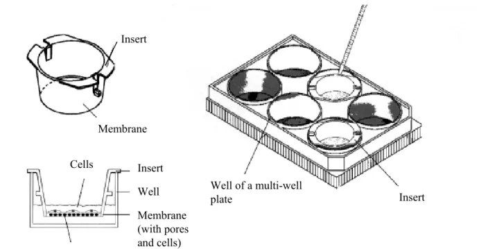
Differentiation of EBs was induced by plating them on gelatine-coated cover slips.
Cover slips (12 mm diameter) were placed in each well of a 24-multi well plate (Falcon), covered with gelatine (0.1% in PBS; gelatine from porcine skin, Sigma- Aldrich, Steinheim, Germany) and incubated overnight at 37°C. Before plating EBs, the gelatine solution was removed, cover slips were air-dried for 2 minutes and each well was filled with 1 ml 15% D-MEM. One to three embryoid bodies were isolated out of suspension and placed onto a gelatine-coated cover slip. EBs were supplemented with fresh medium every 3-4 days. Figure 3 schematically depicts the preparation of embryoid bodies.
2.1.4 Endothelial cells
2.1.4.1 Isolation of murine microvascular endothelial cells via Magnet associated cell sorting (MACS) and culture
Magnet associated cell sorting (MACS)-sorted microvascular endothelial cells were isolated as previously described (Schmidt et al., 2004a; Schmidt et al., 2004b; Muller- Ehmsen et al., 2006).
After a period of 5+7 days EBs were dissociated by treatment with Accutase. For immunomagnetic selection a single cell solution of 106 cells in 1 ml 15% D-MEM was stained with the endothelial-specific marker PECAM-1 (rat anti-mouse-PECAM- 1/CD31, 1:800, mAb, Pharmingen, San Diego, USA) for 30 minutes under growth conditions. Cells were washed twice with 2 ml MACS buffer (0.5% BSA and 2 mM EDTA in 0.1 M PBS). Cells were incubated with secondary antibody goat anti-rat IgG- conjugated micro beads (1:4 in MACS buffer, Miltenyi Biotec GmbH, Bergisch Gladbach, Germany) for 15 minutes at 4°C. After washing in MACS buffer, up to 108 cells were resuspended in 500 µl MACS-buffer. The mini-MACS system with MS separation columns (Miltenyi Biotec GmbH) was equilibrated using 500 µl MACS buffer and loaded with the cell suspension. After washing thrice with MACS buffer the MS separation column was removed from the magnetic holding device. The endothelial cell fraction was removed into a reaction cup (Eppendorf, Wesseling, Germany). Cells were centrifuged (5 minutes, 800 rpm) and resuspended in 1 ml 15% D-MEM. The isolated endothelial cell fraction was cultured on gelatine-coated
1000
5
5+7
Embryonic stem cells D3 (cultivated on feeder layer)
[days]
Suspension with 1000 embryonic stem cells/20 µl medium in hanging drops
Formation of embryo-like aggregates (embryoid bodies, EBs) in hanging drops
Collection of EBs and further cultivation in suspension
Plating of EBs on gelantine-coated cover slips in 24-well tissue plates
Appearing of first vessel-like structures in D3-EBs
2 0
Time schedule
dishes. After 2 to 3 weeks, the murine microvascular MACS-sorted endothelial cells were passaged the first time, afterwards twice a week. The purity of cells was tested by Dr. Annette Schmidt, German Sport University Cologne, with dil-Ac-LDL (Paesel + Lorei GmbH & Co, Hanau, Germany) and different endothelial specific markers (e.g.
PECAM-1, VE-Cadherin, flk-1, flt-1, VEGF, Lectin and eNOS) and was amounted to 89% (Schmidt, 2003). Murine microvascular MACS-sorted endothelial cells are referred to as MACS cells throughout this work.
Figure 3: Scheme of the preparation of embryoid bodies
Scheme adapted from Schmidt, 2003 (Schmidt, 2003).
2.1.4.2 Culture of human umbilical vein, coronary artery and aortic endothelial cells
Human umbilical vein endothelial cells (HUVEC), human coronary artery endothelial cells (HCAEC) and human aortic endothelial cells (HuAoEC) were obtained from PromoCell (Heidelberg, Germany). HUVEC were cultured in ECG-medium (PromoCell), HCAEC and HuAoEC in ECG-MV-medium (PromoCell) (Schmidt et al., 2004a). All three types of cells were incubated until confluence at 37°C, 95%
humidity and 5% CO2. The cells were detached upon treatment with Accutase, centrifuged and resuspended in medium. For every passage, cells were distributed to 25-cm2 cell culture flasks (200.000 cells/flask) and were supplemented with fresh medium twice a week. The cells were utilized within passages 3 to 4.
2.2 Quality control of MSCs
The quality of MSCs was ensured by microscopic observations for their characteristic spindle-shaped morphology, flow cytometry, colony forming unit-fibroblast assays and differentiation assays.
2.2.1 Flow cytometry (FACS)
For flow cytometric analysis, MSCs were detached with Accutase, centrifuged at 1300 rpm for 3 minutes, washed with PBS and stained in 100 µl PBS with CD106- Fluorescein isothiocyanate (FITC) (1:20, Becton Dickinson), CD105-Phycoerythrin (PE) (1:50, Ancell, Bayport, USA), CD45-energy-coupled dye (ECD) (1:20, Beckman Coulter, Krefeld, Germany), CD34- Phycoerythrin-Cyanine 5 (PC5) (1:20, Beckman Coulter) and CD14-PC5 (1:20, Beckman Coulter) at room temperature in dark for 15 minutes. The excess dye was removed by washing with PBS and cells were resuspended in 200 µl PBS. FACS analyses were performed by means of a Beckman Coulter Epics XL/MCL with Expo 32 software (Beckman Coulter) (Klinz et al., 2005; Schmidt et al., 2006a). According to published data, human MSCs were defined to be CD14, CD34 and CD45 triple-negative and CD105, CD106 double- positive (Pittenger et al., 1999).
2.2.2 Colony forming unit-fibroblast (CFU-F) assay
To determine the proliferative potential of cultured MSCs, CFU-F assays were performed as described before (Shur et al., 2004). Immediately after isolation (passage P0) 100.000 MSCs were seeded onto a 60-mm culture dish. Cells were cultured for 14 days without changing the media. MSC colonies were washed with PBS and stained with cresyl-violett (Merck, Darmstadt, Germany) for 10 minutes.
After repeated washing steps, blue colonies were macroscopically counted.
2.2.3 Differentiation assays
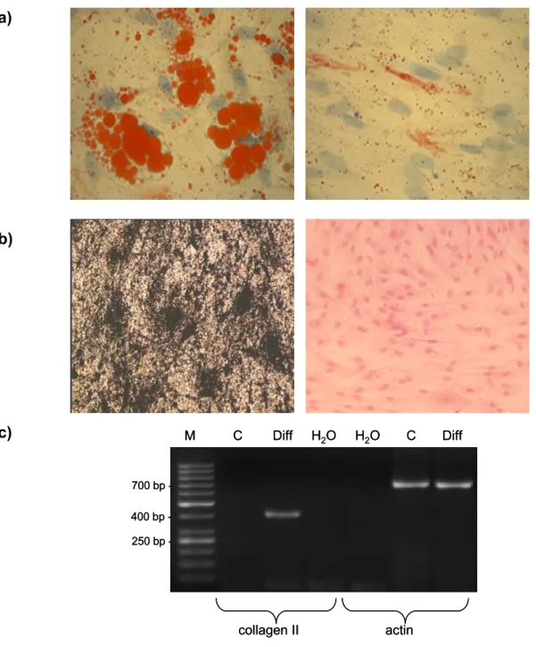
The differentiative potential of MSCs was evaluated by growing the cells under conditions favorable for adipogenic, osteogenic and chondrogenic differentiation (Pittenger et al., 1999; Arnhold et al., 2006).
2.2.3.1 Adipogenic differentiation
For adipogenic differentiation 1000 MSCs/cm2 were seeded onto 4-well plates (Nunc GmbH, Wiesbaden, Germany) and cultured under adipogenic-specific culture conditions in growth medium α-MEM supplemented with 2 mM L-glutamine, 0.5%
antibiotic-antimycotic solution (Fungizone, Gibco), 1 µM dexamethasone (Sigma- Aldrich), 5 µg/ml insulin (ITS Liquid Media supplement 100x, Sigma-Aldrich) and 15%
(v/v) of a selected batch of non heat-inactivated fetal calf serum (Gibco). Additionally 60 µM indomethacin (Sigma-Aldrich) was added shortly before use. Cells growing in 20% α-MEM served as control. Cells were cultured at 37°C in a humidified 5% CO2
atmosphere for 2 weeks and were supplemented with fresh pre-warmed medium thrice a week.
Adipose differentiation was analyzed by Oil Red O-staining. To prepare the working solution a stock solution of 0.17% Oil Red O (Merck) in 60% isopropanol (Merck) was diluted 3+2 with distilled water. After shaking for 5 minutes the working solution was filtered. Unfixed cells were incubated with fresh Oil Red O working solution for 15 minutes at room temperature. Cells were washed thrice with distilled water and counterstained for 4 minutes using Mayer`s hematoxylin stain. Cells were mounted with Kaiser`s glycerol gelatine (Merck). Staining resulted in red lipid drops and blue nuclei. Staining was analyzed using a Zeiss Axiophot microscope (Oberkochem, Germany).