AUS DEM LEHRSTUHL FÜR MUND-, KIEFER- UND GESICHTSCHIRURGIE PROF. DR. DR. TORSTEN E. REICHERT
DER FAKULTÄT FÜR MEDIZIN DER UNIVERSITÄT REGENSBURG
PHYSIOLOGISCHE KONZENTRATIONEN DENOSUMAB STEIGERN DIE OSTEOGENE DIFFERENZIERUNG VON HUMANEN MESENCHYMALEN
STAMMZELLEN
Inaugural – Dissertation zur Erlangung des Doktorgrades
der Zahnmedizin
der
Fakultät für Medizin der Universität Regensburg
vorgelegt von Alexander Mosch
2019
Dekan: Prof. Dr. Dr. Torsten E. Reichert
1. Berichterstatter: PD. Dr. Dr. Christoph Klingelhöffer, M.Sc . 2. Berichterstatter: Prof. Dr. Dr. Peter Proff
Tag der mündlichen Prüfung: 24.07.2019
Deutschsprachige Zusammenfassung der wissenschaftlichen Originalarbeit
„Physiological concentrations of Denosumab enhance osteogenic differentiation in human mesenchymal stem cells of the jaw bone”
Von
Alexander Mosch et al.
publiziert in
Archives of Oral Biology (Impact Factor: 2.050) 2019
vorgelegt von
Alexander Mosch
4
Inhaltsverzeichnis
1. Einleitung und Fragestellung ... ……… .5
2. Material und Methoden ……… 6
2.1 Zellkultur und osteogene Differenzierung ……… .6
2.2 Toxizitätsassay ……… .7
2.3 Alkaline Phosphatase Assay ……… ..7
2.4 Genexpression und quantitative real-time PCR (RT-qPCR) ……… .8
2.5 Statistik ……… ..8
3. Ergebnisse ……… 8
3.1 Nachweis von RANK auf MSCs ……… .8
3.2 Toxizitätsassay ……… ...9
3.3 Alkaline Phosphatase Assay ……… 10
3.4 Genexpression der osteogenen Differenzierungsmarker ……… 11
4. Diskussion ……… ..12
5. Fazit ……… .14
6. Literaturverzeichnis ……… ..15
7. Danksagung ……… ...19
8. Lebenslauf ……… ..20
5
Einleitung und Fragestellung
Die Medikamenten-assoziierte Kiefernekrose (engl.: Medication-related osteonecrosis of the jaw, kurz MRONJ) ist als häufige Nebenwirkung der antiresorptiven Therapie mit Denosumab oder Bisphosphonaten beschrieben. Die American Association of Oral and Maxillofacial Surgeons definierte sie als freiliegenden Knochen, oder Knochen, der durch eine intra- oder extraorale Fistel sondiert werden kann, der länger als 8 Wochen besteht. Eine weitere Voraussetzung zur Diagnosestellung ist eine durchgeführte oder laufende antiresorptive Therapie, zudem darf keine Bestrahlung in der Anamnese vorhanden sein (Ruggiero et al., 2014). Die Pathophysiologie dieser Kiefernekrosen ist noch unklar. Ursächlich ist meist eine lokale Infektion oder ein Trauma des Knochens, zum Beispiel nach dentoalveolärer Chirurgie, bei Prothesendruckstellen oder bei Entzündung des Parodonts (Otto et al., 2018). Medikamente, die im Rahmen einer antiresorptiven Therapie (z.B. bei Osteoporose oder Knochenmetastasen) verabreicht werden, sind beispielsweise der Antikörper Denosumab oder Bisphosphonate wie Zoledronat.
Denosumab bewirkt eine verminderte Ausreifung von Osteoklasten. Bisphosphonate wie Zoledronat führen in bereits entwickelten Osteoklasten zur Apoptose. Die Störung des Knochenstoffwechsels, die hierdurch hervorgerufen wird, wird als Ursache der MRONJ diskutiert (Khan et al., 2014). Krebspatienten, die mit Bevacizumab behandelt werden, welches antiangiogene aber keine antiresorptiven Wirkungen besitzt, zeigten ebenfalls Fälle derartiger Kiefernekrosen (Bettini et al., 2014). Da Bisphosphonate auch antiangiogene Effekte aufweisen, steht auch die verminderte Vaskularisierung des Defekts als mögliche Ursache für die Kiefernekrosen zur Debatte. Dauer und Dosis der antiresorptiven Therapie hat ebenfalls einen signifikanten Einfluss auf die Entstehung des Krankheitsbildes: Hohe Konzentrationen von sowohl Bisphosphonaten als auch Denosumab steigern die Inzidenz signifikant (Khan et al., 2014).
Humane mesenchymale Stammzellen (MSCs) und dentale Follikelzellen
(DFCs) erschienen uns als geeignete in vitro Modelle, da die MSCs im Knochenmark
und die DFCs im dentalen Follikel vorkommen. Beide Zelllinien sind
ektomesenchymalen Ursprungs und entwickeln Strukturen (Morsczeck et al., 2005),
die den Zahnkeim umgeben und sich später in Teilen zum parodontalen Ligament
entwickeln. Sowohl Knochenmark als auch das parodontale Ligament können nach
dentoalveolärer Chirurgie exponiert werden oder bei odontogenen Infektionen als
6
Eintrittspforte für Keime fungieren. Weiterhin besitzen beide Zelllinien osteogenes Differenzierungspotenzial (Jaiswal et al., 1998; Morsczeck et al., 2005). Der Knochenstoffwechsel hängt maßgeblich von der osteogenen Differenzierung von Stammzellen und deren Vitalität ab. Da ein beeinträchtigter Knochenstoffwechsel als Ursache für MRONJ angenommen wird, untersuchten wir die Reaktion der beiden Stammzelllinien hinsichtlich Differenzierung und Vitalität auf antiresorptive Medikamente.
Denosumab, das 2010 auf den Markt kam, wird immer häufiger bei Patienten mit Knochenmetastasen oder Osteoporose angewendet. Es ist ein monoklonaler Antikörper gegen RANKL. RANKL ist ein von Osteoblasten produziertes Protein. Es stimuliert RANK, einen Rezeptor, der üblicherweise auf Prä-Osteoklasten gefunden wird, wodurch es zur Ausreifung von Osteoklasten kommt. Diese Wechselwirkung ist essenziell, um den Knochenstoffwechsel in Balance zu halten. Bei Knochenverlust (z.B. durch Osteoporose) wird durch den Einsatz von Denosumab die Reifung der Prä-Osteoklasten inhibiert. Dabei imitiert Denosumab das humane Protein Osteoprotegerin, das auch von Osteoblasten produziert wird und die Bindungsstelle auf RANKL für RANK blockiert (Schieferdecker et al., 2014). Seine Halbwertzeit beträgt 30 Tage, während Bisphosphonate wahrscheinlich über Jahre im Knochen persistieren (Cremers et al., 2005; Yonemori et al., 2008). Weiterhin hat es einen unmittelbaren Effekt auf den Knochen (Grasemann et al., 2013), im Gegensatz zu Bisphosphonaten, deren maximale Effektivität erst nach 1-3 Monaten erreicht wird (Reid et al., 2002; Cremers et al., 2005). Obwohl Denosumab das Risiko für eine MRONJ im gleichen Maße wie Bisphosphonate erhöht, konzentrieren sich die meisten der aktuellen Studien auf letztere. Währenddessen wächst die Benutzung von Denosumab aufgrund seiner Vorteile gegenüber Bisphosphonaten. Es existieren noch keine Daten über den Effekt von Denosumab auf die Vitalität oder das Differenzierungspotenzial von Stammzellen. Diese zellulären Wirkungen wollen wir mit dieser Studie erforschen.
Material und Methoden
Zellkultur und osteogene Differenzierung
Die MSCs wurden in DMEM mit 10% MSC-ready FBS und 100 µg/ml
Penicillin/Streptomycin ausgesät. Nach einem Tag wurden die Zellen, wie vom
7
Hersteller (Thermo Fisher Scientific) vorgegeben, in Gibco
TMMesenPro RS Medium mit 5 µg/ml Gentamicin subkultiviert. Wir begannen die Zellen osteogen zu differenzieren, indem wir sie in Gibco
TMStempro
TMOsteogenesis Differentiation Kit mit 5 µg/ml Gentamicin kultivierten. Alle Versuche wurden in Zellpassage 6 durchgeführt.
Die DFCs wurden in DMEM mit 10% FBS und 100 µg/ml Penicillin/Streptomycin bis zur Konfluenz kultiviert. Danach induzierten wir die osteogene Differenzierung, indem wir die Zellen in osteogenem Differenzierungsmedium (ODM) kultivierten. ODM enthält DMEM mit 10% FBS, 100 µG/ml Penicillin/Streptomycin, 0,1 mmol/L Ascorbinsäure-2-Phosphat, 20 mmol/L HEPES Puffer, 2,8 mmol/L beta-Glycerophosphat und 1 x 10
-4mmol/L Dexamethason-Natriumphosphat. Die Versuche mit DFCs wurden in Passage 7 durchgeführt.
Toxizitätsassay
MSCs und DFCs wurden in einer 96-well Platte mit 3500 Zellen/cm² ausgesät.
Nach einem Tag inkubierten wir die Zellen mit behandelten Medien. Die Medien wurden in folgenden aufsteigenden Konzentrationen mit Denosumab (XGEVA®, 70mg/ml, AMGEN) versetzt: 0,25 µM, 0,5 µM, 1 µM, 5 µM, 10 µM, 20 µM. Zoledronat (Zometa®, 4mg/5ml, Novartis) als Bisphosphonat der Wahl wurde in ähnlichen Konzentrationen verwendet: 0,25 µM, 0,5 µM, 1 µM, 2 µM, 3 µM, 5 µM, 10 µM.
Zusätzlich enthielt jeder Versuch eine unbehandelte Kontrolle. Die Zellproliferation wurde quantitativ nach 1, 4 und 7 Tagen für alle Konzentrationen erfasst. Dafür benutzten wir das Cell Counting Kit-8 (CCK-8, Dojindo Molecular Technologies Inc.).
Alkaline Phosphatase Assay
Die osteogene Differenzierung wurde mittels des Alkaline Phosphatase
Assays (Thermo Fisher Scientific) quantifiziert. MSCs und DFCs wurden mit 3500
Zellen/cm² in je eine 96-well Platte ausgesät. Nach einem Tag begannen wir mit der
Inkubation durch die jeweiligen Differenzierungsmedien, die in gleichen
Konzentrationen wie die Kulturmedien hergestellt wurden. Zur Kontrolle wurde
sowohl unbehandeltes Differenzierungsmedium, als auch unbehandeltes, nicht
differenzierendes Medium verwendet. Nach 7 Tagen Inkubation erfolgte die
8
Messung. Die quantifizierte ALP-Menge wurde gegen das Gesamtprotein normalisiert, das mittels BCA Assay aus den gleichen Wells evaluiert wurde.
Genexpression und quantitative real-time PCR (RT-qPCR)
Das Vorhandensein von RANK ist eine Voraussetzung für Versuche mit Denosumab. Deshalb untersuchten wir sowohl MSCs als auch DFCs zuerst auf RANK. Beide Zelllinien wurden sowohl nach Inkubation mit normalem Zellmedium, als auch nach Inkubation mit osteogenem Differenzierungsmedium auf RANK getestet. Später evaluierten wir die Genexpression der osteogenen Differenzierungsmarker COL1, ALP, RUNX2 bei MSCs nach 7-tägiger Behandlung mit 0,25 µM Denosumab-behandeltem Differenzierungsmedium. Zur Kontrolle wurde das Gen der Glyceraldehyd-3-Phosphat-Dehydrogenase verwendet.
Statistik
Die Daten wurden mit SPSS Version 25.0 verarbeitet. Bei 2 vorhandenen Gruppen wurde der T-Test verwendet. Bei mehr als 2 Gruppen benutzten wir die one-way ANOVA. Post hoc Analysen wurden durchgeführt, sobald die ANOVA signifikante Unterschiede aufwies. p < 0,05 wurde als signifikant angesehen.
Ergebnisse
Nachweis von RANK auf MSCs
MSCs exprimierten RANK sowohl mit als auch ohne Differenzierung in ähnlicher Menge.
Durchschnittlicher Ct für differenzierte MSCs war 35,93 und für undifferenzierte 35,53, ohne signifikanten Unterschied
untereinander. Die DFCs wiesen keine Expression von RANK auf. Der Unterschied zwischen DFCs und MSCs in ihrer RANK-Expression ist signifikant (Abb. 1).
Abbildung 1: RANK konnte auf MSCs im Gegensatz zu DFCs
nachgewiesen werden.
9
Toxizitätsassay
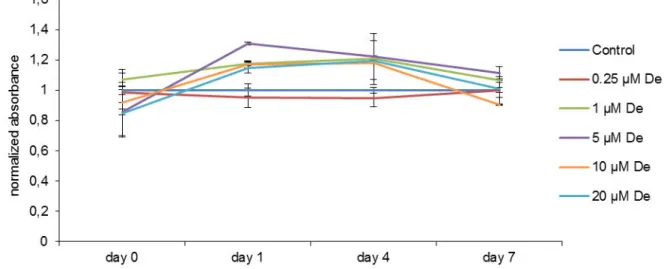
Die DFCs wiesen keine Veränderung der Proliferation nach Behandlung mit Denosumab auf. Das Wachstum der MSCs wurde dosisabhängig ab Tag 4 reduziert.
10 und 20 µM Denosumab verringerten das Wachstum signifikant zur unbehandelten Kontrolle. Nach 7 Tagen war das Wachstum der mit 10 und 20 µM behandelten MSCs signifikant zur unbehandelten Kontrolle und zu allen geringeren Konzentrationen reduziert, außer 10 µM zu 0,25 µM. 0,25 µM, 0,5 µM, 1 µM und 5 µM Denosumab bewirkten keine signifikante Veränderung der Proliferation im Vergleich zur unbehandelten Kontrolle (Abb. 2 & 3).
Abbildung 3: Denosumab zeigt keine Auswirkungen auf die Vitalität von DFCs.
Abbildung 2: 10 µM und 20 µM Denosumab wirken sich signifikant toxisch auf MSCs aus.
10
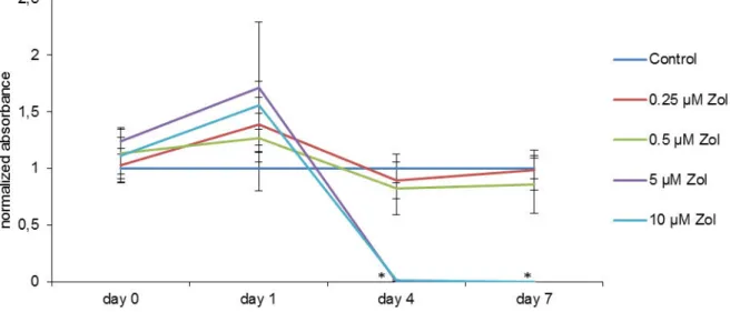
Die Zellproliferation unter Behandlung mit Zoledronat wurde bei DFCs untersucht.
Nach 4 und 7 Tagen zeigte sich eine signifikante Reduktion des Wachstums bei den mit 5 und 10 µM behandelten DFCs im Vergleich zu allen geringeren Konzentrationen und der unbehandelten Kontrolle (Abb. 4).
Alkaline Phosphatase Assay
Nichttoxische Konzentrationen von Denosumab hatten einen positiven Effekt auf die osteogene Differenzierung. Die ALP-Aktivität der MSCs wurde nach 7 Tagen bei 0,25, 1 und 5 µM signifikant im Vergleich zur unbehandelten Kontrolle erhöht. Außerdem erhöhten 1 µM und 5 µM die ALP-Aktivität signifikant mehr als 0,25 µM Denosumab. Bei 20 µM wurde allerdings eine signifikante Reduktion der ALP-Aktivität sowohl zu allen anderen Konzentrationen als auch zur unbehandelten Kontrolle gezeigt (Abb. 5) .
Abbildung 4: Zoledronat senkt konzentrationsabhängig die Vitalität von DFCs signifikant.
Abbildung 5: Denosumab erhöht konzentrationsabhängig die
osteogene Differenzierung. Auch die physiologische Konzentration
(0,25 µM) verstärkt die osteogene Differenzierung signifikant im
Vergleich zur Kontrolle. 20 µM als toxische Konzentration von
Denosumab verringert die osteogene Differenzierung signifikant.
11
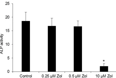
Die DFCs zeigten keine signifikante Veränderung in ihrer ALP-Aktivität nach Behandlung
mit nichttoxischen
Zoledronatkonzentrationen (0,25 µM und 0,5 µM). 10 µM Zoledronat reduzierte die ALP- Aktivität signifikant im Vergleich zur unbehandelten Kontrolle (Abb.6) .
Genexpression der osteogenen Differenzierungsmarker
Obwohl nicht signifikant, war die Hochregulierung der osteogenen Differenzierungsmarker COL1, ALP und RUNX2 im Vergleich zur unbehandelten Kontrolle nach Inkubation mit 0,25 µM Denosumab deutlich erkennbar (Abb. 7).
Abbildung 6: Zoledronat zeigt in nicht-toxischen Konzentrationen (0,25 µM und 0,5 µM) keine Auswirkungen auf die osteogene Differenzierung.
Abbildung 7: Die Auswertung der RT-qPCR spricht für eine klare Stimulierung der Gene der 3 Differenzierungsmarker
Runx2, ALP und COL1.
12
Diskussion
Ziel dieser Studie war, den Effekt, den Denosumab auf die osteogene Differenzierung alveolarer Stammzellen hat, zu evaluieren. MSCs und DFCs sind in der Lage, sich zu Osteoblasten zu differenzieren und sind damit essenziell für den Knochenstoffwechsel (Gamblin et al., 2014; Morsczeck et al., 2017). Vor dem Hintergrund, dass Denosumab die RANK-RANKL-Interaktion inhibiert, wollten wir DFCs und MSCs auf RANK-Expression untersuchen, die eine mögliche Rezeptor- abhängige Wirkung von Denosumab bedingen könnte. Unsere Ergebnisse prädestinierten MSCs für eine Untersuchung Rezeptor-abhängiger Effekte. Da DFCs kein RANK aufwiesen, konnten wir mit den DFCs mögliche Rezeptor-unabhängige Wirkungen von Denosumab untersuchen. Diese wollten wir mit Zoledronat vergleichen, einem Bisphosphonat, das für seine Interaktion mit der Zelle ebenfalls auf keinen Rezeptor angewiesen ist. Dafür errechneten wir im Vorfeld näherungsweise physiologische Konzentrationen der beiden Stoffe, unter Berücksichtigung durchschnittlicher Blutvolumina eines damit behandelten Patienten: 0,25 µM für Denosumab und 0,5 µM für Zoledronat.
Zoledronat hat mehrere Effekte auf verschiedene Arten von Zellen. Sein Einfluss auf die Vitalität von MSCs wurde bereits erforscht und zeigte eine Senkung dieser bei Konzentrationen von 5 und 10 µM (Hu et al., 2017). Weiterhin wurde gezeigt, dass 10 µM und 50 µM Zoledronat nach 3 Tagen die Beweglichkeit und Integrität von alveolaren Osteoblasten senkt. Unsere Ergebnisse zeigen, dass sich hohe Konzentrationen von Zoledronat auch auf die Proliferation von DFCs negativ auswirken. Die Toxizität von Bisphosphonaten wird über den Mevalonatweg vermittelt. Sie blockieren die Farnesylpyrophosphat-Synthase, was zu einer verminderten Reifung des GTP-bindenden Proteins Ras führt und Apoptose in der Zelle induziert (Luckman et al., 1998; Fisher et al., 1999). Sharma et al. (2016) zeigten außerdem, dass Zoledronat die Expression endothelialer Marker und die Integrität und Mobilität von MSCs senkt.
Im Gegensatz dazu und auch zu unseren Ergebnissen wurde bereits von
positiven Effekten von Zoledronat auf Stammzellen berichtet. Park et al. (2017)
vermuteten keine toxischen Wirkungen, nachdem sie Zellen aus dem Knochenmark
und dem Periost des Kiefers 7 Tage lang 1 µM Zoledronat aussetzten. Außerdem
13
wurde gezeigt, dass Zoledronat die Vermehrung hematopoietischer Stammzellen unterstützt und die Anzahl neu entstehender Blutgefäße erhöht (Soki et al., 2013).
Zoledronat senkte zudem die Anhäufung von Fehlern in der DNA (Misra et al., 2016).
Obwohl unterschiedliche Typen von Zellen verschieden auf gleiche Konzentrationen Zoledronat reagieren, scheinen Rahmenbedingungen, wie Passagestadium, Inkubationszeit und Differenzierungsstatus eine Rolle für die Vitalität zu spielen.
Diese Einflussfaktoren sollten künftig untersucht werden.
Nichttoxische Dosen von Zoledronat zeigten keinen Einfluss auf die ALP- Aktivität der DFCs nach 7 Tagen. Unsere Ergebnisse unterstützen die These, dass Zoledronat keinen Einfluss auf die osteogene Differenzierung hat. Die signifikante Abnahme der ALP-Aktivität bei 10 µM Zoledronat schreiben wir seiner Toxizität zu, die wir im Vitalitätsassay ebenfalls nachweisen konnten. Andere Arbeitsgruppen konnten allerdings auch fördernde Effekte auf die osteogene Differenzierung nachweisen. Ribeiro et al. (2014) zeigten gesteigerte Differenzierungsmarker (ALP, BMP2, OPG, OC) in humanen MSCs nach 14 Tagen. Zoledronat konnte außerdem eine steigernde Wirkung auf die Expression von BMP2 und ERKs in Osteoblasten nachgewiesen werden (Pan et al., 2004). Der durch BMP2 induzierte ERK-Signalweg ist wiederum signifikant an der Steigerung der osteogenen Differenzierung beteiligt (Jun et al., 2010). Da DFCs dagegen in ihrer osteogenen Differenzierung nicht durch Zoledronat beeinflusst werden, vermuten wir, dass die Reaktion auf Bisphosphonate vom Zelltyp abhängig ist
Die Ergebnisse des Vitalitätsassays bei Denosumab zeigen, dass es in hohen Konzentrationen (10 µM und 20 µM) einen toxischen Effekt hat. Das erklärt den signifikanten Abfall der ALP-Aktivität nach Behandlung mit 20 µM Denosumab. Dass die DFCs in ihrer Vitalität nicht beeinflusst wurden, führen wir auf das Fehlen eines RANK-Rezeptors bei ihnen zurück. Wir vermuten damit eine starke rezeptorvermittelte Toxizität.
Das ALP Assay zeigte, dass niedrige nichttoxische Dosen von Denosumab die
osteogene Differenzierung erhöhen. Dass diese Konzentrationen die Proliferation
nicht signifikant beeinflussen, spricht dafür, dass Denosumab spezielle Mechanismen
der osteogenen Differenzierung anspricht. Die höchste ALP-Aktivität war bei 1 µM
Denosumab messbar. Eventuell noch höhere ALP-Werte bei höheren
14
Denosumabkonzentrationen könnten durch die Abnahme der Vitalität maskiert worden sein. Auch die im Körper vorzufindende Konzentration an Denosumab, 0,25 µM, erhöhte die osteogene Differenzierung signifikant. Deshalb wählten wir diese Konzentration, um mit RT-qPCR 3 Schlüsselenzyme der osteogenen Differenzierung näher zu untersuchen: COL1, RUNX2 und ALP. Obwohl die Ergebnisse nicht signifikant waren, sprechen sie für eine Stimulierung der osteogenen Marker. Dies wurde auch schon für Zoledronat in MSCs festgestellt (Ribeiro et al., 2014).
Die Mechanismen hinter den positiven Effekten von Denosumab sind noch ungeklärt. Portal-Nunez et al. (2017) beschrieben ein erhöhtes Differenzierungspotenzial bei Osteoblasten, nachdem man sie mit Denosumab behandelte. Sie legten außerdem nahe, dass Denosumab sekundär Kollagenfasern mineralisiert, was dann wiederum zu einer dystrophen Ossifizierung innerhalb der Knochenmasse führt, die sich als erhöhte Knochendichte zeigte. Weiterhin wird RANKL als Einflussfaktor auf reifende MSCs diskutiert. Schena et al. (2017) vermuteten einen autokrinen/parakrinen Effekt von RANKL durch seine Interaktion mit RANK auf MSCs. Bei Knockoutmäusen ohne RANKL zeigten sie eine verringerte osteogene Differenzierung im Vergleich zu Wildtyp-MSCs. Unsere Ergebnisse sprechen in ähnlicher Weise für eine Verringerung der osteogenen Differenzierung bei hohen Denosumabkonzentrationen. Wir vermuten, dass auto- und parakrine Loopmechanismen demnach bei geringen Denosumabkonzentrationen zu einer Hochregulierung der osteogenen Differenzierung in MSCs führen.
Fazit
Unsere Versuche zeigten rezeptor- und dosisabhängige Wirkungen von Denosumab. Physiologische Konzentrationen von Denosumab (0,25 µM) hatten keinen toxischen Effekt. Höhere Konzentrationen (10 µM und 20 µM) reduzierten die Zellvitalität der MSCs signifikant. Weiterhin vermuten wir aufgrund erhöhter ALP- Aktivität und Stimulierung der Schlüsselenzyme einen positiven Effekt auf die osteogene Differenzierung.
Weitere Studien sind notwendig, um die Mechanismen zu untersuchen, wie
Denosumab die osteogene Differenzierung beeinflusst. Dosisabhängige Wirkungen
von Denosumab sollten ebenfalls untersucht werden, um die Behandlung von
15
Patienten mit Denosumab und ihre Folgen besser zu erforschen und deren Sicherheit zu verbessern. Letztendlich können weitere Experimente die Pathomechanismen hinter der Medikamenten-assoziierten Osteonekrose aufklären.
Literaturverzeichnis
Bettini, G., Blandamura, S., Saia, G., & Bedogni, A. (2012). Bevacizumab-related osteonecrosis of the mandible is a self-limiting disease process. BMJ Case Reports, 2012. https://doi.org/10.1136/bcr-2012-007284
Cremers, S. C. L. M., Pillai, G., & Papapoulos, S. E. (2005).
Pharmacokinetics/pharmacodynamics of bisphosphonates: use for optimisation of intermittent therapy for osteoporosis. Clinical
Pharmacokinetics, 44(6), 551 – 570. https://doi.org/10.2165/00003088- 200544060-00001
Fisher, J. E., Rogers, M. J., Halasy, J. M., Luckman, S. P., Hughes, D. E.,
Masarach ia, P. J., … Reszka, A. A. (1999). Alendronate mechanism of action:
geranylgeraniol, an intermediate in the mevalonate pathway, prevents inhibition of osteoclast formation, bone resorption, and kinase activation in vitro. Proceedings of the National Academy of Sciences of the United States of America, 96(1), 133 – 138.
Gamblin, A. L., Renaud, A., Charrier, C., Hulin, P., Louarn, G., Heymann, D., … Layrolle, P. (2014). Osteoblastic and osteoclastic differentiation of human mesenchymal stem cells and monocytes in a miniaturized three-dimensional culture with mineral granules. Acta Biomaterialia, 10(12), 5139 – 5147.
https://doi.org/10.1016/j.actbio.2014.08.033
Grasemann, C., Schündeln, M. M., Hövel, M., Schweiger, B., Bergmann, C., Herrmann, R., … Hauffa, B. P. (2013). Effects of RANK-Ligand Antibody (Denosumab) Treatment on Bone Turnover Markers in a Girl With Juvenile Paget’s Disease. The Journal of Clinical Endocrinology & Metabolism, 98(8), 3121 – 3126. https://doi.org/10.1210/jc.2013-1143
Hu, L., Wen, Y., Xu, J. , Wu, T., Zhang, C., Wang, J., … Wang, S. (2017).
Pretreatment with Bisphosphonate Enhances Osteogenesis of Bone Marrow Mesenchymal Stem Cells. Stem Cells and Development, 26(2), 123 – 132.
https://doi.org/10.1089/scd.2016.0173
16
Jaiswal, N., Haynesworth, S. E., Caplan, A. I., & Bruder, S. P. (1997). Osteogenic differentiation of purified, culture-expanded human mesenchymal stem cells in vitro. Journal of Cellular Biochemistry, 64(2), 295 – 312.
https://doi.org/10.1002/(SICI)1097-4644(199702)64:2<295::AID- JCB12>3.0.CO;2-I
Jun, J. H., Yoon, W.-J., Seo, S.-B., Woo, K.-M., Kim, G.-S., Ryoo, H.-M., & Baek, J.- H. (2010). BMP2-activated Erk/MAP kinase stabilizes Runx2 by increasing p300 levels and histone acetyltransferase activity. The Journal of Biological Chemistry, 285(47), 36410 – 36419. https://doi.org/10.1074/jbc.M110.142307 Khan, A. A., Morrison, A., Hanley, D. A., Felsenberg, D., McCauley, L. K., O’Ryan, F.,
… International Task Force on Osteonecrosis of the Jaw. (2015). Diagnosis and management of osteonecrosis of the jaw: a systematic review and international consensus. Journal of Bone and Mineral Research: The Official Journal of the American Society for Bone and Mineral Research, 30(1), 3 – 23.
https://doi.org/10.1002/jbmr.2405
Luckman, S. P., Hughes, D. E., Coxon, F. P., Graham, R., Russell, G., & Rogers, M.
J. (1998). Nitrogen-containing bisphosphonates inhibit the mevalonate pathway and prevent post-translational prenylation of GTP-binding proteins, including Ras. Journal of Bone and Mineral Research: The Official Journal of the American Society for Bone and Mineral Research, 13(4), 581 – 589.
https://doi.org/10.1359/jbmr.1998.13.4.581
Misra, J., Mohanty, S. T., Madan, S., Fernandes, J. A., Ebetino, F. H., Russell, R. G.
G., & Bellantuono, I. (2016). Zoledronate Attenuates Accumulation of DNA Damage in Mesenchymal Stem Cells and Protects Their Function. STEM CELLS, 34(3), 756 – 767. https://doi.org/10.1002/stem.2255
Morsczeck, C., Moehl, C., Götz, W., Heredia, A., Schäffer, T. E., Eckstein, N., … Hoffmann, K. H. (2005). In vitro differentiation of human dental follicle cells with dexamethasone and insulin. Cell Biology International, 29(7), 567 – 575.
https://doi.org/10.1016/j.cellbi.2005.03.020
Morsczeck, C., & Reichert, T. E. (2017). The dexamethasone induced osteogenic differentiation of dental follicle cells. Histology and Histopathology, 32(12), 1223 – 1229. https://doi.org/10.14670/HH-11-907
Otto, S., Pautke, C., Van den Wyngaert, T., Niepel, D., & Schiødt, M. (2018).
Medication-related osteonecrosis of the jaw: Prevention, diagnosis and
17
management in patients with cancer and bone metastases. Cancer Treatment Reviews, 69, 177 – 187. https://doi.org/10.1016/j.ctrv.2018.06.007
Pan, B., To, L. B., Farrugia, A. N., Findlay, D. M., Green, J., Gronthos, S., … Zannettino, A. C. W. (2004). The nitrogen-containing bisphosphonate,
zoledronic acid, increases mineralisation of human bone-derived cells in vitro.
Bone, 34(1), 112 – 123.
Park, J.-B., Cho, S.-H., Kim, I., Lee, W., Kang, S.-H., & Kim, H. (2018). Evaluation of the bisphosphonate effect on stem cells derived from jaw bone and long bone rabbit models: A pilot study. Archives of Oral Biology, 85, 178 – 182.
https://doi.org/10.1016/j.archoralbio.2017.10.024
Portal-Núñez, S., Mediero, A., Esbrit, P., Sánchez-Pernaute, O., Largo, R., &
Herrero-Beaumont, G. (2017). Unexpected Bone Formation Produced by RANKL Blockade. Trends in Endocrinology & Metabolism, 28(10), 695 – 704.
https://doi.org/10.1016/j.tem.2017.06.003
Reid, I. R., Brown, J. P., Burckhardt, P., Horowitz, Z., Richardson, P., Trechsel, U., … Meunier, P. J. (2002). Intravenous Zoledronic Acid in Postmenopausal Women with Low Bone Mineral Density. New England Journal of Medicine, 346(9), 653 – 661. https://doi.org/10.1056/NEJMoa011807
Ribeiro, V., Garcia, M., Oliveira, R., Gomes, P. S., Colaço, B., & Fernandes, M. H.
(2014). Bisphosphonates induce the osteogenic gene expression in co- cultured human endothelial and mesenchymal stem cells. Journal of Cellular and Molecular Medicine, 18(1), 27 – 37. https://doi.org/10.1111/jcmm.12154 Ruggiero, S. L., Dodson, T. B., Fantasia, J., Goodday, R., Aghaloo, T., Mehrotra, B.,
… American Association of Oral and Maxillofacial Surgeons. (2014). American Association of Oral and Maxillofacial Surgeons position paper on medication- related osteonecrosis of the jaw--2014 update. Journal of Oral and
Maxillofacial Surgery: Official Journal of the American Association of Oral and Maxillofacial Surgeons, 72(10), 1938 – 1956.
https://doi.org/10.1016/j.joms.2014.04.031
Schena, F., Menale, C., Caci, E., Diomede, L., Palagano, E., Recordati, C., …
Sobacchi, C. (2017). Murine Rankl−/− Mesenchymal Stromal Cells Display an
Osteogenic Differentiation Defect Improved by a RANKL-Expressing Lentiviral
Vector. STEM CELLS, 35(5), 1365 – 1377. https://doi.org/10.1002/stem.2574
18
Schieferdecke r, A., Voigt, M., Riecken, K., Braig, F., Schinke, T., Loges, S., … Binder, M. (2014). Denosumab mimics the natural decoy receptor osteoprotegerin by interacting with its major binding site on RANKL.
Oncotarget, 5(16), 6647 – 6653. https://doi.org/10.18632/oncotarget.2160 Sharma, D., Hamlet, S. M., Petcu, E. B., & Ivanovski, S. (2016). The effect of
bisphosphonates on the endothelial differentiation of mesenchymal stem cells.
Scientific Reports, 6, 20580. https://doi.org/10.1038/srep20580
Soki, F. N., Li, X., Berry, J., Koh, A., Sinder, B. P., Qian, X., … McCauley, L. K.
(2013). The effects of zoledronic acid in the bone and vasculature support of hematopoietic stem cell niches. Journal of Cellular Biochemistry, 114(1), 67 – 78. https://doi.org/10.1002/jcb.24301
Yonemori, K., Fujiwara, Y., Minami, H., Kitagawa, K., Fujii, H., Arai, T., … Ohtsu, T.
(2008). Phase 1 trial of denosumab safety, pharmacokinetics, and
pharmacodynamics in Japanese women with breast cancer-related bone metastases. Cancer Science, 99(6), 1237 – 1242.
https://doi.org/10.1111/j.1349-7006.2008.00803.x
19
Danksagung
Ich bedanke mich zuallererst bei meinem Doktorvater und Betreuer Christoph Klingelhöffer. Du hast diese Studie entworfen und mich bis zum Ende sehr tatkräftig unterstützt. Ohne deine Erfahrung wäre diese Arbeit nicht publiziert worden. Du bist stets geduldig und genau geblieben, und ich habe dank dir vieles über die Wissenschaft gelernt. Die Arbeit mit dir war jederzeit unbeschwert.
Ich bedanke mich zudem bei Christian Morsczeck, der mir als Arbeitsgruppenleiter freie Hand in seinem Labor ließ und die nötige wissenschaftliche Weitsicht besaß.
Zudem möchte ich mich bei Anja Reck bedanken, die mir bei einigen Versuchen unter die Arme griff und mich mit größter Geduld im Labor unterwies.
Besonderer Dank gilt ebenfalls den Biologinnen Daniela Schulz, Stephanie Walter und Lena Mischkulnig, die mir alle Fragen im Labor beantworten konnten, mir Kniffe zeigten und die Zeit im Labor für mich persönlich bereicherten.
Eine besondere Rolle kommt Sophia Rohrmüller zu. Erinnerungen an das Labor werden immer an dich geknüpft sein. Ich danke dir für deine Unterstützung in allen Lebensbereichen, die über unsere gemeinsame Zeit im Labor hinausging. Ich wünsche dir nur das Beste.
Zuletzt möchte ich mich bei meinen Eltern, Krystina Meichsner und Andreas Mosch,
meinen Großeltern, Gisela und Martin Mosch sowie Christiane Kschammer, und
meiner Schwester Henrike Mosch bedanken. Ihr habt mich ideell, moralisch und
finanziell unterstützt und mir damit Kraft und Möglichkeit für das Studium der
Zahnmedizin gegeben. Ich kann mich nicht oft genug für euren Rückhalt bedanken.
20
Lebenslauf Alexander Mosch
Geboren am 01.06.1994 in Herzberg (Elster)
Schulausbildung
08/2000 – 04/2001 Grundschule Jessen-Nord
04/2001 – 07/2004 Grundschule „Friedrich Engels“ in Lutherstadt Wittenberg 08/2004 – 07/2012 „Lucas -Cranach- Gymnasium“ in Lutherstadt Wittenberg
Studium
04/2013 – 07/2018 Studium der Zahnheilkunde an der Universität Regensburg
10/2018 – 10/2023 Studium der Humanmedizin an der Universität Regensburg
Wissenschaftliche Arbeit
08/2016 – 12/2018 „Physiologische Konzentrationen D enosumab steigern die osteogene Differenzierung von humanen mesenchymalen Stammzellen “
Publiziert in Archives of Oral Biology (2019)
Berufliche Tätigkeit
04/2017 – 09/2017 Studentische Hilfskraft an der Poliklinik für Kieferorthopädie der Universität Regensburg
10/2019 – dato Ehrenamtliche Mitarbeit im Verein für Migrationsmedizin e.V.
Regensburg, 08.10.2019
Contents lists available atScienceDirect
Archives of Oral Biology
journal homepage:www.elsevier.com/locate/archoralbio
Physiological concentrations of denosumab enhance osteogenic di ff erentiation in human mesenchymal stem cells of the jaw bone
Alexander Mosch
a, Tobias Ettl
a, Andreas Mamilos
b, Stephan Schreml
c, Ste ff en Spörl
a, Gerrit Spanier
a, Christoph Klingelhö ff er
a,⁎aDepartment of Cranio-Maxillofacial Surgery, Hospital of the University of Regensburg, Franz-Josef-Strauß-Allee, 93053 Regensburg, Germany
bDepartment of Pathology Hospital of the University of Regensburg, Franz-Josef-Strauß-Allee, 93053 Regensburg, Germany
cDepartment of Dermatology, Hospital of the University of Regensburg, Franz-Josef-Strauß-Allee, 93053 Regensburg, Germany
A R T I C L E I N F O
Keywords:
Denosumab
Medication-related osteonecrosis of the jaw Osteogenic differentiation
Zoledronate
Mesenchymal stem cells Dental follicle cells
A B S T R A C T
Objective:The aim of this study was to evaluate the possible influence of denosumab and zoledronate on pro- liferation and osteogenic differentiation of alveolar bone stem cells.
Design:Mesenchymal stem cells (MSCs) and dental follicle cells (DFCs) were grown under osteogenic differ- entiation with concentrations from 0.25μM to 10μM (zoledronate) and to 20μM (denosumab). Vitality was assessed after 7 days by CCK-8 Kit. Osteogenic differentiation was measured by alkaline phosphatase (ALP) assay and additionally by RT-qPCR of key enzymes COL1, RUNX2 and ALP.
Results:MSCs expressed receptor activator of NF-κB (RANK), as requirement to interact with denosumab. DFCs did not express RANK. Denosumab significantly reduced proliferation and ALP activity of MSCs in high con- centrations (10μM and 20μM). Growth of DFCs was not influenced at all by denosumab. Zoledronate reduced proliferation of DFCs in higher concentrations (5μM and 10μM) (p > 0.05). Physiological and medium con- centrations of denosumab (0.25μM, 1μM 5μM) significantly enhanced ALP activity in MSCs and COL1, RUNX2 and ALP were upregulated. Zoledronate had no effect on ALP activity in DFCs.
Conclusion:Our evaluations suggest receptor and dose depending effects of denosumab in MSCs. High con- centrations mediate toxic effects, whereas physiological and medium concentrations enhance osteogenic dif- ferentiation.
1. Introduction
Medication-related osteonecrosis of the jaw has been described as a serious side effect of treatment with antiresorptive medication like denosumab or bisphosphonates. It is defined by the American Association of Oral and Maxillofacial Surgeons as exposed bone or bone that can be probed through an intraoral or extraoral fistula in the maxillofacial region existing for more than eight weeks under current or previous antiresorptive or antiangiogenic treatment and no history of radiation therapy (Ruggiero et al., 2014). The pathophysiology of the medication-related osteonecrosis of the jaw has not been completely enlightened so far. Generally, the development of the medication-re- lated osteonecrosis of the jaw starts with local infection or trauma to the bone, which includes periodontal inflammation, pressure sores from prostheses or dentoalveolar surgery (Otto, Pautke, Van den Wyngaert,
Niepel, & Schiødt, 2018). Suppression of bone turnover due to inhibi- tion of osteoclastogenesis by denosumab and initiation of osteoclast apoptosis caused by potent bisphosphonates like zoledronate are probably the reasons for the development of the medication-related osteonecrosis of the jaw (Khan et al., 2015). It has also been described in cancer patients treated with non-antiresorptive but antiangiogenic drugs like bevacizumab (Bettini, Blandamura, Saia, & Bedogni, 2012).
Therefore, and because of similar attributes of bisphosphonates, vas- cularity may be a factor for pathogenesis. Duration and dose of anti- resorptive medication have an influence on developing a medication- related osteonecrosis of the jaw. The incidence is significantly different between low and high doses of denosumab or bisphosphonates, whereas high or low doses of both treatments compared to each other show no difference (Khan et al., 2015).
Human bone-marrow derived mesenchymal stem cells (MSCs) and
https://doi.org/10.1016/j.archoralbio.2019.03.005
Received 9 November 2018; Received in revised form 5 March 2019; Accepted 6 March 2019
Abbreviations:MSCs, mesenchymal stem cells; DFCs, dental follicle cells; ODM, osteogenic differentiation medium for DFCs; ALP, alkaline phosphatase; RANK(L), receptor activator of NF-κB (ligand); COL1, collagen type I A; RUNX2, runt-related transcription factor 2; ERK, extracellular-signal-related kinase
⁎Corresponding author.
E-mail address:christoph.klingelhoeffer@ukr.de(C. Klingelhöffer).
Archives of Oral Biology 101 (2019) 23–29
0003-9969/ © 2019 Elsevier Ltd. All rights reserved.
T
dental follicle cells (DFCs) are suitable models for evaluating cellular effects of denosumab and zoledronate. MSCs are found in the bone marrow. DFCs can be found in the dental follicle, an ectomesenchy- mally derived connective structure surrounding the tooth germ, which derives to the periodontal ligament in mature teeth (Morsczeck et al., 2005). Bone marrow and periodontal ligament are exposed after dental surgery or are impaired due to odontogenic inflammations. Another essential factor that makes MSCs and DFCs adequate models is their osteogenic differentiation potential. Both MSCs and DFCs are able to derive into human alveolar osteoblasts (Jaiswal, Haynesworth, Caplan,
& Bruder, 1997; Morsczeck et al., 2005). Modified osteogenic differ- entiation or precursor cell vitality may be an influencing factor for impaired bone turnover, which is discussed to be the main reason for developing a medication-related osteonecrosis of the jaw.
Denosumab, firstly licensed in 2010, becomes more common as treatment to bone metastases of solid tumors or osteoporosis (Amgen Reports Fourth Quarter And Full Year 2017 Financial Results, n.d.). De- nosumab is a monoclonal antibody to RANKL. RANKL is a protein usually produced by osteoblasts. It stimulates RANK receptor usually found on pre-osteoclasts, who mature in consequence. This is a key factor to keep bone modelling in balance. Under bone loss conditions denosumab inhibits maturing of pre-osteoclasts so that bone loss is decreased. Hence, it mimics the human decoy receptor OPG that is also produced by osteoblasts and interacts with binding site for RANK on RANKL (Schieferdecker et al., 2014). Denosumab has a half-life of 30 days, whereas bisphosphonates remain for years in the bone (Cremers, Pillai, P, & apapoulos, 2005;Yonemori et al., 2008). Furthermore, it has an immediate effect (Grasemann et al., 2013), opposed to bispho- sphonates, whose maximum of effectiveness is reached after 1 month with intravenous bisphosphonates to 3 months with oral bispho- sphonates (Cremers et al., 2005; Reid et al., 2002). Although it in- creases the risk of medication-related osteonecrosis of the jaw the same amount than bisphosphonates, most studies focus on the latter ones.
Usage of denosumab increases due to its benefits compared to bispho- sphonates. There are no data existing about effects of denosumab on vitality or differentiation potential. Hence, we want to explore cellular effects of denosumab in this study.
2. Material and methods
2.1. Cell culture and osteogenic differentiation
MSCs were purchased from Thermo Fisher Scientific. Cells were seeded in Dulbecco’s Modified Eagle Medium (DMEM, Sigma-Aldrich) with 10% MSC-approved fetal bovine serum and 100μg/ml Penicillin/
Streptomycin and maintained there for 1 day. According to the sup- plier, cells were then cultivated and subcultured in Gibco™MesenPro RS (MSC medium) (Thermo Fisher Scientific) containing 5μg/ml Gentamicin. After cultivating the MSCs until sub-confluence, osteogenic differentiation was carried out by changing medium to Gibco™ StemPro™Osteogenesis Differentiation Kit (MSC-ODM) containing 5μg/
ml Gentamicin. Cells were at passage 6 when used for experiments.
DFCs were purchased from ALLCells (Alameda, CA, USA). Cells were seeded and cultivated in DMEM with 10% fetal bovine serum and 100μg/ml Penicillin/Streptomycin. After cultivating the DFCs until sub-confluence, osteogenic differentiation was ensured by changing medium to osteogenic differentiation medium (DFC-ODM) containing DMEM with 10% fetal bovine serum, 100μg/ml Penicillin/
Streptomycin, 0,1 mmol/L ascorbic acid 2-phosphate, 20 mmol/L HEPES, 2,8 mmol/L β-glycerophosphate and 1 × 10⁻⁴ mmol/L dex- amethasone sodium phosphate (Sigma-Aldrich). Cells were on passage 7 when being studied. Both cell lines were incubated in a humidified atmosphere of 95% air and 5% CO2at 37 °C.
2.2. Cell proliferation and toxicity assay
MSCs and DFCs were passaged to a 96-well plate with a density of 3500 cells/cm². Exposure to treated mediums started 1 day after. Cells were incubated with increasing concentrations of denosumab (XGEVA®, 70 mg/ml, AMGEN): 0.25μM, 0.5μM, 1μM, 5μM, 10μM and 20μM.
With likewise concentrations zoledronate (Zometa®, 4 mg/5 ml, Novartis) was used (0.25μM, 0.5μM, 1μM, 2μM, 3μM, 5μM, 10μM).
Cell proliferation was quantitatively assessed after 1, 4 and 7 days of treatment with concentrations of zoledronate and denosumab men- tioned above. Each experiment contained also samples with untreated mediums used as a control. We used Cell Counting Kit-8 (CCK8, Dojindo Molecular Technologies Inc., Rockville, USA). For evaluation, mediums were substituted with 100μL regular medium consisting 10% of WST-8 solution. After being incubated for 2 h absorbance was read at 450 nm with iMark™Microplate Absorbance Reader (Bio-Rad).
2.3. Alkaline phosphatase activity
The osteogenic differentiation was measured with Alkaline phos- phatase (ALP) activity assay. Both MSCs and DFCs were passaged in a 96-well plate with a density of 3500 cells/cm². After 24 h media was changed to treated osteogenic differentiation media (concentrations mentioned above). Each experiment contained also samples with un- treated osteogenic differentiation medium and samples with untreated non-osteogenic medium, used as controls. Measurement took place after 7 days of osteogenic differentiation. Media were disposed and cells were lysed by incubation with 60μL 0.1% Triton-X for 10 min at 37 °C.
After that, 25μL of each well was dispensed to another 96-well plate for BCA quantification. Then, cells were incubated with 40μL Alkaline Phosphate Yellow Liquid Substrate (Thermo Fisher Scientific) for 1 h at 37 °C before stopping the reaction with 40μL NaOH and in following being measured with iMark™Microplate Absorbance Reader at 405 nm.
Total protein content was measured with Pierce™ Rapid Gold BCA Protein Assay Kit (Thermo Fisher Scientific). The BCA assay wells were incubated for 30 min with 200μL of BCA kit solution at 37 °C before being measured with the same microplate reader at 480 nm. ALP ac- tivity was then normalized to total protein content (BCA).
2.4. Gene expression by quantitative real-time PCR (RT-qPCR)
Expression of RANK in MSCs and DFCs was evaluated on untreated cells after being grown to sub-confluence in both MSC medium and MSC-ODM. Osteogenic differentiation markers (COL1, ALP, RUNX2) were assessed after 7 days of treatment with 0.25μM denosumab on MSCs. RNeasy Mini Kit (QIAGEN, Venlo, NL) and reverse transcribed via iScript™ Advanced cDNA Synthesis Kit (Bio-Rad, Hercules, USA) were used. We purchased iTaq™Universal SYBR®Green Supermix and corresponding primers from Bio-Rad. RT-qPCR was carried out with StepOnePlus™Real-Time PCR System (Thermo Fisher Scientific, Waltham, USA). The housekeeping gene Glyceraldehyde 3-phosphate dehydrogenase was used as a control.
2.5. Statistical analysis
Data were analyzed with SPSS for Windows, version 25.0 (SPSS, IBM, Ehningen Germany).t-test was used if there were two groups. If there were more than two groups statistical analyses were performed using one-way analysis of variance (ANOVA). Post hoc (LSD) analysis was performed, if ANOVA showed significant differences and is pro- vided in detail as supplementaryfiles (Tables 1–3). Apvalue < 0.05 was considered as significant. Results were presented as mean +/−
S.D. Each treatment had 3 samples, except the evaluation of prolifera- tion and the ALP quantification with denosumab on MSCs (5 samples).
A. Mosch, et al. Archives of Oral Biology 101 (2019) 23–29
24
3. Results
3.1. RANK detection on MSCs
MSCs were found to express RANK in both MSC-ODM and MSC medium in a likewise amount. Average Ct for MSC-ODM was 35.93 and for MSC medium 35.53. There was no significant difference in RANK values between cells treated with MSC medium and MSC-ODM. DFC samples showed almost no expression of RANK in neither DMEM nor ODM samples. We found a significant difference between RANK ex- pression in MSCs and DFCs. Different gene expression after 7 days of treatment with DFC-ODM and MSC-ODM is shown inFig. 1.
3.2. Cell proliferation and toxicity assay
DFC cultures showed no significant increase or decrease in cell proliferation under denosumab treatment compared to the untreated control. Growth of MSCs was dose dependent reduced after 4 days.
Concentrations of 10μM and 20μM denosumab significantly reduced growth compared to untreated control and 20μM compared to 0.25μM.
After 7 days concentrations of 10 μM and 20 μM denosumab sig- nificantly decreased growth of MSCs compared to the untreated control and also to all lower concentrated samples, except 10μM to 0.25μM denosumab. 0.25μM, 0.5μM, 1μM and 5μM samples did not show any significant change in proliferation compared to the untreated control.
The CCK-8 results of denosumab are shown inFig. 2&3.
Cell proliferation under zoledronate treatment was assessed with DFCs. It was shown, that on day 4 and on day 7 there was a significant decrease in growth of cells treated with 5μM and 10μM zoledronate (Fig. 4) compared to all lower concentrated treatments and also to the untreated control.
3.3. Alkaline phosphatase activity
Nontoxic concentration of denosumab had a positive effect on the osteogenic differentiation of MSCs. ALP activity of MSCs was found to be increased significantly after 7 days of treatment with 0.25μM, 1μM and 5μM denosumab compared to the untreated control (MSC-ODM).
Also, 1μM and 5μM samples enhanced ALP activity significantly more than 0.25μM denosumab. On the other hand, after treatment with 20 μM denosumab a decrease in ALP activity was shown to untreated control and compared to any other denosumab treated sample (Fig. 5).
DFCs showed no significant change in ALP activity after treatment with nontoxic zoledronate concentrations (0.25μM and 0.5μM) com- pared to the untreated control (DFC-ODM). 10 μM zoledronate sig- nificantly reduced ALP activity compared to the untreated control (Fig. 6).
Fig. 1.RANK expression in MSCs is significantly higher than in DFCs. Results were presented as mean +/−S.D.pvalue < 0.05 is considered as significant (*).
Fig. 2.CCK-8 assay. DFC proliferation is not influenced at all by denosumab (De). Measurements were normalized on the results of control, which was considered as 100% in every point of time. Results were presented as mean +/−S.D.pvalue < 0.05 is considered as significant (*).
A. Mosch, et al. Archives of Oral Biology 101 (2019) 23–29
25
3.4. Gene expression by RT-qPCR
Although not significant, there was a clear upregulation of key en- zymes of the osteogenic differentiation ALP, COL1 and RUNX2 after treating MSCs with 0.25μM denosumab compared to untreated controls (MSC-ODM) (Fig. 7).
4. Discussion
The main aim of this study was to investigate the effects that
denosumab has on the osteogenic differentiation of alveolar bone stem cells. Stem cells like MSCs or DFCs are able to differentiate to osteo- blasts and therefore are mandatory for bone turnover (Gamblin et al., 2014;Morsczeck & Reichert, 2017). By examining expression of RANK on DFCs and MSCs we wanted to clarify if there is a possible receptor- dependent effect of denosumab on these stem cells, regarding the fact that it inhibits RANK-RANKL interaction. According to our results, we chose MSCs for evaluation of receptor-dependent effects and DFCs for evaluation of receptor-independent effects. In accordance with calcu- lated concentrations appearing in an average human body’s circulation Fig. 3.CCK-8 assay. 10μM and 20μM denosumab sig- nificantly decreased MSC proliferation compared to the control and to lower concentrations (0.25μM, 1μM, 5μM).
Measurements were normalized on the results of control, which was considered as 100% in every point of time.
Results were presented as mean +/−S.D.pvalue < 0.05 is considered as significant (*). Detailed information of p- values at day 7 is provided as supplementary files (Table 1).
Fig. 4.CCK-8 assay. Zoledronate (Zol) rapidly decreases cell proliferation in DFCs. After 4 days and after 7 days there is a significant reduction of cell proliferation with 5 μM and 10μM samples. Measurements were normalized on the results of control, which was considered as 100% in every point of time. Results were presented as mean +/−
S.D. p value < 0.05 is considered as significant (*).
Detailed information of p-values at day 7 is provided as supplementaryfiles (Table 2).
Fig. 5.ALP assay. Denosumab was found to increase ALP activity after 7 days of treatment in each con- centration from 0.25μM to 5μM in comparison to the untreated control (MSC-ODM). 20μM denosumab re- duced ALP activity. Results were presented as mean +/−S.D.pvalue < 0.05 is considered as significant (*). Detailed information of p-values at day 7 is pro- vided as supplementaryfiles (Table 3).
A. Mosch, et al. Archives of Oral Biology 101 (2019) 23–29
26
(6 l blood volume) under treatment, we used concentrations increasing from 0.25μM (denosumab) and 0.5μM (zoledronate).
Zoledronate has various effects on different types of cells. Influence of zoledronic acid on proliferation of MSCs has been already evaluated and showed a decrease with concentrations of 5 and 10μM in vitro (Hu et al., 2017). It also has been shown that zoledronate decreases cell viability and migration of human alveolar osteoblasts in concentrations from 10 μM to 50μM after 72 h of incubation (Zafar et al., 2016).
According to our results high concentrations of zoledronic acid also reduce the proliferation of DFCs. Cell toxicity of bisphosphonates are mediated by interfering with the mevalonate pathway. By blocking the farnesyl pyrophosphate synthase bisphosponates inhibit the post- translational modification of GTP-binding protein Ras, which leads to apoptosis in these cells (Fisher et al., 1999; Luckman et al., 1998).
Furthermore, application of zoledronate leads to reduced endothelial marker expression and decreased cell migration in MSCs (Sharma, Hamlet, Petcu, & Ivanovski, 2016).
However, in contrast to our results, beneficial effects of zoledronic acid on stem cells have been reported. Park et al. suggested no toxic effects after treating both bone-marrow derived cells and periosteum derived cells from the jaw with 1μM for 7 days (Park et al., 2018).
Additionally, zoledronate induced hematopoietic stem cell expansion and increased vessel numbers (Soki et al., 2013). Furthermore,
zoledronate was shown to decrease accumulation of DNA damage in MSCs (Misra et al., 2016). Although various cell types react different on similar concentrations of zoledronic acid, environmental factors like, passage of cells, culturing period and stage of differentiation may also have an impact on cell vitality. Further investigations should target these potential influencing factors.
Non-toxic doses of zoledronate showed no effect on ALP activity at DFCs after 7 days. Therefore our results do not support an influence on osteogenic differentiation. We attribute the significant decrease in ALP activity with 10μM zoledronate to its general toxic effects which was also shown in our CCK-8 assay on zoledronate. However, other in- vestigations showed a positive effect of zoledronic acid and alendronate on osteogenic differentiation markers (ALP, bone-morphogenetic pro- tein 2, osteoprotegerin, osteocalcin) in human MSCs after 14 days (Ribeiro et al., 2014). Zoledronate was shown to increase bone-mor- phogenetic protein 2 (Pan et al., 2004) and extracellular signal-regu- lated kinases (ERKs) (Mathov, Plotkin, Sgarlata, Leoni, & Bellido, 2001) in osteoblastic cells. A bone-morphogenetic protein 2 -induced ERK signaling pathway is known for enhancing osteoblastic differentiation (Jun et al., 2010). Due to DFCs’ osteogenic differentiation not being influenced at all by zoledronate treatment, we suggest cell-type de- pending reactions to bisphosphonates.
The CCK8 assay results indicate, that denosumab has a toxic effect Fig. 6.ALP assay. 10μM zoledronate significantly reduced ALP activity after 7 days of treatment in DFCs. Results were presented as mean +/−S.D.pvalue < 0.05 is considered as significant (*).
Fig. 7.Gene expression with RT-qPCR: 0.25μM as the smallest effective concentration of denosumab increase the amount of 3 key enzymes of the osteogenic differentiation ALP, COL1 and RUNX2. Results were presented as mean +/−S.D.pvalue < 0.05 is considered as significant (*).
A. Mosch, et al. Archives of Oral Biology 101 (2019) 23–29
27
at higher doses (10μM and 20μM) on MSCs. This explains the sig- nificant decrease in ALP activity after incubation with 20 μM deno- sumab (Fig. 4). Due to the lack of RANK receptor at DFCs, vitality was not influenced at all by denosumab. Consequently, we assume a highly specific receptor mediated toxicity.
According to the ALP assay, low and non-toxic concentrations of denosumab increase osteogenic differentiation. As these low doses of denosumab do not enhance cell proliferation we attributed this effect on denosumab targeting mechanisms of osteogenic differentiation. The highest ALP activity was measured at 1μM denosumab. Possible effects of higher dosed denosumab on ALP activity might be counteracted by decrease in cell vitality. Interestingly, the average concentration in a human blood circulation (0.25μM) after administration of denosumab significantly enhances osteogenic differentiation. Hence, we chose a concentration of 0.25μM for osteogenic gene expression experiments with the key enzymes COL1, ALP and RUNX2. Although not significant, there was an upregulation of these 3 key enzymes of osteogenic dif- ferentiation by low dosed denosumab on MSCs, which has also been shown for zoledronate treatment with MSCs by earlier investigations (Ribeiro et al., 2014).
The mechanisms behind denosumab having beneficial effects on bone cells are not enlightened so far. Portal-Núñez et al. (2017)de- scribed an increased differentiation potential of osteoblasts after in- cubation with denosumab. They also suggested that denosumab pro- mote secondary mineralization of collagen fibers, which leads to a dystrophic ossification as a mechanism for increased bone density.
RANKL has also been considered as influencing factor for MSCs ma- turing.Schena et al. (2017)suggested an autocrine/paracrine effect of RANKL through the interaction with its receptor RANK on MSCs. In a RANKL knockout mice model they found a reduced osteogenic differ- entiation potential in MSCs, compared to wild type MSCs. Our results suggest likewise to the knockout model, that high concentrations of RANKL binding denosumab (20 μM) decrease the osteogenic differ- entiation in MSCs. Due to autocrine and paracrine loop mechanisms, we speculate that lower concentrations of denosumab therefore resulting in an increase of osteogenic differentiation potential in MSCs.
In conclusion, our evaluations showed a receptor and dose de- pending effect of denosumab. Physiological concentrations of deno- sumab (0.25μM) showed no toxic effects on MSCs. Higher concentra- tions (10μM and 20μM) reduced the cell vitality of MSCs significantly.
Furthermore, we suggest positive effects on osteogenic differentiation of low dosed denosumab due to increased ALP activity and upregulated key enzymes expression (ALP, COL1 and RUNX2).
Further studies are necessary to investigate the mechanisms behind denosumab influencing osteogenic differentiation. Dose-dependent toxic effect of denosumab should also be targeted in further research to improve, secure and specify its treatment to patients. This might be helpful to finally understand the pathogenesis of medication-related osteonecrosis of the jaw.
Funding
This research did not receive any specific grant from funding agencies in the public, commercial or not-for-profit sectors.
Conflicts of interest
The authors confirm that there are no conflicts of interest.
Acknowledgements
We thank Anja Reck for assistance with RT-qPCR and Christian Morsczeck for conception of the study and interpretation of data.
Appendix A. Supplementary data
Supplementary material related to this article can be found, in the online version, at doi:https://doi.org/10.1016/j.archoralbio.2019.03.
005.
References
Amgen Reports Fourth Quarter And Full Year 2017 Financial Results (2019).Amgen re- ports fourth quarter and full yearfinancial results(n.d.). Retrieved November 7, 2018, fromhttps://www.amgen.com/media/news-releases/2018/02/amgen-reports- fourth-quarter-and-full-year-2017-financial-results/.
Bettini, G., Blandamura, S., Saia, G., & Bedogni, A. (2012). Bevacizumab-related osteo- necrosis of the mandible is a self-limiting disease process.BMJ Case Reports, 2012.
https://doi.org/10.1136/bcr-2012-007284.
Cremers, S. C. L. M., Pillai, G., & Papapoulos, S. E. (2005). Pharmacokinetics/pharma- codynamics of bisphosphonates: use for optimisation of intermittent therapy for os- teoporosis.Clinical Pharmacokinetics, 44(6), 551–570.https://doi.org/10.2165/
00003088-200544060-00001.
Fisher, J. E., Rogers, M. J., Halasy, J. M., Luckman, S. P., Hughes, D. E., Masarachia, P. J., ... Reszka, A. A. (1999). Alendronate mechanism of action: geranylgeraniol, an in- termediate in the mevalonate pathway, prevents inhibition of osteoclast formation, bone resorption, and kinase activation in vitro.Proceedings of the National Academy of Sciences of the United States of America, 96(1), 133–138.
Gamblin, A. L., Renaud, A., Charrier, C., Hulin, P., Louarn, G., Heymann, D., ... Layrolle, P. (2014). Osteoblastic and osteoclastic differentiation of human mesenchymal stem cells and monocytes in a miniaturized three-dimensional culture with mineral granules.Acta Biomaterialia, 10(12), 5139–5147.https://doi.org/10.1016/j.actbio.
2014.08.033.
Grasemann, C., Schündeln, M. M., Hövel, M., Schweiger, B., Bergmann, C., Herrmann, R., ... Hauffa, B. P. (2013). Effects of RANK-ligand antibody (Denosumab) treatment on bone turnover markers in a girl with juvenile Paget’s disease.The Journal of Clinical Endocrinology and Metabolism, 98(8), 3121–3126.https://doi.org/10.1210/jc.2013- 1143.
Hu, L., Wen, Y., Xu, J., Wu, T., Zhang, C., Wang, J., ... Wang, S. (2017). Pretreatment with bisphosphonate enhances osteogenesis of bone marrow mesenchymal stem cells.Stem Cells and Development, 26(2), 123–132.https://doi.org/10.1089/scd.2016.0173.
Jaiswal, N., Haynesworth, S. E., Caplan, A. I., & Bruder, S. P. (1997). Osteogenic differ- entiation of purified, culture-expanded human mesenchymal stem cells in vitro.
Journal of Cellular Biochemistry, 64(2), 295–312.https://doi.org/10.1002/(SICI) 1097-4644(199702)64:2<295::AID-JCB12>3.0.CO;2-I.
Jun, J. H., Yoon, W.-J., Seo, S.-B., Woo, K.-M., Kim, G.-S., Ryoo, H.-M., ... Baek, J.-H.
(2010). BMP2-activated Erk/MAP kinase stabilizes Runx2 by increasing p300 levels and histone acetyltransferase activity.The Journal of Biological Chemistry, 285(47), 36410–36419.https://doi.org/10.1074/jbc.M110.142307.
Khan, A. A., Morrison, A., Hanley, D. A., Felsenberg, D., McCauley, L. K., O’Ryan, F., ...
International Task Force on Osteonecrosis of the Jaw (2015). Diagnosis and man- agement of osteonecrosis of the jaw: A systematic review and international con- sensus.Journal of Bone and Mineral Research, 30(1), 3–23.https://doi.org/10.1002/
jbmr.2405.
Luckman, S. P., Hughes, D. E., Coxon, F. P., Graham, R., Russell, G., & Rogers, M. J.
(1998). Nitrogen-containing bisphosphonates inhibit the mevalonate pathway and prevent post-translational prenylation of GTP-binding proteins, including Ras.
Journal of Bone and Mineral Research, 13(4), 581–589.https://doi.org/10.1359/jbmr.
1998.13.4.581.
Mathov, I., Plotkin, L. I., Sgarlata, C. L., Leoni, J., & Bellido, T. (2001). Extracellular signal-regulated kinases and calcium channels are involved in the proliferative effect of bisphosphonates on osteoblastic cells in vitro.Journal of Bone and Mineral Research, 16(11), 2050–2056.https://doi.org/10.1359/jbmr.2001.16.11.2050.
Misra, J., Mohanty, S. T., Madan, S., Fernandes, J. A., Ebetino, F. H., Russell, R. G. G., ...
Bellantuono, I. (2016). Zoledronate attenuates accumulation of DNA damage in mesenchymal stem cells and protects their function.Stem Cells, 34(3), 756–767.
https://doi.org/10.1002/stem.2255.
Morsczeck, C., & Reichert, T. E. (2017). The dexamethasone induced osteogenic differ- entiation of dental follicle cells.Histology and Histopathology, 32(12), 1223–1229.
https://doi.org/10.14670/HH-11-907.
Morsczeck, C., Moehl, C., Götz, W., Heredia, A., Schäffer, T. E., Eckstein, N., ... Hoffmann, K. H. (2005). In vitro differentiation of human dental follicle cells with dex- amethasone and insulin.Cell Biology International, 29(7), 567–575.https://doi.org/
10.1016/j.cellbi.2005.03.020.
Otto, S., Pautke, C., Van den Wyngaert, T., Niepel, D., & Schiødt, M. (2018). Medication- related osteonecrosis of the jaw: Prevention, diagnosis and management in patients with cancer and bone metastases.Cancer Treatment Reviews, 69, 177–187.https://doi.
org/10.1016/j.ctrv.2018.06.007.
Pan, B., To, L. B., Farrugia, A. N., Findlay, D. M., Green, J., Gronthos, S., ... Zannettino, A.
C. W. (2004). The nitrogen-containing bisphosphonate, zoledronic acid, increases mineralisation of human bone-derived cells in vitro.Bone, 34(1), 112–123.
Park, J.-B., Cho, S.-H., Kim, I., Lee, W., Kang, S.-H., & Kim, H. (2018). Evaluation of the bisphosphonate effect on stem cells derived from jaw bone and long bone rabbit models: A pilot study.Archives of Oral Biology, 85, 178–182.https://doi.org/10.
1016/j.archoralbio.2017.10.024.
Portal-Núñez, S., Mediero, A., Esbrit, P., Sánchez-Pernaute, O., Largo, R., & Herrero- Beaumont, G. (2017). Unexpected bone formation produced by RANKL blockade.
A. Mosch, et al. Archives of Oral Biology 101 (2019) 23–29
28