Novel redox indicators for optical detection of NADH Dissertation
zur Erlangung des Doktorgrades der Naturwissenschaften (Dr. rer. nat.)
an der Fakultät Chemie und Pharmazie der Universität Regensburg
vorgelegt von Maksim Fomin
Roche Diagnostics GmbH under the supervision of Dr. Dieter Heindl and Prof. Dr. Burkhard König.
Submission of thesis: 12.11.2015 Date of colloquium: 14.01.2016
Board of Examiners:
Prof. Dr. Frank-Michael Matysik (Chairman) Prof. Dr. Burkhard König (1st Referee) Prof. Dr. Joachim Wegener (2nd Referee) PD Dr. habil. Axel Dürkop (Examiner)
This dissertation is the result of my own work and includes nothing, which is the outcome of work done in collaboration except where specifically indicated in the text. It has not been previously submitted, in part or whole, to any university of institution for any degree, diploma, or other qualification.
Regensburg, den 12.11.2015
Maksim Fomin
In memory of Helga Kammerl 6. Mai 1948 — 6. Januar 2015
S UMMARY
The rapid, sensitive, and selective sensing of nicotinamide adenine dinucleotides NAD(P)H is of significant interest for applications in diagnostic assays, synthesis of chiral compounds and drug discovery. The redox couple NAD(P)+/NAD(P)H plays a crucial role in energy metabolism and has been used as a biomarker to detect altered cancer-cell metabolism. Furthermore, NAD(P)H detection is desirable for in vitro analysis of NAD+-dependent enzymes, which is important in the context of high- throughput screening and the development of these enzymes for industrial applications.
Moreover, NAD(P)+-dependent glucose dehydrogenase (GDH) has been widely utilized for glucose sensors in point-of-care (POC) testing.
Optical techniques proved to be a powerful tool for quantitation of analytes in biological fluids. Both NADH and NADPH absorb light at 340 nm and have intrinsic fluorescence.
Direct NAD(P)H detection does not provide adequate sensitivity for most applications.
In this regard, colorimetric and fluorescent indicators are particularly attractive, because they are able to change their absorption spectra to a longer wavelength upon reaction with a target analyte. The key considerations in a search for a tailor made NADH indicator include water-solubility, rate of reaction with NADH and sensitivity. Up to now, it still remains a challenge to develop an indicator meeting all these demands.
The aim of this thesis was to discover a small molecule indicator for NADH detection.
Chapter 1 introduces the concepts of test strip development for blood glucose monitoring and gives a brief overview of research activities in this area.
Chapter 2 describes, for the first time, that a colorimetric indicator — directly accepting electrons from an enzymatic reaction — can be utilized for glucose sensing on a dry chemistry test strip. The glucose assay reagent included a quinone- functionalised Ru (II) complex, the artificial cofactor cNAD+ and mutated GDH.
An absorbance response was observed at 840 nm in proportion to glucose concentration.
Chapter 3 presents a new optical NADH indicator to analyze intracellular metabolic pathways at the single-cell level. The design is based on an electron-deficient heterocycle (active acceptor) conjugated to a polymethine chain of the cyanine dye scaffold. Such compounds are nearly colourless and nonfluorescent. Upon hydride transfer from NADH to the active acceptor moiety, cyanine chromophore is unmasked leading to a sensitive absorbance and fluorescence response.
Z USAMMENFASSUNG
Ein schneller, empfindlicher und selektiver Nachweis von Nicotinamidadenindinukleotid NAD(P)H ist für Anwendungen im Bereich diagnostischer Assays, der Synthese von chiralen Produkten und der Wirkstoffentwicklung von großem Interesse. Das Redoxpaar NAD(P)+/NAD(P)H spielt eine entscheidende Rolle im Energiestoffwechsel und wurde als Biomarker für die Untersuchung des veränderten Stoffwechsels von Krebszellen verwendet. Darüber hinaus ist die Detektion von NAD(P)H für die in vitro Analyse NAD+-abhängiger Enzyme bedeutend. Dies spielt eine Rolle im Zusammenhang mit Hochdurchsatz- Screening und der Entwicklung solcher Enzyme für industrielle Anwendungen.
Außerdem wird die NAD(P)+-abhängige Glukosedehydrogenase (GDH) in Glukosesensoren im Bereich der patientennahen Labordiagnostik intensiv genutzt.
Optische Methoden erwiesen sich als geeignete Werkzeuge für die Quantifizierung von Analyten in biologischen Fluiden. Sowohl NADH als auch NADPH absorbieren Licht bei 340 nm und zeigen intrinsische Fluoreszenz. Ein direkter NAD(P)H Nachweis weist allerdings keine ausreichende Sensitivität für die meisten Anwendungen auf. Deswegen sind kolorimetrische und fluoreszierende Indikatoren teilweise attraktiv, da sich ihr Absorptionsspektrum nach Reaktion mit einem gewünschten Analyten zu längeren Wellenlängen hin verschiebt. Die wichtigsten Überlegungen bei der Suche nach einem maßgeschneiderten NADH Indikator beinhalten dessen Wasserlöslichkeit, die Reaktionsgeschwindigkeit mit NADH und die Sensitivität. Bis zum jetzigen Zeitpunkt stellt die Entwicklung eines solch gewünschten Indikators eine Herausforderung dar.
Das Ziel dieser Arbeit war es einen niedermolekularen Indikator für die NADH- Detektion zu finden.
Kapitel 1 führt das Konzept der Teststreifenentwicklung für die Blutzuckermessung ein und gibt einen kurzen Überblick der Forschungsarbeiten auf diesem Gebiet wieder.
Kapitel 2 zeigt zum allerersten Mal, dass ein kolorimetrischer Indikator, der Elektronen direkt aus einer enzymatischen Reaktion aufnimmt, für die Glukosemessung auf einem trockenen Teststreifen verwendet werden kann. Dazu wurden ein Quinon- funktionalisierter Rutheniumkomplex, der künstliche Kofaktor cNAD+ und eine GDH-
Kapitel 3 zeigt einen neuen optischen NADH Indikator für die Analyse intrazellulärer Stoffwechselwege bei Einzelzelluntersuchungen. Dieser basiert auf einem elektronenarmen Heterozyklus (aktiver Akzeptor), der zu einer Polymethinkette des Cyaninfarbstoffgerüsts konjugiert vorliegt. Solche Verbindungen sind nahezu farblos und nicht fluoreszent. Nach einem Hydridtransfer von NADH auf den aktiven Akzeptorteil des Cyaninfarbstoffs erzielt man eine sensitive Absorption- und Fluoreszenzantwort.
C ONTENTS
1 Introduction to blood glucose test strip development and project objectives ... 11
1.1 Diabetes ... 12
1.2 Glucose monitoring ... 13
1.3 A photometric test strip ... 15
1.4 Glucose biosensors: the challenge and opportunity ... 17
1.5 In a search for an ideal glucose converting enzyme ... 19
1.6 Chemistry ... 22
1.7 A tailor made redox indicator ... 27
1.8 Aims of the study ... 27
1.9 References ... 28
2 A “mediatorless” indicator for colorimetric detection of glucose on a dry chemistry test strip ... 35
2.1 Introduction ... 36
2.2 Results and Discussion ... 38
2.3 Conclusions ... 56
2.4 Experimental part ... 57
2.5 References ... 73
2.6 Supplementary UV-Vis material ... 76
2.7 Supplementary NMR spectra ... 80
3 A rapid response "turn-on" fluorescent and colorimetric indicator for NADH detection and its application in tumour cell imaging ... 89
3.1 Introduction ... 90
3.2 Results and discussion... 92
3.3 Applications ... 108
3.4 Conclusions ... 118
3.5 Experimental part ... 119
3.6 References ... 146
3.7 Supplementary UV-Vis and fluorescence spectra ... 150
3.8 Supplementary NMR spectra ... 153
List of abbreviations ... 173
Page intentionally left blank.
1 I NTRODUCTION TO BLOOD GLUCOSE TEST STRIP
DEVELOPMENT AND
PROJECT OBJECTIVES
1.1 Diabetes
A long time ago, Aretaeus of Cappadocia, one of the most celebrated physicians of the late Hellenistic period, gave the first clear and complete description of diabetes:[1]
"Diabetes is a remarkable affliction, not very frequent among men… The course is the common one, namely, the kidneys and the bladder; for the patients never stop making water, but the flow is incessant, as if from the opening of aqueducts… The nature of the disease, then, is chronic, and it takes a long period to form; but the patient is short- lived, if the constitution of the disease be completely established; for the melting is rapid, the death speedy. Moreover, life is disgusting and painful; thirst, unquenchable…”, the term comes from the Greek verb διαβαίνω (diabaino), which means "to go or run through". Communications of Aretaeus remained unknown in the West until 1552, when the first Latin edition was published in Venice, and so it was introduced into medical nomenclature. In 1675 Thomas Willis added the Greek word
"mellitus".[2] He noted that diabetes — also known as "the pissing evil" — produced urine which was "wonderfully sweet, like sugar or honey".[2] The major turning point in the history of diabetes mellitus (commonly referred to as diabetes) happened centuries later. In the summer of 1921, Banting and Best discovered insulin — a treatment for diabetes and already in the winter of 1922, the life of a young boy was saved by the treatment. This can be considered to be one of the most spectacular events in medicine.[3-4]
There are two major types of diabetes, type 1 diabetes (T1D) and type 2 diabetes (T2D).
Both of them are defined by high blood glucose levels (hyperglycaemia).[5] This requires continuous management of glucose levels and on-time treatment in order to prevent dangerous complications, which can provoke damage to the eyes, kidneys, feet and heart. In T1D, hyperglycaemia results from insulin deficiency caused by autoimmune destruction of pancreatic ß-cells.[6] In T2D, the body does not use insulin properly.[7] This is known as the insulin resistance. At first, ß-cells in pancreas produce extra insulin to compensate for it. Over time the pancreas is not able to produce enough insulin to keep glycaemia at normal levels. T1D is caused by genetics and environmental factors that trigger the disease; T2D results from combination of genetic, environmental and lifestyle factors.[8-9] No cure has yet been found for diabetes — lifelong treatment is needed for the patients.
Modern day medical practices rely on self-monitoring of blood glucose levels (SMBG).[10] Routine glucose measurements (often several times a day) are required to
monitor abnormal glucose levels, indicating illness, and to determine changes in diet and physical exercise, or in the medical treatment. A key challenge in diabetes care is supporting patients in their efforts to efficiently self-manage the disease, while maintaining a good quality of life.[10]
The World Health Organization (WHO) estimates 347 million people living with diabetes in 2013; and WHO projects that it will be the 7th leading cause of death in 2030. Three out of four people with diabetes now live in low- and middle-income countries. Over the next 20 years, Africa, Middle East and South-East Asia regions will be affected with diabetes most.[11],[12]
In 2013, globally, healthcare costs for diabetes reached $548 billion, equivalent to 11%
of total health spending. Unsurprisingly, high-income countries spent vastly more on diabetes-related costs than lower-income countries: only 20% of global health spending on diabetes was made in low- and middle-income countries, where 80% of people with diabetes live.[12] In resource-poor countries, high-tech diagnostic aids are inaccessible to the bulk of population. The first Global Diabetes Plan 2011–2021 agreed actions on diabetes, setting as the highest priority the improvement of the health of people who already have diabetes.[13] Monitoring of blood glucose levels is currently the only recognized and widely used method for the diagnosis and management of diabetes.[14]
Up to now, manufacturing of glucose measuring devices, meeting all the demands, still remain a significant challenge. Design and development of low-cost and technologically advanced glucose sensors would definitely have a beneficial impact on the global health situation.
1.2 Glucose monitoring
The history of glucose monitoring could be tracked back in 1500 BC. An ancient Hindu writing described "a disease of honeyed urine" — as it was observed, ants were attracted to urine of certain people. Probably it was the first method to test glucose in urine, as a sign of diabetes. During mediaeval times the technique was refined — physicians would taste urine to look for the sweet taste of sugar.[15] Luckily for the physicians, chemical analysis made the tasting unnecessary in the 20th century as Stanley Benedict devised a copper reagent for measurement of glucose in urine.[16] The
comparing the colour formed with a colour chart provided. This became, with modifications, the mainstay of glucose monitoring in diabetes for over 50 years.[17]
Another big step for the physicians was launch of urine reagent strip, Clinistix, in 1957.[18] The strips utilized an enzyme, for more specific reaction, and a chromogenic reagent to provide a read-out. When glucose was present in urine, a deep blue colour was observed on the strip. There was no longer need to heat the urine. Despite these advances, urine testing had a number of significant limitations. The method only shows elevated glucose levels, while hypoglycaemia remains undetected, and urine glucose can only be retrospective to the current glucose status.[19]
In 1965, Ames research team under Ernie Adams went on to develop the first blood glucose test strip, Dextrostix.[20] The paper reagent strip utilized a glucose oxidase/peroxidase reaction, and it was designed for visual evaluation.[21] In late 1960s Anton Clemens at Ames developed an instrument to produce quantitative blood glucose results with Dextrosix. The key idea was using of reflected light from a surface of a solid strip, which was captured by a photoelectric cell to produce a signal, which was displayed by a moving pointer. The original Ames Reflectance Meter (ARM) weighed 1.2 kg, cost around $495 and was available mainly for a doctor's office.
The first generation of blood glucose meters allowed to access current level of glycaemia.[17, 22] The "revolution" for patients and physicians came, when the test strip based glucose meters were widely introduced in 1980s, and the SMBG practice became a commonplace in the 1990s. Patients were taught how to use blood glucose readings to guide their decisions on immediate treatment. Nowadays it is an essential component of intensive management of T1D.[23-24]
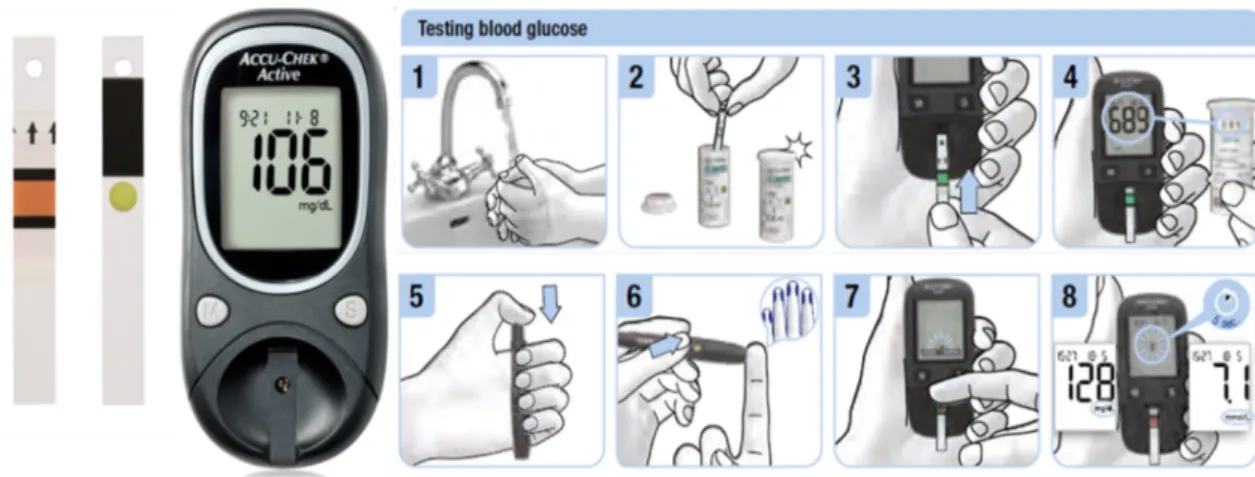
Since the 1980s, continuous development by many competing companies has led SMBG devices to an ease of use, high technical performance, and affordability. SMBG instruments have evolved into almost standard size and shape, and have become nice examples of consumer electronics (Figure 1.1). The strips have not changed their appearances for many years, but the active area of the strips (chemical matrix) has universally become smaller, enabling lower sample volume to be analyzed (typically around 1 µL). Improvements in technology have facilitated the test strips to do this difficult job in as little as five seconds.[23]
Figure 1.1 Example of a glucose meter (Accu-Chek Active) and SMBG routine.
There are now about forty glucose meters available on the market. By 2008, worldwide sales of these products climbed to an astonishing $8.8 billion, which accounted for approximately 22% of the entire in vitro diagnostics industry.[25] Due to constant technological improvements, SMBG became readily available for a vast number of patients. The recently published norm ISO 15197:2013 serves as a yardstick for glucose meter manufacturers.[26],[27] This specifies that 95% of results should be within
±0.83 mmol/L of the reference glucose method for concentrations <5.6 mmol/L, and within 15% for higher concentrations. The American Diabetes Association (ADA) has suggested that the systems should achieve an analytical performance <5% with blood glucose levels between 30 and 400 mg/dL.[28]
Enzymatic methods have been proven to be a powerful approach and have attracted much attention, on account of their simplicity and practicality. Even though good selectivity and high sensitivity are obtained with these enzymatic sensors,[29] inevitable drawbacks still limit their analytical applications such as chemical and thermal instabilities, interferences originating from complex nature of the blood.[30-31]
1.3 A photometric test strip
Photometric techniques are usually considered as the "gold standard" signal generation method in point-of-care (POC) devices.[32-35] In comparison to the other market leader, the electrochemical detection, optical methods may have a lower cost per test, especially in view of the increasing availability of cheap, high-quality optoelectronic components such as charge-coupled device (CCD) detectors and light emitting diodes (LEDs).[36-42]
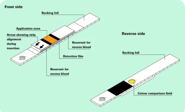
Figure 1.2 Glucose measurement principle. The test consists of multiple layers.
Enzyme reaction takes place in the bottom layer and the colour change observed from below.[23]
Roche photometric glucose meters employ a measurement principle shown in Figure 1.3. A drop of blood is placed on top of a test strip, where it diffuses rapidly (in about 0.5 s) into a chemistry layer. The "mirror" layer also acts as a filter to prevent erythrocytes from entering the chemistry layer. A few seconds after the dye formation, a photometric signal is observed from the bottom side of the strip. The photometric measurement is done by illumination with LED light. A part of the diffuse reflection arrives at a photodetector and is converted to a current. Volumes down to a few nanoliters can be measured with state of the art layers.
Figure 1.3 Illustration of the setup for the measurement of glucose levels in blood. A — Dry chemistry layer containing an enzyme, a coenzyme and an indicator, before addition of the blood sample; B — the chemistry layer after addition of the blood sample, showing a colour forming reaction which reduces the amount of reflected light.
Chemistry layer Blood sample
LED Detector LED
A B
Detector
"Mirror" layer
1.4 Glucose biosensors: the challenge and opportunity
The first-generation glucose biosensors are based on oxidase enzymes, which employ oxygen as a co-substrate to generate hydrogen peroxide (Scheme 1-1). The electrons are then transferred to an acceptor, enabling colorimetric and/or amperometric detection.
Today most commercially available glucose sensors are of the second generation.[43] In a colorimetric version of the detection method: an oxidoreductase enzyme oxidizes glucose to gluconolactone; electrons from glucose are then transferred to an oxidized form of a mediator molecule, thereby converting it to a reduced form; this mediator in turn delivers the electrons to a redox indicator molecule, which in turn forms colour.
Scheme 1-1 Enzymatic detection of glucose. Electrons from the glucose oxidation reaction are first taken up by the enzyme's cofactor and then transferred to either oxygen (first generation), or the artificial mediator (second generation), or directly to the acceptor (third generation). The acceptor is ether an electrode or a redox indicator.
The redox indicators show a bathochromic shift (a change of spectral band position in the absorption spectrum to a longer wavelength) upon reduction. The indicators commonly used in the test strips are phosphomolybdic acids (PMO)[44] or tetrazolium salts (MTT, WST etc).[45-46] Both are not directly reduced by the enzyme, instead they require a mediator compound.
Artificial mediators were one of the key breakthroughs, which took glucose biosensors forward and allowed them for home testing of blood glucose. The mediator is usually a
Glucose
Glucono-
lactone reduced oxidized
H2O2
O2 H2O
oxidized reduced
Direct electron
transfer Enzyme
Cofactor FAD, NAD(P)+, PQQ
Mediator
1stgeneration
2ndgeneration
3rdgeneration Acceptor
e− Acceptor
Acceptor
The main enzymes used in commercial glucose test strips are glucose oxidase (GOx) or glucose dehydrogenases (GDH). These enzymes are coupled to a cofactor (Figure 1.4), such as flavin adenine dinucleotide (FAD), nicotinamide adenine dinucleotide (NAD+), or pyrrolo-quinoline quinine (PQQ).[29]
Figure 1.4 Cofactors used by GOx and GDHs. The structures of FAD, PQQ, and NAD are shown in their oxidized state. Regions that undergo reduction are in orange, with the reduced form drawn nearby, redox active sites are marked with blue colour.
Modern photometric test strips achieve the required sensitivity and selectivity. The test strip chemistry has good shelf-life stability under normal storage conditions. However, it is essential for the companies to further improve robustness of the strips.[51] In order to achieve longer shelf-life, their storage and distribution should include protection from moisture, freezing, excessive heat and from light. The test strips are usually packaged in containers with desiccant materials, incorporated into the cap of the container, to prevent undesired exposure to moisture from the environment. However, this method has not been effective for eliminating the risks of sensor degradation due to faulty storage by the patient. It often happens, that users forget to reseal the container between uses or leave test strips inside a parked car on a sunny day (light and high temperature) or in a bathroom (high humidity). Another known measure is the use of a deliquescent material in the chemical matrix to decrease the sorption of water in the material. Both methods increase production costs. It is disadvantageous for low-income countries. For example in tropical climate zones, the storage often has to be carried out under harsh conditions (high temperature and humidity).[52]
FAD / FADH2 NAD+/ NADH
PQQ / PQQH2
In the past years considerable effort has been done to develop enzymes with improved stability for diagnostic purposes.[29, 53-54] However, the mediators and/or redox indicators used in the test strips generally react sensitively to extreme storage conditions, and are degraded over time.[51] This circumstance certainly affects accuracy of the measurement. In the case of SMBG, inaccurate results can be easily overlooked and may even cause clinical risk to the patient.
The development of a new redox indicator, accepting electrons directly from an enzyme, will lead to a simpler colorimetric technique and therefore lower cost. This is the so called third-generation glucose biosensor (Scheme 1-1).[22] Until now only a few cases have been reported where enzymes transfer electrons directly to an indicator for photometric detection.[55-57] The elegance of the solution, in developing a novel system with a reagent combining both functions of the mediator and the chromogenic agent, would possibly allow storage and worldwide shipping at ambient temperature, securing analytical performance of the enzyme-linked assays.
1.5 In a search for an ideal glucose converting enzyme
When searching for an enzyme for diagnostic purposes, such as glucose measurements, one of the most important parameters is substrate specificity. Relying on an enzyme that recognizes also saccharides other than glucose (such as mannose, maltose, D-galactose, and D-xylose) would lead to an overestimation of glucose levels in blood and result in incorrect therapeutic decisions. In addition, stability of the enzyme is also an important factor, as its inactivation during storage would also result in inaccurate and inconsistent measurements. Luckily, a multitude of different enzymatic systems derived from bacterial and fungal microorganisms are now available on the market. Ferri et al gave a good overview of the present knowledge on redox enzymes currently utilized in commercially available glucose monitoring systems.[29]
Glucose oxidase
Glucose oxidase (FAD-GOx, EC 1.1.3.4) is the “gold standard” enzyme used in commercial glucose sensors. FAD molecule binds to the active sites of this enzyme, forming the adduct (FAD-GOx), which is reduced to FADH2-GOx by glucose.
The enzyme normally returns to its active oxidized state by transferring the electrons to
Stable FAD-GOx is commercially available at low cost and seems to be advantageous in terms of sugar specificity.[58] However, the natural second substrate of this enzyme is oxygen. Consequently, for the same patient, glucose measures of venous, capillary, and arterial blood, would give different values because of various oxygen content. To this account, natural or artificial mediators were proposed to re-oxidize FADH2-GOx.
Obviously, the redox indicators would also have to compete with oxygen for the electrons from FADH2-GOx.[59] As an alternative to GOx, GDH family of enzymes brought our attention.
Pyrroloquinoline quinone glucose dehydrogenase
PQQ-dependent GDH (PQQ-GDH, EC 1.1.5.2) is a dimeric enzyme composed of two identical protein monomers with each monomer binding PQQ molecule and three calcium ions. One of the three calcium ions activates the PQQ cofactor. The oxidation mechanism of glucose by PQQ-GDH is similar to that of FAD-GOx with the exception that the reduced form (PQQH2-GDH) is not oxidized by O2.
𝑔𝑔𝑔𝑔𝑔𝑔𝑔+𝑃𝑃𝑃 − 𝐺𝐹𝐹 → 𝑔𝑔𝑔𝑔𝑔𝑔𝑔𝑔𝑔𝑔𝑔𝑔𝑔𝑔+𝑃𝑃𝑃𝐹2 − 𝐺𝐹𝐹
PQQ-GDH is a particularly efficient enzymatic system, with a rapid electron transfer rate. However, native PQQ-GDH oxidizes also a variety of saccharides such as mannose, maltose and lactose, etc. In August of 2009, the US FDA agency's public health notification stated that the PQQ-GDH based sensors can cause potentially fatal errors in the glucose measurements in patients on medications which contain nonglucose sugars. Therefore, FDA recommended the public and healthcare facilities to avoid PQQ-GDH based glucose test strips, which were used by many commercial glucose meters.[60]
Intensive work has been dedicated to improve the specificity of PQQ-GDH. From the patent literature,[61] it is known that mutations have been used to reduce interferences to below 2% of the native enzyme.
Flavin adenine dinucleotide dependent glucose dehydrogenases
The FAD-dependent GDHs (FAD-GDH, EC 1.1.99.10) have received much attention for their potential applications in sensor development. These enzymes combine the oxygen independence of PQQ-GDH with the high specificity of GOx toward glucose.
Moreover FAD-GDHs utilize a variety of external electron acceptors.[62]
Bacterial FAD-GDH is a thermostable hetero-oligomeric enzyme complex made up of a catalytic subunit harboring FAD in its redox center, a multiheme cytochrome-complex
electron-transfer subunit, and a chaperone-like subunit required for proper folding and secretion of catalytic subunit. Although the catalytic subunit alone shows high catalytic activity, the electron-transfer subunit facilitates the transfer of electrons between the active-site cofactor and the artificial electron acceptors, giving this enzyme a catalytic activity similar to or even higher than that of PQQ-GDH. Bacterial FAD-GDH's unique direct electron transfer ability makes this enzyme an interesting candidate for the third generation glucose sensors.[63] Although sensors employing bacterial FAD-GDH are on the market, the enzyme is not commercially available.
Fungi derived FAD-GDH was successfully expressed in E. coli and, despite not having their native glycosylation, showed GDH activity and a high specificity for glucose.
Diagnostic reagent-grade fungal FAD-GDH is commercially available.
FAD-GDH enzymes have only a single active center.[63] An ordered sequence of reactions is necessary to transfer the electrons from glucose to an external electron acceptor, before another molecule of glucose could be again oxidized to gluconolactone.
The transfer of electrons from a reduced state (FADH2-GDH) to the acceptor is the rate limiting step in the reaction. Also mediator inhibition phenomena have been reported for FAD-GDH.[23]
Nicotinamide adenine dinucleotide (phosphate) dependent glucose dehydrogenase NAD(P)+-dependent GDH (NAD(P)+-GDH, EC 1.1.1.47) is different from the aforementioned dehydrogenases. The mechanism of catalysis also involves the binding of an oxidized cofactor and glucose. However, after an electron transfer from glucose, the ordered release of gluconolactone is followed by the cofactor — after reduction, NAD(P)H is not bound to the enzyme.[53]
𝑔𝑔𝑔𝑔𝑔𝑔𝑔+𝑁𝐹𝐹(𝑃)+ → 𝑔𝑔𝑔𝑔𝑔𝑔𝑔𝑔𝑔𝑔𝑔𝑔𝑔𝑔+𝑁𝐹𝐹(𝑃)𝐹+𝐺𝐹𝐹
NAD(P)+ cofactors are abundant and inexpensive substrates, and they demonstrate very high activity up to 550 U/mg with GDH. In addition, the enzymatic oxidation of glucose is not reversible, due to the spontaneous hydrolysis of gluconolactone.[64]
NAD(P)H is produced first-hand depending on glucose concentration. The interaction with the artificial electron acceptors is a non-enzymatic one, and thus an inhibition of the enzymatic reaction is not expected. This makes NAD(P)+-GDH a very attractive
1.6 Chemistry
We chose NAD(P)+-dependent GDH to develop a simplified assay for optical determination of blood glucose on the test strip. The glucose detection mechanism is shown in Scheme 1-2A. This involves an ordered binding of cofactor NAD(P)+ and glucose, followed by a release of glucono-δ-lactone and reduced NAD(P)H.
The reduced cofactor then transfers electrons to the redox indicator. The reduction induces a shift of the absorbance band to a longer wavelength in the visible range (Scheme 1-2B). Hence, a remarkable change in the absorbance strength could be observed. This could be conveniently monitored by UV-Vis spectroscopy (Scheme 1-2B) and even observed by the naked eyes (Scheme 1-2C). The increase in absorbance corresponds to an increase of glucose concentration in a sample.
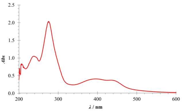
Scheme 1-2 Colour forming reaction of a redox indicator in the presence of glucose. A
— The NAD(P)H can be generated from NAD(P)+ by oxidation of a glucose catalyzed by GDH; B — UV-Vis spectra example of oxidized (blue) and reduced (red) forms of a redox indicator; C — Colour forming reaction can be observed by naked eyes:
a solution of a redox indicator before (left) and after (right) glucose addition.
In principle, the amount of NAD(P)H formed according to Scheme 1-2 can be directly followed at 340 nm. However, the sensitivity of NAD(P)H-coupled assays is not high, due to their low extinction coefficient.[65] A number of optical NAD(P)H sensors has been previously proposed, which include noble metal (e.g. Au and Ag)
300 500 700 900
A
Glucose
Glucono-
δ-lactone NAD(P)H
NAD(P)+
oxidized reduced
Redox indicator
e−
- with glucose - blank
GDH
B C
nanoparticles(NPs),[55, 57, 66-68] quantum dots (QDs)[56-57] and genetically-encoded glucose sensing enzymes.[69-70] However, these sensors require from minutes to hours for analysis making them unsuitable for SMBG test strips. Moreover, their implementation in dry chemistry is difficult.
An elegant alternative would be the use of a small organic molecule, where electrons could be directly transferred from NAD(P)H, causing a colorimetric signal at longer wavelengths. Molecules able to oxidize NAD(P)H had been previously reported;[48, 71-77]
but the majority of them do not show a bathochromic shift upon reduction. Examples of redox indicators showing the desired colorimetric response are nitrosoaniline[48] and quinone[77] derivatives. The drawback of these compounds is the low extinction coefficient of their reduced forms. A very well-known redox indicator is phosphomolybdic acid (PMO). This compound however has the disadvantage that the redox states are not well defined and the reduced form of PMO tends to have multiple broad absorption lines.[78] Moreover, PMO is only slowly reduced by NADH, therefore a mediator has to be used.[44] Tetrazolium salts[79] and resazurin[80-81] are used as redox indicators as well, but these compounds are not very stable and also require the use of a mediator like 5-methylphenazine methosulfate (PMS) or diaphorase to work in presence of dehydrogenases.
It is therefore still necessary to design an easy to obtain and efficient indicator for NAD(P)H. Several parameters have to be considered.
Dry chemistry
Typical coating formulations are described in patent literature.[82-85] Common components of the chemistry layer include reactive ingredients (e.g. enzymes, cofactors, indicators etc), dispersants, organic fillers, mineral fillers, pigments, surfactants, pH modifiers, buffer agents, viscosity modifiers, stabilizers, defoamers, flow modifiers etc.
Roche employs a blade coating technique to place a detection film on a plastic strip, which is followed by a drying process. Short intensive drying (50–85°C, 15 min) is advantageous to avoid denaturation of the enzyme and unwanted pre-reactions of the mediator and the indicator. The enzyme survives well in the drying process.
The critical phase is the time when temperature rises towards the end of drying process,
The reactive ingredients are also able to work in classical wet analytics. However, there are obviously differences compared to an analytical test running in a solution.
Preparation of test strip compositions is a rather complex technological process; hence such experiments in laboratory are time-consuming, tedious, and costly. To demonstrate feasibility of our strategy, it would be more convenient to investigate new redox indicators for optical monitoring of glucose in a solution.
Enzyme
A mutant of NAD(P)+-GDH suitable for applications in the test strips was identified by previous investigators at Roche.[85] Double mutant of GDH (E170K/Q252L = GlucDH2) from Bacillus subtilis is able to provide extraordinary catalytic performance:
it has a high affinity to glucose under ambient conditions of pH and temperature,[86] as in solution and in the dry chemistry.[84] GlucDH2 from Bacillus subtilis has an isoelectric point of 4.7–4.8, relative molecular weight (MW) of 126 kDa and is comprised of four subunits of MW 31.5 kDa each.
Baik et al discovered the role of residues Q252L and E170K for GDH stabilization.
Assisted by crystal structures from the wild-type and mutant GDHs from Bacillus megaterium IWG3,[87-88] Baik et al determined that a cooperative effect between Q252L and E170K stabilizes the tetramer structure by strengthening the hydrophobic interactions within the interface. Eduardo Vazquez-Figueroa et al demonstrated improvement in stability of GlucDH2 from B. subtilis in comparison to the wild-type GDH: from ca. 20 min at 25°C to ca. 3.5 days at 65°C, a 106-fold improvement.
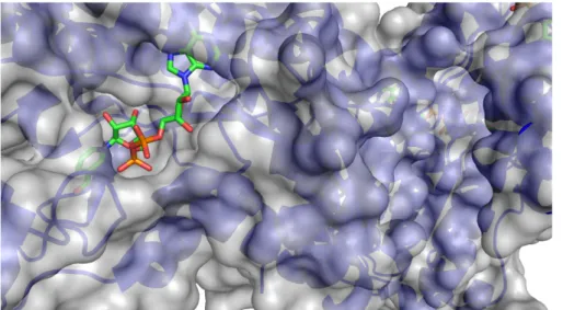
The enzyme shows a pH optimum at 8.0, and a broad activity range in pH 6.0–10.5. To assure its functionality an NAD(P)+ cofactor needs to be present (Figure 1.5).[86]
Figure 1.5 Crystal structure IWG3 of the GlucDH2 enzyme with a close-up of the region harbouring NAD+.
Coenzyme
Out of two natural cofactors, only the NAD+ cofactor has been commonly utilized in the test strip chemistry. The long-term stability of NAD+ in dry test turned out to be restricted. Luckily, its stable analogue — carba nicotinamide adenine dinucleotide (cNAD+) — has proven to be a conceivable alternative as the co-enzyme for enzymatic reactions with GDH on the test strips.[89-90] In cNAD+, the 2,3-hydroxycyclopentane ring replaces the β-D-ribonucleotide ring (Figure 1.6).
Figure 1.6 Structure of Carba-NAD.
As the result, cNAD+ is more stable concerning hydrolytic cleavage than NAD+. Furthermore, cNAD+ in combination with GlucDH2 is a rapid, selective and robust system for quantitative determination of glucose.[84-85]
Effect of pH
Enzyme activity is always affected by several parameters, the pH of the solution being one of them. To our best knowledge, the full pH-activity profile of GlucDH2 was not reported previously. For other characterized GDH enzymes from Bacillus subtilis, an optimal pH in a neutral to slightly alkaline pH is described.[64, 91-92]
The oxidized cofactor (NAD+) has long been known to be unstable toward basic conditions, as NAD+ hydrolysis rate dramatically increases above pH 8.0.[93-94] Several publications describe the catalytic decomposition of the reduced cofactor (NADH) as a function of pH and temperature.[93, 95-96] Wu and co-workers reported that NADH is undergoing rapid hydration below pH 6.[95] In addition, a number of reports described that electron acceptors for NADH often have lower stability at alkaline pHs.[97-99] It is also known that the mechanism of NADH oxidation by electron accepting molecules could be affected by pH.[73]
Optimum pH for the assay is a combination of kinetic parameters for the enzymatic
cNAD+
Salt concentration
Most GDH variants are stabilized with increased NaCl concentration. In addition, the enzymes are well compatible with positively charged small organic electron acceptors (e.g. phenazines), also in high concentrations.[100]
Effect of temperature
The rate of enzymatic glucose detection can be increased by raising temperature, with a corresponding increase in sensitivity. However, blood glucose monitoring is usually carried out at ambient temperature.
Blood composition and interferences
The components of serum are very complex, which often interferes with blood glucose sensing.[30] Serum contains various amino acids, peptides, proteins, carbohydrates, vitamins etc. The selectivity of a glucose biosensor depends on two major factors:
the enzyme–substrate reaction and the following reaction to generate a signal.
The enzyme–substrate reaction is very specific due to the nature of enzyme functionality. GDH can only react with glucose without any substantial interference from other types of sugars. However, there are other possible interferences in enzymatic glucose determination, such as reducing species in blood (ascorbic acid and glutathione) and nucleophiles (e.g. cysteine and lysine). Such substances could provoke a non-enzymatic reduction of a redox indicator. The blood samples, especially those from patients under specialized treatment in hospitals, may contain up to 5 mM of ascorbic acid.[101] Plasma glutathione levels are typically in micromolar range.[102]
Wavelength consideration
Absorbance maxima for NADH and cNADH are found at 340 nm and 360 nm, respectively. The extinction coefficient of cNADH equals that of NADH and amounts to 6.22 mM−1cm−1. The direct monitoring is limited due to interferences by other chromogenic species in blood, because the sensitivity of NADH-coupled tools is low.[65]
One of the most important components of blood is haemoglobin. It can be found in two forms, deoxygenated (Hb) and oxygenated (HbO2). The absorption spectrum is different in these two types: HbO2 has two maxima at 541 nm and 577 nm within the visible range, while Hb has only one peak at 555 nm. Longwave sensing is highly desirable in view of the strong intrinsic absorbance and fluorescence of serum.[31, 103]
1.7 A tailor made redox indicator
Considerations in a search for a tailor made indicator include water-solubility, rate of reaction with NADH, stability in mixtures with enzymes, redox potential, interferences (mainly with ascorbic acid), availability and cost, and intellectual property rights.
The rate of the reaction with NADH is the key parameter, because the fastest assays are now completed in five seconds. An oxidized form of the indicator should be nearly colourless, and a reduced form should have an absorption band at longer wavelengths (preferably more than 600 nm). It would help to minimize the interference from background absorbance of blood or serum samples. Otherwise, the chromophore should have an extinction value significantly higher than of the reduced coenzymes (6.22 mM−1cm−1). This would allow the assay to achieve better sensitivity than the direct spectrophotometric measurement of NADH or cNADH, at 339 and 360 nm, respectively. For the application on the test strips, the desired indicator should be compatible with aqueous buffer solutions. In addition, the best electron acceptors for NADH, from a prior state of art, commonly utilize a positive charge at the redox centres. The above mentioned considerations provided a basis for the design of a new NADH indicator.
1.8 Aims of the study
The overall aim of the study was to design and synthesize new redox indicators and evaluate their possible application for glucose sensing on a dry chemistry test strip. The study was focused on the development of a small organic indicator for NADH detection via enzymatic redox reactions.
More specifically, the aims were:
I To design and synthesize new redox indicators compatible with aqueous buffer solutions, and to evaluate their spectral properties in an oxidized and reduced form.
II To investigate new compounds for optical NADH sensing via direct electron transfer.
III To explore the use of such indicators in a GlucDH2-coupled enzymatic
1.9 References
[1] K. Laios, M. Karamanou, Z. Saridaki, G. Androutsos. Aretaeus of Cappadocia and the first description of diabetes. Hormones 2012, 11, 109-113.
[2] D. R. Owens. History and Vision: What is Important for Patients with Diabetes?
Diabetes Technol. Ther. 2008, 10, S-5-S-9.
[3] L. Rosenfeld. Insulin: Discovery and Controversy. Clin. Chem. 2002, 48, 2270- 2288.
[4] H. Dominiczak Marek. Linking Research and Innovative Clinical Practice: The Story of Diabetes Mellitus. Clin. Chem. Lab. Med. 2003, 41, 1104–1106.
[5] K. G. M. M. Alberti, P. f. Zimmet. Definition, diagnosis and classification of diabetes mellitus and its complications. Part 1: diagnosis and classification of diabetes mellitus. Provisional report of a WHO consultation. Diabetic Med.
1998, 15, 539-553.
[6] D. Daneman. Type 1 diabetes. The Lancet, 367, 847-858.
[7] S. E. Inzucchi, R. M. Bergenstal, J. B. Buse, M. Diamant, E. Ferrannini, M.
Nauck, A. L. Peters, A. Tsapas, R. Wender, D. R. Matthews. Management of Hyperglycemia in Type 2 Diabetes: A Patient-Centered Approach: Position Statement of the American Diabetes Association (ADA) and the European Association for the Study of Diabetes (EASD). Diabetes Care 2012, 35, 1364- 1379.
[8] Frances M. Ashcroft, P. Rorsman. Diabetes Mellitus and the β Cell: The Last Ten Years. Cell 2012, 148, 1160-1171.
[9] J. P. Crandall, W. C. Knowler, S. E. Kahn, D. Marrero, J. C. Florez, G. A. Bray, S. M. Haffner, M. Hoskin, D. M. Nathan. The prevention of type 2 diabetes.
Nat. Clin. Pract. End. Met. 2008, 4, 382-393.
[10] S. Garg, I. B. Hirsch. Self-monitoring of blood glucose. Int. J. Clin. Pract. 2010, 64, 1-10.
[11] International Diabetes Federation. IDF Diabetes Atlas update poster, 6th edn.
Brussels, Belgium: International Diabetes Federation, 2014.
[12] T. Scully. Diabetes in numbers. Nature 2012, 485, S2-S3.
[13] International Diabetes Federation. Global Diabetes Plan 2011-2021. Brussels:
IDF, 2011. Available online:
http://www.idf.org/sites/default/files/Global_Diabetes_Plan_Final.pdf (accessed on 26 October 2015).
[14] World Health Organization. Definition and diagnosis of diabetes mellitus and intermediate hyperglycaemia: report of a WHO/IDF consultation. Geneva
(Switzerland): WHO, 2006. Available online:
http://www.who.int/diabetes/publications/Definition%20and%20diagnosis%20o f%20diabetes_new.pdf (accessed on 26 October 2015).
[15] L. Ratheau, N. Jeandidier, F. Moreau, S. Sigrist, M. Pinget. How technology has changed diabetes management and what it has failed to achieve. Diabetes Metab. 2011, 37 Suppl 4, S57-64.
[16] H. Pollack. Stanley Rossiter Benedict: Creator of Laboratory Tests for Glycosuria. Diabetes 1953, 2, 420-421.
[17] S. F. Clarke, J. R. Foster. A history of blood glucose meters and their role in self-monitoring of diabetes mellitus. Br J Biomed Sci 2012, 69, 83-93.
[18] P. Voswinckel. A marvel of colors and ingredients. The story of urine test strip.
Kidney Int Suppl 1994, 47, S3-7.
[19] S. Walford, E. A. M. Gale, S. P. Allison, R. B. Tattersall. Self-monitoring of blood-glucose. Improvement of diabetic control. The Lancet 1978, 311, 732- 735.
[20] R. J. Jarrett, H. Keen, C. Hardwick. “Instant” Blood Sugar Measurement Using Dextrostix and a Reflectance Meter. Diabetes 1970, 19, 724-726.
[21] M. A. Genshaw, W. I. White. Glucose indicator and method. U.S. Patent 4,211,845 A, 24 November 1978.
[22] E.-H. Yoo, S.-Y. Lee. Glucose Biosensors: An Overview of Use in Clinical Practice. Sensors 2010, 10, 4558-4576.
[23] J. Hönes, P. Müller, N. Surridge. The technology behind glucose meters: test strips. Diabetes Technol. Ther. 2008, 10, S-10-S-26.
[24] V. Scognamiglio. Nanotechnology in glucose monitoring: Advances and challenges in the last 10 years. Biosens. Bioelectron. 2013, 47, 12-25.
[25] M. D. Hughes. The Business of Self-Monitoring of Blood Glucose: A Market Profile. J. Diabetes Sci. Technol. 2009, 3, 1219-1223.
[26] International Organization for Standardization . Geneva: International Organization for Standardization; 2013. ISO 15197:2013: In vitro diagnostic test systems -- requirements for blood-glucose monitoring systems for self-testing in managing diabetes mellitus.
[27] M. Link, C. Schmid, S. Pleus, A. Baumstark, D. Rittmeyer, C. Haug, G.
Freckmann. System Accuracy Evaluation of Four Systems for Self-Monitoring of Blood Glucose Following ISO 15197 Using a Glucose Oxidase and a Hexokinase-Based Comparison Method. J. Diabetes Sci. Technol. 2015.
[28] K. Tonyushkina, J. H. Nichols. Glucose Meters: A Review of Technical Challenges to Obtaining Accurate Results. J. Diabetes Sci. Technol. 2009, 3, 971-980.
[29] S. Ferri, K. Kojima, K. Sode. Review of Glucose Oxidases and Glucose Dehydrogenases: A Bird's Eye View of Glucose Sensing Enzymes. J. Diabetes Sci. Technol. 2011, 5, 1068-1076.
[30] H. A. Krebs. Chemical Composition of Blood Plasma and Serum. Annu. Rev.
Biochem. 1950, 19, 409-430.
[31] O. S. Wolfbeis, M. Leiner. Mapping of the total fluorescence of human blood serum as a new method for its characterization. Anal. Chim. Acta 1985, 167, 203-215.
[32] A. K. Yetisen, in Holographic Sensors, Springer International Publishing, 2015, pp. 1-25.
[34] L. Gervais, N. de Rooij, E. Delamarche. Microfluidic Chips for Point-of-Care Immunodiagnostics. Adv. Mater. 2011, 23, H151-H176.
[35] F. B. Myers, L. P. Lee. Innovations in optical microfluidic technologies for point-of-care diagnostics. Lab Chip 2008, 8, 2015-2031.
[36] G. Comina, A. Suska, D. Filippini. Autonomous Chemical Sensing Interface for Universal Cell Phone Readout. Angew. Chem. Int. Ed. 2015, n/a-n/a.
[37] V. Gubala, L. F. Harris, A. J. Ricco, M. X. Tan, D. E. Williams. Point of Care Diagnostics: Status and Future. Anal. Chem. 2012, 84, 487-515.
[38] A. W. Martinez, S. T. Phillips, M. J. Butte, G. M. Whitesides. Patterned Paper as a Platform for Inexpensive, Low-Volume, Portable Bioassays. Angew. Chem.
Int. Ed. 2007, 46, 1318-1320.
[39] A. W. Martinez, S. T. Phillips, G. M. Whitesides. Three-dimensional microfluidic devices fabricated in layered paper and tape. Proc. Natl. Acad. Sci.
2008, 105, 19606-19611.
[40] Y. Xia, J. Ye, K. Tan, J. Wang, G. Yang. Colorimetric visualization of glucose at the submicromole level in serum by a homogenous silver nanoprism-glucose oxidase system. Anal. Chem. 2013, 85, 6241-6247.
[41] L. Shen, J. A. Hagen, I. Papautsky. Point-of-care colorimetric detection with a smartphone. Lab Chip 2012, 12, 4240-4243.
[42] A. W. Martinez, S. T. Phillips, E. Carrilho, S. W. Thomas, H. Sindi, G. M.
Whitesides. Simple Telemedicine for Developing Regions: Camera Phones and Paper-Based Microfluidic Devices for Real-Time, Off-Site Diagnosis. Anal.
Chem. 2008, 80, 3699-3707.
[43] R. Tirimacco, G. Koumantakis, R. Erasmus, A. Mosca, S. Sandberg, D. Watson Ian, B. Goldsmith, P. Gillery. Glucose meters – fit for clinical purpose. Clin.
Chem. Lab. Med. 2013, 51, 943.
[44] J. Hoenes, H. Wielinger, V. Unkrig. Use of a sparingly soluble salt of a heteropoly acid for the determination of an analyte, a corresponding method of determination as a suitable agent therefor. U.S. Patent 5,240,860 A, 2 December 1989.
[45] M. Ishiyama, Y. Miyazono, K. Sasamoto, Y. Ohkura, K. Ueno. A highly water- soluble disulfonated tetrazolium salt as a chromogenic indicator for NADH as well as cell viability. Talanta 1997, 44, 1299-1305.
[46] A. Zhu, R. Romero, H. R. Petty. An enzymatic colorimetric assay for glucose-6- phosphate. Anal. Biochem. 2011, 419, 266-270.
[47] A. Heller, B. Feldman. Electrochemical Glucose Sensors and Their Applications in Diabetes Management. Chem. Rev. 2008, 108, 2482-2505.
[48] J. Hoenes, V. Unkrig. Colorimetric determination of an analyte using benzyl alcohol dehydrogenase and a chromogenic redox-indicator. U.S. Patent 5,445,943 A, 8 April 1993.
[49] C. D. Wilsey, M. Ghoshal, H. Wieder. Matrix composition with alkylphenazine quaternary salt and a nitrosoaniline. U.S. Patents 8,008,037 B2, 27 March 2008.
[50] X. Gao, X. Li, Q. Wan, Z. Li, H. Ma. Detection of glucose via enzyme-coupling reaction based on a DT-diaphorase fluorescence probe. Talanta 2014, 120, 456- 461.
[51] S. Duvall, S. Kachel, G. C. Lica, W. Schabel, P. Scharfer. Improved matrix stability compositions and methods. WO2014/037372 A1, Sep. 6, 2012.
[52] M. Ducorps, L. Papoz, J. C. Cuisinier-Raynal, D. Simon. Reliability of glucose measurement by glucose test strips in tropical conditions. Diabetes Res. Clin.
Pract. 1992, 17, 51-54.
[53] A. Basner, G. Antranikian. Isolation and Biochemical Characterization of a Glucose Dehydrogenase from a Hay Infusion Metagenome. PLoS ONE 2014, 9, e85844.
[54] H. Aiba, Y. Nishiya, M. Azuma, Y. Yokooji, H. Atomi, T. Imanaka.
Characterization of a thermostable glucose dehydrogenase with strict substrate specificity from a hyperthermophilic archaeon Thermoproteus sp. GDH-1.
Biosci., Biotechnol., Biochem. 2015, 1-9.
[55] L. Zhang, Y. Li, D. W. Li, C. Jing, X. Chen, M. Lv, Q. Huang, Y. T. Long, I.
Willner. Single gold nanoparticles as real-time optical probes for the detection of NADH-dependent intracellular metabolic enzymatic pathways. Angew. Chem.
2011, 50, 6789-6792.
[56] R. Freeman, R. Gill, I. Shweky, M. Kotler, U. Banin, I. Willner. Biosensing and Probing of Intracellular Metabolic Pathways by NADH-Sensitive Quantum Dots. Angew. Chem. 2009, 121, 315-319.
[57] L. Bahshi, R. Freeman, R. Gill, I. Willner. Optical Detection of Glucose by Means of Metal Nanoparticles or Semiconductor Quantum Dots. Small 2009, 5, 676-680.
[58] R. Wilson, A. P. F. Turner. Glucose oxidase: an ideal enzyme. Biosens.
Bioelectron. 1992, 7, 165-185.
[59] T. Semashko, R. Mikhailova, A. Ramanaviciene, A. Ramanavicius. Specificity of Glucose Oxidase from Penicillium funiculosum 46.1 Towards Some Redox Mediators. Appl. Biochem. Biotechnol. 2013, 171, 1739-1749.
[60] U.S. Food and Drug Administration. FDA public health notification: potentially fatal errors with GDH-PQQ glucose monitoring technology. Available online:
http://www.fda.gov/MedicalDevices/Safety/AlertsandNotices/PublicHealthNotif ications/ucm176992.htm (accessed on 26 October 2015).
[61] M. Boenitz-Dulat, D. Beck, P. Kratzsch, R. Schmuck, H. von der Eltz. Mutants of pyrroloquinoline quinone dependent soluble glucose dehydrogenase. U.S.
Patent 2009/0148874 A1, 13 April 2006.
[62] K. Mori, M. Nakajima, K. Kojima, K. Murakami, S. Ferri, K. Sode. Screening of Aspergillus-derived FAD-glucose dehydrogenases from fungal genome database. Biotechnol. Lett 2011, 33, 2255-2263.
[63] M. N. Zafar, X. Wang, C. Sygmund, R. Ludwig, D. Leech, L. Gorton. Electron- Transfer Studies with a New Flavin Adenine Dinucleotide Dependent Glucose Dehydrogenase and Osmium Polymers of Different Redox Potentials. Anal.
Chem. 2012, 84, 334-341.
[64] A. Weckbecker, W. Hummel, in Microbial Enzymes and Biotransformations,
[66] L. Tang, X. Lei, G. Zeng, Y. Liu, Y. Peng, M. Wu, Y. Zhang, C. Liu, Z. Li, G.
Shen. Optical detection of NADH based on biocatalytic growth of Au–Ag core–
shell nanoparticles. Spectrochim. Acta Mol. Biomol. Spectrosc. 2012, 99, 390- 393.
[67] S. Liu, Z. Du, P. Li, F. Li. Sensitive colorimetric visualization of dihydronicotinamide adenine dinucleotide based on anti-aggregation of gold nanoparticles via boronic acid-diol binding. Biosens. Bioelectron. 2012, 35, 443- 446.
[68] B. Shlyahovsky, E. Katz, Y. Xiao, V. Pavlov, I. Willner. Optical and Electrochemical Detection of NADH and of NAD+-Dependent Biocatalyzed Processes by the Catalytic Deposition of Copper on Gold Nanoparticles. Small 2005, 1, 213-216.
[69] Y. Zhao, J. Jin, Q. Hu, H.-M. Zhou, J. Yi, Z. Yu, L. Xu, X. Wang, Y. Yang, J.
Loscalzo. Genetically Encoded Fluorescent Sensors for Intracellular NADH Detection. Cell Metab. 2011, 14, 555-566.
[70] S. Siedler, G. Schendzielorz, S. Binder, L. Eggeling, S. Bringer, M. Bott. SoxR as a Single-Cell Biosensor for NADPH-Consuming Enzymes in Escherichia coli. ACS Synth. Biol. 2013, 3, 41-47.
[71] T. Yomo, I. Urabe, H. Okada. Electrostatic and redox potential effects on the rate of electron-transfer reaction of nicotinamide adenine dinucleotides with 1- substituted 5-ethylphenazines. BBA - Bioenergetics 1990, 1017, 139-142.
[72] C. J. F. Noorden, J. Tas. Advantages of 1-methoxyPMS as an electron carrier in dehydrogenase cytochemistry. Histochem. J. 1982, 14, 837-842.
[73] G. Hilt, T. Jarbawi, W. R. Heineman, E. Steckhan. An Analytical Study of the Redox Behavior of 1,10-Phenanthroline-5,6-dione, its Transition-Metal Complexes, and its N-Monomethylated Derivative with regard to their Efficiency as Mediators of NAD(P)+ Regeneration. Chem. Eur. J. 1997, 3, 79- 88.
[74] B. Persson, L. Gorton. A comparative study of some 3,7-diaminophenoxazine derivatives and related compounds for electrocatalytic oxidation of NADH. J.
Electroanal. Chem. Interfacial Electrochem. 1990, 292, 115-138.
[75] J. W. Bunting, A. W. C. Ng. Rates of Oxidation of 1-Benzyl-1,4- dihydronicotinamide by Pyrazinium, Quinoxalinium, and Phenazinium Cations.
Bioorg. Chem. 1993, 21, 156-169.
[76] J. F. J. Engbersen, A. Koudijs, H. C. van der Plas. Reaction of NADH models with methylene blue. Recl. Trav. Chim. Pays-Bas 1985, 104, 131-138.
[77] D. Heindl, R. Herrmann, J. Hones, H.-P. Josel, M. Junius-Comer, H. Merdes, A.
Schmidt, E. Selbertinger. Redox-active compounds and their use. U.S. Patent 6,057,120 A, Sep. 24, 1996.
[78] J. E. Going, S. J. Eisenreich. Spectrophotometric studies of reduced molybdoantimonylphosphoric acid. Anal. Chim. Acta 1974, 70, 95-106.
[79] J. P. Albarella, S. W. Felman, J. J. Landi, K. L. Marfurt. Water-soluble tetrazolium salts. U.S. Patent 7,767,822 B2, 14 January 2005.
[80] L. P. Candeias, D. P. S. MacFarlane, S. L. W. McWhinnie, N. L. Maidwell, C.
A. Roeschlaub, P. G. Sammes, R. Whittlesey. The catalysed NADH reduction of resazurin to resorufin. J. Chem. Soc., Perkin Trans. 2 1998, 2333-2334.
[81] A. Zhu, R. Romero, H. R. Petty. An enzymatic fluorimetric assay for glucose-6- phosphate: application in an in vitro Warburg-like effect. Anal. Biochem. 2009, 388, 97-101.
[82] W. R. Knappe, F. Wittmann, D. Mosoiu, C. Horn, J. Hoenes. Transparent support; quantitative analysis. U.S. Patent 2006/0003397 A1, Feb. 18, 2004.
[83] W. R. Knappe. Mediators for photometric tests and means and methods relating to use thereof. U.S. Patent 2008/0213808 A1, Sep. 4, 2008.
[84] W. Roedel, C. Horn, N. Steinke, N. Bucci, T. Meier, R. Schmuck, R. Nagel, D.
Heindl. Stabilization of enzymes with stable coenzymes. U.S. Patent 2012/0276565 A1, Aug. 20, 2009.
[85] C. Horn, C. Gaessler-Dietsche, D. Heindl, J. Hoenes, T. Meier, R. Schmuck.
Fast reaction kinetics of enzymes having low activity in dry chemistry layers.
U.S. Patent 2014/0322737 A1, Feb. 19, 2009.
[86] E. Vázquez-Figueroa , J. Chaparro-Riggers , A. S. Bommarius. Development of a Thermostable Glucose Dehydrogenase by a Structure-Guided Consensus Concept. ChemBioChem 2007, 8, 2295-2301.
[87] S.-H. Baik, F. Michel, N. Aghajari, R. Haser, S. Harayama. Cooperative Effect of Two Surface Amino Acid Mutations (Q252L and E170K) in Glucose Dehydrogenase from Bacillus megaterium IWG3 on Stabilization of Its Oligomeric State. Appl. Environ. Microbiol. 2005, 71, 3285-3293.
[88] K. Yamamoto, G. Kurisu, M. Kusunoki, S. Tabata, I. Urabe, S. Osaki. Crystal Structure of Glucose Dehydrogenase from Bacillus megaterium IWG3 at 1.7 Å Resolution. J. Biochem. 2001, 129, 303-312.
[89] D. Heindl, J. Hoenes, C. Horn, C. Gaessler-Dietsche. Stable NAD/NADH derivatives U.S. Patent 8,809,013 B2, 28 July 2005.
[90] J. T. Slama, A. M. Simmons. Carbanicotinamide adenine dinucleotide: synthesis and enzymological properties of a carbocyclic analog of oxidized nicotinamide adenine dinucleotide. Biochemistry 1988, 27, 183-193.
[91] R. F. Ramaley, N. Vasantha. Glycerol protection and purification of Bacillus subtilis glucose dehydrogenase. J. Biol. Chem. 1983, 258, 12558-12565.
[92] W. Hilt, G. Pfleiderer, P. Fortnagel. Glucose dehydrogenase from Bacillus subtilis expressed in Escherichia coli I: purification, characterization and comparison with glucose dehydrogenase from Bacillus megaterium. BBA - Protein Struct. M. 1991, 1076, 298-304.
[93] O. H. Lowry, J. V. Passonneau, M. K. Rock. The Stability of Pyridine Nucleotides. J. Biol. Chem. 1961, 236, 2756-2759.
[94] N. Oppenheimer. NAD hydrolysis: Chemical and enzymatic mechanisms. Mol.
Cell. Biochem. 1994, 138, 245-251.
[95] J. T. Wu, L. H. Wu, J. A. Knight. Stability of NADPH: effect of various factors on the kinetics of degradation. Clin. Chem. 1986, 32, 314-319.
[96] L. Rover Jr, J. C. B. Fernandes, G. d. O. Neto, L. T. Kubota, E. Katekawa, S. l.
[97] B. W. Carlson, L. L. Miller. Mechanism of the oxidation of NADH by quinones.
Energetics of one-electron and hydride routes. J. Am. Chem. Soc. 1985, 107, 479-485.
[98] T. Yomo, H. Sawai, I. Urabe, Y. Yamada, H. Okada. Synthesis and characterization of 1-substituted 5-alkylphenazine derivatives carrying functional groups. Eur. J. Biochem. 1989, 179, 293-298.
[99] V. Simanek, V. Preininger. Pseudobase formation from quaternary pyridinium, quinolinium and isoquinolinium cations. Heterocycles 1977, 6, 475-497.
[100] H. Freitag, C. D. Wilsey. Reagents and assay methods including a phenazine- containing indicator. EP0606296 B1, 19 September 1991.
[101] S. Ohno, Y. Ohno, N. Suzuki, G.-I. Soma, M. Inoue. High-dose Vitamin C (Ascorbic Acid) Therapy in the Treatment of Patients with Advanced Cancer.
Anticancer Res. 2009, 29, 809-815.
[102] P. S. Samiec, C. Drews-Botsch, E. W. Flagg, J. C. Kurtz, P. Sternberg Jr, R. L.
Reed, D. P. Jones. Glutathione in Human Plasma: Decline in Association with Aging, Age-Related Macular Degeneration, and Diabetes. Free Radical Biol.
Med. 1998, 24, 699-704.
[103] M.-S. Steiner, A. Duerkop, O. S. Wolfbeis. Optical methods for sensing glucose.
Chem. Soc. Rev. 2011, 40, 4805-4839.
2 A “ MEDIATORLESS ”
INDICATOR FOR
COLORIMETRIC DETECTION OF GLUCOSE ON A DRY
CHEMISTRY TEST STRIP
2.1 Introduction
The detection of glucose dehydrogenase (GDH)-catalyzed reactions is an active research area due to industrial applications. NAD+-dependent GDH is widely used in glucose sensing dry chemistry test strips. In the strips, glucose is oxidized to gluconolactone by GDH while NAD+ is reduced to NADH. Electrons from NADH are then transferred to a mediator, which in turn delivers the electrons to a redox indicator, thus providing a colorimetric read-out.[1] However, the mediators and/or redox indicators used in the test strips generally react sensitively to extreme storage conditions, and degrade over time.[2-3] In tropical climate zones test strips are stored often under harsh conditions of high temperature and humidity.[4] A reagent, combining both functions of the mediator and the chromogenic agent, may lead to a more stable system. Of particular interest in this regard are small molecule indicators, where electrons can be directly transferred from NADH, causing a colorimetric signal at longer wavelengths. Such compounds would provide a simpler colorimetric detection method. This would possibly allow storage and worldwide shipping of glucose test strips at ambient temperature, securing the analytical performance of the enzyme-linked assays.
Quinones are a good starting point in the search for a new redox indicator.
The quinone/semiquinone/hydroquinone (Q/SQ•−/H2Q) triad is a vital link in the movement of electrons through cells and tissues.[5] The presence of quinone derivatives in interstellar stardust has led experts to hypothesize a crucial role for pyrroloquinoline quinone (PQQ) in the evolution of life on Earth.[6] PQQ shows an extraordinary molecular stability and its analogues can act as NADH indicators.
Hilt et al reported that transition-metal complexes of 1,10-phenanthroline-5,6-dione are efficient catalysts for NADH oxidation at physiological pHs.[7] Heindl and colleagues showed that quinone functionalized acceptors are useful for the colorimetric NADH detection.[8] Around the same time, Ambroise et al investigated spectral properties of a Ru(II) complex with the aqphen[9] ligand, where its reduction with dithionite resulted in a new absorption band in the visible range.[10]
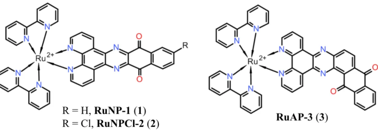
Figure 2.1 Molecular structure of Ru (II) complexes studied in this report.
Despite extensive literature on the reduction of 1,10-phenanthroline-5,6-dione, its transition-metal complexes, and its N-monomethylated derivatives by NADH, we have been unable to locate any study on the reaction of larger quinone-functionalized polypyridinic ligands with NADH. This finding motivated us to investigate Ru(II) complexes with the ligands nqphen[11] (1–2) and aqphen (3) for quantitative NADH sensing in aqueous buffer solutions (Figure 2.1). It is known that both complexes, RuNP-1 and RuAP-3 have high electron acceptor character. In addition, we designed RuNPCl-2 to explore the impact of the Cl group in the reduction kinetics.
The Cl-connected aromatic ring may change the electronic density of the nqphen moiety through its electron-withdrawing effect. The new ligand can also be functionalized by displacement of the Cl group as a potential route to new derivatives. Looking at the published data, the electrochemical results indicate that aqphen is a stronger electron acceptor than the nqphen ligand.[12] There are two possible pathways to transfer the electrons from NADH to a quinone moiety.[13] Cyclic voltammetry studies of aqphen show a favoured one-electron reduction mechanism compared to the alternative mechanism via hydride transfer. We hypothesized that if the quinone moiety is reduced with NADH by a pathway similar described by Hilt et al,[7] a coloured semiquinone would be formed.[14-16]
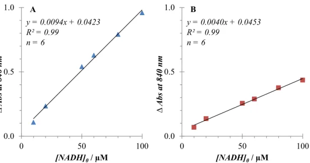
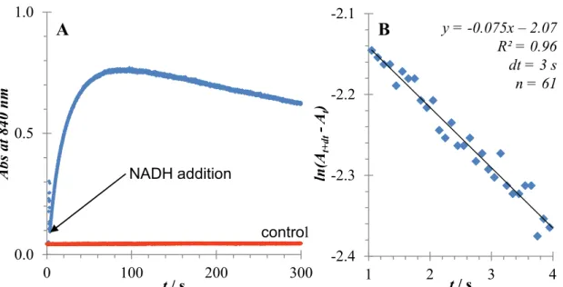
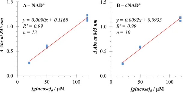
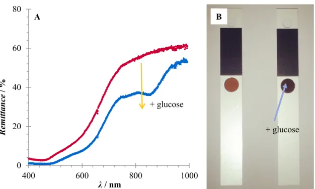
Here we show that RuAP-3 displays sensitive colorimetric response upon reduction with NADH, a rapid reaction time and good stability in phosphate buffer at neutral pH.
This facilitated its application in an enzymatic glucose assay in solution as well as in dry chemistry. In this proof-of-principle study, we showed that RuAP-3 could be
R = H, RuNP-1(1)
R = Cl, RuNPCl-2(2) RuAP-3(3)
2.2 Results and Discussion
2.2.1 Synthesis of ruthenium complexes
The synthetic routes for 1–3 are outlined in Scheme 2-1. The RuAP-3 was kindly provided by Roche Diagnostics GmbH as water soluble TFA salt. Complexes RuNP-1 and RuNPCl-2 were synthesized in two steps and isolated as hexafluorophosphate salts using adapted literature procedures.[10, 17] Each synthetic step presented here is straightforward and provides yields of 27 to 94% of the desired product in pure form.
Scheme 2-1 Synthesis of the complexes 1, 2 and 3. Reagents and conditions: (i) AcOH, 75°C, 3 h; (ii) MEG, reflux, Ar, overnight.
The key building blocks 5 and 6 were synthesized according to the efficient two-step procedure previously reported by Chesneau et al (Scheme 2-2).[17] While 2,3-dichloronaphtoquinone (11) is commercially available material, 2,3,6-trichloronaphtoquinone (12) was synthesized in two steps (43%) as described previously (see experimental part for details).[18-19]
Scheme 2-2 Synthesis of the 2,3-diaminonaphtoquinones. Reagents and conditions: (i) potassium phthalidimide, anhydrous MeCN, reflux, 3 h; (ii) 64% aqueous hydrazine, 70°C, 3 h.
R = H, nqphen (7), 94%
R = Cl, nqphen-Cl (8), 86%
R = H (5) R = Cl (6)
(4)
aqphen (10)
Ru(bpy)2Cl2(9)
Ru(bpy)2Cl2(9)
RuNP-1(1), 66%
RuNPCl-2(2), 27%
RuAP-3(3) (i)
(ii)
(i) (ii)
R = H (11)
R = Cl (12) R = H (13), 74%
R = Cl (14), 36% R = H (5), 96%
R = Cl (6), 40%