Surveillance of mRNP composition during translation
termination regulates gene expression via nonsense-mediated mRNA decay
Inaugural-Dissertation
zur
Erlangung des Doktorgrades
der Mathematisch-Naturwissenschaftlichen Fakultät der Universität zu Köln
vorgelegt von
Volker Böhm aus Bergisch Gladbach
Köln, 2015
Berichterstatter/in: PD Dr. Niels H. Gehring Prof. Dr. Karin Schnetz
Tag der mündlichen Prüfung: 16.06.2015
Content
1. Introduction ... 1
1.1 Surveillance of gene expression ... 1
1.2 Translation‐coupled mRNP quality control ... 3
1.3 Mechanism of eukaryotic translation termination ... 5
1.4 Models of NMD activation and substrate definition ... 7
1.5 Factors involved in NMD assembly ... 11
1.5.1 The RNA helicase UPF1 plays a central role in NMD ... 12
1.5.2 UPF2 provides the scaffold for the NMD assembly ... 14
1.5.3 UPF3 acts as the link between UPF proteins and the EJC ... 15
1.5.4 UPF1 is phosphorylated by the SMG1 kinase ... 16
1.5.5 Initiation of mRNA degradation via phospho‐UPF1 interactions ... 16
1.5.6 Initiation of exonucleolytic degradation ... 17
1.5.7 Dephosphorylation of UPF1 is initiated by decay factors ... 18
1.5.8 Endonucleolytic cleavage is executed by SMG6 ... 19
1.6 Model of the EJC‐NMD mechanism ... 20
1.7 Physiological function of NMD and importance in diseases ... 21
1.8 Aims of this work ... 23
2. Publications ... 25
2.1 The interaction of cytoplasmic poly(A)‐binding protein with eukaryotic initiation factor 4G suppresses ... nonsense‐mediated mRNA decay ... 26
2.2 CWC22 connects pre‐mRNA splicing and exon junction complex assembly ... 41
2.3 Structural and functional analysis of the three MIF4G domains of nonsense‐mediated decay factor UPF2 ... 55
2.4 3' UTR length and messenger ribonucleoprotein composition determine endocleavage efficiencies ... at termination codons ... 96
3. Discussion ... 124
3.1 The long 3′ UTR mRNP composition influences NMD activation ... 124
3.2 EJC loading on the mRNP and the involvement in NMD... 128
3.3 Comparison of EJC‐ and long 3′ UTR‐induced NMD... 130
3.4 Degradation of the mRNA via endonucleolytic cleavage ... 132
4. References ... 136
5. Summary ... 146
6. Zusammenfassung ... 147
7. Author contribution ... 149
8. Acknowledgement ... 150
Erklärung ... 151
Lebenslauf ... 152
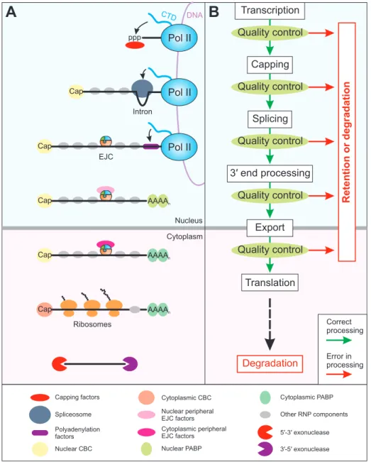
According to the central dogma of molecular biology, access to the genetic information contained within the DNA requires the synthesis of messenger RNA (mRNA), which is decoded in order to generate proteins (Crick, 1970; Crick, 1958; Nirenberg and Matthaei, 1961). In eukaryotic cells, this process of gene expression consists of several consecutive, but integrated steps (Moore and Proudfoot, 2009). The first step is the RNA polymerase II (Pol II) mediated transcription of the DNA into pre‐mRNA, which is generally accompanied by three co‐
transcriptional processing actions to modify the transcript (Bentley, 2014; Lee and Tarn, 2013).
These involve the addition of a 7‐methylguanosine cap to the 5′ end, the splicing of intronic sequences, and the cleavage at the 3′ end followed by addition of the poly(A) tail. Upon completion of these steps, the mature mRNA is exported through the nuclear pores into the cytoplasm where ribosomes translate the transcript into a polypeptide chain. Eventually the mRNA is degraded, which represents the final step in the lifecycle of an mRNA (Figure 1A) (Moore, 2005).
1.1 Surveillance of gene expression
As each of the individual processes during gene expression are carried out by specific complex machineries with inherent error rates, mistakes can occur, which need to be detected in order to prevent the generation of faulty RNA or proteins (Doma and Parker, 2007; Schmid and Jensen, 2010; Shoemaker and Green, 2012). These mistakes are, for example, the misincorporation of nucleotides or amino acids by Pol II or the ribosome during transcription or translation, respectively. Moreover, different stimuli, chemical agents or environmental influences can increase the error frequency of these processes, thereby potentially producing more aberrant gene expression products (Drummond and Wilke, 2009; Jack et al., 2011;
Remenyi et al., 2004; Wurtmann and Wolin, 2009; Zaher and Green, 2009). Eukaryotic cells employ several quality control mechanisms at basically every nuclear and cytoplasmic gene expression step in order to detect abnormalities (Figure 1B) (Ghosh and Jacobson, 2010;
Muhlemann and Jensen, 2012). Although failsafe mechanisms are also implemented in the
gene expression machineries themselves, quality control commonly involves the handover of
only correctly processed products from one step to the other, whereas faulty intermediates are
retained or degraded (Doma and Parker, 2007; Hagiwara and Nojima, 2007; Maniatis and Reed,
2002). On the molecular level, this is achieved by the interplay of specific proteins, which bind
ppp
Cap
Pol II
CTD
Pol II
Intron
Cap Pol II
EJC
Cap AAAAn
Cap AAAAn
Transcription
Capping
Splicing
3′ end processing
Cap AAAAn
Nucleus
Cytoplasm Export
DNA
Ribosomes
Capping factors Spliceosome Polyadenylation factors Nuclear CBC
Cytoplasmic CBC Nuclear peripheral EJC factors Cytoplasmic peripheral EJC factors Nuclear PABP
Cytoplasmic PABP
5 -3 exonuclease′ ′ 3 -5 exonuclease′ ′ Other RNP components
Degradation Translation
A B
Quality control Quality control
Quality control
Quality control Quality control
Retention or degradation
Correct processing Error in processing
the mRNA to form ribonucleoproteins (RNP) (Muller-McNicoll and Neugebauer, 2013;
Rodriguez-Navarro and Hurt, 2011). The composition of the mRNP changes dramatically during the progression of gene expression and determines the fate of the transcript (Figure 1A) (Mitchell and Parker, 2014; Singh et al., 2015).
Figure 1: Overview of mRNP composition and quality control during gene expression. (A) Central steps of gene expression are depicted schematically. mRNA processing factors are recruited to the mRNA co-transcriptionally by the C-terminal domain (CTD) of Pol II. The Legend of mRNP components is shown at the bottom (A). (B) The fidelity of each step of the gene expression cascade (white boxes) is monitored by quality control mechanisms, which initiate the degradation or retention of erroneous product. Only correctly processed or quality control evading gene expression intermediates are handed over to the next step (green arrows). Finally, the mRNA is translated in the cytoplasm and eventually degraded. Abbreviations: Pol II = RNA polymerase II; ppp = triphosphate; EJC = exon-junction complex; CBC = cap-binding complex; PABP = Poly(A) binding protein.
In metazoan cells, one key regulator of gene expression is the exon-junction complex (EJC),
which is a well-studied example of a
multi-protein complex that shapes the mRNP and influences many subsequent gene expression steps. EJCs are deposited in the nucleus onspliced mRNAs closely upstream of the exon-exon junction (Figure 1A) (Le Hir et al., 2000). They
displaced by the translating ribosome in the cytoplasm (Gehring et al., 2009b). The core of the EJC is comprised of the DEAD‐box helicase eIF4A3, Barentsz (BTZ; also MLN51) and the heterodimer Y14/MAGOH. Of the core EJC proteins, ATP‐loaded eIF4A3 directly binds the RNA in a sequence‐independent manner due to interaction with the phosphate‐sugar backbone (Andersen et al., 2006; Bono et al., 2006). Interaction with the RNA stimulates the hydrolysis of ATP, which leads to dissociation of eIF4A3. To lock the EJC stably on the RNA, the Y14/MAGOH dimer binds to RNA‐interacting eIF4A3 and keeps it in a state that prevents the completion of ATP hydrolysis (Ballut et al., 2005). Specific disassembly of EJCs is achieved by ribosome‐
associated PYM (partner of Y14 and MAGOH), which lifts Y14/MAGOH from the EJC resulting in the release of eIF4A3 from the RNA (Bono and Gehring, 2011).
At certain steps during the mRNP metabolism, additional EJC proteins can join and leave the core factors (Bono and Gehring, 2011; Tange et al., 2004). By recruitment of specific protein factors, the dynamic composition of the EJC changes and allows for the activation of downstream processes in the mRNP lifecycle, such as mRNA export or translation (Chazal et al., 2013; Gudikote et al., 2005; Le Hir et al., 2001; Nott et al., 2004; Wiegand et al., 2003).
Therefore, the EJC is not only an example for the tight interplay of the gene expression processes, it also represents an integral component of mRNP quality control, because only correctly spliced mRNAs benefit from the enhancing effects of the EJC.
1.2 Translation‐coupled mRNP quality control
The mRNPs being translated in the cytoplasm have gone through multiple controlled processing steps and are therefore supposed to contain the proper mRNA modifications and mRNP composition needed for the synthesis of functional protein. Major errors should have been corrected at this point, for example newly transcribed mRNA, which fail to be correctly 5′‐
capped, will be degraded or retained in the nucleus until properly processed (Doma and Parker, 2007). However, not all potentially occurring errors can be recognized and corrected by the quality control mechanisms, especially if the mistakes are subtle. For instance, nucleotide misincorporations during transcription by Pol II, which evade the inherent proofreading mechanism, are difficult to detect by the downstream surveillance machineries (Li et al., 2011).
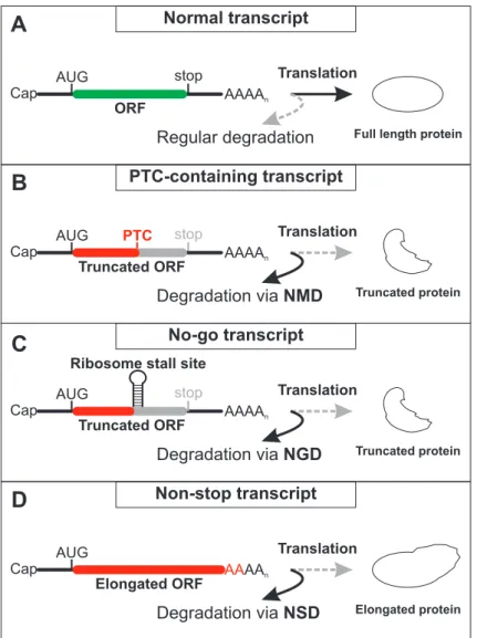
One of the frequent and subtle errors that occur during gene expression is the acquisition of
premature translation termination codons (PTC) (Savas et al., 2006). The presence of a PTC in
the mRNA leads to the abortion of protein synthesis before the complete open reading frame has been translated and, therefore, results in the synthesis of truncated, non‐functional or even harmful proteins (compare Figure 2A and B) (Frischmeyer and Dietz, 1999; Holbrook et al., 2004). Possible ways to generate PTCs on the DNA level are somatic rearrangements, nonsense mutations, as well as deletions and insertions that shift the reading frame. Furthermore, mutations in functional elements or motifs such as splice sites or splicing regulatory sites can lead to differently spliced, PTC containing transcripts (Nicholson et al., 2010). PTCs can also arise on the RNA level by transcription errors (incorporation of incorrect bases or loss of register) or alternative splicing events (e.g. intron inclusion or exon skipping). It has been calculated that about one‐third of all alternative splicing events in human multi‐exon genes result in PTC containing mRNA (Lewis et al., 2003).
Early studies discovered that the truncated proteins encoded by the PTC‐transcripts are not efficiently produced, but that the mRNA itself is degraded (Chang and Kan, 1979). Active translation is required for this process, as the presence of translation inhibiting antibiotics or stable secondary structures in the 5′ UTR of the PTC‐containing mRNA result in increased PTC‐
mRNA levels (Belgrader et al., 1993; Carter et al., 1995). This implicates that a translation‐
coupled surveillance system monitors the identity of the stop codon and decides whether the ribosome stalls at a normal or a premature termination codon. This mechanism was termed nonsense‐mediated mRNA decay (NMD) and represents one of the three characterized translation‐dependent mRNA quality control systems (Figure 2) (Shoemaker and Green, 2012).
The other pathways, non‐stop decay (NSD) and no‐go decay (NGD), detect and degrade mRNAs lacking a termination codon or containing strong ribosome stalling sites, respectively (Figure 2C and D) (Isken and Maquat, 2007; Wilson et al., 2008). These two systems are more similar to each other, compared to NMD, since they do not terminate translation upon encountering a stop codon. Moreover, NGD and NSD utilize the same factors for recognition and clearance of the erroneous transcript, whereas NMD relies on different proteins.
Degradation via NMD
Cap AAAAn
Elongated ORF AUG
Cap AAAAn
Truncated ORF
AUG stop
Ribosome stall site
Truncated protein PTC stop
Cap AAAAn
Truncated ORF AUG
Full length protein
Regular degradation
Cap AAAAn
ORF
AUG stop Translation
PTC-containing transcript Translation
Truncated protein
No-go transcript
Degradation via NGD Translation
Elongated protein
Non-stop transcript
Degradation via NSD Translation
B
C
D
Figure 2: Comparison of translation-coupled quality control mechanisms. (A) Normal mRNAs are translated and give rise to full length, functional proteins. These mRNAs are not targeted for accelerated degradation in a translation-dependent manner. (B) The presence of a premature translation termination codon (PTC) disrupts the open reading frame (ORF) and results in shortened proteins upon translation. PTC-containing mRNA are removed by nonsense-mediated mRNA decay (NMD) during translation. (C) Strong secondary structures or other components of the mRNP can stall ribosomes upstream of the termination codon. This results, similarly to (B), in the potential generation of truncated protein. No-go decay (NGD) detects and degrades these mRNAs. (D) Transcripts without stop codons are translated until the ribosome reaches the 3ʹ end of the mRNA. The potential production of elongated protein is prevented by degradation of the mRNA by non-stop decay (NSD). Alternatively, translation of the poly(A) tail leads to stalling of the ribosome and induction of NGD.
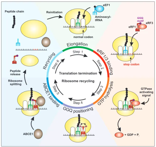
1.3 Mechanism of eukaryotic translation termination
Since NMD has the potential to discriminate between normal and abnormal
terminationcodons, it is important for the understanding of NMD to analyze the molecular events occurring during
translation termination. During the elongation phase of translation, the eukaryotic elongation factor 1 (eEF1) complex guides cognate aminoacyl-tRNA to the A site of theribosome in order to elongate the peptide chain (Figure 3, step 1) (Sasikumar et al., 2012).
However, when the ribosome encounters a stop codon (UAA, UGA or UAG) in the A site, this
codon is not recognized by tRNA, but by the eukaryotic release factors 1 and 3 (eRF1 and eRF3)
(Jackson et al., 2012; Klaholz, 2011).
eRF1
stop codon
eRF3 GGQmotif
normal codon
GTPase activating
signal
ABCE1
gati
n o
o n
El
eRF1/3 binding nilg
cecy R
Translation termination Ribosome recycling Peptide
release Ribosome
splitting
Reinitiation
Aminoacyl- tRNA
eEF1
pet6 S
p e t 1 S
Step 2
+ GDP + Pi
Peptide chain
Figure 3: Schematic overview of eukaryotic translation termination. After initiation of translation, aminoacyl-tRNAs are recruited to the ribosome by eEF1 in the elongation phase in order to generate the peptide chain (Step 1). Upon entry of a stop codon in the A site, eRF1 and eRF3 interact with the ribosome and decode the stop codon (Step 2). Hydrolysis of the eRF3- bound GTP can be activated by interacting factors and results in the dissociation of eRF3 from the ribosome (Step 3). Thereby, the GGQ motif of eRF1 can be positioned properly to enable the hydrolysis of the tRNA-peptide bond (Step4). This positioning is further enhanced by the association of the recycling factor ABCE1, which fills the space previously occupied by eRF3 (Step 5). Translation termination is completed by ATP-mediated splitting of the ribosomal subunits, accompanied by peptide hydrolysis mediated by eRF1 (Step 6). Of note, peptide release can also take place during steps 4 and 5. Finally, the single components are used for another round of translation.
The proteins eRF1 and eRF3 form a complex which is structurally reminiscent to the tRNA-eEF1
complex, with eRF1 decoding the stop codon via multiple conserved sequence motifs and eRF3serving as the eRF1-delivering factor (Figure 3, step 2) (Kong et al., 2004; Song et al., 2000).
Furthermore, eRF1 catalyzes the hydrolysis of the peptidyl-tRNA ester bond using a GGQ motif
that can be positioned in the peptidyl transferase center of the ribosome (Cheng et al., 2009).
To accomplish this step, the GTPase eRF3 has to hydrolyze GTP and dissociate from the
ribosome, therefore making space for the main recycling factor ABCE1 (Figure 3, step 3)(Pisarev et al., 2010; Salas-Marco and Bedwell, 2004). Either because of the GTP hydrolysis and
dissociation of eRF3 or because of the association of the ATPase ABCE1 with the ribosome,hydrolysis (Figure 3, steps 4 and 5) (Becker et al., 2012; Franckenberg et al., 2012). The final step is the ATP‐hydrolysis induced splitting of the ribosome followed by the recycling of the ribosomal subunits for another round of translation (Figure 3, step 6). At some point during these steps of translation termination, the decision whether the stop codon is considered normal or aberrant has to be made. Due to its central role in regulating the progression in the translation termination pathway, eRF3 is considered to be involved in this decision‐making process (Franckenberg et al., 2012). This is further supported by structural data obtained by cryo‐electron microscopy (cryo‐EM), suggesting that the flexible N‐terminus and the GTPase domain of eRF3 are positioned outside of the ribosome and are solvent‐exposed (Preis et al., 2014; Taylor et al., 2012). Thereby, these domains are likely available for binding of potential GTPase‐modulating factors, which influence the further advancement in translation termination (Figure 3, step 3). It is therefore conceivable that, depending on the type of the eRF3 interaction partner, either the current termination event proceeds normally and without mRNA degradation or the mRNA is marked as aberrant and is subsequently degraded.
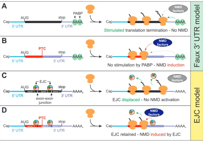
1.4 Models of NMD activation and substrate definition
As discussed above, certain factors or elements need to exist on the mRNP, which initiate NMD during translation termination. Despite NMD being a general and evolutionary conserved mRNA surveillance mechanism, different models for the activation in various organisms have been proposed (Rebbapragada and Lykke‐Andersen, 2009; Schweingruber et al., 2013).
Nevertheless, the key underlying determinant for NMD‐induction is similar: translation is
terminated at an unusual or aberrant position on the mRNP. In lower eukaryotes, the distance
of the stop codon to the poly(A) tail at the 3′ end of the mRNA is a critical determinant for the
recognition of PTCs (Amrani et al., 2004; Muhlrad and Parker, 1999). When a PTC is introduced
in the transcript, the resulting elongated 3′ UTR is believed to disturb interactions between the
terminating ribosome and downstream factors, which are required for proper termination
(Figure 4A; also see 1.3). The poly(A) binding protein (PABP, in yeast Pab1 and in mammals
PABPC1), which binds the poly(A) tail of mRNAs via its two first RNA recognition motifs (RRM),
is one of these potential downstream factors (Adam et al., 1986; Deo et al., 1999). Specifically,
the direct interaction of eRF3 and PABP is believed to be impaired due to the long distance
between stop codon and poly(A) tail (Figure 4B). Interestingly, this interaction is differently
mediated in yeast and mammals, as the mammalian PAPBC1 interacts via its C‐terminal MLLE
PTC
Cap AAAAn
AUG stop
Cap AAAAn
exon-exon junction
5′ UTR 3′ UTR
exon 1 exon 2 exon 3
EJC
Cap AAAAn
AUG stop
Cap AAAAn
5′ UTR 3′ UTR
exon 1 exon 2 exon 3
EJC displaced - No NMD activation
EJC retained - NMD induced by EJC
C
D
PTC
Cap AAAAn
AUG stop
Cap AAAAn
5′ UTR 3′ UTR
Cap AAAAn
AUG stop
Cap AAAAn
5′ UTR 3′ UTR
Stimulated translation termination - No NMD
No stimulation by PABP - NMD induction
A
B
PABP
Faux 3′ UTR model EJC model
factorsNMD
factorsNMD
factorsNMD
factorsNMD
Cap stop AAAAn
5′ UTR 3′ UTR
exon 1 exon 2 exon 3
C
domain with the N-terminal PAM2 motifs of eRF3, whereas the
yeast bindings sites are less welldefined (Cosson et al., 2002; Kozlov and Gehring, 2010; Kozlov et al., 2001; Roque et al., 2015).
Since PABP binding
stimulates the GTP hydrolysis of eRF3, loss of this interaction results in decreased translation termination efficiency (Amrani et al., 2006; Hoshino et al., 1999;Kononenko et al., 2010; Uchida et al., 2002). This in turn is supposed to enable proteins from the NMD machinery to interact with eRF3 in order to
activate NMD (Czaplinski et al., 1998;Ivanov et al., 2008; Kashima et al., 2006; Singh et al., 2008; Wang et al., 2001). This
“faux 3ʹUTR” model of NMD
activation was further supported by the observation that artificialrecruitment of PABP closely downstream of a PTC suppresses NMD (Amrani et al., 2004; Behm-
Ansmant et al., 2007a; Silva et al., 2008). Despite many observations being in agreement withthe
faux 3ʹ UTR model, recent studies revealed discrepancies, which are not covered by thismodel (Kervestin et al., 2012; Meaux et al., 2008; Roque et al., 2015).
Figure 4: Key concepts of NMD activation. (A) In transcripts with short 3ʹ UTR, the stop codon and the downstream poly(A) tail populated by cytoplasmic poly(A) binding proteins (PABP) are positioned in close proximity. Translation termination is stimulated by PABP and therefore prevents NMD factors to initiate degradation. (B) According to the faux 3ʹ UTR model, translation of PTC-containing mRNA results in ribosome stalling at a position where PABP is unable to efficiently promote translation termination. In turn, NMD factors can initiate the NMD pathway. (C) EJCs deposited on the mRNA are displaced and removed by the translating ribosome, preventing the EJC-induced recruitment of NMD factors. (D) The presence of a PTC sufficiently upstream of an exon-exon junction results in incomplete EJC removal. This enables the EJC to initiate NMD.
Contrary to the situation in
yeast, early studies showed that PTCs are distinguished from normal stop codons in mammalian cells when a EJC is located downstream of the terminating ribosomeaccordance with the observation that normal stop codons are positioned either in the last exon or are followed by introns not more than 50 nucleotides downstream (Brocke et al., 2002;
Maquat and Li, 2001; Nagy and Maquat, 1998). As a consequence of this so‐called position rule, PTCs introduced in the last exon will escape detection, whereas PTCs in any other exon will elicit NMD, if not positioned too close to the last splice site. The explanation for this rule is that during translation all EJCs in the ORF are removed by the ribosome, whereas EJCs in the 3′ UTR are unaffected and their position is maintained (Figure 4C and D) (Dostie and Dreyfuss, 2002;
Lejeune et al., 2002). The required minimum distance between stop codon and EJC is necessary, because closer positioning would already result in EJC dismantling due to steric reasons.
According to the current model, downstream EJCs recruit NMD factors and thereby define an upstream stop codon as premature (Figure 4D).
It was initially believed that the EJC‐induced NMD activation can only occur on mRNAs which are translated for the very first time (Chiu et al., 2004; Ishigaki et al., 2001; Matsuda et al., 2007;
Sato et al., 2008). This so‐called “pioneer round of translation” is the first loading of ribosomes on the mRNA and is characterized by the nuclear cap‐binding complex (CBC) heterodimer, consisting of CBP80 and CBP20, still attached to the 7‐methylguanosine cap (Maquat et al., 2010). After export of the mRNP from the nucleus, the CBC is replaced by the cytoplasmic eIF4F complex, consisting of eIF4A, eIF4E and eIF4G (Figure 1A) (Gross et al., 2003). Recent studies showed that EJC‐induced NMD takes place also on mRNPs bound by eIF4F, therefore NMD is not limited to the very first translation event (Durand and Lykke‐Andersen, 2013; Rufener and Muhlemann, 2013).
Several studies reported that EJC‐independent NMD exists in mammalian cells as well, which exhibits features comparable to the yeast “faux 3′ UTR” model (Buhler et al., 2006; Eberle et al., 2008; Singh et al., 2008). Substrates for this NMD pathway are normally devoid of EJCs downstream of the stop codon but contain an elongated 3′ UTR. Reporter mRNAs with artificially inserted, unspliced regions of various lengths (600‐1700 nucleotides) in the 3′ UTR resulted in decreased reporter levels and accelerated degradation via NMD (Buhler et al., 2006;
Eberle et al., 2008; Huang et al., 2011; Singh et al., 2008). Interestingly, endogenous transcripts with long 3′ UTRs, which encode for full length protein, are also targeted for degradation.
Therefore, NMD is not restricted to degrade faulty mRNAs that arise due to errors during gene
Cap AAAAn
5′ UTR 3′ UTR
Cap AAAAn
PTC
Cap AAAAn
Cap AAAAn
uORF
3′ UTR intron
Cap AAAAn
UGA
Cap Long 3′ UTR AAAAn
Sec-tRNASec
A B
C
EJC Long 3UTR
′
+ + + + -
-
+ - + + -
+
expression, but it also regulates wild type transcripts. Accordingly, genome-wide analyses in various eukaryotic organisms showed that about 3-10% of all cellular mRNAs are upregulated upon NMD inhibition (Guan et al., 2006; He et al., 2003; Johansson et al., 2007; Lelivelt and Culbertson, 1999; Mendell et al., 2004; Ramani et al., 2009; Rehwinkel et al., 2005; Tani et al., 2012; Wittmann et al., 2006; Yepiskoposyan et al., 2011).
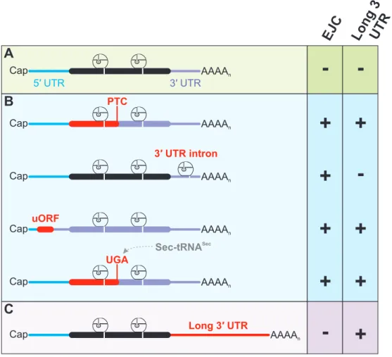
Figure 5: Examples of mRNPs targeted by NMD. (A) Normal mRNP composition with no EJC or long 3ʹ UTR downstream of the stop codon represents a NMD-resistant transcript. (B) Different causes exist for irregular EJC positioning relative to the stop codon, leading to NMD. Mutations, errors during transcription or splicing, and induced frameshifts are examples for generating a PTC in the regular ORF of the mRNP (top). Regulated splicing of 3ʹ UTR introns results in the deposition of EJC downstream of the physiological stop codon (second from top). Usage of upstream ORF (uORF) for translation initiation normally leads to premature translation termination, rendering all downstream EJC as potentially NMD-active (second from bottom). Lack of aminoacyl-tRNASec which would encode the stop codon UGA for selenocysteine, results in premature translation termination.
(C) Elongated distance between stop codon and poly(A) tail activates NMD.
In the light of the several molecular circumstances, which can potentially lead to NMD
activation, NMD targets represent a heterogeneous class of mRNPs (Figure 5). NMD-resistantmRNPs in general contain the correct 3ʹ UTR architecture required for proper translation
termination and lack downstream EJCs (Figure 5A). In contrast, various reasons for EJC-induced degradation of mRNPs exist (Figure 5B). As already discussed, PTC-containing transcripts, whichcan be generated by a multitude of potential errors during gene expression, frequently exhibit
EJCs downstream of the PTC. In specific cases, introns are positioned in the regular 3ʹ UTR and
mRNA encoding for the serine/arginine‐rich (SR) splicing factor SC35 (also referred to as SRSF2).
SC35 regulates alternative splicing of its own mRNA in a concentration‐dependent manner (Sureau et al., 2001). High SC35 levels lead to the excision of a 3′ UTR‐located intron, resulting in degradation of the mRNA via EJC‐induced NMD. SC35 therefore utilizes the NMD pathway for autoregulatory purposes by specifically activating NMD when required. A different class of NMD targets is degraded not because the ORF or 3′ UTR is modified, but because an upstream ORF (uORF) located in the 5′ UTR is translated. Since translation initiated at uORFs normally terminates upstream of the original ORF, not only the 3′ UTR is massively elongated, but also all normally displaced EJCs are still present. As long as no reinitiation of the ribosome occurs further downstream, this transcript will be degraded by NMD (Neu‐Yilik et al., 2011). One class of NMD targets encode for selenoproteins, which are characterized by the incorporation of selenocysteine (Sec) by the UGA codon. Upon low selenium levels in the cell, the tRNA
Seccannot be aminoacylated and the UGA codon will be recognized as a stop codon (Moriarty et al., 1998).
This can in turn lead to NMD activation, given that the UGA codon location results in an elongated and/or EJC‐populated 3′ UTR. EJC‐independent NMD targets, as mentioned earlier, are normally degraded because of their unusually long 3′ UTR (Figure 5C).
1.5 Factors involved in NMD assembly
Once a termination codon has been identified as aberrant, the NMD machinery has to properly assemble to execute the degradation of the target. Understanding of this process requires the detailed knowledge of the involved proteins and their molecular functions. The first proteins critical for NMD were discovered in nonsense suppression screens performed in S. cerevisiae
and
C. elegans(Culbertson et al., 1980; Hodgkin et al., 1989). The identified yeast
upf(up‐frameshift) and worm smg (suppressor with morphogenetic effect on genitalia) mutations were characterized later and the responsible genes were termed UPF1‐3 (Cui et al., 1995; Leeds et al., 1991; Leeds et al., 1992) and SMG1‐7 (Cali et al., 1999; Hodgkin et al., 1989; Pulak and Anderson, 1993), respectively. UPF1‐3 are the evolutionary central core of the NMD factors, as homologs have been identified in all late‐branching eukaryotes (Behm‐Ansmant et al., 2007b;
Chen et al., 2008; Culbertson and Leeds, 2003; Kadlec et al., 2006). The SMG proteins seem to
have evolved later and are found, with exceptions, mostly in metazoans. The initially
characterized
C. elegansSMG2‐4 proteins are homologous to the yeast UPF1‐3 proteins,
therefore the extended mammalian NMD core factors consist of UPF1‐3, SMG1, and SMG5‐7
(Applequist et al., 1997; Aronoff et al., 2001; Denning et al., 2001; Lykke‐Andersen et al., 2000;
Ohnishi et al., 2003; Page et al., 1999; Yamashita et al., 2001). To date, the number of proteins involved in NMD has doubled, although for many the specific role in NMD has not been characterized in detail. In mammalian cells, these include the proteins SMG8, SMG9, PNRC2, DHX34, NBAS, RUVBL1, RUVBL2, MOV10, GNL2 and SEC13 (Casadio et al., 2015; Gregersen et al., 2014; Hug and Caceres, 2014; Izumi et al., 2012; Longman et al., 2013; Longman et al., 2007;
Yamashita et al., 2009).
1.5.1 The RNA helicase UPF1 plays a central role in NMD
Research on NMD has so far been consistent in the point that the evolutionary highly conserved UPF1 is the most essential NMD factor in all investigated organisms. This is because UPF1 represents the center of the NMD machinery as it interacts with a multitude of other core factors and is functionally involved in all stages from the initiation until the disassembly of the NMD complex.
Early studies proposed that the release factors eRF1 and eRF3 directly recruit UPF1 to mRNA targets in order to initiate the NMD pathway (Kashima et al., 2006). This would imply that UPF1 is loaded onto the transcript in a translation‐dependent and regulated manner. However, individual‐nucleotide‐resolution UV cross‐linking and immunoprecipitation (iCLIP) experiments showed that UPF1 has the ability to bind mRNAs even in the absence of active translation (Zund et al., 2013). Furthermore, UPF1 binds NMD targets and those that are NMD‐resistant to an equal extent, suggesting that a regulated loading on NMD targets is unlikely. Furthermore, UPF1 occupies preferentially the 3′ UTR region of mRNA due to displacement from the 5′ UTR and coding region by scanning and translating ribosomes, respectively (Hurt et al., 2013;
Kurosaki and Maquat, 2013; Zund et al., 2013). Since UPF1 is able to compete with PABPC1 for binding to eRF3, it was proposed that long 3′ UTRs as NMD‐activating elements not only increase the distance between PABPC1 and eRF3, but also increase the local concentration of the competitor UPF1 (Hogg and Goff, 2010; Singh et al., 2008; Zund et al., 2013). The molecular details of this mechanism, specifically how the eRF3‐UPF1 interaction could initiate the NMD pathway, is still unclear.
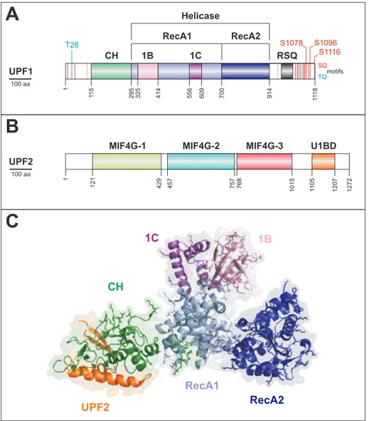
Concerning the domain architecture, the central part of UPF1 comprises two functional
domains, the N‐terminal zinc knuckle cystidine‐histidine‐rich CH domain followed by the central
helicase domain formed by two RecA‐like domains (Figure 6A) (Culbertson and Leeds, 2003).
nucleic acid duplexes in the 5′‐3′ direction in vitro, the helicase belongs to the superfamily 1Bα (SF1Bα) (Bhattacharya et al., 2000; Cheng et al., 2007; Fairman‐Williams et al., 2010; Singleton et al., 2007). Besides conferring potential unwinding ability, the helicase domain also mediates the direct binding to RNA (Bhattacharya et al., 2000; Chamieh et al., 2008). The overall importance of a functional UPF1 helicase domain is represented by the fact that the ATPase activity and direct RNA binding ability are both required for NMD (Mendell et al., 2002; Weng et al., 1996a, b). It remains controversial whether UPF1 utilizes ATP hydrolysis to translocate on the mRNA or uses it to remodel the mRNP after NMD execution is finished. In the first scenario, it was proposed that the helicase activity could help to bridge the distance between a terminating ribosome and the downstream‐located EJC (Shigeoka et al., 2012). In the latter, it would help to recycle NMD factors and allow the execution of full exonucleolytic degradation of the mRNA once initial decay steps have taken place (Cheng et al., 2007; Franks et al., 2010;
Singleton et al., 2007).
It was shown that both the CH domain as well as a C‐terminal region of UPF1 (regulatory SQ
region, RSQ) can regulate the helicase activity, which ensures that UPF1 clamps to the RNA and
does not translocate during the earlier stages of NMD (Chakrabarti et al., 2011; Fiorini et al.,
2013). Therefore, usage of the helicase domain for enabling a direct interaction with
downstream factors on the mRNP in the activation phase of NMD seems unlikely. More
specifically, conformational changes induced by the direct interaction of the UPF1 CH domain
with the RecA2 domain results in tighter RNA binding, which represses the helicase activity
(Chakrabarti et al., 2011). In order to initiate the unwinding activity of UPF1, the CH domain has
to be removed from the helicase core, which is achieved by the interaction with the C‐terminal
UPF1‐binding domain (U1BD) of UPF2 (Figure 6B and C) (Chakrabarti et al., 2011; Chamieh et
al., 2008).
UPF1
CH 1B 1C
RecA1 RecA2
Helicase
1118
115 295
1 414325 556 609 700 914
S1078
S1116 S1096 T28
SQ TQmotifs 100 aa
RSQ
MIF4G-3 U1BD
1272121 429
1 457 757 768 1015 12071105
MIF4G-2 MIF4G-1
UPF2
100 aa
A
B
RecA2 UPF2
CH
RecA1 1C 1B
C
Figure 6: Interaction and domain architecture of UPF1 and UPF2. (A) Schematic domain representation of UPF1, indicating functional domains. The SQ and TQ motifs, which are potentially phosphorylated in the N- and C-terminus are indicated, the major functional ones are highlighted. Two insertions in the RecA1 domain, called 1B and 1C are unique for UPF1. (B) UPF2 domains are depicted as in (A), highlighting the three MIF4G domains and the UPF1-binding domain (U1BD). (C) Crystal structure of UPF1 core domains and co-crystallized UPF2 C-terminus. Atomic coordinates of PDB accession code 2WJV were modeled with PyMol (Schrodinger, 2010) according to Clerici et al. (2009).
1.5.2 UPF2 provides the scaffold for the NMD assembly
As a core factor for NMD, UPF2 has additional roles besides the above-discussed stimulation of UPF1 helicase
activity. UPF2 consists of three tandem MIF4G domains (Middle portion ofeIF4G), followed by the U1BD (Figure 6B) (Aravind and Koonin, 2000; Clerici et al., 2014;
Ponting, 2000). MIF4G domains frequently provide the surface for
critical interactions forfactors involved in general mRNP metabolism (Ponting, 2000). In line with this role, the MIF4G-
3 domain of UPF2 interacts with UPF3, establishing a physical bridge between UPF1 and UPF3
(Chamieh et al., 2008; Kadlec et al., 2004; Serin et al., 2001). Cryo-EM studies identified that
the three N-terminal MIF4G domains form a ring-like structure together with the C-terminal
U1BD (Melero et al., 2012). Besides providing potential structural functions, the role of the two
N-terminal MIF4G domains in mammalian NMD is unclear. In S. cerevisiae, conserved residues
although potential interaction partners were identified, the function of these interactions in the molecular pathway of NMD remain uncertain (Fourati et al., 2014). Although UPF2 is widely accepted as an essential NMD component in mammalian cells, UPF2‐independent NMD has been observed in tethering assays (Gehring et al., 2005).
1.5.3 UPF3 acts as the link between UPF proteins and the EJC
Whereas in yeast and other invertebrates only one UPF3 protein exists, higher eukaryotes contain two UPF3 paralogs with high sequence similarity, UPF3a and UPF3b, the latter being expressed from the X chromosome in mammals (Lykke‐Andersen et al., 2000; Serin et al., 2001).
UPF3b was found to be the predominant NMD factor of both paralogs. However, a cross‐
regulatory circuit was described, which mainly involves the regulation of UPF3a stability as a consequence of the competition of both UPF3 proteins for binding to UPF2 (Chan et al., 2009;
Gehring et al., 2003; Kunz et al., 2006). UPF3b is a nucleocytoplasmic shuttling protein and contains a conserved N‐terminal RNA recognition motif (RRM). This domain is the binding site for the MIF4G‐3 of UPF2 and does not mediate RNA binding (Kadlec et al., 2004; Lykke‐
Andersen et al., 2000; Serin et al., 2001). At the C‐terminus, a short linear motif termed EJC‐
binding motif (EBM) is responsible for the interaction of UPF3b with a composite binding site of the EJC formed by the core components eIF4A3, MAGOH and Y14 (Buchwald et al., 2010;
Chamieh et al., 2008; Gehring et al., 2003; Kashima et al., 2010). UPF3b likely associates with the EJC in the nucleus and remains bound until it is displaced by PYM during ribosome‐mediated EJC disassembly (Bono and Gehring, 2011; Chamieh et al., 2008; Gehring et al., 2003; Tange et al., 2004). It was proposed that for mammalian NMD, EJCs downstream of a translation termination event could increase the concentration of UPF1‐UPF2 in the mRNP due to the specific recruitment via UPF3b (Kervestin and Jacobson, 2012). Yet, the exact molecular function of UPF3 in NMD remains elusive, since this UPF1‐UPF2 recruiting function does not explain the function of UPF3 in EJC‐independent NMD (Chamieh et al., 2008; Melero et al., 2012; Metze et al., 2013). This is especially interesting in case of organisms that do not employ EJC‐enhanced NMD as the standard pathway, but still rely on UPF3 for NMD. Examples are yeast, flies and worms, which either contain a very small number of spliced transcripts, lack EJC proteins and the EBM in the C‐terminus of UPF3, or do not require EJC core components for NMD, respectively (Culbertson and Leeds, 2003; Gatfield et al., 2003; Gehring et al., 2003;
Longman et al., 2007; Spingola et al., 1999; Wen and Brogna, 2010). Similar to the UPF2‐
independent NMD described, UPF3‐independent pathways were observed also, suggesting that these proteins are not absolutely necessary for certain NMD events (Chan et al., 2007).
1.5.4 UPF1 is phosphorylated by the SMG1 kinase
It was first observed in C. elegans that UPF1 (called SMG2 in C. elegans) is a phosphoprotein (Page et al., 1999). The phosphorylation status of UPF1 was found to be positively regulated by
SMG1, UPF2 and UPF3 (SMG3 and SMG4 in
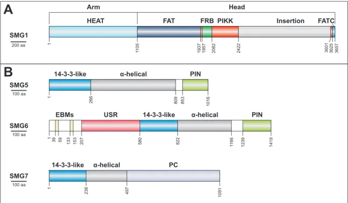
C. elegans)and negatively by SMG5‐7. SMG1 belongs to the phosphatidylinositol (PI) 3‐kinase‐related kinase (PIKK) family and was characterized as the responsible kinase for UPF1 phosphorylation in metazoan cells (Denning et al., 2001; Grimson et al., 2004; Page et al., 1999; Yamashita et al., 2001). Structural studies showed that the domain arrangement of the 410 kDa SMG1 protein is divided into a catalytic head structure and a flexible arm (Figure 7A) (Arias‐Palomo et al., 2011; Melero et al., 2014).
The binding of regulatory proteins termed SMG8 and SMG9 to the arm region of SMG1 modulates the kinase activity of SMG1 (Arias‐Palomo et al., 2011; Fernandez et al., 2011;
Yamashita et al., 2009). The head region including the catalytic PIKK domain and the FRB domain mediates the interaction with UPF1 and UPF2, respectively (Melero et al., 2014). The UPF2 binding to SMG1 is believed to modulate and positively stimulate the kinase activity, resulting in the phosphorylation of UPF1 (Ivanov et al., 2008; Kashima et al., 2006). Thereby, the list of potentially essential roles for UPF2 can be extended, since UPF2 not only forms the linear interaction cascade from UPF1 to UPF3 and modulates the helicase of UPF1, but it also positively influences the phosphorylation of UPF1.
1.5.5 Initiation of mRNA degradation via phospho‐UPF1 interactions
PIKK members, like SMG1, preferentially phosphorylate serines and threonines followed by glutamines (SQ and TQ motifs) (Bensimon et al., 2011; Yamashita et al., 2001). The SMG1‐
phoshorylated SQ and TQ motifs of mammalian UPF1 are clustered in the extended and unstructured N‐ and C‐terminus (Figure 6A) (Chakrabarti et al., 2014; Page et al., 1999;
Yamashita et al., 2001). Even though phosphorylation was also reported for yeast Upf1, the
mechanism and responsible kinase are different, since yeast Upf1 lacks most of the clustered
SQ and TQ motifs in the C‐terminus and no orthologue of SMG1 has been found (Lasalde et al.,
2014; Wang et al., 2006). The phosphorylation sites in mammalian UPF1 act as recruitment
platforms for the remaining core NMD factors, SMG5, SMG6 and SMG7. The three proteins
100 aa
100 aa
SMG5
SMG7 SMG6
100 aa
PIN
2661 809 1016853
α-helical 14-3-3-like
PC
2361 1091
α-helical 14-3-3-like
497
PIN
391 1166 14191239
α-helical 14-3-3-like
USR EBMs
59 133 153 207 580 822
SMG1
200 aa
3657
2422 3601 3625
2082
19571937
Arm Head
HEAT FAT FRB PIKK Insertion FATC
1 1105
A
B
and is able to interact with phosphorylated peptides (Figure 7B) (Fukuhara et al., 2005).
Figure 7: Domain structure of SMG proteins. (A) The complex domain architecture of SMG1 is depicted schematically. The N- terminal HEAT repeats form the arm, whereas the C-terminal domains form the globular head. (B) The decay inducing SMG5- 7 proteins share a 14-3-3-like domain, which is followed by α-helical extensions required for the stabilization of the domain.
The remaining domains or functional regions are indicated. PC = proline-rich region.
SMG5 and SMG7 form a heterodimer by perpendicular back-to-back interactions of their N- terminal 14-3-3-like domains. This is an uncommon arrangement, compared to the normal head-to-head interaction found in most 14-3-3 dimers and could explain why normal 14-3-3 proteins do not interact with UPF1 (Gardino et al., 2006; Jonas et al., 2013; Obsil and Obsilova, 2011). The 14-3-3-like domain of SMG7 is mostly responsible for the
phosphorylation-dependent interaction between phosphorylated amino acids (e.g. S1096) in the C-terminus of UPF1 and the heterodimer SMG5-SMG7 (Chakrabarti et al., 2014; Fukuhara et al., 2005; Jonas et al., 2013; Okada-Katsuhata et al., 2012). The 14-3-3-like domain of SMG5, which by itself is not able to interact with UPF1, supposedly provides additional binding strength and specificity (Jonas et al., 2013; Okada-Katsuhata et al., 2012).
1.5.6 Initiation of exonucleolytic degradation
Early work showed that artificial recruitment of full length SMG7 or the C-terminal proline-rich
(PC) region to tethering reporter mRNA induces mRNA degradation in a position-independent
and XRN1-/DCP2-dependent manner (Unterholzner and Izaurralde, 2004). DCP2 is the catalytic
subunit of the decapping complex, whereas XRN1 is the major cytoplasmic 5ʹ-3ʹ exonuclease,
suggesting that SMG7 induces accelerated decapping (Ghosh and Jacobson, 2010). Recently, the direct interaction of the PC region of SMG7 with the catalytic subunit of the CCR4‐NOT deadenylase complex POP2 has been shown (Loh et al., 2013). Therefore, SMG7 recruitment, mediated by its 14‐3‐3‐like domain, to the C‐terminus of phospho‐UPF1 induces deadenylation followed by decapping and degradation of the mRNA in the 5′‐3′ direction (Loh et al., 2013).
Early reports showed that the N‐ and C‐terminus of UPF1 can interact with decapping proteins, however, it was unclear if this interaction is direct or mediated by another factor (He and Jacobson, 1995, 2001; Lejeune et al., 2003; Lykke‐Andersen, 2002). The proline‐rich nuclear receptor coregulatory protein 2 (PNRC2) interacts with UPF1 and the decapping complex component DCP1, thereby providing a potential link for deadenylation‐independent decapping during NMD (Cho et al., 2009; Lai et al., 2012).
1.5.7 Dephosphorylation of UPF1 is initiated by decay factors
NMD is impaired under conditions where UPF1 accumulates in the hyper‐ or hypo‐
phosphorylated form, suggesting that a cycle of phosphorylation and dephosphorylation is essential (Grimson et al., 2004; Ohnishi et al., 2003; Okada‐Katsuhata et al., 2012; Page et al., 1999; Yamashita et al., 2001). Protein phosphatase 2A (PP2A) associates with the SMG5‐SMG7 heterodimer via the interaction with SMG5 and was identified as the phosphatase required for the dephosphorylation of UPF1 (Anders et al., 2003; Ohnishi et al., 2003). SMG5 contains a C‐
terminal PilT N‐terminus (PIN) domain, which is potentially involved in the interaction with PP2A. Deletion of the very C‐terminal amino acids or the replacement of a conserved aspartate at position 860 in this domain increased the phosphorylation of UPF1 (Ohnishi et al., 2003). PIN domains are commonly found in proteins executing endonuclease activity, however, the catalytic triad normally consisting of three aspartate residues is absent in the SMG5 PIN domain
and no endocleavage activity was reported neither
in vivonor in vitro (Clissold and Ponting, 2000; Glavan et al., 2006; Schoenberg, 2011). Interestingly, D860 is the one remaining aspartate residue in the active site, which was implicated in the regulation of UPF1 phosphorylation status (Ohnishi et al., 2003). Of note, evidence for SMG6 association with the
PP2 complex was provided as well, suggesting that, in line with the initial observation in
C.elegans,
all three SMG5‐7 proteins mediate UPF1 dephosphorylation by recruiting
phosphatases (Chiu et al., 2003).
Studies on the preferred nucleolytic degradation pathway of PTC containing mRNA in
Drosophila melanogaster S2 cells showed that the
knockdown of exonucleolytic machineries employing deadenylation, decapping, 3′‐5′ and 5′‐3′ degradation could not stabilize reporter mRNA levels (Gatfield and Izaurralde, 2004). However, evidence for PTC‐dependent endonucleolytic cleavage was found due to the accumulation of 3′ and 5′ fragments upon depletion of XRN1 and components of the 5′‐3′ degrading exosome complex, respectively (Gatfield and Izaurralde, 2004). In metazoans, SMG6 was identified as the endonuclease responsible for cleavage of the NMD targets in the vicinity of the stop codon (Eberle et al., 2009; Gatfield and Izaurralde, 2004; Huntzinger et al., 2008). SMG6 contains a C‐terminal PIN domain similar to SMG5. In contrast to SMG5, all catalytically important residues are present in the active site and the SMG6 PIN domain exhibits nucleolytic activity in vitro (Glavan et al., 2006). Mutations of any of the catalytic aspartate residues, which are required to coordinate divalent metal ions for the nucleophilic attack of H
2O on the phosphodiester bond of the RNA, renders the protein inactive and abolishes endonucleolytic degradation of NMD targets (Eberle et al., 2009; Glavan et al., 2006; Huntzinger et al., 2008; Kashima et al., 2010; Nicholson et al., 2014). Like SMG5 and SMG7, SMG6 contains a 14‐3‐3‐like domain, which is located centrally in the protein and does not form hetero‐ or homodimers (Chakrabarti et al., 2014; Fukuhara et al., 2005). This domain was also suggested to bind phosphorylated UPF1 and mutational analysis showed that mutation of the residues in the phosphopeptide binding pocket abolished the interaction with UPF1 (Okada‐Katsuhata et al., 2012). Similarly, alanine exchange of T28 in the N‐terminus of UPF1 greatly reduces the interaction with SMG6, suggesting that the 14‐3‐
3‐like domain of SMG6 interacts with the phosphorylated N‐terminus of UPF1 (Okada‐
Katsuhata et al., 2012). In recently reported in vitro experiments with phosphorylated UPF1,
the phospho‐dependent interaction with SMG5‐SMG7 was confirmed, however, no interaction
of the isolated 14‐3‐3‐like domain with hyperphosphorylated UPF1 was observed (Chakrabarti
et al., 2014). This is in line with recent data showing that phosphorylated UPF1 preferentially
occupies the 3′ UTR of NMD targets in a complex with SMG5 and SMG7, but not SMG6 (Kurosaki
et al., 2014). However, the unstructured region preceding the 14‐3‐3‐like domain of SMG6 was
observed to bind UPF1 in a phospho‐independent manner in vitro, which was complemented
by functional studies of SMG6 tethering and UPF1 complementation assays performed in
another recent publication (Chakrabarti et al., 2014; Nicholson et al., 2014). In addition, two
Cap AAAAn
Interaction eRF1/3
AAAAn
UPF1 UPF2
UPF3 C-term
N-term SMG1
AAAAn
UPF1UPF2 UPF3 SMG1 EJC
Phosphorylation
Phosphorylation Terminating ribosome
A UPF1UPF2
UPF3 SMG1 SMG5
SMG7
SMG6
Ribosome disassembly ?
Endocleavage
Deadenylation
eRF1/3 bindingUPF assembly PhosphorylationmRNA degradation
1 2
4 3
Cap Cap
Cap
EBMs were characterized in the very N-terminus of SMG6, which similarly to the EBM of UPF3b, mediate the interaction with the EJC (Kashima et al., 2010). These EBM motifs were found to be essential for NMD (Kashima et al., 2010). Given the multitude of possible interactions, the exact mechanisms by which SMG6 is recruited to the target mRNA remain elusive.
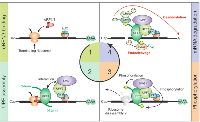
1.6 Model of the EJC-NMD mechanism
As the details of functions and interplay between NMD factors has been discussed above, the following model aims to present the most important steps from NMD
initiation to mRNA degradation (Figure 8). This is exemplified for EJC-induced NMD, because the molecular eventsduring long 3ʹ UTR-induced NMD are only poorly understood in mammalian cells.
Figure 8: Schematic model of EJC-induced NMD. (1) For EJC-NMD to be initiated, the ribosome has to stall at a stop codon upstream of an EJC. (2) Following the interaction of UPF1 with eRF3, UPF2 and SMG1 are recruited. This is enhanced due to the EJC-bound UPF3 recruitment of UPF2. (3) Activated SMG1 phosphorylates UPF1 at the N- and C-terminus. It is currently unclear, if the ribosome is already disassembled at this point. (4) Phosphorylated UPF1 recruits SMG5/7 and SMG6, which initiate degradation via deadenylation or endocleavage, respectively.