• Keine Ergebnisse gefunden

Inhibition of nonsense-mediated mRNA decay by the cytoplasmic poly(A)-binding protein

N/A
N/A
Protected

Academic year: 2021

Aktie "Inhibition of nonsense-mediated mRNA decay by the cytoplasmic poly(A)-binding protein"

Copied!
102
0
0

Wird geladen.... (Jetzt Volltext ansehen)

Volltext

(1)

Inhibition of nonsense-mediated mRNA decay by the cytoplasmic poly(A)-binding protein

Inaugural-Dissertation

zur

Erlangung des Doktorgrades

der Mathematisch-Naturwissenschaftlichen Fakultät der Universität zu Köln

2016

vorgelegt von Tobias Fatscher

aus Heidelberg

(2)
(3)

Berichterstatter/in: PD Dr. Niels H. Gehring

Prof. Dr. Karin Schnetz

Tag der mündlichen Prüfung: 05. Juli 2016

(4)
(5)

Parts of this thesis have been published as:

1) Fatscher T, Boehm V, Weiche B, Gehring NH. The interaction of cytoplasmic poly(A)- binding protein with eukaryotic initiation factor 4G suppresses nonsense-mediated mRNA decay. RNA. 2014; 20(10):1579-92.

2) Fatscher T, Boehm V, Gehring NH. Mechanism, factors, and physiological role of

nonsense-mediated mRNA decay. Cell Mol Life Sci. 2015; 72(23):4523-44.

(6)

i

Table of Contents

1 Abstract ... v

2 Deutsche Zusammenfassung ... vi

3 Introduction ... 1

3.1 Poly(A)-binding proteins: A protein family regulating mRNA fate ... 1

3.1.1 More than one PABP is present in human cells ... 1

3.1.2 PABPC1 structure and interaction partners ... 2

3.2 EJC-mediated NMD: deposition, factors, and pathway ... 4

3.2.1 Characteristics of the EJC ... 4

3.2.2 Central factors involved in NMD and their interplay ... 5

3.2.3 Phosphorylation of UPF1 regulates NMD activity ... 6

3.2.4 Exonucleolytic degradation via SMG5/7 ... 7

3.2.5 Endonucleolytic degradation via SMG6 ... 7

3.3 EJC-independent activation of NMD ... 10

3.3.1 NMD activation due to a long 3′ UTR ... 10

3.3.2 The faux 3′ UTR model in yeast ... 11

3.4 The link between translation and NMD ... 11

3.4.1 Translation initiation ... 11

3.4.2 Proper translation termination ... 12

3.4.3 Steps in aberrant translation termination and the link to NMD ... 14

3.4.4 Active translation is necessary for NMD ... 16

3.5 Additional NMD targets present in the cell ... 17

3.6 Physiological implications of NMD ... 19

3.6.1 Importance of NMD factors on cell viability ... 19

3.6.2 NMD and genetic disorders ... 19

3.6.3 Treatment of physiological conditions resulting from NMD ... 20

3.7 Aim of this thesis ... 21

(7)

ii

4 Results ... 23

4.1 PABPC1 is able to antagonize long 3′ UTR-mediated NMD ... 23

4.1.1 Increased reporter abundance is a consequence of PABPC1-mediated NMD suppression ... 24

4.2 Interaction partners of PABPC1 necessary for NMD suppression ... 27

4.2.1 The interaction between PABPC1 and eRF3a is not required for NMD suppression ... 27

4.2.2 PABPC1 needs the interaction with eIF4G to antagonize NMD ... 30

4.3 PABPC1 does not affect EJC-mediated NMD ... 31

4.4 eRF3a requires the interaction with PABPC1 for NMD suppression ... 32

4.5 eIF4G antagonizes NMD independently of PABPC1 ... 35

4.6 PABPC1 acts as a general suppressor of long 3′ UTR-mediated NMD ... 38

4.7 NMD inhibition by PTBP1 and other proteins known to interact with PABPC1 39 4.8 Peptide-mediated fold-back of the poly(A)-tail stabilizes NMD-targeted mRNA 41 4.8.1 eRF3a and eRF1 suppress NMD by folding back the poly(A)-tail ... 41

4.8.2 Heterodimer formation of synthetic peptides suppresses NMD by folding back the poly(A)-tail ... 42

4.9 PAM2 motifs are sufficient to antagonize NMD ... 44

4.9.1 PABPC1 recruitment via PAM2 motifs efficiently antagonizes NMD ... 44

4.9.2 Tethering PAM2 motifs has no negative effect on translation of the reporter transcript ... 46

4.9.3 Not all PAM2 motifs show the expected interaction with PABPC1 ... 47

4.9.4 NMD suppression by PAM2 motifs is a specific tethering effect ... 48

4.9.5 EJC-factor mediated NMD is antagonized by PAM2 motifs ... 49

4.9.6 PAM2 motifs suppress NMD of an intron-carrying reporter ... 51

4.10 PABPC1 suppresses NMD of the β -globin PTC39 mRNA... 52

5 Discussion ... 54

(8)

iii

5.1 NMD suppression of long 3′ UTR substrates ... 55

5.1.1 PABPC1 is a general suppressor of long 3′ UTR mediated NMD ... 55

5.1.2 Implication of translation termination in NMD suppression by PABPC1 .... 55

5.1.3 Protein-mediated NMD inhibition ... 58

5.1.4 Peptide-mediated fold-back of the poly(A)-tail ... 59

5.1.5 PAM2 motifs strongly suppress long 3′ UTR-triggered NMD ... 60

5.2 EJC-activated NMD is antagonized by PAM2 peptides ... 62

5.3 Medical implications of NMD inhibition and potential therapeutic approaches 64 5.4 Concluding remarks ... 66

6 Material and Methods ... 67

6.1 Molecular biology... 67

6.1.1 Mammalian expression plasmids ... 67

6.2 Molecular cloning ... 67

6.2.1 Transformation ... 67

6.2.2 Plasmid preparation ... 68

6.3 Cell culture ... 68

6.3.1 Cell lines ... 68

6.3.2 Plasmid DNA transfection ... 68

6.4 Immunoprecipitation ... 69

6.4.1 Transfection of HTO cells ... 69

6.4.2 Preparation of cell lysates ... 69

6.4.3 Co-immunoprecipitation ... 69

6.4.4 SDS-polyacrylamide gel electrophoresis (SDS-PAGE) ... 70

6.4.5 Western blot analysis ... 70

6.5 Northern blot analysis ... 71

6.5.1 Transfection of HTO cells ... 71

6.5.2 Harvesting cells ... 71

(9)

iv

6.5.3 RNA extraction ... 71

6.5.4 Northern blotting ... 72

6.5.5 Protein extraction ... 73

7 Appendix ... 75

7.1 List of Antibodies ... 75

7.2 List of Primers ... 75

8 References ... 77

9 Acknowledgement ... 89

10 Erklärung ... 90

(10)

v

1 Abstract

The expression of a gene from transcription of the DNA into pre-messenger RNA (pre-

mRNA) over translation of messenger RNA (mRNA) into protein is constantly monitored

for errors. This quality control is necessary to guarantee successful gene expression. One

quality control mechanism important to this thesis is called nonsense-mediated mRNA

decay (NMD). NMD is a cellular process that eliminates mRNA transcripts harboring

premature translation termination codons (PTCs). Furthermore, NMD is known to

regulate certain transcripts with long 3′ UTRs. However, some mRNA transcripts are

known to evade NMD. The mechanism of NMD activation has been subjected to many

studies whereas NMD evasion or suppression still remains rather elusive. It has

previously been shown that the cytoplasmic poly(A)-binding protein (PABPC1) is able to

suppress NMD of certain transcripts. In this study I show that PABPC1 is able to

suppress NMD of a long 3′ UTR-carrying reporter when tethered immediately

downstream of the termination codon. I further am able to show the importance of the

interaction between PABPC1 and eIF4G for NMD suppression, whereas the interaction

between PABPC1 and eRF3a seems dispensable. These results indicate an involvement

of efficient translation termination and potentially ribosome recycling in NMD

suppression. I am able to show that if PABPC1 is too far removed from the terminating

ribosome NMD is activated. After showing the importance of PABPC1 recruitment

directly downstream of a terminating ribosome in NMD suppression, I am further able to

demonstrate several different methods by which PABPC1 can be recruited. Fold-back of

the poly(A)-tail mediated by two interacting proteins on opposite ends of a 3′ UTR

manages to bring PABPC1 bound to the poly(A)-tail into close proximity of the

terminating ribosome and therefore suppress NMD. Furthermore, small PAM2 peptides

that are known to interact with the MLLE domain of PABPC1 are able to strongly

suppress NMD initiated by either a long 3′ UTR or an EJC. I am also able to show the

NMD antagonizing power of recruited PABPC1 for the known endogenous NMD target β-

globin PTC39, which is responsible for the disease β-thalassemia. This shows the

potential medical implications and application of suppressing NMD by recruiting

PABPC1 into close proximity of a terminating ribosome.

(11)

vi

2 Deutsche Zusammenfassung

Die Expression eines Gens angefangen mit der Transkription der DNA in prä-Messenger RNA (prä-mRNA) über Translation von Messenger RNA (mRNA) in ein Protein ist ein stark überwachter Prozess. Diese Qualitätskontrolle ist notwendig, um fehlerfreie Genexpression zu gewährleisten. Ein wichtiger Mechanismus zur Qualitätssicherung ist Nonsense-mediated mRNA decay (NMD) genannt. NMD ist ein zellulärer Prozess, der mRNA-Transkripte die ein vorzeitiges Stoppcodon (PTC) beherbergen eliminiert.

Weiterhin ist bekannt, dass NMD bestimmte Transkripte mit langen 3′ UTRs regulieren kann. Allerdings erkennt NMD nicht alle Transkripte und es sind einige mRNA- Transkripte bekannt die sich NMD entziehen. Der Mechanismus der NMD-Aktivierung wurde bereits vielen Studien unterzogen, während der Mechanismus der NMD Umgehung oder Unterdrückung noch relativ unerforscht ist. Es wurde zuvor schon gezeigt, dass das zytoplasmatische Poly(A)-bindende Protein (PABPC1) imstande ist NMD bestimmter Transkripte zu unterdrücken. In dieser Studie zeige ich, dass PABPC1 in der Lage ist, NMD eines langen 3′ UTR-tragenden Reporters zu unterdrücken, wenn es unmittelbar nach dem Terminationscodon gebunden ist. Ich bin weiter in der Lage, die Bedeutung der Interaktion zwischen PABPC1 und eIF4G in der NMD Unterdrückung zu zeigen, während die Interaktion zwischen PABPC1 und eRF3a hierfür entbehrlich erscheint. Diese Ergebnisse deuten auf eine Beteiligung von effizienter Translationstermination und Ribosom-Recycling in der Unterdrückung von NMD hin.

Nachdem ich die Bedeutung der Rekrutierung von PABPC1 direkt hinter einem Terminationscodon in der Unterdrückung von NMD zeigen konnte, war es mir weiterhin möglich verschiedene Methoden zu zeigen, mit denen PABPC1 rekrutiert werden kann.

Das Zurückfalten des Poly(A)-Schwanzes durch zwei interagierende Proteine an gegenüberliegenden Enden einer 3′ UTR ermöglicht es PABPC1, welches am Poly(A)- Schwanz gebunden ist, in die Nähe des terminierenden Ribosoms zu bringen und somit NMD zu unterdrücken. Weiterhin zeige ich, dass kleine PAM2 Peptide, die mit der MLLE Domäne von PABPC1 interagieren, in der Lage sind NMD aktiviert durch entweder einer lange 3′ UTR oder eines EJC zu unterdrücken. Die PAM2 Peptide unterdrücken NMD durch die Rekrutierung von PABPC1 in die direkte Nähe des terminierenden Ribosoms.

Außerdem bin ich in der Lage NMD des endogenen Transkripts β-Globin PTC39, welches

für die Krankheit β-Thalassämie verantwortlich ist, durch die Rekrutierung von PABPC1

(12)

vii

zu unterdrücken. Dies zeigt die potentiellen medizinischen Auswirkungen und mögliche

Anwendung der NMD Unterdrückung durch PABPC1 auf.

(13)

1

3 Introduction

Eukaryotic gene expression is a complex and highly regulated process. Messenger RNAs (mRNAs) are transcribed from DNA in the nucleus and serve as blueprints for protein synthesis before being degraded. Synthesis of mRNA starts in the nucleus in the form of a pre-mRNA (Moore, 2005; Moore and Proudfoot, 2009). Transcription from DNA to pre-mRNA is undertaken by RNA polymerase II (Pol II). Pre-mRNAs are subjected to co- transcriptional 5′ end capping, splicing, 3′ end cleavage, and polyadenylation before the processed mRNA is exported to the cytoplasm. In the cytoplasm the mature mRNAs are translated into proteins by ribosomes and are eventually degraded. To ensure error-free gene expression, several quality control mechanisms exist in the living cell. One major quality control mechanism is called nonsense-mediated mRNA decay (NMD). NMD is responsible for recognizing and degrading mRNA transcripts with premature termination codons (PTCs) or other aberrant translation termination events. PTCs are known to arise through different mechanisms such as alternative splicing events, transcription errors, or due to mutations on the DNA level (Nicholson et al., 2010).

Without NMD the aberrant transcripts could lead to the production of C-terminally truncated proteins with potential dominant-negative effects on the cell. In addition to the quality control function, NMD also regulates many so-called endogenous NMD targets. Thus, NMD is not only responsible for mRNA surveillance but also influences general gene expression.

3.1 Poly(A)-binding proteins: A protein family regulating mRNA fate

Poly(A)-binding proteins (PABPs) are a set of highly conserved proteins among eukaryotes. PABPs are RNA-binding proteins, specifically recognizing the poly(A)-tail of virtually all mRNAs in the cell. PABPs do not feature any catalytic or enzymatic activity themselves. However, they serve as interaction partner to a multitude of other proteins, which highlights the role of PABPs in many different cellular events such as translation, mRNA stability, or quality control.

3.1.1 More than one PABP is present in human cells

Metazoan cells express more than one PABP. Whereas there is more than one

cytoplasmic PABP (PABPC), metazoan cells encode a single nuclear PABP (PABPN).

(14)

2

PABPCs and PABPN only share the ability to bind poly(A)-tails of RNAs but have no other common functions and they do not show any structural similarities. The most well researched PABPC is called cytoplasmic poly(A)-binding protein 1 (PABPC1) (Kuhn and Wahle, 2004). Even though PABPC1 is mostly localized in the cytoplasm, it was also found to be able to shuttle into the nucleus. PABPN1 is known to bind to the poly(A)-tail of mRNAs in the nucleus and is required for mRNA export into the cytoplasm (Apponi et al., 2010). Since both PABPC1 and PABPN1 are known to shuttle between the nucleus and the cytoplasm it is difficult to determine the exact time point when PABPC1 replaces PABPN1 on mRNA transcripts. Mammalian cells encode three additional PABPs besides PABPC1 and PABPN1. Two of these PABPs are restricted to the germ line and are called testis-specific PABP (tPABP, PABPC2/PABPC3) and embryonic PABP (ePABP, ePAB).

The third PABP is termed PABP4 or inducible PABP (iPABP). These three PABPs share the same domain structure with PABPC1. An additional PABP termed PABPC5 or PABP5 is also found in mammals. However, the function of PABP5 has not been addressed and remains subject of further research (Gray et al., 2015; Kuhn and Wahle, 2004).

3.1.2 PABPC1 structure and interaction partners

PABPC1 is a 636 amino acid long protein with a molecular weight of about 70kDa. The N-terminal region of PABPC1 is comprised of four RNA recognition motifs (RRMs) that are connected by short linker regions. PABPC1 also harbors a C-terminal region connected to the N-terminal region by an unstructured linker region that is proline- and glutamine-rich. PABPC1 has a high binding affinity for poly(A), however it does exhibit a lower binding affinity for poly(U) and poly(G) as well (Kuhn and Wahle, 2004). PABPC1 requires at least 12 adenosines for a high affinity interaction and covers about 25 nucleotides on a saturated stretch of poly(A) (Kuhn and Wahle, 2004). The interaction between PABPC1 and the poly(A)-tail is mediated via the RRMs 1 and 2 of PABPC1 (Deo et al., 1999). The major roles of PABPC1 are in mRNA translation and stability. An additional important characteristic of PABPC1, which is of interest in this work, lies in its ability to affect the nonsense-mediated mRNA decay pathway.

PABPC1 can interact with a multitude of other proteins that can influence mRNA fate.

The interaction with other proteins is mediated either by some of the four different

RRMs or by the C-terminal region. One such important interaction partner of PABPC1

that binds to the RRMs is eIF4G. eIF4G is part of the eIF4F complex and plays a vital role

in mRNA circularization and translation initiation. It has been shown that eIF4G binds to

(15)

3

RRM2 of PABPC1 and this interaction is also believed to enhance the binding affinity of PABPC1 for the poly(A)-tail (Kuhn and Wahle, 2004; Safaee et al., 2012).

Another important interaction partner of PABPC1 is eRF3a, which plays a role in translation termination. eRF3a is known to interact with PABPC1 via a so called PABP- interacting motif 2 (PAM2 motif). The C-terminal region of PABPC1 harbors a specific stretch termed Mademoiselle (MLLE) domain, which has been shown to specifically recognize PAM2 motifs of other proteins (Kozlov et al., 2004). The PAM2 motifs comprise only a stretch of about 12 amino acids. Essential amino acids in the PAM2 motif for the interaction with the MLLE domain of PABPC1 are known to be EFxP. These three amino acids are mostly conserved among PAM2 motifs originating from different PABPC1-interacting proteins (Kozlov and Gehring, 2010). The PAM2 motif was first discovered in PABP-interacting protein 1 (Paip1), Paip2, and eRF3a (Kozlov et al., 2001).

After the initial discovery of PAM2-containing proteins, many more proteins harboring PAM2 have been found (Albrecht and Lengauer, 2004; Kozlov et al., 2001). Interestingly, even though PAM2-containing proteins interact with PABPC1 in the same way, they play roles in different cellular processes. This also explains why PABPC1, even though not a catalytically active protein itself, can influence so many different aspects of an mRNA.

The interaction between PABPC1 and eRF3a is very important as it has been shown to

play a role in polypeptide release and ribosome recycling (Hoshino et al., 1999; Kozlov

and Gehring, 2010; Uchida et al., 2002). In contrast, other PAM2-containing proteins like

PAN3 and TOB1 are known to play a role in deadenylation. PABPC1 is known to recruit

the PAN2-PAN3 deadenylase complex directly by interacting with the PAM2-harboring

protein PAN3 (Siddiqui et al., 2007). The other major deadenylase complex, CCR4-NOT,

can also be recruited by PABPC1. However, this interaction is not mediated directly by

PABPC1. The CCR4-NOT complex is recruited to the mRNA through the interaction

between TOB1 and PABPC1 via the PAM2 motif and MLLE domain, respectively

(Ezzeddine et al., 2007). This shows that PABPC1 can interact with proteins that

influence the mRNA fate by, for example, either stabilizing the mRNA or by promoting

deadenylation. Furthermore, the PABPC1-interacting protein PAIP2 is a known

repressor of translation by interfering with the ability of PABPC1 to interact with the

poly(A)-tail (Khaleghpour et al., 2001). It is interesting to note that PABPC1 can interact

with a multitude of PAM2-containing proteins, although with only one at a time, which

can give rise to potentially contradictory functions. The determinant for an interaction

(16)

4

preference between PABPC1 and the many PAM2-containing proteins is currently unknown.

In summary, PABPC1 is able to interact with many different proteins and plays an important role in multiple cellular processes. It does not influence the fate of an mRNA with direct catalytic activity but does so by acting as a scaffolding protein. Interestingly, PABPC1 can interact with proteins both favoring mRNA stability as well as proteins that will eventually lead to the degradation of the mRNA.

3.2 EJC-mediated NMD: deposition, factors, and pathway 3.2.1 Characteristics of the EJC

Pre-mRNAs undergo several co-transcriptional processing steps in the nucleus. One such processing step important for NMD is splicing. During splicing the introns of a pre- mRNA are excised and the exons are subsequently joined. Furthermore, an exon- junction complex (EJC) is deposited 20 to 24 nucleotides upstream of the splice site.

NMD targets mRNAs that harbor a PTC 50 to 55 nucleotides upstream of the final exon- exon junction and fails to do so for many mRNAs carrying a stop codon downstream of this critical border (Nagy and Maquat, 1998; Thermann et al., 1998; Zhang et al., 1998a;

Zhang et al., 1998b). The core EJC complex consists of four proteins: the heterodimer MAGOH/Y14, eIF4A3, and Barentsz (BTZ). The EJC remains bound to the mature mRNA during export into the cytoplasm until it is displaced by a translating ribosome (Dostie and Dreyfuss, 2002; Le Hir et al., 2000; Lejeune et al., 2002). Although a translating ribosome is sufficient to remove an EJC from an mRNA, an additional protein named PYM is known to aid in the displacement of the EJC from the mRNA (Gehring et al., 2009). The EJC binds RNA not by recognizing a specific RNA sequence, but does so via the eIF4A3 component of the EJC. eIF4A3 is an ATPase known to strongly bind RNA in an ATP-dependent manner. The reason why the EJC is able to stay on the mRNA during export as well as thereafter, is due to the partial inhibition of the ATPase activity of eIF4A3. This is achieved by the heterodimer Y14 and MAGOH that are able to lock the EJC onto the mRNA by stabilizing a closed formation of eIF4A3 (Andersen et al., 2006;

Ballut et al., 2005; Bono et al., 2006; Mishler et al., 2008). Besides the four core EJC

components mentioned above, there are several other known EJC factors that are not

part of this core complex. One of these so called peripheral EJC components is the RNA-

binding protein with serine rich domain 1 (RNPS1). RNPS1 is known to function as a

(17)

5

general co-activator of splicing and is also able to communicate the position of the EJC on the mRNA to other factors of the NMD machinery, which are discussed in more detail later (Le Hir et al., 2000; Lykke-Andersen et al., 2001; Mayeda et al., 1999). Furthermore, the EJC serves as a binding platform for a multitude of other proteins involved in the NMD pathway (Chang et al., 2007).

3.2.2 Central factors involved in NMD and their interplay

There are many factors known to be involved in NMD with more factors most likely still unknown. Of all the factors involved in NMD arguably the most important one is the central NMD factor UPF1 (up-frameshift 1), which is involved in the pivotal stages of the NMD mechanism (Chang et al., 2007). UPF1 was first discovered in yeast more than 30 years ago alongside UPF2 and UPF3 (Cui et al., 1995; Leeds et al., 1991; Leeds et al., 1992). UPF1 has several different functions. It harbors a helicase domain which is able to unwind double stranded nucleic acids in a 5′ - 3′ directionality with the help of ATP hydrolysis. It has been shown that the helicase activity as well as the ability of UPF1 to bind RNA is essential for NMD (Mendell et al., 2002; Weng et al., 1996a, b). The ability of UPF1 to bind RNA directly is, according to the current model, necessary for the assembly of other NMD factors on UPF1. The RNA-binding ability of UPF1 is also mediated by its helicase domain (Bhattacharya et al., 2000; Cheng et al., 2007; Fairman-Williams et al., 2010; Singleton et al., 2007). The first model of UPF1 recruitment to the NMD targeted mRNA proposed that the interaction between UPF1 and the release factor eRF3 (will be discussed in more detail later) at the terminating ribosome were responsible for this recruitment. This suggested that UPF1 recruitment is directly linked to active translation and that UPF1 is bound exclusively in close proximity of a termination codon.

The complex formed by this interaction involved an additional protein called SMG1 (discussed in more detail later) and the resulting complex was termed SURF-complex.

(Kashima et al., 2006). However, it was more recently shown that UPF1 is able to bind

over the whole 3′ UTR of an mRNA. UPF1 was additionally shown to also bind to other

regions than the 3′ UTR of an mRNA, but the scanning and translating ribosome is able

to displace UPF1. Displaced UPF1 is believed to increasingly bind to the 3′ UTR of a

transcript as the ribosome stalls at the termination codon upstream of the 3′ UTR,

unable to displace any UPF1 further downstream. This model further proposes that

UPF1 occupancy of the 3′ UTR is responsible for NMD activation and not the direct

(18)

6

recruitment of UPF1 via the release factors eRF1 and eRF3 (Hurt et al., 2013; Kurosaki and Maquat, 2013; Zund et al., 2013).

Additional to UPF1, there are two more UPF proteins termed UPF2 and UPF3. Together these proteins form the central set of NMD factors (Cui et al., 1995). An important role of UPF2 is to regulate the helicase activity of UPF1. UPF2 is able to do so by directly interacting with UPF1, causing conformational changes in UPF1, which results in the activation of the UPF1 helicase activity (Chakrabarti et al., 2011; Chamieh et al., 2008).

UPF2 is further able to directly interact with UPF3, therefore providing a physical link between UPF1 and UPF3 (Chamieh et al., 2008). It is worth mentioning that higher eukaryotes contain two paralogs of UPF3, namely UPF3a and UPF3b (Lykke-Andersen et al., 2000; Serin et al., 2001). Besides both paralogs displaying some cross-regulation, UPF3b has been shown to be the dominant of the two paralogs (Kunz et al., 2006).

UPF3b is able to directly interact with the EJC via a so called EJC-binding motif (EBM) in its C-terminal region (Buchwald et al., 2010; Chamieh et al., 2008; Gehring et al., 2003).

This interaction provides a molecular link between UPF1 and the EJC complex and was proposed to play an important role during NMD (Chamieh et al., 2008). However, it has also been shown that UPF3b plays a role in EJC-independent NMD (Metze et al., 2013).

As a result, the exact function of UPF3b in NMD remains subject of further research.

3.2.3 Phosphorylation of UPF1 regulates NMD activity

Besides the UPF proteins, the NMD machinery consists of several SMG (suppressor with

morphogenetic effect on genitalia) proteins. In humans, the proteins SMG1 and SMG5-7

have been shown to be part of the NMD core machinery (Denning et al., 2001; Ohnishi et

al., 2003; Yamashita et al., 2001). Phosphorylation of UPF1 is known to be catalyzed by

SMG1, a member of the phosphatidylinositol 3-kinase-related kinase (PIKK) family

(Denning et al., 2001; Grimson et al., 2004; Yamashita et al., 2001). SMG1 is able to

phosphorylate SQ and TQ motifs in the N- and C-terminal regions of UPF1 (Chakrabarti

et al., 2014; Page et al., 1999; Yamashita et al., 2001). These phosphorylation sites of

UPF1 act as binding platforms for other SMG proteins in the NMD pathway (Fukuhara et

al., 2005). Specifically, SMG6 and SMG5/7 are able to interact with phosphorylated

UPF1. Furthermore, it is important to note that SMG5 and SMG7 form a heterodimer and

SMG5 alone is unable to interact with phosphorylated UPF1. SMG5 is therefore believed

to offer additional binding strength between SMG7 and phosphorylated UPF1

(Chakrabarti et al., 2014; Jonas et al., 2013; Obsil and Obsilova, 2011; Okada-Katsuhata

(19)

7

et al., 2012). The SMG proteins are known to activate two different pathways of mRNA degradation: exonucleolytic degradation via SMG5/7 and endonucleolytic degradation via SMG6 (Nicholson and Muhlemann, 2010).

3.2.4 Exonucleolytic degradation via SMG5/7

It has previously been shown that SMG7 is able to position-independently activate degradation of the NMD-targeted mRNA via XRN1 and DCP2 (Unterholzner and Izaurralde, 2004). XRN1 is a known 5′ - 3′ exoribonuclease and DCP2 is part of the decapping complex responsible for the removal of the 5′ cap structure before mRNA degradation (Ghosh and Jacobson, 2010). SMG7 is unable to initiate exonucleolytic degradation by directly recruiting XRN1 or DCP2. Instead, SMG7 interacts with POP2, the catalytic subunit of the CCR4-NOT complex, which is part of the deadenylation machinery in the cell (Loh et al., 2013). This recruitment of the CCR4-NOT complex via SMG7 initiates deadenylation of the transcript, followed by the recruitment of XRN1 and DCP2 as a result of deadenylation. This provides a link between UPF1 and phosphorylation-mediated activation of the SMG5/7-initiated exonuceolytic decay pathway. Furthermore, since SMG5/7 is known to interact and recruit part of the deadenlyation machinery, it is also feasible that the mRNA targeted by NMD can be degraded not only via XRN1-mediated 5′ - 3′ degradation but also in a 3′ - 5′ direction via the exosome.

3.2.5 Endonucleolytic degradation via SMG6

An additional degradation pathway of NMD targets was found when knockdown of the exonucleolytic machinery failed to stabilize NMD-targeted reporter mRNA levels in flies.

At the same time, PTC-mediated endonucleolytic cleavage was found to occur on these targets (Gatfield and Izaurralde, 2004). It was then found that SMG6 is responsible for cleaving the target in close proximity of the PTC via its catalytically active PIN domain leading to degradation of the NMD target (Eberle et al., 2009; Gatfield and Izaurralde, 2004; Glavan et al., 2006; Huntzinger et al., 2008). Similar to the heterodimer SMG5/7, SMG6 is recruited via a phospho-dependent interaction with UPF1. Additionally, SMG6 can also interact with UPF1 in a phospho-independent manner (Chakrabarti et al., 2014;

Okada-Katsuhata et al., 2012).

Since NMD exhibits two different degradation pathways, it is of interest to examine the

interplay between the two pathways. Recent information gathered through high-

(20)

8

throughput sequencing experiments suggests that the SMG6-mediated endocleavage

pathway is the major of the two NMD degradation pathways (Lykke-Andersen et al.,

2014; Schmidt et al., 2015). Even though SMG6-mediated degradation is most likely the

more prominent of the two NMD degradation pathways, there seems to be a connection

between SMG6- and SMG5/7-mediated degradation. It has been shown that knocking

down either SMG6 or SMG5/7 alone is not enough to sufficiently inhibit NMD. Instead,

both pathways need to be impaired simultaneously to achieve complete NMD inhibition

(Jonas et al., 2013). These findings seem to indicate that inactivating one NMD pathway

will result in the upregulation of the other, thereby ensuring effective NMD even when

one pathway is compromised. It is still unknown, however, whether or not both

pathways are able to act independently, or are able to affect each other either directly or

indirectly through other proteins. For an activation overview of both degradation

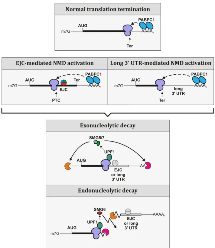
pathways see Figure 1.

(21)

9

Figure 1: NMD is able to activate two different degradation pathways: exonucleolytic and endonucleolytic.

(Top) During normal translation termination PABPC1 interacts with ribosome-bound proteins to initiate

translation termination. (Middle) During EJC- or long 3′-UTR-mediated NMD activation the terminating

ribosome is too far removed from poly(A)-bound PABPC1, which results in inefficient translation

termination and ultimately NMD activation. (Bottom) Both exonucleolytic and endonucleolytic decay

pathways are activated by UPF1 interacting with the terminating ribosome, followed by SMG5/7- or

SMG6-mediated degradation.

(22)

10 3.3 EJC-independent activation of NMD

Critical criteria for NMD activation due to an EJC downstream of a PTC have been discussed in the previous sections. However, adding to the complexity of the NMD mechanism in general is the fact that NMD can also be activated without an EJC downstream of a termination codon. It has been shown that a reporter carrying an intron downstream of a PTC was degraded via NMD even after knocking down the core EJC component eIF4A3. A second experiment in the same study showed that a reporter without an intron downstream of a PTC was unaffected by an eIF4A3 knockdown and still degraded via NMD (Buhler et al., 2006). These results showed that NMD can in fact be activated without an EJC downstream of a stop codon.

3.3.1 NMD activation due to a long 3′ UTR

It has been proposed that the length of a 3′ UTR can be responsible for activation of NMD independently of an EJC downstream of the termination codon (Singh et al., 2008).

Complementing these results are a set of reporter constructs harboring the 3′ UTRs of SMG5, UPF3b, or the heterologous GFP coding sequence as 3′ UTRs in separate triosephosphate isomerase (TPI) reporters for which active degradation via NMD has been shown (Boehm et al., 2014). The fact that the 3′ UTRs of the NMD factors SMG5 and UPF3b can elicit NMD indicates a self-regulatory mechanism of NMD. Furthermore, taking into account that a rather arbitrary sequence, in this case the heterologous GFP coding sequence, is actively targeted by NMD suggests that the mere length of a 3′ UTR is the determining factor for whether or not NMD is activated in the absence of an EJC downstream of a termination codon. In contrast, another study has recently shown that not all long 3′ UTRs are actually targeted for degradation via NMD (Toma et al., 2015).

This study showed that several reporters contained certain NMD-inhibiting cis elements

located within the first 200 nucleotides of the 3′ UTR. These cis elements were shown to

be A/U rich and were able to inhibit NMD in a positional dependent manner. This was

made evident by the fact that these 200 nucleotide A/U rich elements were able to

suppress NMD when inserted directly downstream of a termination codon of otherwise

NMD targeted reporters. Furthermore, there seems to be a second pathway of NMD

evasion of long 3′ UTR reporters. Another set of mRNAs showed no NMD inhibiting

effects of the first 600 nucleotides of a long 3′ UTR, however the full length 3′ UTR was

able to suppress NMD. Taken together these results suggest that there are at least two

pathways responsible for NMD inhibition of a long 3′ UTR.

(23)

11

As discussed earlier when describing the central NMD factor UPF1, the position of UPF1 on the 3′ UTR can also influence the fate of an mRNA. Since UPF1 occupation of a 3′ UTR can differ from transcript to transcript, it is feasible why some mRNAs are targeted by NMD whereas others are not. Taken together, these results are an indication that the mere length of a 3′ UTR is not the determining feature for NMD activation.

3.3.2 The faux 3′ UTR model in yeast

Another model predating the findings in the previous section is termed the faux 3′ UTR model in yeast (Amrani et al., 2004). This model is based on the fact that a transcript harboring a PTC inevitably contains an elongated 3′ UTR. The model states that with increasing length of the 3′ UTR the interaction between the poly(A)-binding protein (in yeast called Pab1) and the terminating ribosome is becoming less efficient. Due to this inefficient or completely absent interaction between the ribosome at the termination codon and Pab1, the translation termination event is believed to be compromised and as a result NMD is activated. Furthermore, tethering Pab1 directly downstream of a PTC so that Pab1 is upstream of the long 3′ UTR is able to antagonize NMD. This is believed to be because of the re-established translation termination due to proximity of Pab1 to the ribosome. Since yeast lack EJCs in general, it is believed that NMD targets in yeast are exclusively recognized based on 3′ UTR length (Muhlrad and Parker, 1999). This model was the first to implicate 3′ UTR length as a result of translation termination in NMD.

3.4 The link between translation and NMD 3.4.1 Translation initiation

To fully understand the link between NMD and translation, it is necessary to take a separate look at translation initiation as well as translation termination events.

Translation initiation is the step-wise process in which a ribosome is assembled on a mature mRNA in the cytoplasm in order to start translating the mRNA into protein.

Several eukaryotic initiation factors (eIFs) play a pivotal role in the complex process of translation initiation. The first step in translation initiation is the recognition of the 5′

m 7 G (methyl-7-guanosine) cap structure on the cytoplasmic mRNA by the eIF4F

complex (Jackson et al., 2010). The eIF4F complex is comprised of the cap-binding

protein eIF4E, the scaffolding protein eIF4G, and the DEAD-box helicase eIF4A. This

complex is essential for translation initiation as it is responsible for the recruitment of

(24)

12

the small ribosomal subunit (Sonenberg and Dever, 2003). Additionally, eIF4G is responsible for the interaction between the cap-bound eIF4F complex and the cytoplasmic poly(A)-binding protein that is bound to the 3′ poly(A)-tail of mRNAs (Imataka et al., 1998). This interaction between 5′-bound eIF4G and 3′-bound PABPC1 was shown to give rise to a circularized form of the mRNA, which is believed to facilitate recycling of terminating ribosomes as well as enhance translation initiation (Amrani et al., 2008; Kahvejian et al., 2005; Wells et al., 1998). Before the complete 80S ribosome can be assembled on the mRNA, the 43S pre-initiation complex (PIC) is assembled on the mRNA by interacting with the eIF4F complex. This 43S PIC consists of several eIFs and the 40S ribosomal subunit (Jackson et al., 2010). An important function of the helicase component eIF4A of the eIF4F complex is believed to be the scanning of the 5′ UTR in a 5′ to 3′ directionality. This action is hypothesized to clear the mRNA 5′ UTR of any unwanted secondary structures or bound proteins to free a space for the 43S PIC- binding to the mRNA (Pestova and Kolupaeva, 2002). Scanning of the 43S PIC commences until an AUG start codon is decoded by the initiator methionine tRNA. After recognition of the start codon, the remaining eIFs are released from the mRNA, the 60S ribosomal subunit is able to join the PIC to form the complete 80S ribosome, and the elongation process is starting (Jackson et al., 2010).

3.4.2 Proper translation termination

Protein synthesis does not only require translation initiation but also efficient translation termination. Translation termination ensures that ribosomes are being freed from the mRNA to start another round of translation as well as the proper release of a finished polypeptide chain. A simplified overview of translation termination can be seen in figure 2. The translating ribosome stops the active translation process when encountering a stop codon (UAA, UAG, and UGA) in the ribosomal A-site. The termination process requires two important proteins: eukaryotic release factor 1 (eRF1) and eukaryotic release factor 3 (eRF3). The eRF3 protein, a GTPase, exists as two paralogs called eRF3a and eRF3b in mammalian cells. It has been shown that in human cells eRF3a is the main factor implicated in translation termination (Chauvin et al., 2005;

Hoshino et al., 1998; Hoshino et al., 1989). The direct involvement of eRF3 in translation

termination has been shown by eRF3 being required for eRF1 stability in the cell

(Chauvin et al., 2005; Janzen and Geballe, 2004). Structurally, eRF1 mimics a tRNA and

can therefore decode stop codons by binding in the ribosomal A-site (Chavatte et al.,

(25)

13

2001; Frolova et al., 1999). After binding the ribosomal A-site, eRF1 promotes hydrolysis of the synthesized polypeptide chain via its GGQ motif. This process is supplemented by eRF3 as the hydrolytic activity of eRF1 is stimulated by GTP hydrolysis of eRF3. This stimulation is co-dependent, as eRF3 has an inherently low GTP hydrolyzing activity that is stimulated in turn by the formation of a quaternary complex consisting of GTP, eRF1, eRF3 and the ribosome (Alkalaeva et al., 2006; Frolova et al., 1996; Frolova et al., 1999; Salas-Marco and Bedwell, 2004; Song et al., 2000;

Zhouravleva et al., 1995). After the peptide is successfully released, the ribosome

remains bound to the mRNA. To free up the termination codon for any approaching

upstream ribosomes, the ribosome at the termination codon needs to be removed and

potentially recycled for another round of translation. The stalled ribosome is dissociated

from the mRNA by the ATP-binding cassette subunit family E member 1 (ABCE1)

ATPase. The process of ribosome splitting involves ATP hydrolysis by ABCE1. The exact

mechanism by which the ribosome is removed and recycled via ABCE1 is currently

unknown. However, several key steps have been elucidated. An important requirement

for ribosome splitting by ABCE1 is the interaction with eRF1 after eRF3 dissociates from

eRF1 (Pisarev et al., 2010). Additionally, ABCE1 is also known to interact with the

ribosome directly (Barthelme et al., 2011; Becker et al., 2012). The process of ribosome

removal and recycling is an important mechanism of translation termination as a whole,

as it ensures continuous use of the recycled ribosomes as well as an increase in mRNA

survivability. An additional factor important for translation termination is PABPC1,

which is known to interact with eIF4G (Safaee et al., 2012; Tarun and Sachs, 1996). As

mentioned above, this interaction mediates the closed loop formation of the mRNA,

which is believed to facilitate ribosome recycling and translation termination in general

by bringing the 5′ end and the 3′ end of the mRNA into close proximity (Amrani et al.,

2008; Kahvejian et al., 2005; Wells et al., 1998). Additionally, PABPC1 is also known to

be a potent interaction partner of eRF3. This interaction plays a vital role in translation

termination as it allows for efficient polypeptide release as well as ribosome recycling

(Hoshino et al., 1999; Kozlov and Gehring, 2010; Uchida et al., 2002).

(26)

14

Figure 2: Steps during normal translation termination.

(A) After translation initiation, the ribosomes translate along the ORF until it encounters a termination codon. PABPC1 interacts with eIF4G of the eIF4F complex resulting in a circularized mRNA. (B) Once the ribosome stalls at a termination codon it binds the two release factors eRF1 and eRF3a. Poly(A)-bound PABPC1 is able to interact with eRF3a bound to eRF1 at the ribosome, signaling a proper translation termination event. (C) As a result of a proper translation termination, the finished protein is released from the ribosome and the latter is removed from the mRNA, opening up the termination codon for any subsequent approaching ribosome.

3.4.3 Steps in aberrant translation termination and the link to NMD

Not all termination events are created equal and although some similarities between

normal and aberrant translation termination events exist, there are several significant

differences that can lead to the activation of NMD. The most important similarity is that

(27)

15

a PTC, just like a normal termination codon (NTC), enters the A-site of the ribosome.

However, the subsequent steps are different. An indication for a significant difference between ribosomes encountering a PTC and an NTC can be seen by ribosomal profiles.

Toeprinting assays have shown that there are no signals for ribosomes encountering an NTC unless eRF1 is removed. In stark contrast to this are the toeprinting signals generated for a PTC-carrying mRNA. In this case, typical signals for a stalled ribosome with an occupied A-site are shown independently of eRF1 (Amrani et al., 2004). An additional indication for an aberrant translation termination event is shown by toeprinting assays for the well-known NMD target β -globin NS39, which carries a PTC at amino acid position 39. In this case, toeprinting assays show signals for a stalled ribosome for β -globin NS39, whereas the signals for β -globin wildtype mRNA are normal (Peixeiro et al., 2012). All these results point towards errors during translation termination that ultimately result in the ribosomes being stuck on the mRNA. Without the removal of the ribosome from the mRNA, the degradation of the mRNA is initiated.

The exact mechanism by which this degradation is initiated is still unknown. However, a

current model suggests a possible mechanism that could be responsible for the

degradation of a PTC-carrying transcript (Singh et al., 2008). This model suggests that a

stalled ribosome at a PTC is still able to bind the release factors eRF1 and eRF3 in the

same manner as when a ribosome encounters an NTC. The major difference, however, is

in the interaction partner of eRF3. It has been shown that the central NMD factor UPF1 is

able to interact with eRF3, however, the interaction between eRF3 and PABPC1 is

normally favored (Kashima et al., 2006; Singh et al., 2008). The fact that UPF1 is able to

interact with eRF3, which plays a vital role in translation termination, suggests that

there might be a competition between PABPC1 and UPF1 for eRF3 binding. In case of a

PTC, just like the faux 3′ UTR model in yeast suggests, the ribosome is further removed

from the poly(A)-tail and therefore PABPC1. This could lead to an inefficient interaction

between PABPC1 and eRF3, which would allow for the less favored interaction between

3′ UTR-populated UPF1 and ribosome-bound eRF3 to occur more readily. In turn, this

would allow UPF1 to recruit other NMD factors and eventually initiate NMD. Supporting

this competition model is an experiment that shows when increasing amounts of

PABPC1 are added to an in vitro reaction, the interaction between UPF1 and eRF3 can be

prevented (Singh et al., 2008). Furthermore, reporter mRNAs carrying different PTCs at

various 5′ and 3′ positions show gradually less efficient NMD depending on the location

(28)

16

of the PTC (Eberle et al., 2008). Interestingly, in this set of data a PTC close to the start codon is able to evade NMD. This observation provides further evidence for the importance of the physical distance of PABPC1 from the terminating ribosome. In case of a PTC close to the start codon, the ribosome would stall close to the translation initiation site. Due to the closed loop form of the mRNA, PABPC1 would be in close proximity of the terminating ribosome due to its interaction with eIF4G. Furthermore, the discussed study uses a reporter with complementary RNA sequences in the 3′ UTR. On the one hand, the complementary sequences are positioned close to the PTC and on the other just upstream of the poly(A)-tail. Due to the complementary sequences interacting with each other, the 3′ UTR folds back the poly(A)-tail. This again brings PABPC1 bound to the poly(A)-tail into close proximity of the terminating ribosome and subsequent interaction with eRF3 can signal a normal translation termination event (Eberle et al., 2008).

These results show a possible model of NMD activation of a PTC by physically enlarging the distance between the terminating ribosome and poly(A)-bound PABPC1. Further research has to be performed to illuminate additional factors and intricacies of this mechanism.

3.4.4 Active translation is necessary for NMD

Early models of NMD stated that NMD was limited to the first or pioneer round of translation. This was believed to be the case because mRNA with cap-binding protein 80 (CBP80) attached was targeted by NMD (Ishigaki et al., 2001). CBP80 is part of the nuclear cap binding complex that is added during transcription and stays on the mRNA until after the mRNA is exported into the cytoplasm and replaced by the eIF4F complex (Izaurralde et al., 1994; Lewis and Izaurralde, 1997; Sonenberg and Dever, 2003). Later studies refuted the notion that NMD is limited to the pioneer round of translation as it was shown, that NMD is also able to degrade mRNAs bound by eIF4E, which is part of the eIF4F complex (Durand and Lykke-Andersen, 2013; Rufener and Muhlemann, 2013).

Furthermore, it has been shown that active translation is mandatory for the activation of

NMD. Several experiments have highlighted this requirement. For example, structures in

the 5′ UTR of an mRNA that are able to block ribosomal translation have been implicated

in NMD inhibition (Belgrader et al., 1993). Another set of experiments established that

protein synthesis inhibitors like cycloheximide, anisomycin, emetine, pactamycin, and

puromycin are able to efficiently block NMD activation of a T-cell receptor mRNA

(29)

17

harboring a PTC (Carter et al., 1995). These examples highlight the need for active translation in order for NMD to be activated on an NMD-targeted mRNA.

3.5 Additional NMD targets present in the cell

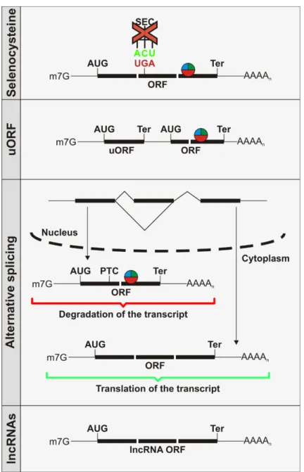
There are several other classes of NMD substrates in the cell (Figure 3). One example are mRNAs for so called selenoproteins. Selenoproteins are a class of polypeptides that incorporate the unusual amino acid selenocysteine (Sec). The reason why selenoprotein coding transcripts can be targeted by NMD is because the UAG stop codon triplet can under specific circumstances also encode for Sec. Previous work showed that the stability of selenoprotein transcripts rely on the selenium concentration in the cell.

Under low selenium conditions the transcript can be degraded via NMD since not enough Sec is present in the cell to be incorporated into the polypeptide. As a result the UAG codon is treated as a premature termination codon (Bermano et al., 1995; Berry et al., 1993; Hadley and Sunde, 2001; Lee et al., 1989).

However, selenoprotein-coding mRNAs are not the only additional NMD target.

Furthermore, transcripts containing an upstream open reading frame (uORF) can be targeted by NMD. A transcript harboring a uORF still contains a regular ORF downstream of the uORF. Since the uORF contains a stop codon, there will still be EJCs present in the regular ORF as the ribosome cannot translate the ORF. In this case, a uORF-containing transcript will be targeted and degraded by the NMD machinery (Mendell et al., 2004). Interestingly however, not all uORF transcript are targeted by NMD (Stockklausner et al., 2006). This indicates that even though a transcript harboring a uORF can be targeted by NMD, a uORF cannot generally be considered as an NMD activator.

Additionally, certain genes can regulate their own expression levels by means of alternative splicing (Sureau et al., 2001). Alternative splicing leads to different mRNA transcript isoforms depending on which splice sites are utilized (Brett et al., 2000).

Alternatively spliced pre-mRNAs can harbor a PTC, for example due to intron inclusion,

which would result in the degradation of the transcript via NMD (Lewis et al., 2003).

(30)

18

Figure 3: Additional NMD substrates in the cell.

Last but not least, long non-coding RNAs (lncRNAs) can be targeted by NMD as well. As discussed earlier, NMD requires active translation to degrade mRNAs. However, lncRNAs are believed to be untranslated as the name non-coding already implies.

Therefore, it was surprising to see that the lncRNA growth arrest factor 5 (GAS5) was upregulated under UPF1 knockdown conditions (Tani et al., 2013). Later studies showed, however, that lncRNAs can actually be translated contrary to what was earlier believed (Chew et al., 2013). This could explain why some lncRNAs are targeted by NMD.

These examples listed here show that NMD is not only a quality control mechanism in

the cell but the role of NMD is more complex and broader than simply quality control.

(31)

19 3.6 Physiological implications of NMD

3.6.1 Importance of NMD factors on cell viability

NMD factors do not only play a vital role in the degradation of faulty mRNA transcripts but several factors also seem to be important for cell viability. It has been shown that multiple NMD factors are essential for embryonic development in mice (Hwang and Maquat, 2011). For example, knocking out the central NMD factor Upf1 in mice causes failure of embryonic development in early stages. After uterine implantation, mouse embryos lacking Upf1 die due to apoptosis. Additionally, homozygous deletion of Upf1 in ES cell lines could not be established and Upf1 null cells have never been observed (Medghalchi et al., 2001). Besides Upf1, Upf2 and Smg1 have also been implicated in cell survivability in mice. Upf2 deficient mice die in utero between embryonic day 3.5 and embryonic day 7.5, whereas Smg1 deficient mice die before embryonic day 12 (McIlwain et al., 2010; Weischenfeldt et al., 2008). Smg6 has also recently been shown to play a role in early development as Smg6 knockout mice have displayed embryonic lethality at the blastocyst stage (Li et al., 2015).

These results show the important functions of several NMD factors besides their role in mRNA degradation during development and on cell viability. This makes studying the function of individual NMD factors difficult as many carry a role in cell survivability and knockout experiments are simply not viable. Using inducible systems where the NMD factors could be depleted at a later stage could help study the function of these proteins.

3.6.2 NMD and genetic disorders

Many inherited genetic disorders as well as cancers are caused by nonsense or frameshift mutations that lead to PTC-carrying transcripts. Over 2400 genetic disorders result from a nonsense mutation. Furthermore, a recent study suggests that about 11%

of human genetic disorders are due to nonsense mutations (Culbertson, 1999; Mort et

al., 2008; Peltz et al., 2013). NMD is able to modify clinical phenotypes by degrading

PTC-containing transcripts. This ensures that the cell does not express any C-terminally

truncated proteins with potential dominant negative effects. One example showcasing

this protective side of NMD can be seen in erythrocytes. Individuals suffering from the

blood disorder called β -thalassemia have in many cases nonsense mutations in the first

or second exon of the β -globin gene. These mutations result in a PTC in the β -globin

mRNA transcript, which is recognized and therefore degraded by NMD. In heterozygous

(32)

20

individuals with one functional allele of the β -globin gene, the levels of truncated protein are kept very low by NMD and enough functional protein is synthesized from the wild type allele, ensuring that the individuals remain symptom free. Nonsense mutations in the final exon of β -globin are however not recognized by NMD as they lack an EJC downstream of the PTC. As a result, the transcript evades NMD and heterozygous individuals present an intermediate from of the disease called thalassemia intermedia (Hall and Thein, 1994).

Although NMD can certainly be beneficial as seen in the example of β -thalassemia, there are physiological conditions that are worsened by NMD. NMD is not beneficial in cases where a truncated version of the protein would lead to a less severe medical condition of a genetic disorder. A specific example would be Duchenne muscular dystrophy (DMD). A nonsense mutation in the dystrophin gene leads to a PTC-carrying transcript which activates NMD. This leads to a severe form of DMD. However, there are other known mutations in the dystrophin gene that evade NMD and produce a C-terminally truncated version of the protein. This C-terminally truncated protein is known to be partially functional and results in a less severe form of DMD called Becker muscular dystrophy (BMD) (Kerr et al., 2001). These examples show that although NMD has a protective function in the cell, it is not always favorable to activate NMD. In some cases it would be in the patients’ interests to subdue NMD in order to increase levels of partially functional C-terminally truncated proteins.

3.6.3 Treatment of physiological conditions resulting from NMD

As described above it can be beneficial to deactivate NMD for certain transcripts to

lessen clinical manifestations of a disease. There are two ways in which NMD can either

directly or indirectly lead to a more severe form of a genetic disorder: directly, by

degrading a transcript leading to an otherwise beneficial truncated protein, or indirectly

by transcripts that escape NMD where a dominant negative effect protein will be

expressed and active NMD would be beneficial. It would be of interest to generally

stabilize a transcript that is targeted by NMD but would produce a favorable medical

condition if it escaped NMD. Since blocking the NMD machinery in general could do

more harm than good as many dominant-negative proteins could hypothetically be

expressed, a potential read-through approach at PTCs is the more favorable approach

(Kerem, 2004). Administering a drug that would potentially lead to read-through at a

certain PTC could theoretically be used as treatment (Peltz et al., 2013). At first,

(33)

21

aminoglycosides were used to achieve translational read-through. In patients with cystic fibrosis or DMD the use of aminoglycosides was shown to be successful in increasing the respective protein levels. However, the aminoglycosides administered had severe side effects in patients such as renal failure, which limits the usage of aminoglycosides in humans (Kovesi et al., 1998). An alternative drug instead of aminoglycosides was proposed with the drug Ataluren. It has been proposed that Ataluren only selectively promotes read-through and does not affect translation termination at normal stop codons (Welch et al., 2007). The mechanism by which Ataluren acts has recently been challenged and the future of this drug remains to be seen as more clinical trials need to be carried out (Auld et al., 2009; McElroy et al., 2013). Generally, a treatment where specific NMD-targeted transcripts could be altered in a way that they evade NMD, coupled with efficient read-through drugs with minimal side effects could prove to be a favorable approach to treat genetic disorders caused by nonsense mutations.

3.7 Aim of this thesis

Nonsense-mediated mRNA decay plays a pivotal role in mRNA surveillance by targeting aberrant mRNA transcripts. Furthermore, NMD is also able to regulate endogenous mRNAs of non-aberrant nature. The factors involved in NMD as well as the mechanism of activation and the degradation pathways have been subjected to many studies. In contrast, the mechanism by which certain transcripts are able to escape NMD and important proteins involved in this process remain elusive.

One protein that was previously shown to be able to suppress NMD is PABPC1. In this thesis I aim to elucidate the role of PABPC1 during NMD suppression. I further aim to characterize potential important and necessary interaction partners of PABPC1 involved in the suppression of NMD. After identifying other proteins and interactors of PABPC1, I aim to shed light on the potential mechanism by which PABPC1 is able to antagonize NMD.

After elucidating the potential pathway, I further explore several different methods of

suppressing NMD by recruiting PABPC1 to the correct position of an NMD-targeted

substrate. Since NMD is implicated in regulating the clinical phenotype of many diseases

I try to modulate disease associated transcripts with PABPC1 to discuss a potential

outlook of NMD suppression via PABPC1 in a medical environment.

(34)

22

Combining these results I try to elucidate a part of the NMD suppression mechanism and

factors involved as well as show multiple routes to achieve the antagonizing NMD effect.

(35)

23

4 Results

NMD as an mRNA quality control mechanism targets a multitude of transcripts in the organism. Two of the main NMD target classes are degraded either due to an EJC downstream of a terminating ribosome or due to characteristics of certain 3′ UTRs. The mechanism by which some NMD targets are able to evade NMD is currently not fully understood. It is known that NMD of certain transcripts can be suppressed by PABPC1.

Here, I aim to elucidate the role of PABPC1 in NMD suppression and propose a mechanism by which PABPC1 achieves this.

4.1 PABPC1 is able to antagonize long 3′ UTR-mediated NMD

PABPC1 plays an essential role in gene expression. It is required for mRNA circularization, thereby facilitating ribosome recycling, and for suppressing NMD at normal translation termination codons (Behm-Ansmant et al., 2007; Wells et al., 1998).

Tandem tethering assays have previously shown that PABPC1 is able to suppress NMD

when tethered upstream of an NMD-activating factor (Ivanov et al., 2008). However, the

mechanism by which PABPC1 is able to antagonize NMD is not well understood. In light

of this, I started investigating NMD suppression of long 3′ UTR-mediated NMD targets. It

has previously been shown that the mRNA encoding for the NMD factor SMG5

undergoes NMD due to its 3′ UTR (Singh et al., 2008). To study how PABPC1 can

suppress NMD I used a triosephosphate-isomerase (TPI) reporter construct harboring

the first 1037 nucleotides of the 3′ UTR of SMG5 downstream of 4MS2 binding sites,

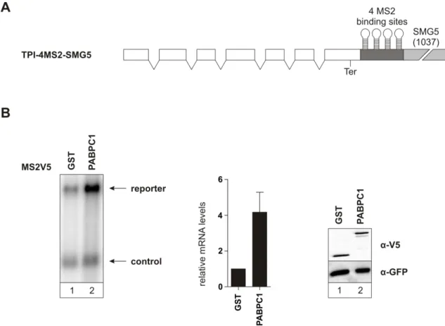
which are located directly downstream of the stop codon (Figure 4A). This reporter has

been shown to be degraded due to the SMG5 3′ UTR (Boehm et al., 2014). Co-expressing

and thereby tethering MS2V5-tagged PABPC1 to the reporter increased the steady-state

mRNA levels by a factor of about four compared to the negative control MS2V5-GST

(Figure 4B). The stabilization seen in this experiment suggests that PABPC1 is able to

antagonize NMD induced by a long 3′ UTR.

(36)

24

Figure 4: PABPC1 stabilizes an NMD-targeted long 3′ UTR-containing reporter mRNA

(A) Schematic representation of the TPI reporter carrying 4MS2-binding sites upstream of a shortened version of the SMG5 3′ UTR. The TPI ORF is represented as white boxes and introns are shown as lines connecting the boxes. (B) Northern blot of total RNA extracted from HeLa cells showing the stabilizing effect of MS2V5-PABPC1 when tethered to the reporter mRNA (left). Quantification of the northern blot with standard deviation represented as black bars (middle). Protein expression was detected by western blotting with anti-V5 and anti-GFP antibody (right).

4.1.1 Increased reporter abundance is a consequence of PABPC1-mediated NMD suppression

As mentioned in the introduction, PABPC1 is known to have a positive effect on general

mRNA stability. I made use of two control reporters (Figure 5) to exclude the possibility

that the stabilization effect observed when PABPC1 is tethered to the NMD reporter is

not a general stabilization effect but instead is a consequence of NMD suppression. To

this end, I used a reporter for which the NTC was mutated and a new stop codon appears

downstream of the 4MS2 sites (Figure 5). This reporter is called TPI- ∆ ter-4MS2-SMG5. A

lack of reporter stabilization was expected since the translating ribosome should

displace the tethered PABPC1. Furthermore, the other reporter carried the 4MS2

tethering sites downstream of the SMG5 3′ UTR instead of upstream and was called TPI-

(37)

25

SMG5-4MS2 (Figure 5). Tethering PABPC1 to this reporter should fail to stabilize the transcript as PABPC1 is too far removed from the terminating ribosome.

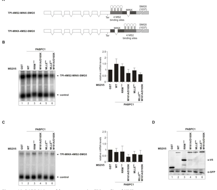
Figure 5: Schematic representation of TPI control reporters

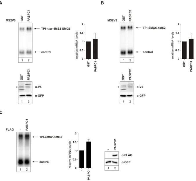
MS2V5-tagged PABPC1 tethered to the TPI- ∆ ter-4MS2-SMG5 reporter failed to

antagonize NMD (Figure 6A). The same held true when MS2V5-PABPC1 was tethered to

the reporter carrying the 4MS2 sites downstream of the SMG5 3′ UTR (Figure 6B). In

both cases the reporter mRNA abundance was unchanged compared to the negative

control. I performed an additional experiment to exclude any trans effects of PABPC1 on

the initially used reporter construct. To this end, I co-expressed FLAG-tagged PABPC1

with the TPI-4MS2-SMG5 reporter (Figure 6C). In this scenario FLAG-PABPC1 should

not be directly tethered to the reporter as it carried a FLAG-tag and not an MS2V5-tag

and should therefore fail to stabilize the reporter. The results of the northern blot

showed that PABPC1 was unable to suppress NMD of the reporter in trans and only

caused a slight increase in reporter levels (Figure 6C). This slight increase of about 1.5

fold was negligible compared to MS2V5-PABPC1 tethered to the same reporter

construct. Western blots for all three experiments showed that PABPC1 is expressed and

therefore excluded the lack of PABPC1 expression as the reason for failing to suppress

NMD (Figure 6). Combining the results obtained so far shows that PABPC1 is able to

antagonize NMD activated by a long 3′ UTR specifically when tethered downstream and

in close proximity of terminating ribosomes.

(38)

26

Figure 6: PABPC1 stabilization of a long 3′ UTR-carrying NMD reporter is position and tethering specific

(A) Northern blot showing the stabilizing effect of MS2V5-PABPC1 tethered to the reporter mRNA.

Quantification of the northern blot with standard deviation is represented with black bars. Protein

expression was detected with anti-V5 and anti-GFP antibodies (Asterisk represents unspecific bands). (B)

Northern blotting result of tethering MS2V5-PABPC1 to the TPI-SMG5-4MS2 reporter. Quantification and

standard deviation is shown with black bars. Protein expression was detected with anti-V5 and anti-GFP

antibody. (C) Northern blot showing trans effects of co-expressed FLAG-PABPC1 alongside the TPI-4MS2-

SMG5 reporter. Quantification and standard deviation are represented with black bars. Protein expression

was detected using anti-FLAG and anti-GFP antibodies (Asterisk represents unspecific bands).

(39)

27

4.2 Interaction partners of PABPC1 necessary for NMD suppression

4.2.1 The interaction between PABPC1 and eRF3a is not required for NMD suppression

Current data suggest that PABPC1 competes with the central NMD factor UPF1 for eRF3a binding (Singh et al., 2008). It is suggested that UPF1 outcompetes PABPC1 for eRF3a binding when a ribosome is stalling at a termination codon upstream of a long 3′

UTR. This is speculated to be due to the large physical distance between poly(A)-tail bound PABPC1 and ribosome bound eRF3a. To elucidate which domains and interaction partners of PABPC1 play a role in the NMD suppression effect, I created several PABPC1 mutants (Figure 7). It is known that PABPC1 interacts with the PAM2 motifs of eRF3a via its own C-terminal domain called MLLE (Mademoiselle) (Kozlov and Gehring, 2010).

Therefore, I first made use of two PABPC1 mutants: PABPC1 MLLE Mut , which carries a mutation of the MLLE motif to GAAR, and the PABPC1 RRM1234 mutant, which lacks the entire C-terminal region (Figure 7).

Figure 7: Schematic representation of the PABPC1 mutants used in this work

(40)

28

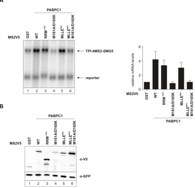
Surprisingly, tethering these two PABPC1 mutants to the TPI-4MS2-SMG5 reporter mRNA still led to an increase of about 3.5-fold in mRNA abundance (Figure 8A, lanes 3 and 5). Next, I tested two additional PABPC1 mutants with impaired PAM2 interaction.

The first mutant contained two additional point mutations (E564R and F567A) that are also known to play a role in PAM2 interaction (Kozlov et al., 2004).

Figure 8: Interaction between PABPC1 and eIF4G necessary to suppress NMD

(A) Northern blot showing the stabilizing effect of the MS2V5-PABPC1 mutants when tethered to the reporter mRNA. Quantification of the northern blot with standard deviation is represented with black bars. (B) Protein expression was detected by western blotting with anti-V5 and anti-GFP antibodies.

This mutant was termed PABPC1 MLLE Mut2 and the additional two point mutations

ensure that the lack of NMD suppression is not due to any residual binding between

(41)

29

PABPC1 and eRF3a due to the lack of these two additional mutations in PABPC1 MLLE Mut (Figure 7). The second additional mutant termed PABPC1 1-496 is similar to PABPC1 RRM 1234 , but carries extra C-terminal amino acids (Figure 7). This mutant was used to show that only the lack of the C-terminal region containing the MLLE domain is implicated in the lack of NMD suppression and not the linker region.

Figure 9: Additional PABPC1 mutants show that the interaction with eRF3a is dispensable for NMD suppression

(A) Northern blot showing stabilization effect of additional PABPC1 mutants tethered to the reporter mRNA. Quantification and standard deviation is shown with black bars. (B) Protein expression was detected with anti-V5 and anti-GFP antibodies.

Tethering these mutants to the SMG5 3′ UTR reporter also led to an increase in reporter

mRNA abundance similar to PABPC1 wild-type tethering (Figure 9A). Western blots

confirmed the expression of the tethered proteins (Figure 8B and figure 9B). These

Abbildung

Figure  1:  NMD is able to activate two different degradation pathways: exonucleolytic and  endonucleolytic
Figure 2: Steps during normal translation termination.
Figure 3: Additional NMD substrates in the cell.
Figure 4: PABPC1 stabilizes an NMD-targeted long 3′ UTR-containing reporter mRNA
+7

Referenzen

ÄHNLICHE DOKUMENTE

We want to discuss the origin of the BRST symmetry in a more general context, and show that, by quoting Zinn-Justin, the ”Slavnov Taylor identities in gauge theories owe less to

3 Im Jahr 2010 werden die Leute nicht mehr rauchen dürfen.. In 2010 people will not be allowed to

The effect of indomethacin treatment on prema- ture labor was evaluated by cessation of uterine contractions and the period of prolongation of gestation.. Thirty minutes

This exercise sheet aims to assess your progress and to explicitly work out more details of some of the results proposed in the previous lectures. Please, hand in your solutions

Sehen Sie einen Zusammenhang mit der

Fachbereich Mathematik und

Adjoint functors between module categories are described by a tensor and a Hom functor and the properties derived from the categorical setting are explained in Section 3.. Algebras

der Universit at M unchen Set