• Keine Ergebnisse gefunden

Characterization of an olfactory receptor mediating aversive behaviour to a death-associated odour

N/A
N/A
Protected

Academic year: 2021

Aktie "Characterization of an olfactory receptor mediating aversive behaviour to a death-associated odour"

Copied!
115
0
0

Wird geladen.... (Jetzt Volltext ansehen)

Volltext

(1)

Characterization of an olfactory

receptor mediating aversive behaviour to a death-associated odour

Inaugural-Dissertation

zur

Erlangung des Doktorgrades

der Mathematisch-Naturwissenschaftlichen Fakultät der Universität zu Köln

vorgelegt von

Venkatesh Krishna Subramanian

aus Modinagar, Indien

Köln 2017

(2)
(3)

Berichterstatter: Prof. Dr. Sigrun I. Korsching PD Dr. Joachim Schmidt

Prüfungsvorsitzender: Prof. Dr. Peter Kloppenburg

Tag der mündlichen Prüfung: April 2016

(4)
(5)

TABLE OF CONTENTS

ACKNOWLEDGEMENT ... 1

ZUSAMMENFASSUNG ... 3

ABSTRACT ... 5

ABBREVIATIONS ... 7

List of Figures ... 9

Introduction ... 11

The biology of olfaction ... 11

The Olfactory System ... 12

Signal transduction events mediating olfaction ... 13

Olfactory sensory neurons ... 14

Olfactory receptor gene family ... 16

Amines as ligands ... 18

Trace Amine-Associated Receptor Subfamily 13 ... 20

TALEN and CRISPR: recent improvements for knockout in zebrafish ... 22

Zinc-finger nucleases ... 23

The Talen approach to genetic engineering ... 24

Clustered regularly interspaced short palindromic repeats/CRISPR- associated 9: ... 25

AIM OF THIS STUDY ... 27

Results ... 28

2.1 Preparation, Purification and characterisation of TAAR13c antibody . 28

(6)

2.2 The purified antibody labels a sparse population of cells in the olfactory epithelium ... 30 2.3 Co-labelling with acetylated Tubulin and G

olf

shows expression of TAAR13c in ciliated neurons ... 31 2.4 Suitability of several neuronal activity markers to show activation of TAAR13c with cadaverine ... 36 2.5 Egr1 as immediate early gene neuronal activity marker, Whole mount in situ on Zebrafish larvae ... 37 2.6 pERK as a neuronal activity marker for TAAR13c activation due to cadaverine. ... 38 2.7 Diamine assay using pERK as neuronal activity marker ... 40 2.8 Search for the TAAR glomerulus with the TAAR13c antibody ... 42 2.8.1 Olfactory Bulb stained with glomerular marker SV2 and co-labelled with TAAR13c antibody ... 42 2.8.2 Attempts to increase sensitivity of the immunological detection of TAAR13c via cadaverine stimulation and different fixation techniques 44 2.9 Ontogenetic onset of avoidance behaviour towards cadaverine ... 45

2.9.1 Observation of behaviour upon sequential increase of cadaverine concentration from 0 mM as control until 300mm threshold ... 48 2.9.2 Method development and optimization for behaviour experiments49 2.11 Attempted knockout of TAAR13c gene using the CRISPR/Cas

method ... 53

2.11.1 Generation of CRISPR gRNAs and CAS9 capped mRNA ... 53

2.11.2 Sequencing revealed high number of background SNP’s resulting

from the amplification of different TAAR13 subfamily genes ... 54

2.11.3 Initiation of TAAR13 gene family cluster knock out ... 61

DISCUSSION ... 63

(7)

Molecular characterization of TAAR13c as an olfactory receptor ... 63

Ontogenetic onset of avoidance behaviour towards cadaverine ... 69

Mutagenesis of TAAR13c using CRISPR-CAS9 ... 71

Key Findings and conclusions ... 74

Materials and Methods ... 76

Animal Strains, Breeding and Maintenance ... 76

Plastic ware ... 76

Chemicals, Enzymes, oligos and Kits ... 77

Reagents and Solutions ... 77

Laboratory equipment ... 78

Bacterial Strains ... 78

Dissection ... 78

Tissue Lysis and protein estimation ... 79

Western Blotting... 79

Signal detection ... 80

Immunohistochemistry ... 80

Preparation of the whole mount zebrafish larvae for in-situ hybridization 81 Genome editing using TALEN ... 82

mRNA transcription in vitro ... 83

Genome editing using CRISP-CAS9 ... 83

Vectors ... 83

Production of Cas9 mRNA ... 83

Production of TAAR13c specific guide RNA ... 84

Microinjection of CRISPR/Cas9 vectors and screening for genomic

alterations ... 86

(8)

T7 Endonuclease I assay ... 86

Subcloning and Sequencing of T7 Endonuclease I assay-samples ... 87

Primers and target sites used in TALEN and Crispr-Cas mediated mutations ... 87

REFERENCES ... 90

APPENDIX ... 104

ERKLÄRUNG ... 105

Lebenslauf ... 107

(9)

1

ACKNOWLEDGEMENT

It gives me great pleasure to acknowledge and thank the people who were part of my Ph.D journey which I started in 2012. Firstly I would like to thank Prof.Dr. Sigrun Korsching who gave me the opportunity to pursue my Ph.D in her Neurobiology lab of at the Institute for Genetics, University of Cologne, Germany. Her mentorship, guidance and criticism helped me develop my scientific acumen. Working under her was a learning experience in scientific writing, presentation and communication, the three most important things I needed improvement upon. I would like to thank her especially for not giving up on me during a tumultuous phase of my Ph.D.

I extend my sincere gratitude to PD Dr. Joachim Schmidt for accepting to be my second referee. I thank Prof.Dr. Peter Kloppenburg for accepting to chair my defense.

I would like to thank AG Hammerschmidt for sharing the Loligotrack software and valuable inputs in Zebrafish behaviour tracking. I would like to thank Dr. Hans-Martin Pogoda for his inputs and suggestions regarding TALEN and Immunostaining experiments.

I extend my gratitude to Kathy Joergens from the graduate school for biological sciences for her cooperation in visa and contract related issues. I also thank Dr. Isabell Witt for her timely help and facilitating my move from contract to the graduate school for biological sciences.

My parents Mr.K.Subramanian and Mrs.Parvathi Manian are the most

important people in my life to whom any amount of gratitude would be

insufficient. I thank them for being the moral and motivational support

throughout my life. I am greatly indebted to my brother Dr.Santosh Krishna

for being a pillar of strength and bearing my worst outbursts at times of

stress. His constant guidance and constructive criticism shaped my Ph.D and

taught me several aspects of research life. I also extend my heartfelt gratitude

(10)

2

to my Bhabi Mrs.Kavita Kawathekar Krishna for her constant support and motivation during times of stress and sharing many moments of joy. I also thank my sweet niece Swara Krishna for her love towards her Chachu, Her chachu will always love her evermore.

I extend my heartfelt gratitude to Hemal Bhasin for her support and belief in me. She was an amazing friend who stood by me in times of stress and motivated me.

I would like to thank my close friends Vimal Rawat, Deepak Bhandari, Daniel Kowatschew, Aathmaja Rengarajan, Rohan Raut and many other friends for interesting conversations and fun moments spent with them.

I thank my international labmates Ivan Ivandic, Milan Dieris, Vladimir Shiryagin Gaurav Ahuja Adnan Syed Kanika Sharma Shahrzad Bozorgnia for being nice colleagues. Their cultural diversity and unique personalities gave me a wonderful learning experience.

I thank our lab manager Mr. Mehmet Saltuerk for taking good care of the zebrafish and help in some experiments and for the wonderful conversations on diverse topics.

Last but not the least I thank the people of Cologne for their warm and

gracious hospitality.

(11)

3

ZUSAMMENFASSUNG

Der Riechsinn beeinflusst das Verhalten in der gesamten Tierwelt und ist oftmals ein unverzichtbarer Sinn der für Organismen, falls nicht vorhanden, auch lebensbedrohlich sein kann. Geruchsstoffe werden im Nasenepithel von Riechepithelzellen, den sogenannten Rezeptor Neuronen erkannt. Diese Rezeptorneuronen exprimieren G-Protein gekoppelte Rezeptoren, zu welchem auch die TAARs (Trace Amine Associated Receptors) gehören.

TAARs sind oftmals in der Regulierung und Steuerung von Sozialverhalten involviert. Die vorliegende Arbeit handelt über die molekulare Charakterisierung von TAAR13c, einem Mitglied der TAAR Familie.

Kadaverin ist chemisch gesehen ein di-Amin und entsteht bei der bakteriellen dekarboxylierung von Lysin, meist in verrottendem Gewebe. Es ist bekannt das Kadaverin der Primärligand für den TAAR13c Rezeptor ist und Verhaltensversuche im Zebrabärbling haben gezeigt, dass es wenn vorhanden zum Aversen Verhalten führt. In der vorliegenden Arbeit wurde herausgefunden, dass TAAR13c im Nasenepithel des Zebrabärblings von zillierten Rezeptorneuronen exprimiert wird. Doppelmarkierungs- Experimente mit einem neuronalen Aktivitätsmarker (cFos) haben gezeigt, dass Kadaverin nach Applikation, eine Vielzahl von Neuronen aktiviert, mitunter auch TAAR13 positive Neuronen. Ein anderer Aktivitätsmarker, Egr1 stellte sich auch als nicht selektiv heraus. Histologische Färbungen mit einem dritten Neuronalen Aktivitätsmarker namens pERK, der nicht von der Genexpression abhängt sondern über Phosphorylierung reguliert wird und somit viel schneller ist, zeigte spärliche neuronale Aktivität nach der Stimulation mit Kadaverin und anderen di-Aminen.

Doppelmarkierungsexperimente mit einem TAAR13c Antikörper und pERK

nach einer Kadaverinstimulation zeigte eine Kolokalisierung auf zellulärer

Ebene. Der ontogenetische Beginn von aversivem Verhalten des larvalen

Zebrabärblings wurde anhand eines Präferenz-Apparats getestet und es wurde

(12)

4

ein Trend zum Aversen Verhalten im larvalen Zebrabärbling festgestellt. Es

wurden Versuche unternommen mit der TALEN und der CRISPR/Cas9

Methode das TAAR13c Gen auszuschalten, leider führte dies nicht zum

Erfolg aufgrund von fehlenden Überprüfungsmethoden. Die sehr große

Sequenzhomologie zwischen den Subtypen der TAAR Gene und eine sehr

große A-T region machten es unmöglich eine spezifische

Überprüfungsmethode zu etablieren. Durch eine Optimierung der

Überprüfungsmethode wurden schliesslich zwei putative TAAR13c

Mutanten hergestellt. Letzendlich sollten neue Mutagenese Konstrukte gegen

die TAAR sub- Familie es ermöglichen eine funktionierende TAAR Mutante

im Zebrabärbling zu erstellen.

(13)

5

ABSTRACT

Olfaction or the sense of smell is a strong driver of behavior in many animals and is important for their survival. Odors are perceived through a complex molecular recognition process which involves detection of odorants by odorant receptors in olfactory sensory neurons located in the nasal olfactory epithelium. The odorant receptors belong to the G protein–coupled receptors (GPCRs) class of proteins, which includes the trace amine–associated receptors (TAARs), a class of GPCRs associated with the detection of social cues. The present study focused on molecular characterization of a TAAR receptor, TAAR13c, in zebrafish. Cadaverine, a diamine produced by bacterial decarboxylation of lysine, and thus associated with odor emanating from decaying flesh was reported to be the primary ligand for TAAR13c.

Aversion to cadaverine was observed in adult Zebrafish by behavior experiments.

Here, the cell type expressing TAAR13c receptors was identified as ciliated OSNs. Double-labeling of TAAR13c-expressing neurons with cFos as neuronal activity marker after stimulation with cadaverine showed widespread expression of cFos in many cells, a few of which were also TAAR13c-positive. Another activity marker tested, Egr1, was also found to be nonselective. However, pERK, an activity marker, which does not rely on gene expression, but on phosphorylation and is therefore much faster, showed sparse cells activated after stimulation with cadaverine and a series of other diamines with different chain length. Double-labeling with pERK antibody and TAAR13c antibody showed cellular co-localization of pERK and TAAR13c signals upon cadaverine stimulus.

Ontogenetic onset of avoidance behavior was studied using a two channel

choice apparatus in Zebrafish larvae. A trend towards avoidance of

cadaverine was observed from the larval behavior experiments.

(14)

6

Knockout of TAAR13c was attempted using TALEN and Crispr-Cas9

mediated mutagenesis. Unfortunately it was not possible to design

completely specific screening strategies due to high sequence identity within

the five members of the TAAR13 subfamily and the AT-rich coding region

of TAAR13c. Two putative knockout mutations were obtained with

optimized screening methods. Finally, the design of new constructs for a

TAAR13 subfamily knockout may provide a useful tool in the future for

creating a functional Taar13 knockout in zebrafish.

(15)

7

ABBREVIATIONS

CRISPR Clustered Regularly Interspaced Short Palindromic Repeats DAPI 4',6-diamidino-2-phenylindole

dpf days post fertilization

GPCRs G-protein coupled receptors gRNA single guide RNA

IEG Immediate early gene IHC Immunohistochemistry OE Olfactory epithelium OR Olfactory receptors

OSN Olfactory sensory neurons

pERK phosphorylated Extracellular Signal-Regulated Protein Kinase SV2 synaptic vesicle glycoprotein 2

TAARs Trace Amine-Associated Receptors TALEN Transcription activator-like effector nuclease TCA Trichloroaceticacid

V1Rs Vomeronasal Receptors Family Type1

V2Rs Vomeronasal Receptors Family Type2

(16)

8

(17)

9

LIST OF FIGURES

Figure 1. The olfactory system ... 13 Figure 2.The Schematic representation of cAMP second messenger cascade of olfactory sensory neurons. ... 14 Figure 3. A summary of TAAR ligands, their expression patterns, and

behavioral roles ... 20 Figure 4. The positions of five TAAR13 genes as it appears in ENSEMBL browser ... 21 Figure 5. Schematic representation of various genome-editing platforms .... 26 Figure 6. Purification and characterization of TAAR13c antibody ... 29 Figure 7. Immunohistochemical detection of TAAR13c in ciliated neurons of the olfactory epithelium of adult zebrafish ... 31 Figure 8. Double Immunohistochemistry on Olfactory Epithelium of Adult Zebrafish ... 32 Figure 9. G

olf

labelling of adult zebrafish olfactory epithelium ... 33 Figure 10. TAAR13c Inhibitor peptide assay at 0.5 molar peptide excess &

co-labeling with microvillous neuron marker Calretinin as control ... 35

Figure 11. c-Fos and TAAR13c Co-localization experiment ... 37

Figure 12. Wholemount in situ on zebrafish larvae using the Egr1 probe... 38

Figure 13. The neuronal activity marker pERK labels TAAR13c-expressing

cells in adult zebrafish OE ... 40

Figure 14. Diamines elicit pERK increase in olfactory sensory neurons ... 42

Figure 15. Whole mount IHC with TAAR13c antibody on Adult Zebrafish

Olfactory Bulb ... 44

Figure 16. Whole mount Immunohistochemistry on Zebrafish larvae ... 46

Figure 17. The zebrafish larval behavior apparatus ... 47

Figure 18.Larval behavior dose response with increasing concentration of

Cadaverine stimulus ... 50

(18)

10

Figure 19. Screening of putative mutant larvae at 24hpf after TALEN

injections in fertilized oocytes ... 52

Figure 20. Initial Screening for putative mutants using Surveyor assay ... 54

Figure 21. The high sequence identity within TAAR13 gene family ... 55

Figure 22. Alignment of sequenced amplicons with TAAR13 subfamily .... 56

Figure 23. Screening for putative mutants using the most specific TAAR13c primers possible that also enclose the target site ... 57

Figure 24. Sequence analysis of putative knock outs ... 59

Figure 25. Amino acid sequences of putative knock out within TAAR13 subfamily ... 60

Figure 26. Strategy for creating a TAAR13 subfamily knockout ... 62

Figure 27. In situ hybridisation and TAAR13c IHC (Right) ... 64

Figure 28. CRISPR-CAS cloning strategy ... 85

(19)

11

INTRODUCTION

The biology of olfaction

Olfaction, the sense of smell is one of the primary senses in humans and in other animals. Olfactory sense is pivotal in communicating with our surrounding and provides vertebrates and arthropods with the ability to sense food, evade predators, localise prey, recognize kin (Gerlach et al., 2008), select mates and reproduce, detect changes in habitat and environment and overall is an essential feature of life. Olfactory sense is also a defence and offense mechanism seen in many animals. Animals such as skunks emit repugnant odours in order to evoke avoidance response in their predators (Ferrero and Liberles, 2010).

Olfaction is also tightly linked to emotional states, and certain odours trigger a variety of emotions and behavioural responses. Odours are associated with memory, pleasure, disgust, fear, desire and these emotional states can help the animal find prey or food, evade a predator, find a mate or trigger unique behavioural responses. In many vertebrate species, e.g. rodents, olfaction plays a much larger role than in humans and is central to their survival. The relative prominence of brain centers dedicated to olfactory signal processing is correspondingly larger in such species. From an evolutionary perspective, olfaction is one of the most primitive senses with astounding complexity during the evolution of vertebrates.

Odorants are detected upon interaction with olfactory receptors (ORs) in the

nose. Molecular biology of olfaction has seen tremendous progress since the

discovery of the vertebrate olfactory receptor family in rat by scientists Linda

Buck and Richard Axel (Buck and Axel, 1991), who later won the Nobel

Prize for their discovery. The first olfactory receptors which were cloned as a

part of this monumental study were rodent G-protein coupled receptors

(GPCRs). With further advances in genome sequencing and availability of

(20)

12

whole genomes sequences of various other organisms, the characterisation of olfactory receptor repertoires has expanded massively. As a result a comprehensive insight into the olfactory system in early diverging vertebrates such as fish came to light which was previously unknown.

Zebrafish provides an attractive model system in developmental biology to study the molecular and functional mechanisms underlying the olfactory- driven behaviours (Fishman, 2001). There have been massive advances in elucidating the genetics, developmental processes, neurophysiology, behavioural patterns and structures underlying the zebrafish olfactory system (Braubach et al., 2009; Korsching et al., 1997; Miyasaka et al., 2013).

The Olfactory System

The Olfactory system consists of the olfactory epithelium in the nose, the olfactory bulb as first projection area, and the olfactory cortex (Figure 1).

Furthermore these regions have manifold connections to several other brain regions such as habenula, and amygdala. Olfactory perception is initiated by highly specialized olfactory sensory neurons (OSNs) which house the olfactory receptors (Fleischer et al., 2009; Gaillard et al., 2004). The olfactory receptors are integral membrane proteins. Some olfactory receptors bind odorants with high specificity; others have broad ligands or targets.

These olfactory receptors are present on ciliary structures present on the apex

of the neurons and these ciliary structures are exposed to the environment,

enabling them to capture and bind the odorant. There exists a family of

olfactory binding proteins to which the odorant molecule binds, and which

are believed to assist in transport of the odorants. At the base of the neuronal

cell body an axon extends toward the olfactory bulb. Within the olfactory

bulb, olfactory neurons express a particular receptor bundle and converge to

form synapses with post synaptic cells (Figure 1). The junctions form a

spherical unit called a glomerulus and eventually a complex network of

glomerulus, the glomeruli (Fleischer et al., 2009; Kermen et al., 2013).

(21)

13 Figure 1. The olfactory system

(Adapted from a paper that appears at http://www.leffingwell.com/olfaction.htm).

The scientific advancements in the last few decades, facilitated by improved genetic and molecular tools have identified many multigene families encoding olfactory receptors. Such a diverse array of olfactory receptors accounts for the ability of the olfactory system to detect and distinguish a diverse range of chemical odorants.

Signal transduction events mediating olfaction

Soon after ligand binding, the GPCRs undergo a confirmation change and

initiate an intra-cellular signaling via a G-protein with which it associates

(Figure 2). G-protein is a molecular switch, which can be activated to bind

GTP (guanosine triphosphate) or deactivated to bind GDP. When the

olfactory receptor couples to the G-protein, the GDP in the alpha subunit of

the corresponding G-protein is replaced by GTP. This GTP-bound G-protein

activates the enzyme adenylyl cyclase, which catalyzes the conversion of

(22)

14

ATP to cAMP (cyclic adenosine monophosphate) which acts as secondary messenger. An elevation of cAMP leads to the opening of cyclic nucleotide gated (CNG) ion channels, allowing Na

+

and Ca

2+

ions into the cell. This results in membrane depolarization. The action potentials in axons of the OSN transmits the chemosensory information to the olfactory bulb (Hayden and Teeling, 2014; Munger et al., 2009).

Figure 2.The Schematic representation of cAMP second messenger cascade of olfactory sensory neurons.

Binding of an odorant to its cognate OR results in the activation of heterotrimeric G protein. Activated G protein in turn activates type III adenylyl cyclase (AC3), leading to the production of cyclic AMP (cAMP) from ATP. High cAMP leads to opening of the cyclic nucleotide-gated (CNG) ion channel, leading to the influx of Na

+

and Ca

2+

and subsequent depolarization of the cell. Figure from (Munger et al., 2009).

Olfactory sensory neurons

The model organism of this study is Zebrafish (Danio rerio), in which the olfactory system is highly conserved. Zebrafish Olfactory Sensory Neurons (OSNs) are comprised of three morphologically distinct types of cells:

Ciliated, microvillous and Crypt. While ciliated and microvillous cells are

present in higher vertebrates, crypt cells have only been found in fish

(23)

15

(Kermen et al., 2013). Most OSNs express only one type of receptor (Sato et al., 2007). Ciliated OSNs express Gα

olf

while Microvillous OSNs are hetero geneous, with many expressing Gα

q/11

, whereas crypt OSNs express G α

o

(Hansen et al., 2003).

Ciliated neurons: Ciliated neurons have long dendrites and few cilia and are located in deep layer of Olfactory epithelium (OE) (Hansen and Zielinski, 2005; Kermen et al., 2013). Ciliated neurons express OR family of olfactory receptors and mainly project to the dorsal and medial olfactory bulb (Yoshihara, 2009). Subsets of zebrafish olfactory sensory neurons express members of the Trace Amine-Associated Receptor (TAAR) gene family (Hussain et al., 2009; Kermen et al., 2013). The signal transduction of ciliated OSN uses cyclic nucleotide- gated channel A2 subunit, and olfactory marker protein (OMP) (Sato et al., 2005; Sato et al., 2007). Ciliated cells respond to amino acids, urine extracts with bile acids and have been proposed to be involved in alarm substance detection (Sato and Suzuki, 2001).

Microvillous neurons: Microvillous neurons have shorter dendrites and microvilli and are located in the intermediate layer of OE (Hansen and Zielinski, 2005; Kermen et al., 2013). Microvillous cells express Vomeronasal receptor (VR) family of olfactory receptors and transient receptor potential channelC2 (TRPC2) (Sato et al., 2005; Yoshihara, 2009).

They have been shown to project to the lateral olfactory bulb. Microvillous cells have been shown to respond to amino acids and nucleotides (Hansen et al., 2003).

Crypt neurons: Crypt neurons are pear shaped cells with a crypt-like apical

invagination. These are specific to fish and contain microvilli and few cilia

(Hansen and Zielinski, 2005; Hansen et al., 1999). Crypt cells project to the

ventral olfactory bulb in carp and to the dorsomedial olfactory bulb in

zebrafish (Gayoso et al., 2012). Crypt cells have been shown to express a

member of the Vomeronasal receptor (VR) family in zebrafish (Oka et al.,

(24)

16

2012). Molecular markers characterizing crypt neurons are TrkA- and S100- like immunoreactivity (TrkA-ir, S100-ir) (Catania et al., 2003; Germana et al., 2004). Crypt cells have been proposed to participate in reproductive pheromone detection (Bazaes et al., 2013).

Kappe neurons: Kappe neurons are a recently identified fourth population of olfactory sensory neurons in zebrafish (Ahuja et al., 2014). This novel neuronal population identified by G

o

-ir does not express established markers of ciliated, microvillous and crypt neurons, but appears to have microvilli.

Furthermore, they have a unique cell shape and spatial position which is significantly different from either crypt, ciliated or microvillous neurons.

Olfactory receptor gene family

Four olfactory receptor gene families have been characterized to date in teleost, all of which belong to families of GPCRs which contain seven hydrophobic membrane spanning domains (Korsching, 2009). These include Odorant receptors (ORs), Vomeronasal Receptors Family Type1 (V1Rs), Vomeronasal Receptors Family Type2 (V2Rs) and Trace Amine-Associated Receptor Family (TAARs). The mammals contain an additional fifth family called formyl peptide receptor (FPRs). Around 300 genes in zebrafish encode potential olfactory receptors. The four classes of zebrafish olfactory receptors are described below in detail:

Odorant receptors (ORs): ORs belong to the rhodopsin class of GPCRs with a seven-membrane domain topology (Mombaerts, 2004). OR gene family in zebrafish contains about 140 genes as compared to up to two thousand genes in mammals (Fleischer et al., 2009; Korsching, 2009;

Miyasaka et al., 2013; Venkatesh et al., 2014). ORs are intron less and are

expressed in ciliated neurons. The ORs have a hypervariable region in their

transmembrane domain which is the presumed site of ligand binding enabling

them to detect a wide range of chemical odours in their environment

(25)

17

(DeMaria and Ngai, 2010). Olfactory receptors are expressed according to the “one neuron, one receptor” rule, in which each olfactory neuron expresses a singular odorant receptor (Chess et al., 1994; Serizawa et al., 2003).

Vomeronasal Receptors Family Type1&2 (V1Rs and V2Rs):

Vomeronasal receptor family is expressed in the accessory olfactory organs named Vomeronasal organ (VNO). There are two types of Vomeronasal receptors: V1Rs, which are located in the apical compartment and V2Rs which are located in the basal compartment of the VNO (Dulac, 2000). The vomeronasal receptors too belong to GPCRs and are implicated in detection of pheromones. Unlike the terrestrial vertebrates, teleost fishes do not have a VNO and their V1Rs and V2Rs are expressed in the main olfactory epithelium.

The teleost odorant receptors A (ORA) family is related to mammalian V1Rs and are related to the class A GPCRs (Behrens et al., 2014; Pfister et al., 2007; Pfister and Rodriguez, 2005; Saraiva and Korsching, 2007). ORA receptors exhibit high sequence diversity. The teleost ORA receptor gene family is relatively small with typically 6 members only, compared to over 100 genes in the corresponding rodent V1R gene family. Moreover there are very few gene birth and death events in the ORA family, compared to the rapidly evolving V1R family (Zapilko and Korsching, 2016).

The teleost OlfC is related to Mammalian V2Rs and belongs to class C GPCRs. They have a large (70 kDa) N-terminal extracellular domain (Pin et al., 2003). About 50 V2R genes are present in the zebrafish genome while no intact V2R genes are present in humans (Shi and Zhang, 2009). Fish V2Rs have been proposed to recognize mainly amino acids (Luu et al., 2004).

Mammalian V2Rs may also recognize small peptides that serve as ligands for

major histocompatibility complex (MHC) molecules (Leinders-Zufall et al.,

2004; Leinders-Zufall et al., 2014).

(26)

18

Trace Amine-Associated Receptor Family (TAARs):

TAARs belong to the classA (rhodopsin-like) GPCRs. The fish TAAR gene repertoire appears to be much larger than the mammalian repertoire with 112 TAARs in zebrafish and only 15 characterized TAARs in mice (Hussain et al., 2009). TAAR genes segregate into 3 classes, with the third and youngest class emerging in teleost fish (Hussain et al., 2009). This third class is actually the largest clade in teleost fish TAARs (Hussain et al., 2009;

Liberles, 2015). TAARs share homology with biogenic amine receptors such as serotonin and dopamine receptors which recognize amines through a key salt bridge involving three conserved transmembrane aspartic acid residues (Shi and Javitch, 2002). All TAARs except TAAR1 function as olfactory receptors, based on studies in rodent, primate, and fish where they are involved primarily in detecting social or alarm cues using volatile amines as ligands (Hussain et al., 2009; Liberles, 2015; Pacifico et al., 2012). OSNs expressing TAARs co-express Golf, the G protein to which odorant receptors couple (Liberles and Buck, 2006). TAAR repertoire has undergone expansion, contraction, and mutations across the phylogeny allowing recognition of diverse amines (Hussain et al., 2009; Liberles, 2015).

Amines as ligands

Biogenic amines are an important class of chemical messengers which

regulate a range of behavioral and emotional states. They include

neurotransmitters like dopamine and serotonin and several hormones, many

of them interacting with GPCRs. They are synthesized from aromatic amino

acids and their synthetic pathways include a decarboxylation step that is

catalyzed by one of several aromatic amino acid decarboxylases (Zucchi et

al., 2006). The biogenic amines that are present in the central nervous system

at very low concentrations in the order of 0.1-10nM are referred to as trace

amines. These trace amines were eponymous for the dedicated class of

receptors for amine odors/ligands in the OE called TAARs (Ferrero and

(27)

19

Liberles, 2010; Liberles and Buck, 2006). Ligands have been identified for TAAR1, TAAR3, TAAR4, TAAR5, TAAR7s, TAAR8s, TAAR9, and TAAR13c using high through-put chemical screens and medicinal chemistry approaches (Borowsky et al., 2001; Bunzow et al., 2001; Ferrero et al., 2012;

Liberles and Buck, 2006; Scanlan et al., 2004). Each of these TAARs detects amines. TAAR1, which is not expressed in OE detects various biogenic amines and other TAARs which are expressed in OE detects volatile odors, some also natural products (Figure 3). TAARs have been implicated in mediating both aversion and attraction behavior (Hussain et al., 2013;

Liberles, 2015). Recently, high throughput chemical screens were performed which reports recognition of ligands for 11 TAARs from zebrafish (Li et al., 2015).

A special class of amines is diamines, which possess two amine groups.

Physiologically relevant diamines are cadaverine and putrescine, which arise

from bacterial decarboxylation of the amino acids lysine and arginine,

respectively. Cadaverine is a strongly repulsive odour for zebrafish and

humans, whereas it has been reported to be feeding attractants for rats as well

as goldfish (Heale et al., 1996; Rolen et al., 2003).

(28)

20

Figure 3. A summary of TAAR ligands, their expression patterns, and behavioral roles

Table modified from (Liberles, 2015) Human (h), mouse (m), rat (r), and/or zebrafish (z) genomes contain genes from indicated TAAR subfamilies. Mice and rats have multiple TAAR7s and TAAR8s, as indicated in parenthesis. All TAARs except TAAR1 are expressed in olfactory epithelium (OE). The identities, ecological sources, and evoked behavioral responses of TAAR ligands are shown.

Trace Amine-Associated Receptor Subfamily 13

This study is focused on characterization of an olfactory receptor, TAAR13c

in zebrafish. The TAAR13 sub family belongs to class II TAAR olfactory

receptors, which also have mammalian representatives. There are five genes

within this subfamily, which are clustered side by side on the same

chromosome in the order: TAAR13E, TAAR13A, TAAR13B, TAAR13D,

TAAR13c (Figure 4). The five genes share about 90% identity at the

nucleotide level. Cadaverine has been reported to be a bonafide ligand for

TAAR13c (Hussain 2010; Hussain et al., 2013). Mice lack a TAAR13c

(29)

21

ortholog, yet a role for other TAARs in diamine recognition is supported by the finding that mice lacking all olfactory TAARs fail to avoid cadaverine odor (Dewan et al., 2013; Li et al., 2015; Liberles, 2015). High affinity ligands for other TAAR13 genes have also been reported recently (Li et al., 2015). TAAR13A recognizes histamine, TAAR13D recognizes putrescine, while TAAR13E detects agmatine with highest affinity (Li et al., 2015).

Figure 4. The positions of five TAAR13 genes as it appears in

ENSEMBL browser

(30)

22

TALEN and CRISPR: recent improvements for knockout in zebrafish

Ever since the discovery of DNA double helix and genome sequencing, there has been a quest to study the biological function of these sequences and their transcripts. This has been aided by several breakthroughs in the field of genetics. While forward genetics relies on identification of a phenotype followed by isolation of the gene responsible for the phenotype; reverse genetics relies on mutating a particular gene with targeted mutagenesis and then identify the novel function associated with the gene. With the advent of recombinant DNA technology in 1970’s, manipulation of genomic sequences became possible aiding the further advancement in the field of reverse genetics. The initial developments made use of homologous recombination for targeted gene mutation, which is an endogenous repair mechanism operating in organisms to deal with DNA damage where two similar DNA strands exchange nucleotide information (Carroll, 2014; Gaj et al., 2013;

Smithies et al., 1985). However, this approach was hampered by low frequency of homologous recombination in higher organisms. In mouse, this has been overcome by selection in embryonic stem cells (Mansour et al., 1988), but this approach has not been transferable to most other species.

In zebrafish, before the availability of genome editing techniques, disruption of gene function was mostly achieved by targeted knockdown using anti- sense morpholinos (MO) (Eisen and Smith, 2008). But these knockdowns were temporary, raising the need for a means to generate stable, directed mutations.

The development of targetable nucleases led to the beginning of a new era of genome engineering where virtually any gene could be mutated or deleted.

These nucleases can generate double strand breaks in the DNA, which are

repaired in the cells by two mechanisms: Homologous recombination (HR)

and Non-homologous end joining (NHEJ) (Carroll, 2014; Wyman and

(31)

23

Kanaar, 2006). Homologous recombination along with site specific nucleases can be used to create precise mutations in the target by using a homology- containing donor template. On the other hand, NHEJ does not require a homologous template and re-ligates the broken ends without regard for homology leading to introduction of small insertions and deletions that alter the genome sequence and can thus lead to a knockout of the affected gene (Carroll, 2014; Gaj et al., 2013).

Three major kinds of nucleases are in use for genome editing at present:

ZFN’s (Zinc-finger nucleases); TALEN’s (transcription activator-like effector nucleases) and RNA-guided engineered nucleases (RGENs) derived from the bacterial clustered regularly interspaced short palindromic repeat (CRISPR)–Cas (CRISPR-associated) system. They are briefly discussed in the following sections.

Zinc-finger nucleases

Zinc-finger nucleases (ZFNs) consist of DNA-binding domains derived from natural transcription factors (TFs) that are linked to the nuclease domain of the Type IIS restriction enzyme, FokI. Chandrasekaran and colleagues examined FokI, a Type IIS enzyme, and found that it has physically separable cleavage and binding domains (Carroll, 2011; Li et al., 1992). In addition, the cleavage activity is non-specific and can be re-directed to alternate sites by substituting the natural DNA recognition domains with alternative specific DNA binding domains. In ZFN’s the DNA binding domain consists of a series of different C2H2 zinc-fingers, which are the most common DNA- binding motif found in higher eukaryotes (Figure 5A). Each zinc-finger recognizes a nucleotide triplet sequence, and 3–6 zinc-fingers are used to generate a single ZFN subunit that binds to DNA sequences of 9–18 bp. Two ZFN monomers are required to dimerize for the nuclease activity to occur.

Each monomer binds to their target half site separated by a linker of 5-7 bp.

(32)

24

This requirement for dimerization greatly increases the specificity of target site recognition (Carroll, 2011; Carroll, 2014; Gaj et al., 2013).

As compared to other nucleases, ZFN’s have several limitations. In addition to the laborious designing, not all newly assembled ZFN’s have high activity and cannot cleave DNA efficiently. They also have high off-target effects and suffer from low target density (Ramirez et al., 2008; Schmid and Haass, 2013).

The Talen approach to genetic engineering

Talens are yet another tool which has emerged in recent years with a potential to modify genomes in a regulated manner (Hubbard et al., 2015).

The general composition of TALEN’s is similar to that of ZFN’s. They too

use the same Fok1 endonuclease domain for cleavage. However, they have a

different DNA binding domain called transcription activator-like effectors

(TALEs), which are derived from the plant pathogenic Bacterium

Xanthomonas spp (Carroll, 2014; Kim and Kim, 2014; Schmid and Haass,

2013). The bacterium uses these proteins to import them into host nucleus

and regulate host genes to promote infection. TALE’s are composed of nearly

identical 33–35 amino acids-long repeats each of which recognizes a single

base pair in the major groove (Figure 5B). The repeats possess two

hypervariable amino acids called repeat-variable di-residues (RVDs) each at

positions 12 and 13. The RVDs govern the binding specificity of each repeat

to a single nucleotide in the DNA target sequence. Four different RVD

modules — namely Asn-Asn, Asn-Ile, His-Asp and Asn-Gly — are most

widely used to recognize guanine, adenine, cytosine and thymine,

respectively. Each TALEN is composed of transcription activator-like

effectors (TALEs) at the amino terminus and the FokI nuclease domain at the

carboxyl terminus. Target sequences of TALEN pairs are typically 30–40 bp

in length, excluding spacers. TALEN’s have a simpler design strategy as

compared to ZFN’s and also have considerably lower off-target effects. In

(33)

25

addition, their binding affinities are predictable after the code of TALE DNA binding domain was elucidated (Campbell et al., 2013; Carroll, 2014).

Clustered regularly interspaced short palindromic repeats/CRISPR- associated 9:

CRISPR-CAS9 system has emerged as one of the most powerful and a preferential genome editing tool due to its simplicity, efficacy and specificity.

CRISPR-CAS9 idea is based on an adaptive immune system widespread among bacteria and archaea. The CRISPR-associated protein Cas9 is an endonuclease that uses a guide sequence within an RNA duplex, tracrRNA:crRNA, to form base pairs with DNA target sequences, enabling Cas9 to introduce a site-specific double-strand break in the DNA (Doudna and Charpentier, 2014). A single guide RNA (sgRNA or gRNA) can be constructed by fusing a crRNA containing the targeting guide sequence to a tracrRNA that facilitates DNA cleavage by Cas9 in vitro (Cong et al., 2013;

Jinek et al., 2012) . The gRNA contains a nucleotide sequence at the 5′ end

that determines the DNA target site by Watson-Crick base pairing and a

duplex RNA structure at the 3′ side that binds Cas9 (Doudna and

Charpentier, 2014; Yang, 2015). The gRNA target site must contain a three

nucleotide NGG motif called the protospacer adjacent motif (PAM)

downstream of the recognition site for cleavage by Cas9 to occur (Figure

5C). For targeted mutagenesis in zebrafish, the gRNA can be transcribed

from a plasmid or from an oligo and co-injected with Cas9 mRNA to induce

the mutations at the target site (Schmid and Haass, 2013). CRISPR-CAS9

system has become a preferred genome editing method over TALEN and

ZFN’s due to its simple and inexpensive design, high efficiency, and ease of

multiplexed target recognition.

(34)

26

Figure 5. Schematic representation of various genome-editing platforms

Figure taken from (Mahfouz et al., 2014) (a) Zinc finger nucleases (ZFNs) are composed

of DNA recognition domains and FokI nuclease catalytic domain fusions. Each zinc finger

in the DNA recognition domains binds three nucleotides. On average three to four zinc

fingers are fused to recognize 9–12 nucleotides. Two ZFNs are required to produce

double-strand breaks (DSB) as the FokI domain requires dimerization to be catalytically

active. (b) Transcription activator-like effector nucleases (TALENs) are composed of TAL

central DNA-binding repeat domain and FokI catalytic domain fusions. DNA-binding

specificity is determined by the 12th and 13th hypervariable residues of each repeat

[repeat variable diresidue (RVD)]. Similarly, two TALENs heterodimer binding in a tail-

to-tail orientation with proper spacer length to allow dimerization of the FokI domains are

required for activity and DSB formation. (c) Clustered regularly interspaced short

palindromic repeats (CRISPR)/Cas9 mediates DSBs formation. Cas9 is guided to the 20 nt

DNA target by a synthetic single guide RNA (gRNA) molecule composed of crRNA and

tracrRNA. Cas9 recognizes a specific protospacer associated motif (PAM) sequence on

the DNA (NGG—marked in red) and performs a complete cut with the two active

nuclease domains RuvC and HNH (Mahfouz et al., 2014).

(35)

27

AIM OF THIS STUDY

This study focuses on molecular characterization of TAAR13c, an olfactory receptor from the TAAR family. TAAR13c possesses a high affinity ligand, cadaverine, and also is activated by other diamines (Hussain 2010).

Cadaverine is a foul-smelling compound emanating from decaying flesh and occurs naturally as a bacterial decarboxylation product of the amino acid lysine. Adult zebrafish was shown to have an aversive behaviour to cadaverine (Hussain 2010), which may be mediated by TAAR13c. My study focuses on further characterization of TAAR13c receptor using a combination of cell biological, animal behavioural experimentation and genetic approaches. The aims of my study are:

1. Molecular characterization of an olfactory receptor TAAR13c and characterization of the neurons which express TAAR13c

2. To establish a functional knockout of TAARC13c in zebrafish

3. To test the onset of aversive behaviour in zebrafish larvae in response to

cadaverine as prerequisite for testing a potential loss of aversion towards

cadaverine in larval fishes lacking TAARC13c.

(36)

28

RESULTS

2.1 Preparation, Purification and characterisation of TAAR13c antibody

Since there is no commercially available antibody for TAAR13c a highly specific antibody against a specific Zebrafish (Danio rerio) TAAR13c peptide was synthesized and purified. The peptide corresponding to the 16 amino acids from the TAAR13c protein sequence from amino acid 234 to 250 was used to immunize rabbits. This peptide is 62-81% identical to other members of the TAAR13 subfamily. This represented a compromise between maximal divergence in this highly similar family and at the same time good antigenicity (Van Regenmortel, 2001). The peptide and the polyclonal antibody was generated by a company, Innovagen. The rabbit polyclonal antibody provided as sera by the company was further purified by affinity purification using the TAAR13c peptide columns procured also from Innovagen (see Materials and Methods for details). The affinity purified antibody with a typical yield of 600- 700 ng/ul was further characterised by western blotting including a peptide competition assay to confirm specificity.

Protein extracts from 4 organs and 3 dpf embryo was prepared in RIPA buffer. The immunizing peptide which binds to the epitope recognized by the antibody was used in 10 fold excess to neutralize the antibody from binding to the TAAR13c antigen present in the protein extracts. Western blot was performed using the purified antibody with all lysates with and without TAAR13c peptide.

Olfactory epithelium contained a major band at 55Kda, which would fit to a

glycosylated TAAR13c (protein molecular weight 42 kDa). This band was

absent in the peptide-neutralised sample of the same lysate (Figure 6),

suggesting the purified antibody to be highly specific for TAAR13c. This

band was absent from gills and heart, consistent with the expected tissue

(37)

29

specificity of an olfactory receptor. Heart showed a strongly reacting high molecular weight band, which was not competed by TAAR13c peptide and thus might be due to some cross-reactivity. The aliquots of this antibody were stored at 4°C and used in subsequent experiments.

Figure 6. Purification and characterization of TAAR13c antibody

(A) Immunization with TAAR13c peptide and affinity column purification of the resulting antisera. (B) Peptide sequence comparison between TAAR13c immunizing peptide and other members of the TAAR13 subfamily shows specificity of the peptide.

The peptide sequence used for immunization is between amino acid positions 234 to 250.

(C) Antibody validation by Western blot, using comparison between different organs of

zebrafish and peptide competition. A band at 55 kDa in OE and (weakly) in whole embryo

(Emb) represents most likely the glycosylated form of TAAR13c (42kDa without

glycosylation). The corresponding band is absent in the same organ lysates with the

peptide neutralized antibody (green asterisks). The high molecular weight band seen in the

heart is unaffected by competition with peptide and thus most likely represents a cross-

reacting antigen. (D) Percentage difference of sequence similarity amongst the TAAR13

subfamily members for the immunizing peptide.

(38)

30

2.2 The purified antibody labels a sparse population of cells in the olfactory epithelium

In order to allow a specific labelling of TAAR13c-expressing neurons at the protein level, the affinity purified antibody was used to stain the zebrafish olfactory epithelium. Similar to the western blot the corresponding immunogenic peptide was used to neutralise the antibody in control experiments (refer protocol in materials and methods). The blocking peptide was used in 0.5, 1 and 10 fold molar excess to that of the antibody concentration.

A sparse population of labelled neurons about 2-5 per lamella was seen in the

sensory region of the Olfactory Epithelium. The expression pattern is seen as

neurons distributed in the inner two thirds of the lamella as a ring around the

Central raphae. An IHC was performed on the OE of adult Zebrafish with the

Rabbit Taar 13c antibody at a concentration of 5 ug/mL and imaged under a

fluorescent microscope. A single neuron labelled with the TAAR13c has

been shown (Figure 7A). The cellular morphology strongly suggests a

ciliated neuron and the higher magnification (Figure 7C&D) shows a tuft of

cilia extending from the apical section of the epithelial lamella. The position

of the neurons and pattern of expression was similar to that of expression of

TAAR13c RNA in similar cell population as assessed by in situ hybridization

(Hussain et al., 2009). However, TAAR13c in situ probe labels several fold

more cells than the antibody (Hussain et al., 2013) suggesting that the

antibody is specific for TAAR13c, in contrast to the TAAR13c probe, which

may recognize several members of the subfamily.

(39)

31

Figure 7. Immunohistochemical detection of TAAR13c in ciliated neurons of the olfactory epithelium of adult zebrafish

(A) TAAR13c-expressing neurons are seen labelled with green fluorescence of the secondary antibody. (B) Merged image of TAAR13c-expressing neurons with DAPI used as nuclear counterstain. Panels C and D show a higher magnification (60X). Long protrusions (arrow) at the apical end of a slender cell body constitute the typical morphology of ciliated neurons. Figure modified from (Hussain et al., 2013).

2.3 Co-labelling with acetylated Tubulin and G olf shows expression of TAAR13c in ciliated neurons

Tubulin is a major component of Cilia and it is present in ciliated neurons in

the apical layer of the cilia. This was a major difference from the other cell

types because tubulin is present in the acetylated microtubules of cilia and

Microvilli and crypt neuron are tubulin negative and actin positive. In Figure

8, we observe a co-label between tubulin and TAAR13c at the apical spot

(40)

32

where the cilia is. This suggests that TAAR13c is expressed exclusively in ciliated neurons.

Another approach to categorize the olfactory receptor neurons is by their expression of G alpha proteins. In the zebrafish olfactory epithelium G

olf

, G

01

, G

02

and G

i

have been shown to be present (Oka and Korsching, 2011). Golf has been associated with ciliated neurons, whereas G

01

and G

02

are associated with microvillous receptor neurons and G

i

with crypt neurons (Ahuja et al., 2013; Oka and Korsching, 2011).

Figure 8. Double Immunohistochemistry on Olfactory Epithelium of Adult Zebrafish

(A) Anti-acetylated tubulin (red fluorescence) labels apical structures. Tubulin is a component of microtubuli present in dendrites, dendritic knob and cilia. (B) TAAR13c labelled neurons (green fluorescence) with protruding cilia. (C) Merged Image with blue DAPI nuclear counterstain. (D) (E) and F show higher magnification, 80x, of panels A, B, and C, respectively. (F) Tubulin dots co-localize with the apical tips of TAAr13c- expressing neurons, arrowheads, confirming TAAR13c-expressing cells as ciliated neurons.

Figure 9 shows a Double IHC between an anti-G

olf

antibody and TAAR13c.

G

olf

is seen to label all the cilia in each lamella and also demarcates the

(41)

33

sensory and non-sensory cells of the OE. Here we observe co-labelling between G

olf

positive cells and TAAR13c expressing ciliated neurons indicating that TAAR13c expressing neurons use G-protein G

olf

for signal transduction. This has been observed in ciliated neurons thereby indicating that TAAR13c is expressed in ciliated neurons.

Figure 9. G

olf

labelling of adult zebrafish olfactory epithelium

(A) Shows the expression pattern of G

olf

E-7 antibody (green fluorescence). G

olf

expression is clearly limited to the sensory region of the OE (inner region). (B) Expression pattern of TAAR13c labelled sensory neurons (red fluorescence) (C) Merged Image with DAPI(blue) nuclear counterstain. (D), (E) and (F) Higher magnification at 80X (D) weak TAAR13c labelling is also seen in basal cell bodies, mostly outside the sensory region. (E) Ciliated morphology of a TAAR13c-expressing neuron (red) is seen clearly with the merged image showing co-localization with G

olf

(see arrow).

The next step was to elucidate the presence of TAAR13c exclusively in the

ciliated Olfactory Sensory neurons and also to distinguish the other olfactory

sensory neurons from the ciliated ones. Co-labelling of TAAR13c with

(42)

34

Calretinin as a microvillous cell marker was performed to investigate whether

TAAR13c is expressed in ciliated or microvillous neurons. The labelling of

the TAAR13c expressing cells was somewhat inhibited by the lowest

concentration of the blocking peptide (Figure 10), and below detection at

both higher concentrations. There was no influence of the peptide on

Calretinin staining and moreover none of the Calretinin labelled microvillous

neurons showed expression of TAAR13c, suggesting that TAAR13c is

expressed in ciliated neurons, but not in microvillus neurons. TAAR13c

expression in Crypt neurons was ruled out after V1R related ORA gene

ORA4 was shown as being the only receptor expressed in crypt neurons (Oka

et al., 2012).

(43)

35

Figure 10. TAAR13c Inhibitor peptide assay at 0.5 molar peptide excess

& co-labeling with microvillous neuron marker Calretinin as control

(A) No strongly labeled cells are seen in TAAR13c IHC with 0.5 molar excess of inhibitor peptide, but some faintly labelled cells (arrowheads) are visible, possibly due to incomplete competition. (B) Calretinin IHC labels microvillous neurons (arrow). (C) Merged image with DAPI nuclear counterstain (blue) (D) Higher magnification reveals no co-localization (arrows) between TAAR13c-labelled cells and Calretinin-labelled microvillous neurons.

Taken together, both the co-localisation with ciliated neuron markers such as

tubulin and Golf, and the absence of co-localisation with markers for

microvillous and crypt neurons shows that TAAR13c is expressed in ciliated

receptor neurons.

(44)

36

2.4 Suitability of several neuronal activity markers to show activation of TAAR13c with cadaverine

Immediate early genes are good indicators of neuronal activity in response to an external stimulus, metabolic stress and changes to the physiological state of the organism. c-Fos is a member of the IEG family of transcription factors and a useful indicator of neuronal activity against specific ligands.

Cadaverine has been shown in heterologous expression system to activate TAAR13c therefore c-Fos was used in an attempt to localise neuronal activity in TAAR13c expressing neurons after a cadaverine stimulus at 0.1mM. Adult Zebrafish was stimulated with 0.1mM cadaverine for 1 hr, the time taken for c-Fos to accumulate in sufficient quantity after its translation in the cellular nucleus. IHC was performed to label c-Fos and TAAR13c expressing neurons. In Figure 11 we see c-Fos expression is widespread in the lamellae and far exceeds the number of TAAR13c expressing cells. Co- localisation with TAAR13c expressing neurons occur as one among the several c-Fos expressing cells indicating that neuronal activity was not exclusive to the cadaverine stimulus. c-Fos expression indicates that apart from cadaverine stimulus there are other factors which contribute towards overall stress which trigger neuronal activation. Hence c-Fos was not an ideal neuronal activity marker to ascertain cadaverine’s link to TAAR13c as its specific ligand. This was unexpected, since c-Fos single labelling had been used successfully for stimulation with cadaverine (Hussain et al., 2013).

However, the antibody used in those experiments by Ashiq Hussain had been

discontinued, so I had to scout for another c-Fos antibody which is not of

rabbit origin since the TAAR13c antibody was synthesised in rabbit. The E-8

mouse anti-c-Fos antibody I selected recognised a different epitope than the

K-25 rabbit anti-c-Fos antibody used by Ashiq Hussain.

(45)

37

Figure 11. c-Fos and TAAR13c Co-localization experiment

(A) c-Fos E-8 antibody labelling is seen widespread in Adult Zebrafish OE upon a 0.1mM cadaverine stimulus for 1 hour. (B) TAAR13c labelled neurons (green fluorescence) constitutes a sparse population. (C) Merged image of red and green channels shows some TAAR13c cells also are co-labelled with c-Fos-staining. However, no specificity of c-Fos label for TAAR13c cells is seen. (D) Upon higher magnification, some TAAR13c cells are seen co-localizing with c-Fos (arrow), whereas others are negative for c-Fos (arrowhead).

2.5 Egr1 as immediate early gene neuronal activity marker, Whole mount in situ on Zebrafish larvae

Egr1 has been widely used as an immediate early gene for mapping specific

neuronal populations and it is a regulatory transcription factor (Kress and

Wullimann, 2012). Hence it was also chosen as a candidate to study the

neuronal activity response to Cadaverine at 0.1mM. An Egr1 probe was made

(46)

38

(see materials and methods and appendix) to perform whole mount in situ analysis on Zebrafish larvae. Food and water were used as positive and negative controls respectively. It was seen that both cadaverine and food elicits a strong neuronal activity with expression seen widespread in the olfactory epithelium and mouth regions and other areas like the lateral line and tail (Figure 12). Water did not elicit a response in the nose and mouth region however expression was seen in the lateral line.

Figure 12. Wholemount in situ on zebrafish larvae using the Egr1 probe

(A) Egr1 staining on Water control revealed no staining on the mouth and nose region. (B) Egr1 stains the nose and mouth region upon stimulating with 0.1mM cadaverine for 1 hour. (C) Food stimulus for 1 hour also stains the mouth and nose region in a pattern similar to cadaverine stimulation. Egr1 staining pattern is seen clearly upon higher magnification (80X) of (A), (B) and (C) in panels (D), (E) and (F). Nose region (arrows) shows similar egr1 staining in cadaverine (E) and food stimulated (F) larvae which is absent in the water control (D)

2.6 pERK as a neuronal activity marker for TAAR13c activation due to cadaverine.

Phosphorylation of ERK protein is one of the first cellular activities which

occurs upon an external stimulus. ERK is one of the penultimate kinases of

the MAP-Kinase pathway leading to the activation of the downstream target

genes such as c-Fos and c-Jun. The activation of the pathway is marked by a

(47)

39

rapid phosphorylation of ERK which serves as a marker of neuronal

activation. The robustness of ERK phosphorylation as a readout of neuronal

response to a stimulus in comparison to using expression analysis of

immediate early gene transcription factors like c-Fos and c-Jun (which takes

up to 1 hour for sufficient expression thereby exposing the fish to several

stress inducing factors). Three different anti-phospho ERK antibodies were

tested and the one with reproducible labelling across experiments was

chosen. Co-labelling experiments with anti-TAAR13c and anti-phospho-

ERK antibodies after stimulation of fish with cadaverine showed several

distinct TAAR13c and pERK labelled neurons (Figure 13). The method of

cadaverine stimulus was optimised for maximum pERK detection and co-

labelling of pERK and TAAR13c was observed in one of many experiments

Figure 13). Later in a set of parallel experiments it was shown that at 10µM

cadaverine concentration most pERK expressing neurons also expressed

TAAR13c (Hussain et al., 2013). Hence it was postulated that at low

cadaverine concentration the receptor-ligand specificity could be shown.

(48)

40

Figure 13. The neuronal activity marker pERK labels TAAR13c- expressing cells in adult zebrafish OE

A) TAAR13c-labelled ciliated neuron (green fluorescence) (B) Several cells are stained by pERK antibody after 0.1mM cadaverine stimulation for 5 minute (arrowheads). (C) The TAAR13c-labelled neuron is also stained by pERK (arrow). (D) Merged channels with DAPI (blue) nuclear counter stain.

2.7 Diamine assay using pERK as neuronal activity marker

Next, other diamines were used as stimulus, and phosphorylation of ERK

was analysed, again as a neuronal activity marker. A similar experiment was

earlier performed by another graduate student using cFos as the neuronal

marker. However, in those experiments cFos showed high background, and

so it was advantageous to repeat these experiments using pERK as a marker

for neuronal activity. Olfactory tissue was obtained from zebrafish exposed

to diamines or water control stimuli and stained using standard

immunohistochemical (IHC) techniques. Perk expression was induced by

food odor, cadaverine, and other diamines (approx. 6.0 cells per lamella) but

(49)

41

not tank water alone (<0.5 cells per lamella). The background levels in tank

water are low, and likely result from residual odors in tank water. Low

concentrations of cadaverine and putrescine resulted in very low frequencies

of pERK-labeled cells (Figure 14F), consistent with detection by a single

olfactory receptor (Hussain et al., 2013). The range of diamine chain lengths

(C3 to C10) that stimulate either c-Fos expression or ERK phosphorylation in

olfactory tissue includes all diamines that promote aversive behavior (C4 to

C8), consistent with this behaviour being mediated by olfaction.

(50)

42

Figure 14. Diamines elicit pERK increase in olfactory sensory neurons

Zebrafish (n = 14) were exposed to stimuli indicated (1 mM). Some pERK-labeled cells (red) are emphasized by gray arrowheads; nuclear counterstain (DAPI, blue). Red central stripes in some panels, unspecific label in the basal lamina outside the sensory region. (E and F) Quantification of pERK+ cells/lamella as function of chain length (E) or concentration (F). Values given represent mean ± SEM. Significance in comparison with water was evaluated by Student t test, **P < 0.01. (E) Results from two experiments are shown; 1, water; 2, food extract; 3–8, numbers reflect carbon chain length of diamines; 9, diaminodecane. (F) Evaluation was partly on randomized data, no difference was seen between randomized and nonrandomized evaluation (Hussain et al., 2013).

2.8 Search for the TAAR glomerulus with the TAAR13c antibody

2.8.1 Olfactory Bulb stained with glomerular marker SV2 and co- labelled with TAAR13c antibody

The first step in identifying the neuronal circuit activated by cadaverine

would be the identification of the target area of TAAR13c-expressing

neurons. Due to axonal convergence of same receptor-expressing OSNs

(Bozza et al., 2002; Bozza and Kauer, 1998), the target of TAAR13c-

expressing neurons is expected to be a single glomerulus. Since TAAR13c is

(51)

43

expressed in ciliated OSNs (see above), its target glomerulus is expected among lateral glomeruli and dorso lateral glomeruli, which constitute the target regions of ciliated neurons (Braubach et al., 2012; Braubach et al., 2013).

In the mammalian system axonal transport of olfactory receptor protein into

the target glomerulus has been shown (Wensley 1995). Therefore it was

attempted to stain the TAAR13c target glomerulus using the highly specific

TAAR13c antibody. Whole mount immunohistochemistry was performed on

olfactory bulbs dissected from adult zebrafish. SV2 (synaptic vesicle

glycoprotein 2), a known marker of glomeruli (DeMaria et al., 2013; Koide et

al., 2009) was used as the counter stain to label all the glomeruli of the bulb

and TAAR13c antibody was used in an attempt to label the specific

glomerulus. However, the only staining seen was weak homogenous

background staining and some blood vessel staining, no glomerular staining

could be detected (Figure 15). Most probably the levels of TAAR13c were

not high enough to visualize a distinct glomerulus, both in the expected

regions and elsewhere, even though a high concentration of the TAAR13c

antibody was used (1:50 dilution). This concentration of the antibody is

already 5 times higher than the concentration resulting in intensely stained

neurons in the olfactory epithelium. In contrast, many glomeruli were visible

in the Sv2 labelling, indicating that the immunohistochemical detection per

se worked well in this experiment.

(52)

44

Figure 15. Whole mount IHC with TAAR13c antibody on Adult Zebrafish Olfactory Bulb

(A) Anti SV2 antibody directed against a synaptic vesicle antigen labels glomeruli in the Olfactory bulb (red fluorescence), the nerve endings of the olfactory tract are also seen stained as SV2 is a major component of the synaptic vesicle in neurons. (B) Whole mount labelling with the TAAR13c antibody did not stain any visible fibre bundles or glomerular structures (green fluorescence) (C) The Merged image shows no glomerular co- localization between the SV2 and TAAR13c staining. Some background staining is visible in both the red and green channels, including on blood vessels (yellow arrowhead).

2.8.2 Attempts to increase sensitivity of the immunological detection of

TAAR13c via cadaverine stimulation and different fixation techniques

Since it was not possible to detect TAAR13c antibody staining in the

olfactory bulb, attempts were made to increase the levels of TAAR13c in the

bulb, which would increase the signal from TAAR13c antibody and possibly

enable a clear labelling of the glomerulus. Firstly, zebrafish were exposed to

100 µM cadaverine for one hour before sacrificing them and performing

whole mount IHC on the OB of adult zebrafish. The rationale for this

experiment was that exposure to ligand is able to upregulate the neuronal

activity of responsive neurons (Sullivan and Leon, 1986; Wang et al., 1993;

Abbildung

Figure  3.  A summary of TAAR ligands, their expression patterns, and  behavioral roles
Figure  4. The positions of five TAAR13 genes as it appears in  ENSEMBL browser
Figure 5. Schematic representation of various genome-editing platforms
Figure 6. Purification and characterization of TAAR13c antibody
+7

Referenzen

ÄHNLICHE DOKUMENTE

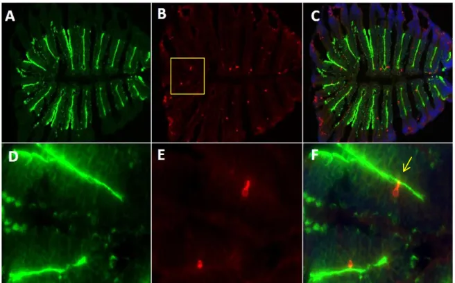
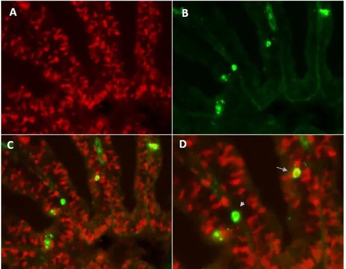
There are various mechanisms of interest speculated to be involved in the observed staining of ORNs namely; via diffusion through open cAMP/CNG cation channels, and

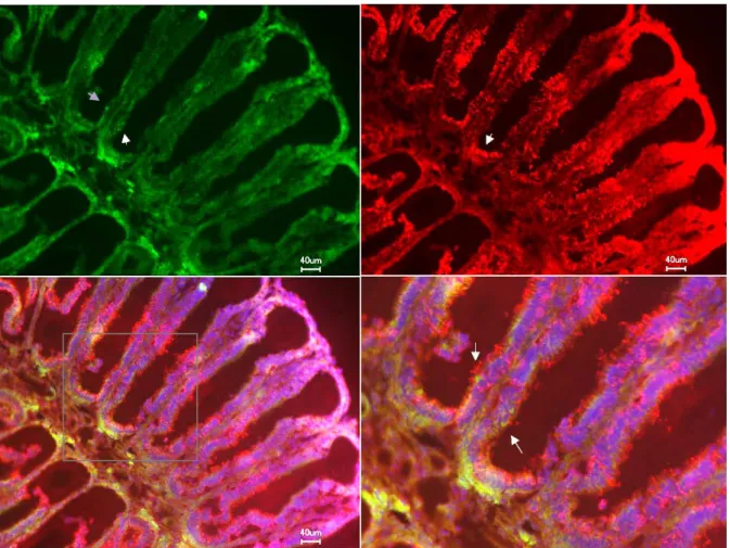
No odorant-induced responses could be obtained from isolated ORNs recorded in the on-cell mode, while recordings in the slice (on-cell and whole-cell) as well as previously

TABLE 1 Average and maximum C stocks in living and dead volumes for forest registered as managed and unmanaged in Germany, based on plot data from the national forest

Correlation analysis demonstrated that MC population activity patterns evoked by Arg and His (pure odors) were initially dissimilar (R = –0.25 at 256 ms) and remained

predictors were centered according to Kraemer and Blasey (2004), and interaction terms were.. residuals-centered according to Little et al. Number of stressful life events were

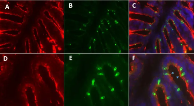
After the expression of OR and V1R genes was verified in the olfactory tissue, a single cell RT-PCR was conducted to validate whether single or multiple chemoreceptors are

Membranes prepared from insect cells expressed the 5-HT 7(a) receptor wild type and different C-terminal cysteine mutants were incubated with [ 35 S]-GTPγS in the presence or

Besides the contribution of mast cells to allergic inflammation [132], they also seem to be involved in non-allergic inflammatory diseases of the gut, since in the intestine of