Characterization of Zebrafish Class C Olfactory Receptor Gene repertoire
Inaugural -Dissertation Zur Erlangung des Doktorgrades
Der Mathematisch -Naturwissenschaftlichen Fakultaet Der Universitaet zu Koeln
Vorgelgt von Aswani Kumar Kotagiri
aus Tanuku, Indien Koeln 2006
Berichterstatter: Prof.Dr.S.I.Korsching Prof.Dr. S. Roth
Tag der muendlichen prufung:5. Mai . 2006
ERKLÄRUNG
Ich versichere, daß ich die von mir vorgelegte Dissertation selbständig angefertigt, die benutzten Quellen und Hilfsmittel vollständig angegeben und die Stellen der Arbeit - einschließlich Tabellen, Karten und Abbildungen -, die anderen Werken im Wortlaut oder dem Sinn nach entnommen sind, in jedem Einzelfall als Entlehnung kenntlich gemacht habe; daß diese Dissertation noch keiner anderen Fakultät oder Universität zur Prüfung vorgelegen hat; daß sie - abgesehen von unten angegebenen Teilpublikationen - noch nicht veröfentlicht worden ist sowie, daß ich eine solche Veröfentlichung vor Abschluß des Promotionsverfahrens nicht vornehmen werde. Die Bestimmungen dieser Promotionsordnung sind mir bekannt. Die von mir vorgelegte Dissertation ist von Prof. Dr. S. I. Korsching betreut worden.
Köln, den 30, Mai 2006 Aswani Kumar
This study was performed in the laboratory of Prof. Dr. Sigrun Korsching in the Institüt für Genetik, Universität zu Köln, Germany and was supported by the International Graduate School and the Deutsche Forschungsgemeinschaft.
I am very thankful to Dr.S.I.Korsching for all the support and guidance. I
appreciate her contribution and all the help she gave in compiling the gene list of the ORC genes.
I would like to thank Prof. Dr. S.Roth, Prof. Dr. Peter Kloppenburg for being in my thesis committee. I extend my thanks to Dr. Mathias Cramer for all his guidance and helping me in generating the ORC 1.1 antibody
Among many of those who would deserve it my special thanks go to Hans Fried, for all his great support and advice. I would take this opportunity to thank Sunil Kumar for his kind and friendly environment. I would extend my thanks to Mehmet saltruek for his kind help and assistance in performing my experiments. Thanks to cheerful discussions of Darko and Ivan and oka yuichiro. They provided the great moments in the lab.
I would like to thank Chemali for being the one who can criticize and who is willing to discuss about my work and share some cola. My many thanks go to Arzu Çelik, Silke and Stefan Fuss, for introducing me to the olfactory system.
I would specially thank Dr.France Weth, who helped me in discussing the results and performing my key experiments.
Thanks to phani and palani for funny moments and being there when needed.
I would always remember my Indian community for their cheerful environment in hard times and good times. I would specially thank Raj Kumar and Soni for their moral support. I would apologize for not able to list all the friends and people for their help and joyful moments.
I would also extend my thanks to Mr and Mrs.Shanmugasundaram who are like parents to me and helped me in taking my decisions.
Finally I would extend my thanks to zebrafish with out which I would not had completed this study.
TABLE OF CONTENTS
I.TABLE OF CONTENTS
II. ABSTRACT 1
III. INTRODUCTION
1.1 Odors are the small chemical molecules 4
1.2 Pheromones are sex specific odors. 5
1.3 Anatomy of olfactory system 6
1.3.1 Zebrafish peripheral olfactory organ 6
1.3.2 Central/tertiary structure in fish 9
1.3.3 Neuronal circuit of the olfactory bulb 9
1.4 Odor ligand molecules are recognized by odorant receptors 12 1.5 Three different types of chemoreceptors are expressed
in the olfactory system 14
1.6 OR gene repertoire 14
1.7 Gene regulatory mechanisms of OR receptor choice 15
1.8 Aim of the thesis 17
IV. RESULTS
1.1 Genomic datamining lead to the identification of ~49 intact V2R like receptors in
zebrafish. 19
2. All fish ORC’s are grouped in to five families 20 2.1 The Uni-ORC family resides with the out groups. 20 2.2. The ORC1 family is evolutionarily conserved 25 2.3. ORC1.1 does not fall in to the class of taste receptors 26 2.4 Human homologue of ORC1.1 is a pseudogene. 28 2.5Other ORC receptors are represented by four families. 29 2.6 V2R like genes in other fish genomes and their relation to zebrafish ORC’s
31
3 .1.2 ORC genes from main group express specifically in the olfactory
epithelium 36
3.2 Cellular distribution of the ORC1.1 gene 38
3.3 Zebrafish genomic BAC filter screening of ORC genes 41 3.4 A specific polyclonal anti serum recognizes the ORC1.1 in the epithelium
43 3.4.1 Preparation of the antigen protein for immunization 44 3.4.2 Charecterization of the polyclonal serum. 45 3.4.3 Immunohistichemistry with the ORC1.1 specific polyclonal antiserum 45
V. DISCUSSION
1.1 Identification of the V2R like gene sequences in the zebrafish genome 48 1.2. The ORC family of receptors are unique with respect to their phylogenetic grouping,
evolutionary conservation. 48
1.3 MHC: signals of individuality 51
1.4 Future applications and studies 52
VI. MATERIALS AND METHODS
1.1. Chemicals and Supplies 54
1.1.1. Enzymes 54
1.1.2. Plasticware 54
1.1.3. Preparation of Solutions 55
1.2. Animals 55
1.2.1. Zebra fish Strains: 56
1.2.2. Maintenance: 56
1.2.3. Microscopic Analysis and Photography 56
1.3. Molecular Biological Techniques 57
1.3.1. Isolation of Plasmid DNA 57
1.3.1.1. Small Scale Plasmid DNA Preparation (Miniprep) 57 1. 3.1.2. Large Scale Plasmid DNA Preparation (Midiprep) 57
1. 3.1.3. Phenol/Chloroform Extraction 58
1. 3.1.4. Ethanol Precipitation 58
1. 3.1.5. Quantitation of DNA and RNA 58
1.3.1.6. Agarose Gel Electrophoresis 59
1.3.2. Enzymatic Modifications of DNA 59
1. 3.2.1. Digestion of DNA 59
1. 3.2.2. Dephosphorylation of Plasmid DNA 59
1. 3.2.3. Ligation of DNA Fragments and PCR Products 59 1. 3.2.4. Isolation of DNA Fragments from Agarose Gels 60 1.3.2.5. Preparation of Electrocompetent Cells and Electroporation 60 1.3.2.6. Colony PCR for Identification of Positive Clones and Determination of
Insert Length 61
1.3.2.7. Preparation of Glycerol Stocks 61
1.3.2.8. Polymerase Chain Reactions (PCR) 61
1.3.2.9. Sequencing Reaction 61
1.3.2.10. Sequence Analysis 62
1.4 .1 Tissue dissection: 62
1.4.2 RNA isolation: 62
1.4.3 RT-PCR 64
1.5. Protein expression studies 65
VII. REFERENCES 67
VIII. APPENDIX 76
IX ABBREVIATIONS 86
X. CURRICULUM VITAE 88
ABSTRACT
To understand the sensory processing of olfactory information it is essential to determine the size and diversity of the olfactory receptor gene repertoire. In the current analysis we performed the genome datamining to estimate the Group C olfactory receptor gene repertoire in zebrafish. A total of 56 receptors were identified. To evaluate the putative receptors as olfactory genes molecular and histochemical studies have been employed. The expression of several genes was detected in the olfactory tissue by RT-PCR. We characterized one of the receptors which is conserved across several species and uniquely clusters with the outgroups in the phylogenetic comparison, unlike the other ORC receptors. This receptor exhibited an broad expression pattern in the olfactory epithelium of zebrafish suggesting that it is co-expressed with other endogenously expressed receptors.
The specific expression of the genes for the members by RT-PCR analysis identified in this approach is reported. These results directs towards the theory that the Class C olfactory receptors might function as a complex of two or more receptors like that of taste receptors.
2
ZUSAMMENFASSUNG
Um die Mechanismen der Genregulation olfaktorischer Rezeptorgene zu untersuchen und das Ligandenspektrum olfaktorischer Rezeptoren zu erfassen, ist es notwendig, das Repertoire an Rezeptorgenen zu bestimmen. Der Zebrabärbling ist ein geeigneter Modellorganismus für diese Fragestellungen, da er im embryonalen Stadium transparent ist und ein genetischer Mutanten-Screen etabliert ist. Das Genom dieses Fisches ist nahezu komplett sequenziert und bietet einen weiteren Vorteil das Geruchssystem in diesem Organismus zu untersuchen. In der vorliegenden Arbeit habe ich Datenschürfung aus der Genomdatenbank betrieben um das Repertoire an olfaktorischen Rezeptorgenen der Klasse C zu bestimmen. Insgesamt wurden 56 Gene ermittelt. Um die Kandidatengene als olfaktorische Rezeptorgene zu identifizieren wurden molekulare und histochemische Methoden verwendet. Die Expression einiger Gene im olfaktorischen Gewebe wurde durch RT-PCR nachgewiesen. Ich habe einen dieser Rezeptoren, der über verschiedenste Speziesgrenzen hinweg konserviert ist und sich in der phylogenetischen Analyse einzigartigerweise von den Klasse C Rezeptorgenen abspaltet, charakterisiert.
Das Expressionsmuster dieses Rezeptors erstreckt sich über das gesamte olfaktorische Epithel des Zebrabärblings, was nahelegt, dass er mit anderen endogen exprimierten Rezeptoren coexprimiert wird.
Das lässt die Vermutung zu, dass die Klasse C Rezeptoren als Komplex mit zwei oder mehr Rezeptoren funktionieren, wie es bei den Geschmacksrezeptoren der Fall ist.
4
Introduction:
Biological communication was defined by Wilson (1970) as “the action on the part of one organism (or cell) that alters the probability pattern of behaviors in another organism (or cell) in an adaptive fashion. Organisms interact with the environment through the sensory organs. Olfactory system is a chemosensory system that informs about the chemical nature of the surroundings. Almost all the organisms rely on the olfactory system for their daily needs and survival. In animals smell can affect feeding and social behaviors such as mating, territoriality and egg laying.
The olfactory system is one of the early evolved sensory systems and is conserved across phyla. The highly invariant anatomical and physiological mechanisms reflect that this sensory system is faithfully performing its function across years of evolution. With its simple anatomical architecture and complex physiological and molecular regulation, olfactory system has been a model neuronal system to study neurological functions.
1.1 Odors are the small chemical molecules
Organisms sense, perceive and respond to enormous number of chemicals that it encounters in daily life. It has been estimated that humans can sense about 5 – 10,000 odors. Odor molecules are small molecules with molecular weights of up to 400da. Even subtle changes in chemical structures are often perceived as different odors; e.g. L-and D - Carvone smell like caraway and spearmint. Odors can be either generalist
odors like food odors or specialist odors like pheromones.
1.2 Pheromones are sex specific odors.
Pheromones are a special class of odors. Pheromones are the chemical cues that regulate the social and reproductive behaviors. The biological function of pheromones extends beyond the social and sexual behavioral modulation. Pheromone communication system is a chemosensory system that is used to communicate with in species; there are some examples where they are sensed outside the species border. Pheromone signaling is a two component system; the source or the signaler and the receiver. (Sorensen PW 1998) There is no commonly agreed definition for pheromones described so far. Some of the properties based on which pheromones are described are as follows.
It is a non volatile compound, so must be sensed by physical contact
It is a sex specific and species specific signal
It evokes a sexual or species specific behavioral pattern in the individual or group of individuals that has come in contact with.
The presence of pheromones does not restrict to vertebrates, it is in fact well documented in insects. A type of pheromone signaling called quorum sensing is an established phenomenon in yeast.
6 1.3 Anatomy of olfactory system
Sensation is described as the registration of the physical stimuli from the environment by sensory organs. The olfactory sensory organ, which has the relevant sensory neurons, samples the olfactory stimuli from the environment. In mouse two anatomically distinct olfactory organs are present. The main olfactory epithelium is located in the nasal cavity and the vomeronasal epithelium is encapsulated in the tubular structure referred to as vomeronasal organ. The olfactory organ is represented in the form of a rosette like structure in fish. The fish does not posses a vomeronasal organ.
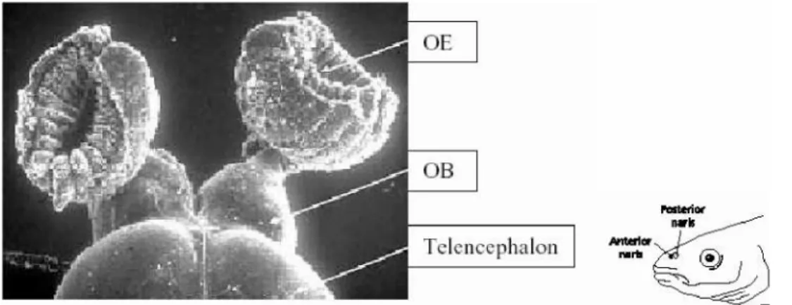
Fig 1: Anatomy of the zebrafish olfactory system: The olfactory epithelium is represented as cup like structure. The olfactory bulb is protrusion from the telencephalon. (Picture kindly provided by Silke Argo)
1.3.1 Zebrafish peripheral olfactory organ
The nasal opening, often referred to as naris allows access of the external water medium to the olfactory organ. The olfactory organ is the nasal epithelium; in zebrafish is rosette like structure enclosed in a cartilaginous structure. The olfactory sensory neurons
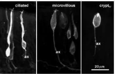
are distributed in lamellae, arranged like a rosette that arises from the base of the naris opening. The lamellae fold in a centrifugal pattern arising from the base of the cup like structure. Morphologically two different sensory neurons are described, microvillous and ciliated cells. Recently a third type of neuron has been described in fish called the crypt neurons. There is no anatomical dichotomy in fish unlike mouse which has vomeronasal and main olfactory epithelium, hence both the olfactory V1R like and the V2R like receptors are expressed in the olfactory epithelium (Stryer et al., 1998). All the sensory neurons are distributed concentric from the centre of the epithelium up to two thirds of the epithelium area defining the sensory zone; the rest marked as non sensory area.
Fig 2: Three morphological types of sensory neurons are described in the fish olfactory system. (Section 1.3.1) (Picture taken from Ann Hanssen et al., 2003)
Ciliated neurons:Ciliated sensory neurons are bipolar neurons that extend their dendrites to the epithelium and project their unmyelinated axons towards the olfactory bulb. Olfactory receptors are expressed in ciliated sensory neurons. The ciliated neurons appear to be distributed more basally in fish epithelium. Sensory neurons expressing different olfactory
8 receptors are differentially distributed across olfactory epithelium. The zone restricted expression of the olfactory receptors came in to light with the expression studies in mice.
In fish several olfactory receptors are shown to express in concentric ring like pattern some what remnant of the of zonal distribution in mice. (Weth F et al., 1996)
Microvillous neurons:The V2R receptors are expressed in the microvillous neurons. (Ryba NJ et al., 1997) Recently expression of a single V1R like receptor has been shown in apical layer of the lamellae, corresponding to the microvillous or crypt neuron distribution. (Pfister P et al., 2005)
Crypt neurons: A third type of microvillous like neuron was identified recently in fish called the crypt cell (Hansen A, 2004). Crypt cells were not described in mouse. These cells are apical in distribution. Crypt neuron has unusual structure, microvilli accompanied by the sunken cilium which does not arise from the dendritic knob (Fig 3). Using S-100 as an marker for crypt cells, S-100 antibody studies showed that the crypt cells are evenly distributed though out the epithelium in zebrafish but in a punctuate manner. (Germana A et al., 2004) No olfactory receptor expression is reported so far in crypt neurons.
In mice the ciliated neurons are located in the main olfactory epithelium. The microvillous neurons are distributed in the vomeronasal epithelium. There are no crypts identified in mouse so far. The V1R and V2R receptors are expressed in the vomeronasal epithelium while the olfactory receptors are expressed in the main olfactory epithelium.
Four different zones olfactory receptor expression were described based on histochemical
studies with associated molecules like OCAM, NCAM and other molecules (Ressler KJ et al., 1993). Some receptors are shown to express exclusively in certain zones. No zonal expression was identified with either V2R or V1R receptors. Immunochemical anatomical studies with G-protein antibodies revealed that in mouse VNO the V1R expressing neurons (stained with Galphai) are epically distributed, while the V2R expressing neurons (stained with Galpha O) are basally distributed in mice. (Berghard A et al., 1996) Microvillous neurons are epically distributed in fish lamellae. The VNO in the humans is a vestigial organ. (Ryba NJ et al., 1997)
1.3.2 The Central/tertiary structure in fish
The synaptic processing of the input sensory information occurs in the olfactory bulb. The olfactory sensory neurons project their axons on to the olfactory bulb where they synapse with the dendrites of the mitral cells, which project their axon to the higher brain centers. The axons of the ciliated sensory neuron were shown to project to the medial part of the medial olfactory tract and mediate alarm reaction. The axons of the microvillous neurons project to the lateral olfactory tract and mediate feeding behavior. The axons of crypt cells expressing G {alpha} o are found to project to the ventral midline of the OB in catfish. (Hansen, A 2003)
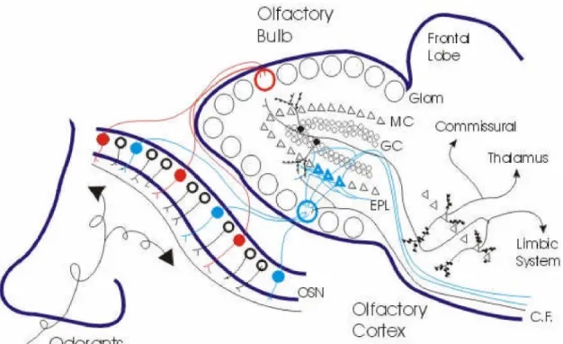
1.3.3 Neuronal circuit of the olfactory bulb
Olfactory bulb is an extrusion of the telencephalon in processing of the sensory olfactory input. Morphologically four layers are distinguishable in zebrafish olfactory bulb. These layers are not well defined compared to that of higher vertebrates. The outer olfactory nerve layer (ONL) where the incoming olfactory sensory neuron axons
10
Fig 3:Neuronal circuit of the olfactory system. The general anatomical principles of the human olfactory system are shown the above illustration. Picture taken from
Strowbridge(http://neurowww.case.edu/faculty/Strowbridge/)
terminate. The glomerular layer (GL) which has morphologically defined spherical structures called glomeruli. They are not uniformly distributed and vary in their size and shape. Aglomerular plexus is a neurophil structure in to which the axons of some OSN terminate. The mitral cell layer, (MCL) is not a well organized domain in zebrafish. The internal cell layer (ICL) which consist of densely packed granule cells that innervate the mistral cell layer. The axons of the olfactory sensory neurons form glutamergic excitatory synapse with dendrites of mitral cells and periglomurular cells of the olfactory bulb. In mice this synaptic structure is surrounded by the periglomurular cells and has a distinct
anatomical distribution in the olfactory bulb. Periglomurular dendrites are GABAergic and dopaminergic to mitral cell dendrites. Periglomurular cells are possibly involved in interglomurular processing and might contribute to the contrast enhancement. The glomurulus is a functional and anatomical unit with several levels of organization. In mice most of the glomeruli have an approximately similar size and are distributed around olfactory bulb. All the sensory neurons expressing the same or similar type of olfactory sensory neuron project their axons to single glomurulus, a phenomenon referred to as convergence. (Korsching.S.I. 2001) The mitral cells of olfactory bulb project their dendrites to a single glomurulus as in mice. The axons of mitral cells project their axons to higher brain centers. In mice, on an average each glomurulus receives input from 25,000 olfactory sensory neurons, where they form synapse with approximately 25 mitral cells and 25 periglomurular cells. Besides mitral cells another cell type was described, tufted cells which also project to glomeruli in mice. The inner layer is constituted by granule cells, which are GABAergic in nature. Zebra fish olfactory bulb circuitry that is involved in synaptic processing of excitatory input from olfactory sensory neurons is mainly constituted by two types of neuronal cell populations; mitral cell and the granule cell. A reciprocal dendritic circuit has been described where a mitral cell activates the granule cells, which in turn inhibits mitral cell. The activated mitral cells mediate feedback inhibition on themselves and lateral inhibition on neighboring mitral cells. (Leon M et al., 2003) This interaction might form the basis of temporal coding, controlling the frequency of impulse output from mitral cells. (Friedrich RW. 2001, Laurent G. 2002)
12 1.4 Odor ligand molecules are recognized by odorant receptors:
The olfactory receptors are seven trans-membrane G-protein coupled receptors.
(Mombaerts P, 1999) The receptors are expressed in the olfactory sensory neurons situated in the nasal epithelium. Odorant receptors generally are oligo specific that is they recognize small molecules often with same odorant feature. Correspondingly each odorant generally gets recognized by more than one receptor. Binding of an odor ligand to its cognate receptor triggers a series of signal transduction cascade events that finally results in depolarization of the olfactory sensory neuron. (Buck LB, 2004) The odorant features, neurological and molecular mechanisms based on which the ligands are discriminated are not yet completely understood.
Table 1: OR genes in the various genomes. (data taken from Nei etal, 2005)
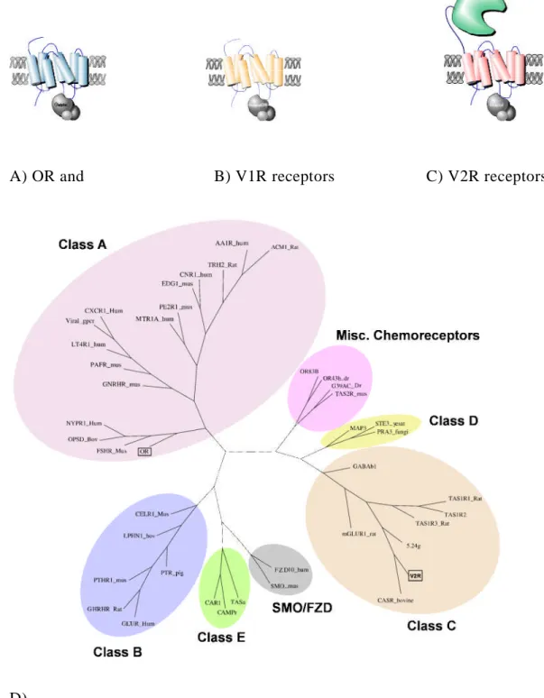
A) OR and B) V1R receptors C) V2R receptors
D)
Fig 4:Receptors that are expressed in the olfactory system belong to the seven transmembrane families of protein. The sub pictures A, B and C illustrates general structure of the olfactory receptors. D) Phylogenetic tree showing the distribution of the
14 olfactory receptors among the GPCR class of proteins.
1.5 Three different types of chemoreceptors are expressed in the olfactory system.
Three types of chemoreceptors are expressed in the olfactory system. All three types of chemoreceptors belong to the family of the seven transmembrane families of receptors.
Odorant receptors falls into the Group A subfamily of receptors, (Fig4) while V1R receptor are classified as putative family of receptor family which include T2RTaste receptors and nematode chemoreceptors. The V2R class of receptors belongs to the group C subfamily.
They have a large extracellular domain apart from typical seven transmembrane structures.
1.6 OR gene repertoire
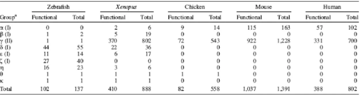
The OR receptors are quite divergent in sequence form mouse to zebrafish. To date 636 human olfactory receptors and 1209 mouse olfactory receptors have been reported by bioinformatics analysis. (Godfrey PA et al., 2004, Malnic B et al., 2004) In fish 136 intact OR genes were reported with 7 partial genes and 10 psuedogenes. (Alioto TS and J.Ngai 2005) In humans almost half of them are psuedogenised with 339 intact coding regions.
(Glusman G, 2000) The mouse has 913 intact OR genes. A total of 172 families and 241 subfamilies were described in human and mouse. The Y chromosome in either species does not have any OR gene. (Table 1) OR genes are classified in to Class I and Class II subtypes depending on the phylogenetic analysis. Initially Class I genes are thought to be involved in sensing airborne odors where as class II genes are for detection of the aquatic odors.
Most genes in mammals and chicken belong to the Class II genes where as Class I genes make up to ~150. Recent analysis found that zebrafish has one Class II gene. (Mombaerts P, 1999)
So far 61 and 57 putative full length V2R genes were identified from mouse and rat genome. (Matsunami H et al., 1997) The human V2R genes are completely disrupted, consistent with the absence of a morphologically well-formed vomeronasal organ in humans. Moreover the TrpC2 gene which is involved in the vomeronasal signal transduction cascade is completely disrupted in humans. (Liman ER, 1999, Berghard A, 1996) The V2Rs belongs to the group C of the GPCR family of proteins. (Ryba NJ, 1997)
V1R receptors are more divergent in sequence than the V2R receptors. The V1R family comprises 187 and 106 intact V1R genes in mouse and rat genome. Both V1R and V2R class of chemoreceptors are expressed in a different neuronal compartment than that of olfactory receptors in mice. (Rodriguez I et al., 2000, Pantages E et al., 2000)In fish how ever does not posses such anatomical dichotomy. So far a single V1R was reported in fish.
(Pfister P et al., 2005) Except 5 receptors of the family V1RL, the V1R families completely in humans are psuedogenized. One member of this family shown to express in human olfactory tissue. (Rodriguez I et al., 2000)
1.7. Gene regulatory mechanisms of OR receptor choice
Each olfactory sensory neuron expresses one of the several hundreds of receptors.
Two models were proposed to explain the OR receptor choice. In a deterministic model,
16 the OR is chosen by the generation of unique combination trans-activators that activate a single receptor bearing the appropriate cis-acting elements. In stochastic model the choice of receptor is random. Presence of a cis-acting element common to all OR genes under regulation would facilitate such mechanism. Expression studies with the P3 OR transgene revealed that it could recapitulate the native OR gene expression. Transgene and the cognate endogenous alleles were rarely co-expressed, thus favoring the stochastic model.
Several attempts were made find a conserved element upstream of the OR gene. Several candidate regulatory factors identified by one-hybrid experiments failed to demonstrate OR specific control or not yet validatedin vivo(Hoppe R et al., 2006)
The cloning of the mice by nuclear transfer form the OR neurons expressing genetically tagged OR revealed that it expressed all repertoire of OR’s (Eggan K et al., 2004). The genomic structure surrounding the selected OR locus remained unaltered. Thus ruling out the mechanism that OR gene choice involves any DNA rearrangements like that of lymphocyte receptor gene families. The lymphocyte receptor genes undergo DNA rearrangement as a one of the mechanism to generate diversity.
The presence of one OR gene expressed suppresses the expression of the other OR gene. A feed back repression mechanism seems to be involved in the OR receptor regulation. Sakano et al., showed that the neuron that choose to express the truncated gene products or disrupted OR gene marked with GFP or LacZ frequently expresses different OR gene. These marked neurons project their neurons diffusively in the olfactory bulb.
(Serizawa S et al., 2003)
It has been well documented that one of the several hundred receptors is chosen by the olfactory sensory neuron. In contrast, co-expression of the some DOR genes has been shown in drosophila. The OR83b receptor was unique in that it is highly conserved across different insect species and is expressed in almost all the OR expressing neuron. It has been demonstrated that OR83b co -expression is required for the functional expression and proper targeting of the endogenous OR gene(Benton R et al., 2006, Jones WD et al., 2005, Larsson MC et al., 2004). T1R class of Taste receptors poses another example where the two receptors are co-expressed.
1.8 Aim of the thesis
To shed a light on the receptors gene regulatory mechanisms and understand the ligand spectrum of the olfactory receptors, it is necessary to estimate the receptor gene repertoire. Zebrafish offers a model organism due to its transparent embryos and established genetic screen for mutants. In the current study we performed the genomic data mining to estimate the Group C olfactory receptor gene repertoire in zebrafish. To evaluate the putative receptors as olfactory gene molecular and histochemical studies have been employed.
IV. RESULTS
RESULTS
1.1. Genomic data mining lead to the identification of ~56 V2R like receptors in zebrafish.
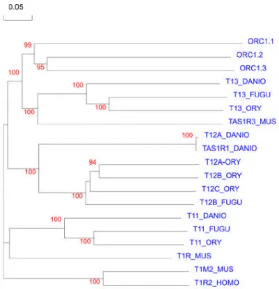
In the first step to retrieve all the possible V2R like sequences, the Mouse, Rat, Fugu, Tetraodon and Zebrafish genomes was searched for the presence of the V2R like genes using the interpro domains (Table 1) and published V2R gene sequences. To prevent any accidental loss of genes even sequences with low e-values are retrieved. The data set thus obtained was subjected to cycles of refinement, undergoing selection and elimination.
The resulting non-redundant sequences were analyzed for the presence of the typical structural properties of group C GPCR proteins; the seven-transmembrane domain and a large extracellular domain. The sequences that were annotated as V2R genes at the ensemble were also included in the analysis. The out groups chosen here are the T2R, GABA receptors, mGLUR and CASR receptors, as they all belong to the same Group C family of GPCR. The verified sequences were then subjected to the phylogenetic analysis to define them as V2R, depending how they cluster in relation with the out-group members (T2R, GABA receptors, mGLUR and CASR) and with known V2R members. The V2R genes are multi exonic, generally several splice variants predicted. In these cases the longest possible transcript in which most of the V2R gene features preserved was chosen.
This approach led to the identification of ~56 intact genes and 4 pseudo genes in the zebrafish (Table 2)
Since there was no common agreed nomenclature described for V2R genes we
20 propose a new nomenclature here for the zebrafish V2R-like genes. The new nomenclature is ORC followed by the family or subfamily indication. (Olfactory Receptor class C). The ORC designation was given to a sequence if it exhibited the sequence features of Group C type GPCR and additionally clustered with known V2Rs relative to the out-group.
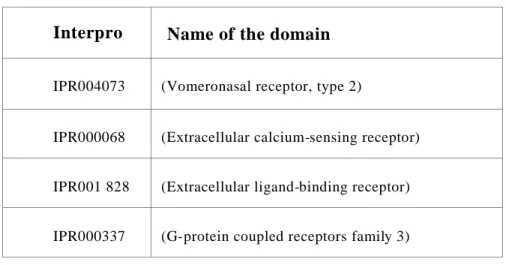
2. All fish ORC’s are grouped in to five families
All the fish ORC receptors share ~29% identity at the protein level (appendix Table1). The receptors were grouped in to 5 families depending on their bootstrap values.
(Fig1) Four pseudo genes were identified in the current analysis at least in the zebrafish version 5 ensemble releases.
2.1 The Uni-ORC family resides with the out-group.
Three genes fall outside the families described above and clusters with the out groups that constitute Taste, GABA, Glutamate and CASR receptors. These three genes the ORC 1.1, ORC1.2 and the ORC1.3, represent the Uni-ORC family. The Gold fish 5.24 receptor and its zebrafish homolog Z09 receptor (which is the ORC1.2 receptor here) was already established as an olfactory receptor by the Ngai’s group(Speca etal., 1997) The ORC 1.1 receptor is an ortholog of mouse V2R2 gene. (Section 2.2) These two recptors are thus considered as olfactory receptors though they reside with the out groups.
Fig1:An unrooted phylogenetic tree showing intact ORC receptors identified in this study in zebrafish. The out groups are CASR, T1R taste receptor Glutamate receptor and the GABA receptor.
22
Interpro Name of the domain
IPR004073 (Vomeronasal receptor, type 2)
IPR000068 (Extracellular calcium-sensing receptor)
IPR001 828 (Extracellular ligand-binding receptor)
IPR000337 (G-protein coupled receptors family 3)
Table 1:The list of the interpro domains used in this study to retrieve the possible V2R like sequences. For more details on the each interprodomain kindly refer to www.ensembl.org and the references there with in.
ORC1.1 is more close to the CASR while the other two receptors ORC1.2 and ORC 1.3 are related to the taste or GABA receptors. The ORC 1.3 has been shown to be expressed in zebrafish olfactory neurons. Moreover a ligand to this receptor was identified.
Thus this gene is a verified ORC. Therefore ORC1.3 may be considered as ORC as well.
The genomic structure of ORC1 .1 is very typical of other V2R genes with 6 or more exons among the different predicted alternative spice forms. The extracellular domain is distributed across 5 exons while the transmembrane domain is represented by a single exon. The Genescan program predicts 6 alternative splice variants for ORC 1.1. (Fig 2) By RT-PCR experiments in olfactory epithelium, we could detect only one of the six predicted variants at least by the set of primers (appendix Seq 2) designed in the extracellular region.
There may be other splice variants expressed in different developmental stages or even have a cellular distribution. These hypotheses need to be tested.
A new member ORC 1.2 has been identified in the present analysis which shares 45% identity with ORC1.3 at the aminoacid level. This gene is more close to ORC1.3 rather than ORC1.1 as revealed by phylogenetic analysis (Fig1). No ortholog has been identified for this receptor neither in mouse nor in human genomic database. The gene has the exonic architecture similar to that of 5.24. The intronic region and the 2 kb upstream and downstream of the showed no resemblance to the corresponding ORC1 .3 gene. In the current database release the gene is assigned to a scaffold rather than to a specific chromosome. The expression of this gene was not verified.
The complete sequence of the BAC harboring the ORC1 .1 gene is available. The ORF prediction program (GENESCAN) revealed the presence of a retro-transposon like element at the end 3’ UTR region of the ORC 1.1. Blast analysis revealed that this retro- transposon like element is having close resemblance to the D1RS family of transposon elements. D1RS transposon element is a fish specific transposon element. The D1RS transposon element or its remnants are not present at similar location in Fugu or Tetraodon fish. It appears that this gene done no damage to the surrounding region as there is no shuffling of sequences in the Tetradon, Fugu, Mouse or Human. The presence of similar elements has been reported in olfactory receptors encoding genomic region by Dugas and Ngai et al., The biological implications of these elements in the olfactory genomic region have not been characterized till date. It need to be verified if the insertion of this particular transposon insertion is specific for the fish strain that was sequenced or it is represented among all species of zebrafish strains.
24
Fig2: Exon-Intron structure of ORC1 .1 gene. The picture shows the position of D1RS transposon element at the end of the gene. Illustration not drawn to scale.
Fig3: The phylogenetic tree showing the evolutionary relationship of the two receptors, ORC1 .1 and ORC1 .3. A) The phylogenetic tree of the ORC1 .3 orthologs from different species and B) shows the phylogenetic tree of the ORC1 .1
2.2. The ORC1 family is evolutionarily conserved
The only V2R receptor for which a ligand and indeed a ligand spectrum have been identified is the ORC 1.3. A blast search in the genomes of other species revealed orthologs for this gene. The ORC1.3 and its homologues from different species share an identity of
~54% at the aminoacid level. Recently the orthologs of 5.24 have been cloned in human
26 and mouse (Fig3a). In humans ORC1.3 (referred to as GPRC6A) has been shown to express in several tissues (prominently in liver, brain and kidney) except in olfactory tissue. In mouse an ortholog has been reported (Genbank Acc: AY101365).
Similarly ORC1.1 homologs in other species were identified in the current analysis.
(Fig3B)
2.3. ORC1.1 does not fall in to the class of taste receptors:
ORC1.1, 1.2 and 1.3 clusters with the out groups that constitutes the Taste receptors. More over VR5.24 was reported to be an amino acid receptor and is expressed in the taste buds of goldfish (Speca DJ et al., 1999). In it necessary to mention that the T2R taste receptors are aminoacid receptors that bind to glutamate and related structures (Nelson G, 2001). Taken together with these observations with that of the expression of VR5.24 in goldfish, it might be possible that ORC1.1 as well as ORC1.3 might be a taste like receptor or related receptor.
Fig 4:The unrooted bootstrapped tree showing the evolutionary relation ships of the ORC1 family of receptor from Danio rerio with taste receptors form with in danio rerio and other species.
To understand how these Uni-ORC genes are related to the taste receptors, phylogenetic analysis of the T1R taste receptors from other organisms was performed. (Fig 4)The Uni-ORC gene share ~39.17% identity with the Taste T1R receptors from other organisms. The phylogenetiuc tree revealed that the Uni-ORC cluster is more related to T13 family with 43.10% identity. T13 family receptors are shown to co-express with other T1R class of receptors.
28 2.4 Human homologue of ORC1.1 is a pseudogene.
Human olfactory and pheromone genes are littered by psuedogenes. There are about ~170 V1R intact genes in mouse and ~150 V2R genes. Similarly there are 340 intact OR genes and more than 300 pseudo genes. In other words more than 50% genes carry disruptions.
In non-human apes the fraction of pseudo genes is ~ 30%. In dog and cow this comprises 20%. In contrast humans have only four V1R genes, one of which shown to express in the nasal epithelium in humans by Ivan et, al. There was no intact V2R gene described except ORC 1.3 homolog in human. (Petrine Wellendorph et al., 2004) In the present analysis a partial homologue of the ORC1 .1 was identified. This gene has 60% homology to the mouse counterpart at the protein level. The gene had deletion in the huge extracellular domain. (Fig5) The bitter taste receptor is a truncated Glutamate receptor. Presence of the N-terminal deletion does not affect the function of this receptor. What is more relevant to ask here is that whether truncated ORC 1.1 homologue in human is still functional? It might be interesting to see the expression of human homologue of ORC 1.1. Extensive search was performed for the presence of the EST clone in all the available Human EST databases. Two EST clones corresponding to the ORC 1.1 was identified in the fetal human lung EST library. There was no olfactory EST or cDNA library made from humans. It might not be relevant to look for the expression in adult olfactory cDNA, as there is no functional vomeronasal organ. Indeed the TRPC2 gene which is a signal transduction molecule mediating the V2R receptor response is disrupted. In humans the vomeronasal organ is present only during the embryonic developmental stages of 14week of post natal embryo, based on the GalphaO staining. Due to limitations in obtaining the fetal olfactory tissue of 14 week embryo in Germany, hampered further study. As a substitute I performed RT-PCR in human embryonic kidney cells revealed the presence of this gene (Data not shown). As the HEK293 cells are immortalized cells and have been maintained in laboratory these results must be interpreted with caution.
2.5 Other ORC receptors are represented by four families.
ORC2 family represents 15 ORC receptors. These receptors exhibit high similarity among themselves. The most divergent member of this family is the ORC2.3. This receptor expression was shown by RT-PCR. No homologs could be identified for ORC2.3 in any of the sequenced fish species. This family might represent authentic ORC receptors as the V2R like receptors that were isolated form cyprinidae species have homologs in this family. A total of 11 receptors are clustered in the ORC5 family. Like the ORC2 family these receptors also share high similarity with in the family. It has been shown that the goldfish GFB8, GFB1 Vr3.3 are expressed in the olfactory epithelium by RT-PCR and insitu experiments. The homologues to these three receptors are present in this family and have been shown to express in the olfactory epithelium of the zebrafish in this study. One might propose that these receptors might be the actively expanding family of receptors.
The rest of the receptors are exhibit less identity with in the member of the family. (Table 2)
30
A
B
Fig 5: A) A multiple alignment of the homologues of the ORC1 .1 from human, rat and mouse. The region of deletion in the extracellular domain of ORC1 .1 is underlined in red.
B) Schematic representation of the region (highlighted in red) of human ortholog of ORC1 .1 that is deleted.
2.6 V2R like genes in other fish genomes and their relation to zebrafish ORC’s
To analyze the representation of V2R like genes in other fish species in relation to the zebrafish ORC gene, a similar BLASTp plus Phylogenetic reconstruction approach was adopted to retrieve the possible V2R genes. Current database release of the Fugu rubripes (v4) and Tetraodon verdis (v6) was searched with custom BLASTp searches. There were 14 V2R like sequences from the Cyprinidae species at the NCBI database; these were also included in the analysis.
2.6.1 Fugu species has 18 full lengths V2R like receptors
The Fugu rubripes genome has 18 full length receptors, with 28 pseudo genes (Table 2). Similarly the other Fugu fish Tetraodon has 18 full length receptors with 10 pseudo genes (Table 3). The more number of pseudo genes in the database is speculated to be due to the contig erroneous assembly. Erroneous assembly is clearly noticed in the zebrafish genome version 4. The puffer fish genes are evenly distributed with in the five families of the zebrafish. (Fig 6)
32
Fig6: The phylogram showing the distribution of the V2R like genes from the other fish among the families of the zebrafish ORC genes. The receptors from cyprinidae species that was found to the public NCBI data base are also included named as Cppr. The Fugu receptors and Tetraodon receptors has the same nomenclature as in ensembl database.
Table 2: List of the V2R like sequences identifies from Fugu rubripes
34
Table 3:List of V2R like sequences identified from the Tetraodon verdis genome.
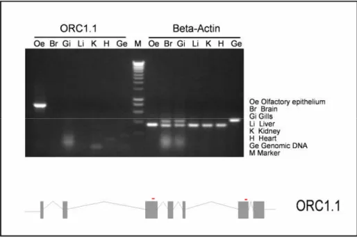
3 .1.1 Tissue specific expression of ORC1.1 in the olfactory epithelium
As a first step to validate the candidate ORC 1.1 receptor gene as authentic olfactory receptor, RT-PCR was performed to examine the expression in olfactory tissue. Total RNA was extracted form the pool of olfactory rosettes prepared from AB/Tu fish that were approximately one year old. A specific set of primers situated in the exons 3 and 6 respectively were designed to amplify ~775 bp corresponding to huge extracellular domain region. The extracellular domain was chosen based on the multiple alignments of all ORC genes of zebrafish; the extracellular domain is more divergent compared to the heptahelical domain.
Fig 7: Determination of tissue distribution of ORC1 .1 gene expression by RT-PCR analysis. A) Shows the tissue distribution of the ORC1 .1 gene. Beta-actin expression was uniformly distributed in all the tissues tested, reflecting the quality of the RNA. The expected band is ~775 bp for ORC1 .1 gene. Beta actin primers amplify ~370 bp. Slight contamination of genomic DNA in RNA preparations of gills and brain was evident presumably due to inefficient DNAse digestion in these samples. The lower panel depicts the genomic structure of ORC1 .1 and the position of the primers used in this experiment.
As shown in the fig 7 the Lane 1 shows amplification of the ORC 1.1 in the olfactory tissue. There was no other receptor amplification. To determine the tissue distribution of ORC 1.1, RT-PCR was performed with cDNA preparation made from several different tissues of zebrafish. The expression of putative ORC 1.1 was restricted to olfactory epithelium. There was no expression detected by RT-PCR in the cDNA preparations of the tissues analyzed, namely heart, liver, gills, kidney, spleen, and total brain. (Fig7) There was no difference in expression between males and females or between different strains (data not shown).
The PCR product was cloned and three independent colonies were sequenced.
36 Sequence comparisons between the sequences provided by the Sanger sequence database and amplified PCR product revealed two point mutations at positions. The first one is a T-
>C change and second was a single cytosine nucleotide deletion in one of the clones. These variations are either PCR induced mutations or may be genuine allelic variations.
3 .1.2 ORC genes from main group express specifically in the olfactory epithelium Three receptor genes form main group was chosen for the examination of the tissue specific expression. Two of them have homologues in goldfish. Similarly expression of other three genes selected; ORC5C. 1, 5a.6 and ORC2.2 was also restricted to the olfactory epithelium. (Fig8) To amplify ORC5c. 1 a two step PCR was performed, first round of PCR at low stringency conditions followed by a booster PCR at high stringency conditions.
To eliminate the possibility that near homologous genes might be amplified by PCR, the resulting PCR products from each respective RT-PCR reaction were cloned and sequenced.
There were no mutations in the sequences. ORC5c. 1 is a homolog of GFB8; ORC5a.6 is homolog of GFB1 by sequence comparison. GFB1 and GFB8 are V2R receptors from goldfish (Cao PNAS etal.), which are expressed in the olfactory epithelium
Fig 8: Olfactory tissue specific expression of three putative ORC genes. Expression of three putative genes, ORC2.2, 5a.6 and 5c. 1 is specific to olfactory tissue. There was no expression detected by RT-PCR in other tissues analyzed. B) Illustrated diagram showing the relative position of the primers used for the three receptors.
38 3.2 Cellular distribution of the ORC1.1 gene
To investigate the cellular localization of the ORC 1.1 expression within the olfactory tissue, insitu experiments were performed using a specific insitu probe was designed from extracellular domain as it is the most divergent region across V2R genes, as compared to the trans-membrane domain. (Appendix Seq2) The primers insituR and insituF were different from the set of primers chosen for the RT-PCR. The resulting PCR product was cloned and verified by sequencing three individual colonies. There were no mutations in the three colonies sequenced. One particular clone, insitu8 (Appendix Fig1) was chosen for the insitu experiments.
The insitu8 probe is 614 bp long, with a GC% of 52. The Tm was calculated to be 80°C. It is evident that the classical olfactory genes express one gene per cell. The same expression mechanism applies to the V2R genes (Dulac C and Axel. R 2000). Interestingly ORC1 .1 showed a broader expression pattern, unlike other ORC genes. The ORC 1.1 sense probe stains the upper half of the lamina of the epithelium. The expression spans all over the sensory part of the epithelium starting from center towards the outer side. (Fig 10) As shown in the figure 11 the staining is still intense even in the section taken from the base of the epithelium. Due to intense staining of the probe the cellular count could not be determined for quantitative analysis. The sense probe does not show any staining. Further more the same antisense probe was used for successful screening of Zebrafish BAC filters asserting the specificity of the probe. The antisense probe stains weakly with out lost of specificity in the epithelium of adult fish of ~2year old (Fig 11).
Fig 9:Insitu probe was designed to corresponding protein sequence in the extracellular domain.
40
Fig 10:Insituhybridization of the olfactory epithelium tissues section with ORC1.1 probe.
A) Shows the labelling of the antisense ORC 1.1 probe in the olfcatory epithelium of the adult zebrafish. B) Sensory and nonsensory region (arrow) coincides with the ORC 1.1 antisense expression.
The staining did not improve even after long incubation times (25 hr) with alkalinephosphate substrate, NBT-BCIP. The weak staining might be due to the less regenerative capacity olfactory sensory neurons in old fish. There was no significant difference in staining pattern between the two epithelia of the same individual or different sexes. In total, 10 epithelium from female zebrafish were tested with antisense probe, two pairs of which are from same individuals and the rest are from different individuals. In the case of female zebrafish 4 epithelium were tested, two of which are from same individual.
The same antisense probe was tested on the tissue section of the olfactory epithelium form Danio malabaricus. The invariant broad expression pattern of ORC 1.1 is noticed even in different species of zebrafish, Danio malabaricus. Similarly there is no variation in the gene expression pattern among different sexes in Danio malbaricus. These results indicate that ORC 1.1 expression pattern is preserved at least among the tested fish species. The expression of ORC 1.1 is restricted to microvillous cells only based on the apical localization of the staining.
Fig11: Insitu picture showing the staining in the adult epithelium of 2 years old zebrafish.
(Panel 1 and 3). The one year old epithelium is shown in 2 and 4. The apical and basal section from the same epithelium is shown here.
3.3 Zebrafish genomic BAC filter screening of ORC genes
It is interesting to study the projection pattern of the ORC genes compared to the classical OR genes. ORC 1.1 promoter offers as valuable tool for specific targeting of molecules of interest like GFP to study the targeting pattern of the ORC genes, Axonal projection of the olfactory sensory neurons
42 In order to obtain the proper promoter region, we performed the genomic screening of the receptor genes of interest. There were several zebrafish genomic libraries available cloned into cosmic, BAC, PAC or YAC vectors. The YAC filters are either not in production at the time of screening for the two zebrafish YAC libraries constructed. The cosmic library was not chosen as the insert size can’t exceed more than 40 kb, more over it becomes unstable. There are currently four BAC libraries and one PAC library available through RZPD. Out of the four libraries the CHORI-21 1 BAC library was chosen for the current screening, as it has 1 0.4X coverage of the genome with an average insert size of ~1 70 kb. The empty vector percentage is 2% compared to 5% in other three BAC libraries.
The four CHORI-21 1 BAC library genomic filters were screened with probes of choice at high stringency conditions. Since the transmembrane domain is highly conserved and shows close homology, this region was omitted for screening as it might result in cross hybridization and false positives. The most variable domain is the extracellular domain.
The probe of an average size of 700 - 800 BP was designed. Table 2.1 shows the probe used, clones identified and that were positive for the respective probe confirmed either by PCR or southern blot analysis. (Fig 13)
Probe Clones Confirmed positive
5.24 CHORB736J1 3210Q2 CHORB736L041 66Q2
CHORB736L04166Q2 CHORB736E17218Q2
CHORB736N05205Q2
CHORB736E17218Q2
CHORB736A21238Q2
ZFB8 CHORB736O201 79Q2 CHORB736O201 79Q2
ORC1.1 CHORB736O12141Q2 CHORB736O12141Q2
Fig 12: BAC (CHORB736O20179Q2) digested with EcoRv and Bgl-2 and probed with ZFB8 probe (appendix fig2)
In the BAC CHORB736O12141Q2 that was positive for ORC1.1, EcoR1 fragment of~9 kb was mapped to contain the promoter region. This was subsequently cloned by Yen Yen.
3.4 A specific polyclonal anti serum recognizes the ORC1.1 in the epithelium
The Insitu and RT-PCR experiments ascertain the ORC 1.1 gene expression at the mRNA level. To detect the gene product at the protein level antibodies serve as valuable tools. There is no zebrafish V2R antibody available either commercially or academically.
The only V2R2 antibody that was prepared was that of mouse. Though V2R2 is a homolog of ORC1 .1 in mice the region to which the antibody was made was diverge, thus compelling to prepare a specific antibody to the ORC 1.1 in zebrafish.
3.4.1 Preparation of the antigen protein for immunization
44 The region spanning the extracellular domain was chosen as antigen segment for raising the antibody. Since this region was in the hinge region it is reasonable to gain access to the antigen. More over, epitope prediction analysis revealed that this is more antigenic. The
Corresponding cDNA fragment was amplified by RT-PCR form olfactory epithelium preparation. The sequence was checked for the presence of any mutations. The fragment was sub cloned in to the PET vector for expression. The cells have difficulty in growing on induction with IPTG, suggesting that resulting protein product is lethal to the E.Coli. It has been shown expression of a cysteine rich protein in E.Coli might be toxic to the cells. The toxicity can be due to improper folding of the over expressed cyteine rich protein. Expressing the toxic protein either as GST or Thioredoxin protein fusion was shown improve the expression. The fragment was then cloned in to the pGEX-4T-2 vector as a GST fusion. The protein was expressed in BL21DE3 pLysE strain of E.Coli. These strain express lysozyme endogenously thus enhancing the cell lysis during protein extraction. The protein was detected in the pellet fraction as inclusion bodies. For generation of the antibody properly folded protein might serve as better antigen for protein detection in immunohistochemistry. Thus a new strategy was adopted to isolate the folded protein. The cells were lysed after induction with appropriate levels of the IPTG. The optimal IPTG concentration was 100mM, at which 50% of soluble protein was obtained.
Higher concentrations of IPTG increased the relative localization of the protein in the inclusion bodies rather than in the soluble fraction. The protein was then loaded on to the GST beads and incubated overnight 4ºC in the presence of DTT. With out DTT the yield of the protein was quite low. Little or no degradation was noticed. The protein was prepared in large quantity of E.Coli culture. The protein was then cleaved with thrombin according to the standard procedures. The 27kd band was gel purified and dialyzed against the PBS to remove any contaminations. Finally the protein was concentrated in the column until a desirable 100μg was achieved.
The purified protein was then injected in to the white rabbit according to the University of Koln animal welfare guidelines. The protein was injected in two booster injections after a primary injection. The rabbit was maintained in hygienic conditions and
was monitored for any toxicity. Approximately 40 ml of blood was drawn per bleed. The blood was kept at room temperature for one hour and kept over night at 4ºC. The blood cells were collected as a pellet by centrifugation at 5000rpm at 4ºC. The resulting serum was stored as aliquots at -20ºC until use.
3.4.2 Characterization of the polyclonal serum.
The polyclonal serum recognized a specific band at around 100 kDa in the olfactory epithelium protein extracts. The predicted size of the protein is ~1 09kDa. Since the ORC 1.1 is expected to be a glycosylated protein it is expected to obtain a diffused band rather than a thin sharp band. (Fig 13) The ORC1.1 has potential N-glycosylation and o -
Glycosylation sites as predicted by the NETGLY program
(www.cbs.dtu.dk/services/NetOGlyc/). No ORC1.1 immunoreactivity was detected in
protein extracts from the olfactory bulb, Brain, Gills, Liver and heart. The antibody is specific to ORC1 .1 of zebrafish. There was no cross reactivity with the mouse homologue of ORC 1.1. in the protein extracts of the mouse VNO and mouse brain.
3.4.3 Immunohistochemistry with the ORC1.1 specific polyclonal antiserum
To investigate the whether the specific polyclonal serum would work in the immunochemistry. Immunohistochemistry experiments were conducted. There was no specific staining in the adult olfactory tissue. (Data not shown) Several experiments were done with different fixation procedures. The Bouin fixative and the Zamboni fixative which has Picric acid fail to increase the specificity. Length of fixation has no effect on the staining. It might be that the antigen was masked either by glycosylation or by interactions with other proteins. To expose the epitope several methods were described. Some of these procedures include denaturation of the protein in sections by heat or by chemical treatment.
The antigen retrieval methods that employ the denaturation by heating the section in the presence of the Tri-sodium Citrate fail to increase the specific staining with the antibody. It might be possible the glycosylation of the protein might mask the epitope. Treating the
46 section with the glycosylases might enhance the accessibility of the epitope. The preliminary experiments using the de-glycosylation procedure fail to increase the specific staining with the polyclonal serum. Thus, more standardization must be done with the deglycosylation and denaturation procedure or a combination of the both to find optimal conditions for specific staining of the ORC 1.1 protein.
Fig13: Immunoreactivity of the specific polyclonal antibody raised against the ORC1.1 protein. Each lane was loaded with ~1 6µg of protein as judged by the commassie staining that was run in parallel(not shown here) The antigen protein was loaded as a positive control in lane 2. Lane 1 was loaded with protein marker.
V. Discussion
48
V. Discussion:
1.1 Identification of the V2R like gene sequences in the zebrafish genome
To characterize the V2R gene repertoire in the zebrafish, we adopted a BLASTp plus Phylogenetic approach to identify all the plausible V2R like sequences. This pragmatic approach leads to the identification of potential 49 V2R like genes in the zebrafish genome. The genes are grouped into five families according to their boostrap values and relative clustering with respect to out group positioning. The genes share about the 40 identity at the protein level. This number is in consistent with the estimated number of genes by Insitu or genomic hybridization studies. The less number of the V2R like gene can be explained by the underestimation of the V2R genes by the bioinformatics approach.
It might be difficult to identify the intact genes owing to their exonic structure and the tandem organization of the genes. One other likely possible explanation of the underestimation of the genes is that most of the genes are shown to be duplicated in the zebrafish, thus making it difficult to identify the alleles from the duplicated genes.
Occasionally some chimera of two or more genes identified a potential gene in the previous data base release. A similar genome data mining in zebrafish revealed that zebrafish has approximately 50 intact genes. The variation in the number of the genes in this study is due the different genomic draft releases used. Nonetheless most of the genes are found in both the genomic draft. There were some noticeable differences with respect to the assigning the genomic location and the identifying as a pseudogene. The most relevant observation is that, in the previous genomic release all the plausible V2R like genes are distributed through out the genome, prominently being in the chromosome 5, 18 and 17. But the version five of the Sanger release designated almost all the genes to a cluster in the chromosome 18. This cluster position remains unaltered in the latest version 6 of the Sanger release.
1.2 The ORC1.1 family of receptors are unique with respect to their Phylogenetic grouping, evolutionary conservation.
Out of the 56 receptors three receptor are very unique with respect to their phylogenic grouping. Unlike other ORC genes these three ORC genes reside with the out groups of receptors that constitute the GABA receptors, mGLUR receptors, taste receptors and the CASR receptors. The ORC 1.1 is more close to the CASR receptor while the other receptor is more close to the Taste receptors. The homologs of these genes are identified in the other fish genomes. The Vr5.24 is the homolog of the ORC1 .3 in goldfish. The Vr5.24 has been shown to be an aminoacid receptor. Infact this is the only V2R gene for which a ligand profile has been characterized.
Interestingly these genes are evolutionarily well conserved. The ORC 1.3 has homologs found in most of the sequenced genomes. It has been shown that this particular receptor is expressed in the olfactory epithelium in mouse and fish. In the current study the ORC 1.1 expression was also shown to express in the olfactory epithelium. Previous studies showed that the homolog of this gene V2R2 related families of genes are clearly expressed in the vomeronasal epithelium.
The most interesting phenomenon with these conserved family of genes in their broad expression pattern. Insitu studies revealed the broad expression pattern of the ORC 1.1 gene. The receptor expression is through out the microvillous receptor neurons but restricted to the sensory area of the olfactory epithelium. These results are consistent with the observation made with the V2R2 gene in the mouse vomeronasal epithelium. In mouse it has been shown that the V2R2 gene is expressed in the basal epithelial layer of the vomeronasal epithelium, a region where the V2R genes are expressed. In general the V2R like genes are expressed one receptor per neuron in a monoallelic fashion. The molecular mechanisms of the receptor choice are not yet deciphered. What might be the other possible functions of this co-expression? These results direct towards the theory that the V2R like genes might function similar to that of the taste receptors, where the two are three taste receptors are expressed in the same cell to form a compels for the ligand identification. It is the most plausible explanation as the taste receptor structure is similar
50 to the of the V2R sequences. Recently a conserved family of receptors has been shown to exhibit similar properties. This Or83b family is shown to co-express with the endogenously expressed or genes, the deletion of which resulted in the impaired targeting, maturation and function of the endogenous gene. It is compelling to propose that this ORC 1 family might server a similar role. It is yet to confirm such roles in cellular studies. The broad expression pattern and co-expression of the ORC and related homologs is evolutionarily well conserved principle. Thus pointing out to the relative importance to this observation.
What are the possible ligands for the V2R like genes? It has been proposed that the V2R genes might be the possible pheromone receptors. Historical anatomical studies and the expression of the V2R genes in the vomeronasal organ further supported this concept.
The fish which does not posses any separate vomeronasal organ express the V2R genes in the same olfactory epithelium, but in a different cellular compartment. The fish V2R were shown to be a aminoacid receptor, thus might suggesting that these receptors might serve for food searching although it might not serve the same function in higher animals.
Several theories are proposed to explain evolution of the V2R genes. The most plausible one and is most applicable to the evolution of the V2R genes in that birth and death theory of evolution. It appears that the V2R and related genes might have evolved from calcium sensing receptor ancestor. The fish and mouse V2R genes are clearly grouped in to two different clutters wit exception of the ORC1 family members. This bifurcation in to two groups in similar to the class I and class II families in olfactory receptors. The only conserved members that are conserved through out the evolution are that of the ORC 1 family. The ORC homolog in the humans appears to be a pseudogene with a deletion in the extracellular domain. It has been proposed that all the V2R genes are psudogenised in the humans. Though the ORC 1.3 is an exception, its expression was not shown in the olfactory epithelium, more precisely in the vomeronasal epithelium. The vomeronasal organ is a vestigial organ in humans. It might be relevant to show the expression of this gene expression in the vomeronasal epithelium of the prenatal embryo.
(14-18 week) in order to verify the abouve hypothesis.
An interesting hypothesis was proposed recently by Zhang etal,. in their article regarding the loss of olfactory perception in higher mammals(Zhang J, 2003). It was proposed that pheromone communication was replaced by color vision. The evolutionary deterioration of the pheromone perception or olfactory sensation has occurred on the emergence of full trichromatic vision development. (Zhang J, 2003)Three genes that encode opsins that are sensitive to green, blue and red confer trichromatic vision.
Trichromacy allows yellow, orange, pink and red hues are perceived distinctly. As supporting to their argument, the birds which has tetrachromatic vision does not have a vomeronasal organ; develop beautiful plumages at sexual maturity. Loss of pheromone function is assessed in terms of loss of TRPC2 gene. TRPC2 is the component of the Pheromone signal transduction pathway. Old world monkeys and hominoids do not posses a functional TRPC2 gene. NW monkeys and prosimians possess a full coding region of the TRPC2 gene and also possess an anatomically distinguishable VNO. On the other hand adults of hominoids and OW monkeys have disrupted TRPC2 and do not have functional VNOs. Despite these hypothesis recent experimental data shows that the olfactory receptors can fnction as pheromone receptor. The attribution of the V2R and V1R for sole pheromone function is extended to the olfactory receptors. The classical description and definition of pheromones is now being changed. It is more relevant what the particular smell biologically means to the organism rather than receptor it b inds.
1.3 MHC: signals of individuality
The function of MHC extends beyond immunological description. The primary immunological function of MHC (Major histocompatibility complex) is to bind and present the antigen on the surface of the presenting cell for recognition of specific T-cell. MHC class I molecules present endogenous peptides, where as MHC class II present exogenous peptides. The role of MHC in vomeronasal system was proposed few decades ago. Several lines of experiments, directly or indirectly hint that MHC may play a role in species specificity. The break through experiments was conducted by two Dulac lab and Mombaerts lab. In their studies both groups showed that MHC class Ib molecules were co- expressed in specific combinations with the native V2R type receptor along with beta-2-
52 microglobulin. These MHC class Ib molecules fall in to two families, M1 (three genes M1, M9 and M11) and M10 ( six genes M10.1 – M10.6). (Loconto J 2003) Does these molecules co-express with the ORC in fish? Is this mechanism conserved across phyla still an open question?
1.4 Future applications and studies.
The current study sets a path to the characterization and identification of the V2R like receptors. This is a ground setting study which might serve as a reference for
molecular characterization of olfactory receptors.
In the first step towards the characterization of the V2R genes, we estimated the V2R gene repertoire in the fish genome combining the Blastp and phylogentic approach to identify the possible 56 V2R sequences in the zebrafish genome. The information rich bioinformatics data generated here in this study can be used for the promoter
characterization and ligand studies.
Several BAC clone positive for selected V2R genes has been identified, which can be used for the promoter based studies (section 3.3). The promoter for ORC1.1 gene has been cloned and might serve as a potential tool for promoter dependent targeting of protein of interest.
These analyses lead to the identification of an evolutionarily well conserved family of receptors. (ORC1.1, 1.2, 1.3) The receptor ORC 1.1 exhibits a broader expression pattern (shown in this study) unlike conventional one receptor and one neuron principle. What factors and elements that contributes to the evolutionarily conserved broader expression of the ORC 1 family is an interesting area of study.
Specific antibody was raised against the ORC1.1 in zebrafish, might serve as a valuable tool as a microvillous neuronal marker. It has potential application in further characterization of the receptor properties and cellular studies.
VI Materials & methods
54
VI Materials & methods 1.1. Chemicals and Supplies
The chemicals were p
urchased from Ambion (Austin, USA), Amersham Pharmacia Biotech (Freiburg), Applichem (Darmstadt), JTBaker supplied by Fisher Scientific (Schwerte), Biozym (Hessisch Oldendorf), Difco (Detroit, USA), Fluka (Neu-Ulm), Merck (Darmstadt), Molecular Probes (Leiden, NL), Roth (Karlsruhe), Serva (Heidelberg), or Sigma (Deisenhofen) unless stated otherwise in the text.
1.1.1. Enzymes
Restriction enzymes were purchased from New England Biolabs (Schwalbach, Taunus). T4 DNA Polymerase, Taq DNA Polymerase, Expand High Fidelity Taq Polymerase, Expand Long Template Taq Polymerase, T4 DNA ligase, T3, T7, and SP6 RNA Polymerase and the Klenow enzymes were purchased from Roche Biochemicals (Mannheim). Shrimp alkaline phosphatase (SAP) was from USB (Cleveland, OH, USA) RNase-free DNase RQ1 was from Promega (Mannheim), RNaseA and Proteinase K were purchased from Sigma.
1.1.2. Plastic ware
All disposable plastic ware like 15 ml and 50 ml Falcon tubes, 6-, 24-, 48-, 96-well plates, Petri dishes in various sizes were from TPP or Castor, both purchased from Fisher Scientific. 96-well plates Polyfiltronics for colony PCR were from Whatman (supplied by Fisher Scientific), 0.2 ml PCR tubes and sterile pipette tips were from MEP supplied by Fisher Scientific. Non-sterile pipette tips were supplied by LaFontaine (Forst/Bruchsal) and Labomedic (Bonn).